cytoplasm dynamics and cell motion: two-phase flow … ‘kinematic energy’ is proposed which, in...
TRANSCRIPT

Cytoplasm dynamics and cell motion:two-phase ¯ow models
Wolfgang Alt a,*, Micah Dembo b
a Theoretical Biology, University of Bonn, Kirschallee 1, D-53115 Bonn, Germanyb Biomedical Engineering, Boston University, 44 Cummington Street, MA 02215-2407, USA
Received 26 February 1998; accepted 12 July 1998
Abstract
The motion of amoeboid cells is characterized by cytoplasmic streaming and by
membrane protrusions and retractions which occur even in the absence of interactions
with a substratum. Cell translocation requires, in addition, a transmission mechanism
wherein the power produced by the cytoplasmic engine is applied to the substratum in a
highly controlled fashion through speci®c adhesion proteins. Here we present a simple
mechano-chemical model that tries to capture the physical essence of these complex
biomolecular processes. Our model is based on the continuum equations for a viscous
and reactive two-phase ¯uid model with moving boundaries, and on force balance
equations that average the stochastic interactions between actin polymers and mem-
brane proteins. In this paper we present a new derivation and analysis of these equations
based on minimization of a power functional. This derivation also leads to a clear
formulation and classi®cation of the kinds of boundary conditions that should be
speci®ed at free surfaces and at the sites of interaction of the cell and the substratum.
Numerical simulations of a one-dimensional lamella reveal that even this extremely
simpli®ed model is capable of producing several typical features of cell motility. These
include periodic `ru�e' formation, protrusion±retraction cycles, centripetal ¯ow and
cell-substratum traction forces. Ó 1999 Elsevier Science Inc. All rights reserved.
Keywords: Cytoplasm dynamics; Cell motion; Two-phase ¯ow; Reactive ¯uids;
Hyperbolic±elliptic system; Free boundary value problem
* Corresponding author. Tel.: +49-228 735 577; fax: +49-228 735 5513; e-mail: wolf.alt@uni-
bonn.de
0025-5564/99/$ ± see front matter Ó 1999 Elsevier Science Inc. All rights reserved.
PII: S 0 0 2 5 - 5 5 6 4 ( 9 8 ) 1 0 0 6 7 - 6
Mathematical Biosciences 156 (1999) 207±228
1. Cell division and cell locomotion: modelling of cytoplasmic interactions
For most biological cells functionality and survival ultimately depend onmotion. This requirement is quite obvious in the case of cell division and celllocomotion. In the case of division, during the later stages of cytokinesis, eu-karyotic cells have to divide into two halves precisely at the `equatorial plane',where cleavage furrows are intensi®ed and successively deepened to form theconstriction ring, which ®nally splits the cell. Responsible for this accuratedynamical event are actin ®laments which preferably assemble near the cellcortex and induce there a quasi-steady cortical ¯ow in the region of the furrow(see Fig. 1 in Ref. [7]).
The molecular systems that produce these motions have been highly con-served in evolution and for the most part are close relatives of the proteins thatconstitute the sarcomeres of muscle cells. In particular, in both muscle cells andamoeboid cells, polymeric actin is present in abundance and forms a meshworkof overlapping ®laments in the cytoplasm. In both cell types the actin ®lamentsare linked to membrane structures and are cross-linked to each other by ho-mologous actin binding proteins. Finally, polymers of myosin are interspersedamong the actin ®laments. These molecular motor proteins exert contractileforces on the actin network and they provide the major source of mechanicalpower in muscle cells. It is clear that the myosin molecules also function toproduce mechanical power for motility in the non-muscle cells, however, onecannot discount the possibility that these cells also employ additional mecha-nisms (see further discussion below).
Typical motions of non-muscle cells are much slower than muscle twitchesand the rates for polymerization and depolymerization of the cytoskeleton aremuch faster. As a result the time scale for a complete chemical cycle of for-mation and breakdown of the cytoskeleton in a typical amoeboid cell is similarto, or even faster than, the functional time scale. Thus, for example, it has beenestimated that the cytoskeleton of a crawling cell can undergo several completechemical cycles during the time it takes to move one cell diameter [26].Moreover, the transient nature of the linkages that interconnect the variouscomponents of the amoeboid cytoskeleton imply that the overall structure isprogrammed to easily relax and adapt itself completely even in the face of verylarge deformations. This conclusion is consistent with those few cases wheredetailed studies of the passive deformation of amoeboid cells have been carriedout. In other words, the cytoskeleton is found to behave very much like aviscous polymeric `¯uid' (reviewed in Ref. [10]).
The isotropy, ¯uidity and reactivity of the amoeboid cytoskeleton has beenthe basis for a simple continuum model using the ideas of two-phase ¯ow. Thebasic idea is that actin polymers can be regarded as a highly viscous polymeric¯uid and that the aqueous portion of the cytoplasm can be regarded as another`interpenetrating' ¯uid. Mathematical simulations with such models [13] have
208 W. Alt, M. Dembo / Mathematical Biosciences 156 (1999) 207±228

revealed that without boundary interactions an isotropic reactive the polymer¯uid tends to generate irregular streaming patterns. These bear some similarityto the chaotic `¯ares' observed in isolated cytoplasmic extracts of Amoeba
Fig. 1. Evaluation of phase-contrast video-images showing the protrusion±retraction dynamics at
the periphery of an adherent (human epidermal) keratinocyte. (A) Steep edges of the 3-dimen-
sionally extended cell body (cb) containing the nucleus (nu), are characterized by a bright halo (cb-
arrows) which at two places coincide with the cell margin appearing stretched there, whereas
elsewhere it is surrounded by a broad and ¯at lamella (1). At their tips there may extend ®lopods
(®l) or lamellipods (lam), and often so-called `ru�es' (ruf). bar: 10 lm. (B) Ru�es are detected as
dark structures of relatively low luminance as seen in the 1-dimensional plot along a section line
drawn over the picture in (A), starting with x� 0 outside the cell and crossing cell edge and lamella
until reaching the cell body near x� 40 sample points. (C) Composite plot of luminance pro®les as
in (B) for increasing times t over 6 min. Moving valleys of low luminance, whose topographical
lines below a certain threshold are plotted in (D), describe the more or less regular appearance of
`ru�es' at the retracting cell edge (lamella tip, whose motion in time is drawn as a bold line). Their
centripetal movement has quite a constant speed (of about 5 lm/min), continuing even when the
lamella tip extends again. (From Ref. [2].)
W. Alt, M. Dembo / Mathematical Biosciences 156 (1999) 207±228 209

proteus [22]. In contrast, by including boundary interactions it is possible toproduce models in which the cytoplasm undergoes predictable and sustainedmotions. This was shown in the case of the regular `fountain streaming' foundin amoeboid motion [9] and in modeling studies of the precise cytoplasmic ¯owand cortical deformation during the ®rst cleavage division of the sea urchin egg[17,18].
In the case of adherent cells, for example keratinocytes, an essential newfeature is introduced due to contact with the substratum and the production oftraction [2,25]. In particular, cells that are almost radially symmetric are bestsuited for a detailed analysis of cell motility, see Fig. 1. The cell has a `fried egg'morphology and the center is essentially stationary but the outer periphery ofthe lamella undergoes continuous cycles of stereotypical motility. Time seriesanalysis of long video records of such keratinocytes reveal that lamellipodialprotrusions at the cell periphery are quite periodic with period of T � 2.5 min.In addition, one-dimensional spatio-temporal correlation analysis along rayspassing through the cell border yields a similar periodicity for so-called `ru�ewaves', see Fig. 1(B)±(D). These waves originate during the retracting phase ofthe cell margin along broad fronts (typically the wave front is 10 or 20 lmwide). The ru�e waves have a wavelength that is only a micron. The frontpropagates normal to its long dimension towards the nucleus at a fairly uni-form rate of about 5 lm/min. The wave speed seems to be independent of theextension retraction cycle of the tip of the lamella. In other words, the ru�econtinues to go centripitally even after the tip has stopped retracting and isagain extending. Side-view electron micrographs of active lamellipods indicatethat ru�e waves represent thickenings of the lamella which are ®lled with highconcentrations of F-actin.
Further studies of the keratinocyte and other similar tissue cells havedemonstrated a general and preexisting steady motion of cytoplasmic ®lamentsdirected from the outer periphery towards the cell body (this is the so calledcentripetal ¯ow). The speed of this ¯ow is very close to the speed of movingru�es [15,16,2], and as a result one may presume that propagation of ru�es issimply another manifestation of this general cytoskeletal current. Hydrody-namic shear between the centripetal current and adhesive sites on the ventralsurface of the cell could be su�cient to provide the traction stresses necessaryfor extension and ¯attening of the lamella. On the other hand one should re-member that such a continuum viewpoint is simply a way of reformulating theaverage e�ects of large numbers of highly speci®c adhesive interactions be-tween the cell and the substratum.
In this paper we present a new, simple derivation of our two-phase ¯owmodel, which we already have used for several applications [13,11,9,10,23,6,19,4]. The new derivation has the advantage of reducing the requiredhypotheses to a minimal set of rules for the mechanics and kinetics of theF-actin polymer system. Indeed, a general minimization principle for the loss
210 W. Alt, M. Dembo / Mathematical Biosciences 156 (1999) 207±228

of `kinematic energy' is proposed which, in addition, allows to compute andvisualize, in which parts of the cell lamella kinematic energy is gained or dis-sipated, and how the resulting cell translocation force depends on the mec-hano-chemical parameters. More details can be found in a further generalarticle by the authors [3].
2. General two-phase ¯ow model for the cytoplasm: model derivation
In accordance with previous model derivations [12,8,1], the actomyosinpolymer network is thought to constitute the so-called ®lament phase which, inthe continuum limit, has a volume fraction h � h�t; x� at time t and at locationx, and a corresponding mean velocity v � v�t; x�. Assuming a disassembly of®laments with rate g and a constant assembly rate b < g, we obtain heq � b=gas the `chemical ®lament equilibrium' and the following hyperbolic mass bal-ance equation
oth�r � �hv� � g�heq ÿ h�: �1�Here we assume that the dissolved ®laments units (e.g. G-actin monomers) arerapidly and uniformly distributed in the other, so-called, solvent phase withvolume fraction (1ÿ h) and mean velocity w � w�t; x�. Then, if no net mass isproduced in the system, the total ¯ux of the two-phase ¯uid is divergence free:
r � �hv� �1ÿ h�w� � 0: �2�Thus, for given time t and ®lament distribution h � h�t; x� over x 2 X � X�t�, apossibly time-dependent spatial domain, this divergence relation is the neces-sary incompressibility side condition for determining the two-phase ¯ow ve-locities v and w, which in the assumed case of negligible inertia is most easilyperformed by just minimizing the following `power functional' J. As usual, it isan integral over X computing the net loss rate of kinematic energy in thecreeping ¯ow as the di�erence between energy dissipation rate and energy gainrate:
J �v;w; p� : � 1
2
ZX
rv ~Mrv� Uhjvj2 � uh�1ÿ h�jvÿ wj2 �rw ~M srw �3�
ÿZX
hpfr � v� �1ÿ h�psr � w� pfs�vÿ w� � rh: �4�
where we used the tensor notation (e.g. for the 2-dimensional case)rv ~Mrv :� ljrvj2 � k�r � v�2 � mjrotvj2.
The positive terms (3) describe energy dissipation due to internal viscosity(M) or external friction (U) of the ®lament phase and due to an interphasefrictional drag (u), as well as (a relatively small) viscosity (Ms) of the solvent
W. Alt, M. Dembo / Mathematical Biosciences 156 (1999) 207±228 211

phase. Substracted from these terms are the other integral terms (4) that des-cribe the net rate of energy gain due to stresses or pressures deforming the two-phase con®guration. Here we model the so-called static contractile stress (anegative pressure) in the ®lament phase as
ÿpf � ÿPf�h� � W�h� :� w2
hexp�ÿh=hsat�; �5�
the static swelling pressure in the solvent phase as
ps � Ps�h� :� rj ln�1ÿ h�j
1ÿ h�6�
and the static interphase pressure as
pfs � Pfs�h� :� ÿ r1ÿ h
: �7�Places where the integrands in Eq. (4) are positive indicate local energy gain,e.g. for the ®rst term, if the ®lament network contracts and induces a negativedivergence r � v. For further interpretation of these terms see Ref. [3].
However, as soon as the two-phase ¯uid starts to ¯ow, we have to accountfor an e�ective hydrostatic pressure p � p�t; x� which is added as a free variableto all the static pressures, i.e. pf � p � Pf�h�; ps � p � Ps�h� and so on. Thisyields an additional Lagrange multiplier term ÿ RX pr � �hv� �1ÿ h�w� inEqs. (3) and (4), thus becoming a functional ~J � ~J �v;w; p� also depending onthe free pressure variable p. Standard arguments reveal that the augmentedfunctional ~J is of `saddle point' type, at least for variations of test functionsv;w; p 2 L2�X�, and that the stationary saddle point (v, w, p) satis®es the di-vergence relation Eq. (2). Moreover, assuming negligably small solvent vis-cosity (Ms� 0) the e�ective hydrostatic pressure p obeys Darcy's law for thetwo-phase ¯ow,
rp � uh�vÿ w� �8�with drag coe�cient u. For a full derivation and further explanations cf. [3].Proceeding as in compressible ¯uid dynamics, namely solving for w and re-placing it in Eq. (2), together with the variational equation for v provides thefollowing linear elliptic system (generalized Stokes equations):
r � �gMrv� ÿ r� �P �h� � p� � Uhv �9�
r � 1ÿ huhrp
�ÿ v�� 0; �10�
where the `averaged two-phase pressure' is de®ned as
�P �h� � h � Pf�h� � �1ÿ h� � Ps�h� � ÿh �W�h� � r � j ln�1ÿ h�j �11�Here we again used the tensor notation (e.g. for the 2-dimensional case)�gMrv� :� lrv� k�r � v�I � m rotv I? with I denoting the unit tensor.
212 W. Alt, M. Dembo / Mathematical Biosciences 156 (1999) 207±228

So far, this model constitutes a viscous, reactive and contractile two-phase¯uid satisfying the hyperbolic±elliptic system of di�erential equations (1), (9)and (10) in a possibly time-varying domain X(t) that might represent, for ex-ample, the projected 2-dimensional cytoplasmic region of a cell. It describesonly intracellular microscopic interactions between ®laments within the acto-myosin network as well as between these ®laments and solvent molecules. Theonly exception concerns the friction term U which is supposed to model in-teractions of actin ®laments with transmembrane proteins that might adhere tothe substratum underneath the domain X(t). Though, in general, the adhesionbonds follow a time dependent kinetics, we assume here, for simplicity, aconstant adhesive friction coe�cient U.
However, we also have to consider mechano-chemical interactions of thetwo ¯uid phases at di�erent parts of the boundary oX. Now, let X � X�t�represent the ¯at lamella region that extends from a resting but active cell, seeFig. 2. This region could have three di�erent types of boundaries, CC, CB andC, where we suppose, with n denoting the outer normal vector:
Fig. 2. Sketch of a 2-dimensional cross-sectional model for the two-phase ¯ow dynamics of a
cellular (pseudopod or) lamella. It is represented by a time variable domain X(t) with three
boundaries: a ®xed boundary CC, where the lamella touches a substratum expressing adhesion
proteins; another ®xed but semi-permeable boundary CB, where the actin ®laments are connected to
the stable cytoskeletal cortex of the cell body, while the solvent can pour through with velocity w;
®nally, a free boundary C�C(t) constituting the cortical membrane±protein complex surrounding
the lamella. The dynamics at the free boundary is determined by a force balance between hydro-
static pressure p and tension s of the actin ®lament network that might be pulled with inward
velocity v.
W. Alt, M. Dembo / Mathematical Biosciences 156 (1999) 207±228 213

· at an `obstacle', i.e. a ®xed impermeable boundary CC, the conditions n � w �0 for the solvent ¯ow and n � v � 0 resp. n � v6 0 for the ®lament ¯ow, de-pending on whether the ®laments sticks to the boundary or not;
· at the `cell body boundary', i.e. a ®xed permeable boundary CB that allowssolvent molecules to pass with a certain drag resistance uB (but the ®lamentsto stick), the condition n � v � 0 and an additional boundary integral termfor energy dissipation in the power functional J, Eqs. (3) and (4), namelyR
CB�1ÿ h��1
2uB�n � w�2 � PB�n � w��, where PB denotes a given `outer pres-
sure' inside the cell body;· at a free impermeable moving boundary C � C�t� the condition
n � �vÿ w�6 0, meaning that the normal ®lament velocity might be less thanthe moving boundary speed _C � n � �hv� �1ÿ h�w� (i.e. n � v < _C de®ningthe case of ®lament disruption from the lamella tip) or equal (n � v � _C, thecase of ®lament attachment), plus a further boundary integral term in thefunctional J, namely ÿ RC sCn � �vÿ w�, where sC models the maximal tensionunder which ®laments can sustain their sticky bonds to membrane proteins.Given this model framework for interactions at the boundaries, we then
have to solve the following variational inequality problem, namely to ®ndfunctions v;wH 1�X� and p 2 L2�X� satisfying these boundary conditions suchthat local variations lead to non-negative di�erential increase of the Lagrangefunctional ~J , i.e. D ~J P 0. It can be shown, for su�ciently smooth boundaries,that this problem has a unique weak solution, at which the original energyloss functional J attains its global minimum in the above de®ned functionsubspace, for more details see Ref. [3]. Again, standard variational principlescan be applied to obtain, in addition to the derived di�erential equationsin the interior of the domain X, a set of (natural) boundary conditions forthe ¯ow velocities v and w as well as for the hydrostatic pressure p, providedthe solutions are smooth enough. Besides the usual tangential Neumannconditions we get the following normal force balances, again for vanishingMs,· on CB the porous ¯ow law for the solvent yielding a boundary condition for
the solvent pressure ÿ�uB=uh��n � rp� � p � Ps�h� ÿ PB;· on the free boundary C an equilibrium between the normal component of
®lament tension s :� n �gMrv � nÿ h�p � Pf�h�� � �1ÿ h��p � Ps�h�� andthe solvent pressure, as well as the (envisaged) inequality condition s6 sC
and the Neumann inequality �1=uh�n � rp6 0.Altogether we can show that this linear elliptic (free) boundary value
problem for determining v and p is well-posed, depending, for ®xed time t, onthe ®lament distribution h�t; x� > 0 in the interior X and smoothly approachingthe boundaries. Notice that the hyperbolic equation (1) for h requires theboundary condition h� 0 at boundary points of `disruption'. Moreover, in apoint of `attachment' at the free boundary, we have _C � n � v and n � rp � 0,whereas a point of `disruption' is characterized by the conditions h � 0; s �
214 W. Alt, M. Dembo / Mathematical Biosciences 156 (1999) 207±228
sC � 0; p � 0 and _C � n � vÿ �1=uh�n � rp > n � v. The boundary conditionsfor �1=h�n � rp have to be understood in a suitable limit from the interior of X.
3. Periodic and chaotic waves for a one-dimensional ®xed domain
3.1. Assumptions
For exploring the dynamical properties of the presented two-phase ¯owmodel let us restrict our analysis and numerical simulations to the one-di-mensional case. Then, viscosity of the ®lament phase can be simply charac-terized by a scalar coe�cient M�h� � l � h, and the interior ¯ow dynamics ofthe hyperbolic±elliptic system equations (1), (9) and (10) is mainly determinedby the following two `state functions' of the actin polymer ¯uid, namely thelinear chemical assembly rate R�h� � g � �heq ÿ h� and the non-linear mechanicalstress rate S�h� � ÿ �P �h�=l � h �W�h�=l� �r=l� ln�1ÿ h�.
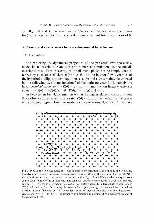
As depicted in Fig. 3, for small as well as for higher ®lament concentrationsh, we observe a decreasing stress rate, S0�h� < 0, and the mechanical system isin its swelling regime. For intermediate concentrations, h� < h < h�, we have
Fig. 3. Plot of the two rate functions (over ®lament concentration h) determining the two-phase
¯ow dynamics, namely the linear chemical assembly rate R(h) and the mechanical stress rate S(h),
see de®nitions in the text. At lower concentrations (h < heq � 0:1) ATP-dependent energy is con-
sumed for assembly of actin ®laments. The relatively sparse network tends to swell and dissipate
energy (S0�h� < 0 for h < h�) de®ning a swelling `sol' state; whereas for intermediate concentrations
(S 0�h� > 0 for h� < h < h�) de®ning the contraction regime, energy is consumed for mutual at-
traction of actin ®laments by APT-dependent action of myosin polymers. For even higher con-
centrations (S0�h� > 0 for h > h�) contractility is inhibited and dominated by dissipative swelling of
the condensed `gel'.
W. Alt, M. Dembo / Mathematical Biosciences 156 (1999) 207±228 215
S0�h� > 0 meaning that an increasing ®lament concentration would inducemore net contractile stress thus de®ning the contractile regime.
Therefore, the condition S0�heq� > 0 at chemical equilibrium implies that,when initially starting with a homogeneous ®lament distribution, i.e. h � heq
and v� 0, a spontaneous instability arises in the linearization of the hyperbolicsystem. For instance, let us consider a ®xed interval X � �0; L�, which could, asa one-dimensional model, describe the in vitro situation of cytoplasm extractsin a cuvette [14], with sticky boundary at x� 0 and non-sticky boundary atx�L, both impermeable for the solvent.
3.2. Results
The mechanical instability leads, for t > 0, to a sudden disruption of theactin ®laments from the boundary x�L and subsequent formation of a re-tracting wave moving towards the sticky boundary x� 0.
For positive drag u > 0 or adhesive friction U > 0 this wave develops anincreasing peak of higher ®lament concentration moving as a growing travel-ling wave, eventually being absorbed into the (already present) peak at x� 0.For quite a large range of parameters a completely periodic solution results byrepetitive disruption and wave formation with period T in the order of minutes,see Fig. 4. This reproduces the experimental observations mentioned above,[14,21], and supports as its underlying mechanism a chemo-mechanical cycle ofdisruption±contraction±relaxation±reassembly. Also, these repetitive contrac-tion waves resemble the observed `ru�es' arising at the lamellipodial tip andbeing retracted towards the cell body, compare Fig. 1. This in vivo modellingsituation with moving boundary is treated in the next section.
Even for the simpler case of ®xed boundaries, our numerical simulationscon®rm earlier results of two-dimensional computations which had producedspatio-temporally irregular or chaotic solutions [13].
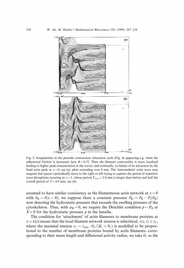
First, when increasing the interphase drag, u > 0, or the adhesive friction,U > 0, or when decreasing contractility, W, the waves intensify in amplitudebut simultaneously slow down (their period increasing T � 2.0), until they donot reach the left hand boundary any more, rather attain an oscillating locationin-between (see Fig. 5(a)). However, as the disruption±contraction±relaxationcycle from the right hand boundary continues, we again observe periodic so-lutions but with double period T� 4.0 (see Fig. 5(b)) or, sometimes, quasipe-riodic behavior with an obvious spatial coupling of two periodic oscillatorsnear both boundaries.
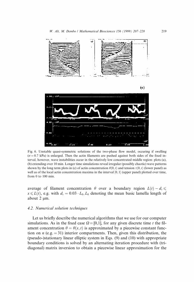
Second, when increasing the swelling rate, r/l, in comparison to the con-traction rate, w/l, so much that the swelling actin network pushes against thenon-sticky boundary x� 1, then we observe quasi-steady solutions with con-tracting ®lament peaks at both boundaries, see Fig. 6. Apparently, thisnon-symmetric con®guration is unstable as it happens that a growing local
216 W. Alt, M. Dembo / Mathematical Biosciences 156 (1999) 207±228

instability arises in the central region with lowest ®lament concentration,forming a slowly varying wave eventually moving in one or the other directionand ®nally being attracted by one of the boundary peaks (Fig. 6(a) and (b)).Longer simulations reveal irregular behavior with no apparent periodicities, sothat we conjecture to observe chaotic wave behavior (Fig. 6(c)).
4. One-dimensional dynamics of lamellipodial protrusion and retraction
4.1. Assumptions
In order to model the in vivo situation of lamella formation and its dy-namics around the periphery of adhering cells, let us again restrict the analysisto the one-dimensional case, thereby trying to match the experimental dataevaluation along transversal rays (see Fig. 1(C) and (D)). For this purpose letX�t� � �0; L�t�� describe the cross-sectional region of a lamella, with x� 0corresponding to the ®xed cell body boundary CB, permeable only to solventmolecules, and with x�L(t) denoting the tip of the lamella as a free imper-meable moving boundary C. Since the cortical cytoskeleton of the cell body is
Fig. 4. Simulations of repetitive contraction waves in a 1-dimensional two-phase ¯ow model on a
®xed domain X� [0,1] with sticky boundary at x� 0 and non-sticky boundary at x� 1. Plot of
actin ®lament concentration h� h(x,t) for t from 0 to 4 min (drawn from back to front) showing
periodic ®lament disruption, wave initiation, and subsequently, new ®lament attachment at x� 1.
The period is T � 1.8 min relative to an assumed actin assembly rate of g� 1/min. Biomechanical
parameters are ®lament viscosity l� 0.1 kPa min, contractility w� 25 kPa, saturating concentra-
tion hsat � 0.2, swelling coe�cient r� 0.5 kPa, drag u� 0.01 and adhesive friction U� 0.26, both in
kPa á min/lm2.
W. Alt, M. Dembo / Mathematical Biosciences 156 (1999) 207±228 217
assumed to have similar consistency as the ®lamenteous actin network at x� 0with hB � h�x � 0�, we suppose there a constant pressure PB :� PB ÿ Ps�hB�now denoting the hydrostatic pressure that exceeds the swelling pressure of thecytoskeleton. Thus, with uB� 0, we require the Dirichlet condition p�PB atX� 0 for the hydrostatic pressure p in the lamella.
The condition for `attachment' of actin ®laments to membrane proteins atx�L(t) means that the local ®lament network tension is subcritical, s�x; t�6 sC,where the maximal tension sC � smax � hC=�Ks � hC� is modelled to be propor-tional to the number of membrane proteins bound by actin ®laments: corre-sponding to their mean length and di�usional activity radius, we take hC as the
Fig. 5. Irregularities in the periodic contraction±relaxation cycle (Fig. 4) appearing e.g. when the
adhesional friction is increased, here U� 0.32. Then the ®lament contractility is more localized
leading to higher peak concentration in the waves, and eventually, to failure of its attraction by the
®xed actin peak at x� 0, see (a), plots extending over 8 min. The `intermediate' actin wave may
stagnate but (quasi-) periodically move to the right or left trying to capture the period of repetitive
wave disruptions occuring at x� 1, whose period Twave� 2.0 min is longer than before and half the
overall period of T� 4.0 min, see (b).
218 W. Alt, M. Dembo / Mathematical Biosciences 156 (1999) 207±228
average of ®lament concentration h over a boundary region L�t� ÿ ds6x6 L�t�, e.g. with ds � 0:03 � L0; L0 denoting the mean basic lamella length ofabout 2 lm.
4.2. Numerical solution techniques
Let us brie¯y describe the numerical algorithms that we use for our computersimulations. As in the ®xed case X� [0,1], for any given discrete time t the ®l-ament concentration h � h�x; t� is approximated by a piecewise constant func-tion on n (e.g.� 31) interior compartments. Then, given this distribution, the(pseudo-)stationary linear elliptic system in Eqs. (9) and (10) with appropriateboundary conditions is solved by an alternating iteration procedure with (tri-diagonal) matrix inversion to obtain a piecewise linear approximation for the
Fig. 6. Unstable quasi-symmetric solutions of the two-phase ¯ow model, occuring if swelling
(r� 0.7 kPa) is enlarged. Then the actin ®laments are pushed against both sides of the ®xed in-
terval, however, wave instabilities occur in the relatively low concentrated middle region: plots (a),
(b) extending over 10 min. Longer time simulations reveal irregular (possibly chaotic) wave patterns
shown by the long term plots in (c) of actin concentration h�0; t� and tension s�0; t� (lower panel) as
well as of the local actin concentration maxima in the interval �0; 1� (upper panel) plotted over time,
from 0 to 100 min.
W. Alt, M. Dembo / Mathematical Biosciences 156 (1999) 207±228 219

®lament velocity v� v(x, t) and a piecewise constant approximation for thepressure p � p�x; t�. At the free boundary x�L(t), the lamella tip, we ®rst (caseI: attachment) search for solutions satisfying the homogeneous Neumanncondition oxp � 0 and accept it if s6 sC�hC�, i.e. if the pressure at the lamella tipful®ls the inequality p6 pmax :� �1=�1ÿ h��sC�hC� ÿ Ps�h�; otherwise (case II;disruption) we declare that the ®lament network has been disrupted from thelamella tip and search for solutions satisfying the homogenous Dirichlet con-dition p� 0 at x�L(t). Then, after having checked the (discretized) inequalityoxp6 0 there, the current speed _L of the free boundary is calculated as de®ned inSection 2. In (case I: attachment) with _L�t� � v�L�t�; t�we transform the domain�0; L�t�� to the standard ®xed interval �0; 1� and apply there, as we did for the ®xeddomain in the last section, the upwind di�erence Euler step in order to solve thenow transformed hyperbolic Eq. (1). In (case II: disruption) we ®rst proceed inthe same way obtaining the ®lament distribution h on an interval �0; ~L�t�� havingmoved along the characteristic curve locally de®ned by _~L�t� � v�~L�t�; t� withinitial condition ~L � L at time t. Afterwards the really extended length of thelamella after dt is determined via the simple Euler step L�t � dt� � ~L�t � dt� �� _L�t� ÿv�L�t�; t� � dt, and the ®lament distribution in the newly covered lamellaarea is adjusted from zero to the newly assembled concentration levelh � g � heq � dt. Thus, the iteration cycle is closed and we restart with t :� t � dt.
4.3. Results
Initially starting with a lamella in the constant equilibrium state h � heq thefree lamella tip will, in general, always move, either by shrinking due to con-traction or by extending due to protrusive pressures. However, under param-eter constellations similar to those in the last section, numerical simulationsshow the already described disruption of actin ®laments from the lamella tipand the formation of a retracting wave. Again, depending on the choice of themechanical parameters, the lamella tip will retract or extend: In Fig. 7(a) weshow a particular simulation, where the lamella tip comes to rest as soon as thebackwards travelling contraction wave has arrived at the ®xed `cell body'boundary x� 0; afterwards the tip very slowly extends until, for long time, itstabilizes leading to a stable stationary actin ®lament distribution �h � h�x��,which decreases from a ®xed `gel concentration' at the `cell body' down to zeroat the lamella tip, see Fig. 7(b). In this steady state, which might model thestable lamella of fast moving keratocytes [25], there is ongoing reassembly ofactin ®laments at the anterior lamella region, from where they are transportedtowards the cell body by the `centripetal' actin ¯ow. The strength of thiscontractile ¯ow �jvj� and the lamella length adjust in such a way, that the lo-cally induced drag in the two-phase ¯uid is equilibrated by the pressure drop(p) from an imposed hydrostatic pressure PB > 0 at the cell body down to zeroat the lamella tip.
220 W. Alt, M. Dembo / Mathematical Biosciences 156 (1999) 207±228

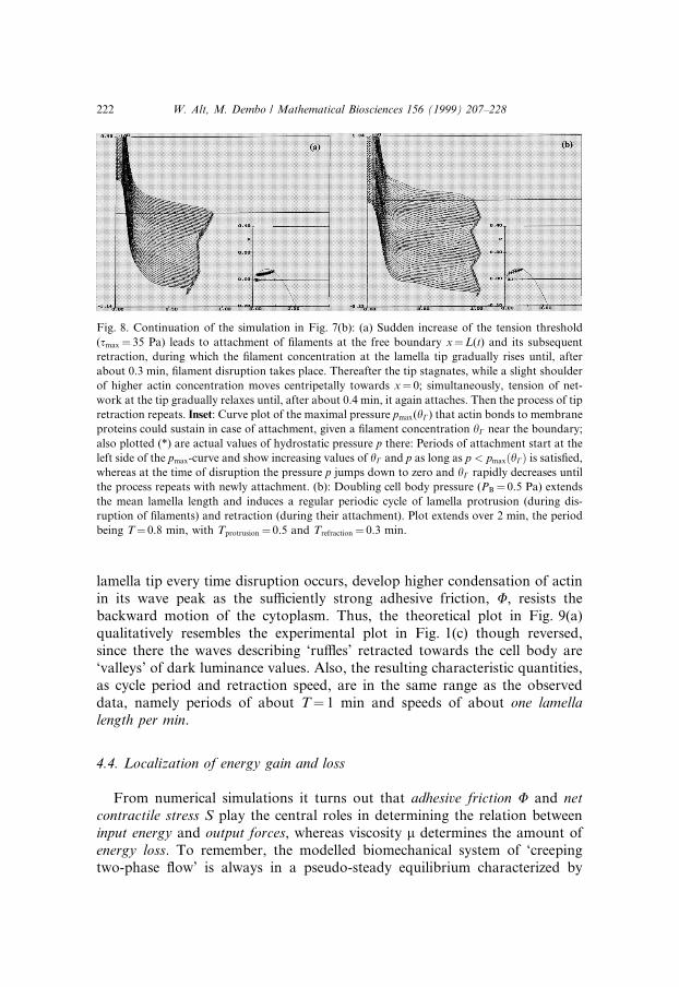
This situation can occur provided the ®lament concentration near the tip(hC) is too small to form enough connections to membrane proteins, and theinduced ®lament tension s at the tip (due to contractile and viscous stresses) issupercritical so that attachment cannot happen. Indeed, when lifting thecritical tension smax a bit, we observe sudden attachment and subsequentcontraction of the whole lamella, see Fig. 8(a). However, due to the rapidlyincreasing ®lament concentration at the retracting tip the ®lament stress be-comes supercritical again, with subsequent ®lament disruption and tip stag-nation. Continuing the simulations we would observe a repetition of thiscontraction±relaxation cycle but with the lamella gradually decreasing inlength. Therefore, in order to balance the mean contractile action of the actinpolymer system and sustain a su�cient protrusion length, we have to providea counteracting protrusion force, for example, by increasing the cell bodypressure PB. Indeed, for the parameters chosen in Fig. 8(b), very similar as inthe ®xed boundary situation (see Fig. 4), we obtain a periodic cycle of alter-nating disruption and attachment of the actin ®laments, now exactly timedwith alternating protrusion and retraction of the lamella. By changing themechanical parameters slightly, the amplitude of these protrusion±retractiondynamics can be enlarged, e.g. by a further increase of the tension thresholdsmax leading to longer periods of contraction, balanced by an increasing pro-trusive pressure at the lamella tip due to lowering the interphase drag, u, seeFig. 9(a). Notice, the periodic contraction waves that are initiated at the
Fig. 7. Simulations of the 1-dimensional contractile two-phase ¯ow model with ®xed sticky
boundary (but permeable for the solvent) at x� 0 and free boundary x�L(t). (a) Actin ®lament
concentration h� h(x, t) is plotted for increasing times from back to front, extending over 2 min.
Parameters are w� 18, r� 0.25, u� 0.008, U� 0.4, with relatively low cell body pressure PB� 0.25
Pa, and tension threshold smax� 30 Pa with saturation constant Ks� 0.1. For times t ® 1 the
concentration pro®le stabilizes and the lamella length converges to a value L1 � 0.97. (b) The
stable steady state distribution of h(x) (upper curve), the modulus jv�x�j of the centripetal ®lament
velocity and the hydrostatic pressure p(x).
W. Alt, M. Dembo / Mathematical Biosciences 156 (1999) 207±228 221
lamella tip every time disruption occurs, develop higher condensation of actinin its wave peak as the su�ciently strong adhesive friction, U, resists thebackward motion of the cytoplasm. Thus, the theoretical plot in Fig. 9(a)qualitatively resembles the experimental plot in Fig. 1(c) though reversed,since there the waves describing `ru�es' retracted towards the cell body are`valleys' of dark luminance values. Also, the resulting characteristic quantities,as cycle period and retraction speed, are in the same range as the observeddata, namely periods of about T� 1 min and speeds of about one lamellalength per min.
4.4. Localization of energy gain and loss
From numerical simulations it turns out that adhesive friction U and netcontractile stress S play the central roles in determining the relation betweeninput energy and output forces, whereas viscosity l determines the amount ofenergy loss. To remember, the modelled biomechanical system of `creepingtwo-phase ¯ow' is always in a pseudo-steady equilibrium characterized by
Fig. 8. Continuation of the simulation in Fig. 7(b): (a) Sudden increase of the tension threshold
(smax� 35 Pa) leads to attachment of ®laments at the free boundary x�L(t) and its subsequent
retraction, during which the ®lament concentration at the lamella tip gradually rises until, after
about 0.3 min, ®lament disruption takes place. Thereafter the tip stagnates, while a slight shoulder
of higher actin concentration moves centripetally towards x� 0; simultaneously, tension of net-
work at the tip gradually relaxes until, after about 0.4 min, it again attaches. Then the process of tip
retraction repeats. Inset: Curve plot of the maximal pressure pmax(hC) that actin bonds to membrane
proteins could sustain in case of attachment, given a ®lament concentration hC near the boundary;
also plotted (*) are actual values of hydrostatic pressure p there: Periods of attachment start at the
left side of the pmax-curve and show increasing values of hC and p as long as p < pmax�hC� is satis®ed,
whereas at the time of disruption the pressure p jumps down to zero and hC rapidly decreases until
the process repeats with newly attachment. (b): Doubling cell body pressure (PB� 0.5 Pa) extends
the mean lamella length and induces a regular periodic cycle of lamella protrusion (during dis-
ruption of ®laments) and retraction (during their attachment). Plot extends over 2 min, the period
being T� 0.8 min, with Tprotrusion� 0.5 and Trefraction� 0.3 min.
222 W. Alt, M. Dembo / Mathematical Biosciences 156 (1999) 207±228
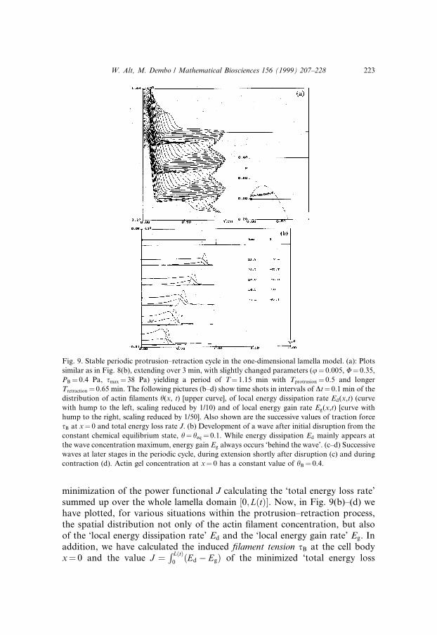
minimization of the power functional J calculating the `total energy loss rate'summed up over the whole lamella domain �0; L�t��. Now, in Fig. 9(b)±(d) wehave plotted, for various situations within the protrusion±retraction process,the spatial distribution not only of the actin ®lament concentration, but alsoof the `local energy dissipation rate' Ed and the `local energy gain rate' Eg. Inaddition, we have calculated the induced ®lament tension sB at the cell bodyx� 0 and the value J � R L�t�
0�Ed ÿ Eg� of the minimized `total energy loss
Fig. 9. Stable periodic protrusion±retraction cycle in the one-dimensional lamella model. (a): Plots
similar as in Fig. 8(b), extending over 3 min, with slightly changed parameters (u� 0.005, U� 0.35,
PB� 0.4 Pa, smax� 38 Pa) yielding a period of T� 1.15 min with Tprotrusion� 0.5 and longer
Tretraction� 0.65 min. The following pictures (b±d) show time shots in intervals of Dt� 0.1 min of the
distribution of actin ®laments h(x, t) [upper curve], of local energy dissipation rate Ed(x,t) (curve
with hump to the left, scaling reduced by 1/10) and of local energy gain rate Eg(x,t) [curve with
hump to the right, scaling reduced by 1/50]. Also shown are the successive values of traction force
sB at x� 0 and total energy loss rate J. (b) Development of a wave after initial disruption from the
constant chemical equilibrium state, h� heq� 0.1. While energy dissipation Ed mainly appears at
the wave concentration maximum, energy gain Eg always occurs `behind the wave'. (c±d) Successive
waves at later stages in the periodic cycle, during extension shortly after disruption (c) and during
contraction (d). Actin gel concentration at x� 0 has a constant value of hB� 0.4.
W. Alt, M. Dembo / Mathematical Biosciences 156 (1999) 207±228 223

rate'. Here, Ed just collects all the integral terms in Eq. (3), but in spite of thechoice of high adhesive friction, U, ®lament viscosity essentially dominatesthe friction and drag terms. On the other hand, Eg contains all integral termsin Eq. (4), but due to the choice of strong contractivity, w, energy gain isdominated by contractile energy which is put into the ®lamenteous networkby the ATP-dependent action of myosin, however, damped by networkswelling, r.
Now, the striking observation is that energy gain Eg is exclusively localizedto the region behind the contraction wave, whereas energy dissipation Ed issigni®cant in all regions where the ®lament network is going to be compressed.Therefore, it seems as if the `contraction wave' is actively pushed and com-pressed by forces in its wake: indeed, the most important and dynamicallye�ective contraction takes place in this region, where actin ®laments from thelower concentrated area at the tip are attracted by the higher concentratedshoulder or peak of the wave.
Finally, we have computed the traction force sB �R L�t�
0Uhjvj onto the cell
body due to the frictional interaction of retracting acting ®laments with
Fig. 9. Continued.
224 W. Alt, M. Dembo / Mathematical Biosciences 156 (1999) 207±228

adhesion proteins connected to the substratum. This force is eventually re-sponsible for and proportional to the rate of cell translocation towards theprotruded lamella tip. During the periodic lamella cycle it increases to amaximal value (sB � 12.8 Pa) at the moment of disruption, see Fig. 9(c),whereas it attains its minimal value (sB � 8.1 Pa) when the contractionwave reaches the cell body, see Fig. 9(d). Simultaneously, the net energyinput (ÿJ), i.e. the di�erence between total energy input and total energydissipation, is always positive meaning that we have negative entropy pro-duction, i.e. entropy reduction, in the active lamella therefore representingthe typical feature of a `biological motor'. It also varies cyclically butcontrarily to the output force sB. After disruption, when the initiated con-traction wave is ¯at, the net energy input is minimal (ÿJ� 11.9 Pa), whereasit is maximal (ÿJ� 30.6 Pa) as the increased wave is approaching the peakat x� 0.
Consistent with the formula for sB, the potential traction force for celltranslocation is reduced, if we lower the adhesive friction (U). In parallel, asimilar reduction of the net energy input (ÿJ) occurs.
5. Discussion: the role of stochastics and outlook to further modelling
These various kinds of spatio-temporally solutions to the one-dimensionalhyperbolic±elliptic boundary value problem seem to con®rm that the presentedtwo-phase ¯uid model, with its main non-linearity S(h) in Fig. 3, has the ca-pacity to produce a broad spectrum of dynamical behavior, already in the one-dimensional situation with ®xed boundaries. Since the derived continuumequations approximate the stochastic `biointeraction' between cytoplasmic®laments and proteins, they might serve as a suitable tool for proving that theobserved richness in the dynamics of cellular motility could be due to themechano-chemical properties of the cytoplasm.
So far, our 1-dimensional model for lamella dynamics assumed that the cellbody (with its boundary x� 0 to the lamella) stays ®xed while the lamella tipprotrudes or retracts. The same assumption holds for a simpli®ed circularmodel that describes tangential cytoplasm ¯ow and radial lamella protrusionsaround the cell periphery, assumed to be a circle with ®xed radius [5]. In aneven more simpli®ed model [23], deformations of cell body shape were con-sidered, though directly dependent on local actin ®lament concentration which,by its varying distribution around the cell periphery, may induce a polarizationof the cell. Indeed, many motile tissue or blood cells (as leukocytes or ®shkeratocytes) soon after displacement on a substratum polarize their shape byexpressing one leading lamella that performs protrusions and determines thedirection of locomotion, whereas the rear of the cell is characterized by cyto-plasmic contraction.
W. Alt, M. Dembo / Mathematical Biosciences 156 (1999) 207±228 225

One unresolved question is whether the experimentally observed ¯uctua-tions in direction and motile activity of the leading lamella can result fromirregular or chaotic cytoplasm dynamics within the deterministic framework ofcontinuum models as presented in this paper; or if they are the consequence ofadditional stochasticity e�ected by random ¯uctuations of the mechanical orchemical parameters. The latter hypothesis has been investigated in [23] as-suming that actin polymerization is stimulated by membrane receptors whichrandomly di�use and bind certain ligands. In this combined deterministic±stochastic model the resulting ¯uctuations of cell polarization and retrogradecytoplasmic ¯ow induce corresponding ¯uctuations of the translocation vector,thus leading to typical cell migration paths which can be characterized as di-rectionally persistent random walks.
Another important candidate for inducing stochasticity in cell motion isthe process of formation and con®guration of adhesion bonds, being essentialfor force transduction between cell and substratum. Hitherto stochasticmodels that took into account the kinetics, di�usion and transport of ad-hesion receptors in order to quantify the dynamics of adhesion and to de-termine the resulting cell translocation speed, see e.g. Ref. [24], lack inconsidering the internal cytoplasm dynamics or, at least, the feedback that itgets from the adhesion kinetics. Thus, we address this as a further openproblem, namely to investigate hybride models which, for example, combinethe presented two-phase ¯ow model for the cytoplasm with stochastic modelsfor the tension dependent kinetics of attachment/disruption between actin®laments and membrane proteins as well as the binding/disruption kineticsbetween adhesion proteins and the substratum; a ®rst attempt is performed inRef. [20].
Acknowledgements
Thanks are due to the Research Program SFB 256 `Non-linear PartialDi�erential Equations', Univ. Bonn, which has sponsered the collaborationbetween the authors, and to Boris Hinz who provided various ®gures. How-ever, most thanks are given to Tanya Kostova and her friendly team for or-ganizing the wonderful and e�ective Conference on Deterministic andStochastic Modelling of Biointeraction in So®a, August 1997.
References
[1] W. Alt, Biomechanics of actomyosin mediated motility of keratinocytes, Biophysics 41 (1996)
181.
226 W. Alt, M. Dembo / Mathematical Biosciences 156 (1999) 207±228

[2] W. Alt, O. Brosteanu, B. Hinz, H.W. Kaiser, Patterns of spontaneous motility in
videomicrographs of human epidermal keratinocytes (HEK), Biochemistry and Cell Biology
73 (1995) 441.
[3] W. Alt, M. Dembo, The dynamics of viscous reactive and contractile two-phase ¯uids,
Biophys. J., submitted.
[4] W. Alt, V. Lendowski, Modelling and simulation of particle movement in reactive two-phase
¯uids, Preprint SFB 256, Univ. Bonn 540, 1998.
[5] W. Alt, R.T. Tranquillo, Basic morphogenetic system modeling shape changes of migrating
cells: How to explain ¯uctuating lamellipodial dynamics, J. Biol. Systems 3 (1995) 905.
[6] W. Alt, R.T. Tranquillo, Protrusion±retraction dynamics of an annular lamellipodial seam, in:
W. Alt, A. Deutsch, G. Dunn (Eds.), Dynamics of Cell and Tissue Motion, Birkh�auser, Basel,
1997, p. 73.
[7] L. Cao, Y. Wang, Mechanism of the formation of contractile ring in dividing cultured animal
cells. II. Cortical movement of microinjected actin ®laments, J. Cell Biol. 111 (1990) 1905.
[8] M. Dembo, Field theories of the cytoplasma, Comments Theoret. Biol. 1 (1989) 159.
[9] M. Dembo, Mechanics and control of the cytoskeleton in Amoeba proteus, Biophys. J. 55
(1989) 1053.
[10] M. Dembo, On free boundary problems and amoeboid motion, in: N. Akkas (Eds.),
Biomechanics of Active Movement and Division of Cells, NATO ASI Ser. H84, Springer,
Berlin, 1994, p. 231.
[11] M. Dembo, F.W. Harlow, Cell motion, contractile networks, and the physics of interpen-
etrating reactive ¯ow, Biophys. J. 50 (1986) 109.
[12] M. Dembo, F.W. Harlow, W. Alt, The biophysics of cell surface mobility, in: F.W. Wiegel,
A.D. Perelson, Ch. DelLisi (Eds.), Cell Surface Dynamics, Concepts and Models, Marcel
Dekker, New York, 1984, p. 495.
[13] M. Dembo, M. Maltrud, F.W. Harlow, Numerical studies of unreactive contractile networks,
Biophys. J. 50 (1986) 123.
[14] R.M. Ezzell, A.J. Brothers, W. Cande, Phosphorylation dependent contraction of actomyosin
gels from amphibian eggs, Nature 306 (1983) 620.
[15] G. Fisher, P.A. Conrad, R.L. DeBasio, R.B. Taylor, Centripetal transport of cytoplasm, actin,
and the cell surface in lamellipodia of ®broblast, Cell Motil. Cytoskeleton 11 (1988) 235.
[16] P. Forscher, H.L. Chi, C. Thompson, Novel form of the growth cone motility involving site-
directed actin ®lament assembly, Nature 357 (1992) 515.
[17] X. He. M. Dembo. A dynamical model of cell division, in: W. Alt, A. Deutsch, G. Dunn
(Eds.), Dynamics of Cell and Tissue Motion, Birkh�auser, Basel, 1997, p. 55.
[18] X. He, M. Dembo, On the mechanics of the ®rst cleavage division of the sea urgin egg, Exp.
Cell Res. 233 (1997) 252.
[19] V. Lendowski, A. Mogilner, Origin of actin-induced locomotion of listeria, in: W. Alt, A.
Deutsch, G. Dunn (Eds.), Dynamics of Cell and Tissue Motion, Birkh�auser, Basel, 1997, p.
93.
[20] J. Lenz, W. Alt, M. Sahm, Model of adhesion kinetics and traction dynamics in cell
locomotion, Manuscript, Univ. Bonn 1999.
[21] Thomas Pohl. Periodic contraction waves in cytoplasmic extracts, in: W. Alt, G. Ho�mann
(Eds.), Biological Motion , vol. 89, Springer, Berlin, 1990, p. 55.
[22] D.L. Taylor, J.S. Condeelis, P.L. Moore, R.D. Allen, The contractile basis of amoe-
boid movement I. The chemical control of motility in isolated cytoplasm, J. Cell Biol. 59
(1973) 378.
[23] R.T. Tranquillo, W. Alt, Stochastic model of receptor-mediated cytomechanics and dynamic
mophology of leukocytes, J. Math. Biol. 34 (1996) 361.
[24] M.D. Ward, D.A. Hammer, Focal contact assembly through cytoskeletal polymerization:
steady state analysis, J. Math. Biol. 32 (1994) 677.
W. Alt, M. Dembo / Mathematical Biosciences 156 (1999) 207±228 227

[25] R. Winklbauer, Andreas Selchow, Beate Boller, J�urgen Bereiter-Hahn, Embryonic mesoderm
cells and larval keratocytes from Xenopus: Structure and motility of single cells, in: W. Alt, A.
Deutsch, G. Dunn (Eds.), Dynamics of Cell and Tissue Motion, Birkh�auser, Basel, 1997, p. 7.
[26] S.H. Zigmond, Recent quantitative studies of actin ®lament turnover during cell locomotion,
Cell Motil. Cytoskeleton 25 (1993) 309.
228 W. Alt, M. Dembo / Mathematical Biosciences 156 (1999) 207±228