cystic fibrosis: the role of airway stem cells in ...€¦ · cystic fibrosis australia phd...
TRANSCRIPT

CYSTIC FIBROSIS: THE ROLE OF
AIRWAY STEM CELLS IN
SUSTAINED GENE EXPRESSION
BY LENTIVIRAL DIRECTED GENE
THERAPY
NIGEL FARROW
BMedSc Flinders University
BHSc Medicine (Hons) first class
The University of Adelaide
Submitted in fulfilment of the Degree of Doctor of
Philosophy (PhD)
28 April 2015
University of Adelaide
School of Paediatrics and Reproductive Health
Robinson Research Institute
Research conducted in the Department of Respiratory and Sleep Medicine at the
Women’s and Children’s Hospital, Adelaide, South Australia.


i
Declaration
I certify that this work contains no material which has been accepted for the award of
any other degree or diploma in my name, in any university or other tertiary
institution and, to the best of my knowledge and belief, contains no material
previously published or written by another person, except where due reference has
been made in the text. In addition, I certify that no part of this work will, in the
future, be used in a submission in my name, for any other degree or diploma in any
university or other tertiary institution without the prior approval of the University of
Adelaide and where applicable, any partner institution responsible for the joint-
award of this degree.
I give consent to this copy of my thesis when deposited in the University Library,
being made available for loan and photocopying, subject to the provisions of the
Copyright Act 1968.
The author acknowledges that copyright of published works contained within this
thesis resides with the copyright holder(s) of those works.
I also give permission for the digital version of my thesis to be made available on the
web, via the University’s digital research repository, the Library Search and also
through web search engines, unless permission has been granted by the University to
restrict access for a period of time.
Signed:
Date: 28 April 2015

ii

iii
This Thesis is dedicated to my daughter
Ella Farrow.
My very special little girl, your determination to enjoy life to its fullest
despite having the insidious disease Cystic Fibrosis is a source of
inspiration which drives me every day. Darling this is for you.

iv

v
Acknowledgements
I would like to thank my supervisor Associate Professor David Parsons for giving
me the opportunity to undertake this PhD and allowing me to pursue a project I have
great interest in. I also thank David for going above and beyond the role of
supervisor and becoming a trusted mentor.
I am also grateful to the people who have taken time from their busy schedules to
share their technical skills and knowledge with me. In particular I would like to
thank my co-supervisor and head of the Lung and Health Research Centre at
Melbourne University, Ivan Bertoncello and his senior researcher Jonathan
McQualter for passing on their vast knowledge in the field of airway stem cells and
teaching me their techniques for isolating and culturing airway stem cells; Members
of the Adelaide Cystic Fibrosis Gene Therapy Group including, Martin Donnelley
for encouragement, emotional support, technical support and providing opportunities
to learn techniques outside of my chosen field; Patricia Cmielewski for
encouragement, advice, assistance and keeping me honest; Ryan Green for assistance
in compiling stacked images of the airway; Harsha Padmanabhan for assistance in
the lab and keeping the place full of energy; Sharnna Devereux for assisting in
mouse tissue collections; Chantelle McIntyre for assistance in the lab and for being
there to bounce ideas around; Bernadette Boog for assistance and encouragement;
the Cure4CF foundation for all the support and everything they do, particularly Greg
Oke, David Coluccio, Jo Close, Mark Evans, Deb Hoskings, Gregg Johnson, Rob
Mills, Jenny Paradiso, and Greg Savage; the MS McLeod Foundation and CF
Australia for financial support throughout my PhD.

vi
I would also like to thank my second co-supervisor Simon Barry and members of his
lab particularly Tim Sadlon for assistance and advice in plasmid construction and
vector production; Lyn Marsden for looking after the mice and providing advice on
animal experiment related matters; member of the Matrix biology lab for assistance,
advice and help, particularly Sharron Byers, Ainslee Derrick Roberts, Carmen
Pyragius, Xenia Kaidonas, Matilda Jackson and Nathan Rout-Pitt; Ruth Williams
and Adelaide microscopy for assistance with histology: Jim Manavis for assistance
with immunohistochemistry.
Finally I would like to express my deepest gratitude to my wife Karen and daughters
Ariah and Ella for encouragement, support, sacrifices and understanding throughout
this journey; my parents, brothers, their partners and my wife’s family for their
ongoing support; Luke and Aaron for laughs, encouragement and guidance; John for
always being there when I needed it; Shaun and the Thursday night group for the
laughs and a place to escape and forget about CF for just a little while each week.

vii
Publications and awards
Publications arising from this work
Cmielewski, P, Farrow, N, et al, Transduction of ferret airway epithelia using a pre-
treatment and lentiviral gene vector. BMC Pulm Med, 2014. 14 (1): p. 183.
DOI:10.1186/1471-2466-14-183.
Cmielewski, P, and Farrow, N, were equal contributing first authors.
N. Farrow, D. Miller, P. Cmielewski, M. Donnelley, R. Bright, D. Parsons, “Airway
gene transfer in a non-human primate: Lentiviral gene expression in marmoset lung”,
Scientific Reports, 3, 2013. P.4. DOI: 10. 1038/srep01287.
Kaye S. Morgan, Martin Donnelley, Nigel Farrow, Andreas Fouras, Naoto Yagi,
Yoshio Suzuki, Akihisa Takeuchi, Kentaro Uesugi, Richard C. Boucher, Karen K.
W. Siu*, and David W. Parsons*. “In vivo x-ray imaging reveals improved airway
surface hydration after Cystic Fibrosis airway therapy” American Journal of
Respiratory and Critical Care Medicine 2014, 190, 4.
Martin Donnelley, Kaye S. Morgan, Karen K. W. Siu, Nigel R. Farrow, Charlene S.
Stahr, Richard C. Boucher, Andreas Fouras & David W. Parsons, “Non-invasive
airway health assessment: Synchrotron imaging reveals effects of rehydrating
treatments on mucociliary transit in-vivo”, Scientific Reports, 4, 2014. DOI:
10.1038/srep03689.
Martin Donnelley, Kaye S. Morgan, Karen K. W. Siu, Andreas Fouras, Nigel R.
Farrow, Richard P. Carnibella and David W. Parsons “Tracking extended
mucociliary transport activity of individual deposited particles: longitudinal

viii
synchrotron X-ray imaging in live mice” Journal of synchrotron radiation, 21, 2014.
DOI: 10.1107/S160057751400856X.
Abstracts Presented At National and International
Conference Meetings
Patricia Cmielewski, Nigel Farrow, Chantelle McIntyre, Harsha Padmanabhan,
Martin Donnelley, Tim Kuchel, and David W Parsons.” Lentiviral airway gene
transfer in normal ferrets” American Society for Gene and Cell Therapy,
Washington, USA.
Nigel Farrow, Martin Donnelley, Patricia Cmielewski, Ivan Bertoncello , David
Parsons. “Gene therapy for CF: is long term expression a consequence of
transducing conducting airway endogenous respiratory stem cells?” presented at the
North America Cystic Fibrosis Conference, Atlanta, Georgia, USA, 2014.
Nigel Farrow, Jonathan L McQualter, David Parsons, Ivan Bertoncello.
“Endogenous stem/progenitor cell compartments of conducting airways differ in
cystic fibrosis and normal mice, presented at the British Society for Gene and Cell
Therapy conference, Royal Holloway, University of London, London.
Nigel Farrow, Jonathan L McQualter, David Parsons, Ivan Bertoncello
, “Analysis of
endogenous airways stem/progenitor cell types in CF and normal mice and the role
of basal cells in sustained gene expression following airway gene transfer”,
presented at the Cystic fibrosis Australia conference, Auckland, New Zealand, 2013
Martin Donnelley, Kaye Morgan, Karen Siu, Andreas Fouras, Nigel Farrow,
Richard Carnibella and David Parsons, “Advances in airway surface imaging for
cystic fibrosis: extended monitoring of individual particle mucocilary clearance”
presented at the Cystic fibrosis Australia conference, Auckland, New Zealand, 201
Ivan Bertoncello, Nigel Farrow, Jonathan McQualter, David Parsons.
2014
2013

ix
Evidence of an Expanded and Dysregulated Airway Epithelial Stem Cell
Compartment in Cystic Fibrosis Mice, Stem Cells and Cell Therapies in Lung
Biology and Diseases conference, Vermont, Burlington, USA, 2013.
Martin Donnelley, Kaye Morgan, Karen Siu, Nigel Farrow, Charlene Chua,
Andreas Fouras and David Parsons, “Non-invasive airway health assessment:
Synchrotron imaging reveals effects of therapeutics on mucociliary transit function”
presented at the Medical Applications of Synchrotron Radiation conference,
Shanghai, China, 2012.
K. Morgan, M. Donnelley, D. Paganin, A. Fouras, N. Farrow, Y. Suzuki, A.
Takeuchi, K. Uesugi, N.Yagi, D. Parsons, K. Siu, “Assessing new treatments for
cystic fibrosis using micron-scale live phase contrast x-ray imaging of the airway
surface liquid”, presented at the Medical Applications of Synchrotron Radiation
conference, Shanghai, China, 2012.
Jonathan L McQualter, Nigel Farrow, David Parsons, Ivan Bertoncello,
“Endogenous Lung Epithelial Stem/Progenitor Cell Compartments Differ In Cystic
Fibrosis and Normal Mice”, presented at the North America Cystic Fibrosis
Conference, Orlando, Florida, 2012.
D Parsons, N Farrow, D Miller, S Le Blanc, R Bright , P Cmielewski and D S
Anson, “One Year Persistence From a Single HIV-1 Lentiviral Vector Delivery Into
Marmoset Lung: LacZ and Vector Gene Presence” presented at the American
Society of Gene & Cell Therapy conference, Seattle, North America, 2011
2012
2011

x
Awards Received
Robinson Research Institute, research travel award 2014
Adelaide University Post Graduate Conference Best Poster Presentation 2012
MS McLeod Foundation PhD scholarship 2011-2014
Cystic Fibrosis Australia PhD scholarship 2011-2014
APA PhD scholarship (declined) 2011
Letter of Commendation from Lord Mayor of Adelaide, South Australia 2010
Golden Key Honour Society 2009
Flinders University Chairperson’s Letter of Commendation 2008 and 2009

xi
Synopsis
In this thesis transduction of airway stem cells (basal cells) in the nasal and tracheal
airways was investigated to determine the causality of sustained transgene
expression following a gene therapy protocol that utilised an LPC pre-treatment and
a HIV-1 VSV-G pseudotyped lentivirus vector treatment, as previously published.
To assess stem cell transduction and epithelial regrowth a forced-injury model was
employed at a number of time points after the gene therapy protocol.
Epithelial remodelling in cystic fibrosis and normal airways of mice was also
assessed. Airway stem cell hyperplasia and goblet cell hyperplasia and hypertrophy,
and epithelial mucin content were assessed in the trachea and in some instance the
nasopharynx in the nasal airways of CF and normal mice.
Additionally, the effectiveness of the LPC / lentiviral gene therapy protocol was
assessed in lung airways of normal ferrets and the marmoset, a non-human primate,
to determine if airway transduction of both differentiated ciliated cells and stem cells
reflected observations noted in previously-published mouse-based studies. These
ferret and marmoset animal studies have been published prior to thesis submission.
Airway stem cells transduction was confirmed in the trachea and nasal airways of
mice following pre-treatment with LPC and subsequent treatment with a HIV-1
VSV-G pseudotyped lentiviral vector carrying the LacZ marker gene. A forced
injury model was employed to force regeneration of the airway epithelium after
vector treatment. Following the ablation and subsequent regeneration of the airway
epithelium, clusters of LacZ positive were observed in both the trachea and nasal
airways suggesting transduction of the airway stem cells and the passing of the

xii
transgene to their progeny upon differentiation.
Airway epithelial remodelling was demonstrated in both airway stem cells and
goblet cells in CF mice. Hyperplasia of airway stem cells and goblet cells in CF mice
was observed. Hypertrophy and change in mucin acidity of goblet cells was also
observed. Additionally, remodelling of the cartilage rings in the trachea was
observed in CF mice. This is the first study to demonstrate the presence of goblet
cell hyperplasia, hypertrophy and change in mucin acidity in the presence of airway
stem cell hyperplasia. Importantly, the hyperplasia of airway stem cells in CF
airways had previously been proposed however, this is the first study to directly
quantitate the airway stem cell compartment using a novel flow cytometry and
clonogenic assay approach.
Finally, the transduction of airway stem cells and ciliated cells is shown in normal
ferrets and marmosets, a non-human primate. Validation of transducing relevant
airway cell type in these animals adds to the gene therapy proof of principle
foundation previously demonstrated in the airways of mice.

xiii
Contents
Declaration .................................................................................................................... i
Acknowledgements ...................................................................................................... v
Publications and awards ............................................................................................ vii
Publications arising from this work ...................................................................... vii
Abstracts Presented At National and International Conference Meetings ........... viii
2014 .................................................................................................................. viii
2013 .................................................................................................................. viii
2012 ..................................................................................................................... ix
2011 ..................................................................................................................... ix
Awards Received ..................................................................................................... x
Synopsis ...................................................................................................................... xi
Contents .................................................................................................................... xiii
Figure list .................................................................................................................. xxi
1 Introduction .......................................................................................................... 1
1.1 Cystic fibrosis: A historical perspective ...................................................... 1
1.2 Mapping, isolating and sequencing of the CF gene ..................................... 3
1.3 The CFTR protein ........................................................................................ 3
1.3.1 Genetic mutations of the Cystic fibrosis gene .......................................... 4
1.4 Pathophysiology of Cystic Fibrosis ............................................................. 8
1.4.1 Current treatments .................................................................................... 9

xiv
1.4.2 Emerging treatments .............................................................................. 10
1.5 Gene Therapy ............................................................................................. 11
1.5.1 Adenoviruses and Adeno-associated viruses ......................................... 13
1.5.2 Retroviral Vectors .................................................................................. 13
1.5.3 Lentiviral Vectors .................................................................................. 14
1.5.4 HIV-1 Biology ....................................................................................... 15
1.5.5 Trans and Cis Acting Elements.............................................................. 16
1.5.6 HIV-1 as a Vector .................................................................................. 17
1.6 Stem cells in Gene Therapy ....................................................................... 20
1.6.1 Respiratory Stem Cells .......................................................................... 22
1.6.2 Respiratory Stem Cell Transduction ...................................................... 24
1.7 Conclusion ................................................................................................. 26
1.8 Aims of Thesis ........................................................................................... 27
2 Materials and methods ....................................................................................... 29
2.1 Materials .................................................................................................... 29
2.1.1 Chemicals and Supplies ......................................................................... 29
2.1.2 Consumables and Suppliers ................................................................... 32
2.1.3 Bacterial Strains and Media ................................................................... 33
2.1.4 Cell Lines ............................................................................................... 33
2.1.5 DNA Plasmids ....................................................................................... 33
2.1.6 Real Time qPCR Assay ......................................................................... 34

xv
2.1.7 Animal Models ....................................................................................... 35
2.1.8 Processing of Mouse Heads ................................................................... 35
2.1.9 ELISA Assay .......................................................................................... 35
2.2 Methods: In Vitro ....................................................................................... 36
2.2.1 Vector Plasmid Preparation .................................................................... 36
2.2.2 Cell Culture ............................................................................................ 36
2.2.3 Lentiviral Production ............................................................................. 37
2.2.4 Establishing Viral Titre by qPCR .......................................................... 39
2.2.5 Lentiviral Vector Instillations ................................................................ 41
2.2.6 Assessing In vivo Vector Dissemination ................................................ 42
2.2.7 Cell Sorting and Clonal Culture of Respiratory Airway Stem Cells ..... 42
3 Airway stem cell transduction by a VSV-G pseudotyped HIV-1 lentiviral vector
............................................................................................................................ 45
3.1 Introduction ................................................................................................ 45
3.2 Methods ...................................................................................................... 46
3.2.1 Animals .................................................................................................. 46
3.2.2 Gene Vector ........................................................................................... 46
3.2.3 Nasal and tracheal gene transfer treatment ............................................ 46
3.2.4 Induced regeneration of the respiratory epithelium via Polidocanol
treatment ............................................................................................................. 47
3.3 Results ........................................................................................................ 49
3.3.1 LPC pre-treatment and vector instillation .............................................. 49

xvi
3.3.2 Effect of PDOC-based ablation on epithelial integrity and regeneration ..
................................................................................................................ 49
3.3.3 Induced regeneration of the respiratory epithelium revealed clonal
clusters of marker gene positive cells ................................................................ 51
3.3.4 Identification of marker gene expressing cell types .............................. 53
3.4 Discussion .................................................................................................. 54
3.5 Conclusion ................................................................................................. 60
4 Airway stem cells and epithelial remodelling in the conducting airways of CF
mice ............................................................................................................................ 61
4.1 Introduction ................................................................................................ 61
4.2 Material and Methods ................................................................................ 63
4.2.1 Mouse models ........................................................................................ 63
4.2.2 Processing of tissue ................................................................................ 63
4.2.3 Tracheal and nasal septum cell preparation and flow cytometry ........... 64
4.2.4 Cell culture ............................................................................................. 64
4.2.5 Histochemistry and Immuno-histochemistry ......................................... 64
4.2.6 Statistics ................................................................................................. 65
4.3 Results ........................................................................................................ 65
4.3.1 Goblet cell hyperplasia and hypertrophy in CF mouse nasal airways ... 65
4.3.2 Proliferation index of airway stem cells ................................................ 67
4.3.3 Stem cell hyperplasia in the respiratory epithelium ............................... 68
4.3.4 Retrospective assessment of the ages of mice in the airway stem cell

xvii
study ................................................................................................................ 70
4.4 Discussion .................................................................................................. 71
4.5 Conclusion .................................................................................................. 77
5 Airway gene transfer and stem cell transduction by a VSV-G pseudotyped HIV-
1 lentiviral vector in the Ferret (Mustela putorius furo) and common Marmoset
(Callithrix jacchus) .................................................................................................... 79
5.1 Preface ........................................................................................................ 79
5.2 PART A: The Ferret (Mustela putorius furo) ............................................ 80
Transduction of ferret airway epithelia using a pre-treatment and lentiviral gene
vector .................................................................................................................... 84
5.3 Abstract ...................................................................................................... 85
5.3.1 Background ............................................................................................ 85
5.3.2 Methods .................................................................................................. 85
5.3.3 Results .................................................................................................... 85
5.3.4 Conclusions ............................................................................................ 85
5.3.5 Keywords ............................................................................................... 86
5.4 Background ................................................................................................ 86
5.5 Methods ...................................................................................................... 87
5.5.1 Gene vector ............................................................................................ 88
5.5.2 In vitro assessment of vector delivery methods ..................................... 89
5.5.3 Ferret in vivo pre-treatment and LV dosing ........................................... 90
5.5.4 Monitoring and tissue harvesting ........................................................... 91

xviii
5.5.5 LacZ gene expression: histology ........................................................... 91
5.5.6 LV vector presence: p24 ELISA analysis of sera .................................. 92
5.5.7 LacZ gene presence: qPCR .................................................................... 92
5.5.8 Statistical analysis .................................................................................. 92
5.6 Results ........................................................................................................ 93
5.6.1 In vitro assessment of vector delivery methods ..................................... 93
5.6.2 Animal health ......................................................................................... 93
5.6.3 LacZ gene expression: Histology ........................................................... 94
5.6.4 LV vector presence: p24 ELISA analysis of sera .................................. 97
5.6.5 LacZ gene presence: qPCR .................................................................... 98
5.7 Discussion .................................................................................................. 98
5.8 Conclusion ............................................................................................... 100
5.9 Competing interests ................................................................................. 101
5.9.1 Authors’ contributions ......................................................................... 101
5.9.2 Acknowledgements .............................................................................. 101
5.10 PART B: The common Marmoset (Callithrix jacchus) ........................... 102
5.11 Introduction .............................................................................................. 102
Airway gene transfer in a non-human primate: Lentiviral gene expression in
marmoset lungs .................................................................................................... 106
Keywords: ........................................................................................................ 106
5.12 Abstract .................................................................................................... 107
5.13 Introduction .............................................................................................. 107

xix
5.14 Results ...................................................................................................... 108
5.14.1 LacZ gene expression: Histology ..................................................... 109
5.14.2 LacZ gene presence: PCR ................................................................ 112
5.14.3 Vector particle dissemination ........................................................... 113
5.15 Discussion ................................................................................................ 113
5.16 Materials and Methods ............................................................................. 116
5.16.1 Airway pre-treatment ....................................................................... 116
5.16.2 Gene vector ...................................................................................... 116
5.16.3 Pre-treatment and LV dosing ........................................................... 117
5.16.4 Monitoring ........................................................................................ 117
5.16.5 Tissue harvesting .............................................................................. 117
5.16.6 LacZ gene expression: Histology ..................................................... 118
5.16.7 LacZ gene presence: qPCR .............................................................. 118
5.16.8 Vector particle dissemination ........................................................... 119
5.17 Acknowledgements .................................................................................. 119
5.18 Author Contribution Statement ................................................................ 119
5.19 Conflict of Interest ................................................................................... 120
5.20 Additional discussion ............................................................................... 120
6 Discussion ........................................................................................................ 123
7 Conclusion ........................................................................................................ 131
8 Appendix .......................................................................................................... 133

xx
8.1 Appendix A .............................................................................................. 133
8.1.1 Tracheal ring remodelling in CF mice ................................................. 133
8.1.2 Methods................................................................................................ 133
8.1.3 Results .................................................................................................. 133
8.1.4 Discussion ............................................................................................ 134
8.2 Appendix B .............................................................................................. 135
8.2.1 Synchrotron based studies: development of new methods for assessing
CF disease and treatment ................................................................................. 135
8.2.2 Airway surface liquid depth assessment, non-invasively .................... 136
8.2.3 Non-invasive measurement of mucociliary transit activity on live and
intact airway surfaces ....................................................................................... 137
8.2.4 In Vivo X-Ray Imaging Reveals Improved Airway Surface Hydration
after a Therapy Designed for Cystic Fibrosis. ................................................. 141
8.2.5 Tracking extended mucociliary transport activity of individual particles:
longitudinal synchrotron X-ray imaging in live mice. ..................................... 146
8.2.6 Non-invasive health assessment: Synchrotron imaging reveals effects of
rehydrating treatments on mucociliary transit in-vivo. .................................... 154
9 References ........................................................................................................ 161

xxi
Figure list
Figure 1-1: The CFTR channel. ................................................................................... 4
Figure 1-2: CFTR class mutations. .............................................................................. 5
Figure 1-3: Schematic diagram of the reduced ASL theory. ....................................... 8
Figure 1-4: Organisation of the HIV-1 genome. ........................................................ 16
Figure 1-5: Cis-acting elements within HIV-1. .......................................................... 17
Figure 1-6: Classical stem cell hierarchy ................................................................... 22
Figure 1-7: Non-classical stem cell hierarchy ............................................................ 24
Figure 2-1: Lentivirus harvest system. ....................................................................... 39
Figure 3-1: Polidocanol .............................................................................................. 48
Figure 3-2: Assessment of PDOC on the airway epithelium. .................................... 50
Figure 3-3: PDOC airway epithelium ablation on the basement membrane and
airway stem cells. ....................................................................................................... 51
Figure 3-4: Forced injury model in the nasal airway. ................................................ 52
Figure 3-5: Forced injury model in the tracheal airway. ............................................ 53
Figure 3-6: Forced injury model histology. ............................................................... 54
Figure 3-7: Pattern of clonal cluster. .......................................................................... 57
Figure 4-1: Goblet cell hyperplasia and hypertrophy. .............................................. 66
Figure 4-2: Goblet cell mucin content in CF mice compared to non CF. .................. 67
Figure 4-3: Ki-67 analysis of CF FABP trachea airway epithelium. ......................... 68
Figure 4-4: Quantification of airway stem cells. ........................................................ 70
Figure 4-5: Average age of mice in the two airway stem cell studies ....................... 71
Figure 4-6: Schematic model of how signalling can effect airway stem cell (basal
cell) behaviour. ........................................................................................................... 76

xxii
Figure 5-1: In vitro assessment of vector delivery methods. ..................................... 93
Figure 5-2: En face tracheal sections. ........................................................................ 94
Figure 5-3: Tracheal histology. .................................................................................. 95
Figure 5-4: High-power tracheal histology. ............................................................... 96
Figure 5-5: Lung histology. ....................................................................................... 97
Figure 5-6: LacZ gene expression (blue stained cells) in marmoset lung one week
after gene transfer. ................................................................................................... 110
Figure 5-7: LacZ histology: ..................................................................................... 111
Figure 5-8: 7-day LacZ gene expression in the alveolar region. ............................. 112
Figure 5-9: p24 assay on marmoset serum demonstrates that the vector components
are present in the blood shortly after dosing, but are cleared prior to day 7. ........... 113
Figure 5-10: Schematic drawing of a tracheal ring cross section showing the pattern
and distribution of ciliated cells in the common marmoset. Adapted from Hoffman et
al 2014 [160] ............................................................................................................ 121
Figure 8-1: Remodelling of the tracheal cartilage in CF mice................................. 134

1
1 Introduction
1.1 Cystic fibrosis: A historical perspective
Cystic fibrosis (CF), a lethal monogenic disorder is the most common chronic
autosomal recessive disease within the Caucasian population affecting
approximately one per 2500 newborns [1]. The first documented scientific history of
CF did not appear until well into the 1930’s in a paper written on the disease by a
Swiss paediatrician [2], however there are previous writings which hint at the
presence of this disease dating back as far as the 16th century. In what is possibly the
earliest accurate medical description of insufficiency and lesions of the pancreas,
associated with CF can be found in an autopsy report of an 11 year old girl by Pieter
Pauw in 1595. This report documented his observations that “the child was meagre
with a swollen, hardened, gleaming white pancreas”[3], a description consistent with
today’s observations of pancreatic deficiency associated with CF. Furthermore there
is anecdotal evidence from the 1600s that suggests the CF hallmark of excessive salt
loss in sweat was associated with early mortality. In 1606, a professor of medicine in
Henares, Spain, Alonso y de los Ruyzes de fonteca wrote “that it was known that the
fingers tasted salty after rubbing the head of the bewitched child” [3]. There is also
similar reference within German folklore from the middle ages “Woe to that child,
which when kissed on the forehead tastes salty; he is ‘bewitched’ and soon must die”
[4]. These writings suggest that while in modern medicine CF wasn’t formerly
classified until the 20th century, Europeans had recognised much earlier the
symptoms of the disease and associated it with early mortality.
With the turn of the 20th century observations began to associate lung disease with
diarrhoea and abnormal familial steatorrhea [5], however the disease still remained
2
nameless and was often thought of as a form of coeliac disease. It was in 1936 that
Fanconi recognised the disease was unconnected to coeliac disease and referred to it
as “cystic fibromatosis with bronchiectasis” [2] and in 1938, Andersen used the term
cystic fibrosis of the pancreas [6] . In 1945 due to Andersens focus on the pancreas
and the belief by Farber [7] that the cause of the disease was a thickening of the
mucus, he coined the term mucoviscidosis and this name for the disease is still
widely used today outside English-speaking European countries [7]. In the ensuing
years Andersen and Hodges refined the observations and provided the first evidence
of a genetic basis of the disease and correctly identified it as a result of an autosomal
recessive mutation [8]. It was at this time that Andersen noticed a high increase in
heat prostration in patients at Columbia Hospital during a heat wave in New York
[9], furthermore 50% of those effected were known to have CF. Intrigued by the salt
loss that had precipitated the heat prostration, a colleague of Andersen’s, di
Sant’Agnese, set about determining the cause for the high incidence amongst CF
patients. His findings concluded that the abnormally elevated electrolyte content of
sweat in CF patients was due to a disturbance in the sweat glands themselves.
Furthermore, and more importantly, he pointed out that it was a hallmark of those
inflicted with this disease [10]. Indeed the elevated electrolyte content soon became
(and still is) a common method of diagnosis of CF in the form of the sweat test [11]
and thus 20th century CF medicine and the observations of medieval Europe had
come full circle.
In the 1950s the life expectancy of infants with CF improved with the introduction of
pancreatic enzyme supplements and at this time the respiratory manifestations of the
disease became more perceptible. The first detailed report discussing the detrimental
effects of CF on the lungs had recently been published [12] and the use of antibiotics

3
to treat the common infections of the lung had begun [13, 14]. It was also at this time
that pulmonary function tests were introduced to monitor the decline of lung health
in CF patients [15] and such tests remain in use today.
1.2 Mapping, isolating and sequencing of the CF
gene
While there had been a breakthrough in the understanding, identification and naming
of CF as a disease unto itself in the first half of the 20th century it became clear that
to gain further insights into the mechanisms behind the disease required exploration
on a genetic and molecular level. The first step was realised in 1985, with the aid of
the polymorphic marker DOCRI-91, the CF gene was first localised and mapped to
chromosome 7q31.2 [16, 17]. The gene was subsequently isolated and sequenced in
1989 using the technique of positional cloning [18-20]. Following sequencing it was
revealed that there are approximately 180,000 base pairs in the CF gene encoding
1480 amino acids [21].
1.3 The CFTR protein
The product of the CF gene was determined to be a membrane protein of 168,173
daltons involved in ion transport and regulation, thus it was named the cystic fibrosis
transmembrane conductance regulator (CFTR) [22]. The CFTR protein is a cAMP-
dependant ATP-gated ion channel and as such belongs to the ATP binding cassette
transporter superfamily of proteins [19, 23], which are involved in the ATP–
dependent transport of large molecules across the cellular membrane (Figure 1-1)

4
Figure 1-1: The CFTR channel.
The CFTR protein consists of 1480 amino acids, folded into globular and transmembrane domains [21]
The relation between CF genotype and phenotype is complicated by the existence of
over 2000 different CF mutations [21] and possible interactions among these
mutations, the environment, and other genetic modifiers [24]. The Mutations can be
grouped into six classes on the basis of CFTR protein alterations: Class I: no
synthesis; Class II: block in processing; Class III: block in regulation; Class IV:
altered conductance; Class V: reduced synthesis and Class VI: decreased CFTR
stability (Figure 1-2).
1.3.1 Genetic mutations of the Cystic fibrosis gene

5
Figure 1-2: CFTR class mutations.
Schematic diagram illustrating the classes of defects in the CFTR gene, their locations within the cell and the
consequence on the mutations within the different classes [25].
Although there are over 2000 different mutations only a small number are prevalent
and even less result in severe lung disease and pancreatic insufficiency (PI) (Table
1-1). The most common CF mutation termed ∆F508 is characterised by both severe
lung disease and PI and is present in approximately 70% of defective CFTR alleles
[21]. ∆F508 is a class II mutation caused by an in-frame deletion of phenylalanine at
position 508 in exon 10 on chromosome 7, resulting in a temperature sensitive
folding defect. The result is retention of the CFTR in the endoplasmic reticulum and
degradation by the proteasome [26], producing a loss of chloride channels in the
lipid bilayer on the apical surface of the epithelial cells [27].
Haplotype analysis of CF chromosomes carrying the ∆F508 CF gene mutation has
suggested that a founder event (single mutation event) is responsible for the
prevalence of this mutation rather than a series of multiple events [18]. While the
incidence of other CFTR mutations is relatively low such founder events can

6
increase the frequency within specific ethnic populations. As an example the class 1
mutation W1282X represents ~ 2% of CF mutations worldwide however, it is
present in 60% of CFTR mutations in the Ashkenazi Jewish group [21]. Furthermore
the W1282X mutation is an example of a premature truncations or nonsense alleles
[21] which are responsible for approximately 5 to 10 percent of all CFTR mutations.

7
Table 1-1: List of common CF mutations, classes, ethnic incidence and symptoms.
Mutation Class Ethnic incidence symptoms
W1282X*
I 60% Ashkenazi Jews
(2.0% worldwide)
Severe lung disease
PI
G542X* I 3.4% worldwide PI
R553X* I 1.3% worldwide PI
N1303K* II 1.8% worldwide PI
ΔF508 II 70 – 75% North America
82% Denmark
32% Turkey
Severe lung disease
PI
G551D* III
2.4% worldwide PI
A455E* IV 8.3% French Canada
1% Dutch
< 1% worldwide
Variable lung disease
PI
3849 + 10 kb C→T* V < 1% worldwide Variable lung disease
PI
* Compound heterozygotes (i.e. one copy of the mutation noted and one copy of
ΔF508). PI = pancreatic insufficiency. [28] [29, 30].

8
1.4 Pathophysiology of Cystic Fibrosis
As previously mentioned CF is due to the defective CFTR channel either
malfunctioning or in some instances being absent altogether. These abnormalities
lead to a disruption in transepithelial Cl- and Na+ ion transport and the subsequent
dysfunction of the epithelium within the respiratory system, pancreas, the sweat
glands and the submucosal glands [23, 31]. Foremost is the disruption to the
respiratory tract which becomes enveloped with thickened mucus due to an
osmotically driven reduction in airway surface liquid (ASL) volume, thus reducing
mucociliary clearance [31, 32] (Figure 1-3)
Figure 1-3: Schematic diagram of the reduced ASL theory.
The ASL layer in the CF airway (bottom) is reduced as a consequence of the osmotically driven cellular retention
of Chloride and hyper absorption of Sodium.
This abnormal environment is ideal for colonisation by pathogenic organisms such
as Staphylococcus aureus, Haemophilus influenzae and Pseudomonas aeruginosa

9
and the enteric organisms Klebsiella pneumoniae and Escherichia coli. However,
airway disease in CF is pathologically characterised by inflammation and airway
remodelling beginning in infants, with neutrophilic airway inflammation being
observed prior to obvious infection [33]. There is supportive evidence that neutrophil
elastase leads to an increase in mucus forming MUC5AC mRNA and protein
expression in the airways [34] which may account in part for the overproduction and
release of mucins. Subsequently, an increase in inflammation is in turn associated
with an increase in infection as pathogenic bacteria proliferate in the thickened
mucus of the conducting airways. Importantly, while there is a failure to clear
thickened mucus there also appears to be a lack of, or dysfunction in, an auto
feedback mechanism preventing goblet cells from continually over producing
mucins leading to mucus plugging and plaques [35]. Tarran and colleagues further
demonstrated that a reduction in ASL volume as opposed to salt content leads to
goblet cell hyperplasia and/or metaplasia in the nasal airways of CF mice [32].
There have also been reports of tracheal cartilage ring remodelling in CF knockout
mice, however while there is a consensus by researchers in some areas of cartilage
ring remodelling there is also a discrepancy. The discrepancy reported is that Bonvin
et al observed no change in cartilage ring numbers between CF knockout mice and
controls while in contrast Wallace et al observed there was a statistical difference
[36, 37].
Current treatments for the respiratory tract are restricted to daily chest physiotherapy,
hypotonic saline inhalation and aggressive antibiotic regimes which although
productive are inadequate at halting the inevitable respiratory failure [38, 39]. With
1.4.1 Current treatments

10
the discovery of the CF gene in 1989, came a shift in CF research towards genetic
therapies, and with the premise that a single gene defect is responsible for the
disease, CF became one of the first targets for a monogenetic gene therapy in an
effort to deal with the morbidity and early mortality caused by respiratory tract
disease and its complications.
Current treatments focus on CF disease manifestations by treating the phenotype of
the mutated genotype. However there has in recent years been an emergence of novel
treatments that aim to correct the underlying CFTR protein defect in an attempt to
prevent or reduce the phenotypic expression of CF disease.
1.4.2.1 Potentiators
Potentiators are a class of small molecules which have shown promise in treating
gating malfunctions of the CFTR protein. One such molecule the compound VX-770
known as Ivacaftor has been shown to restore lung function in CF patients with the
G551D mutation [40, 41]. The mechanisms behind the action of VX-770 are thought
to be a modification of the incorrect phosphorylation, ATP binding, and/or
hydrolysis of the CFTR protein. The result is a return to function of the gating
mechanism of CFTR with a net result of restoring the ASL to a normal state [40] .
1.4.2.2 Correctors
Correctors are aimed at the cellular processing of the CFTR protein and more
specifically as the name suggests correcting the block in processing of the protein
seen in class II mutations. This class includes the ΔF508 mutation which is present
in 70 – 75% North Americans, 82% Danish, and additionally ~90% of CF patients
carry at least one allele [41]. The ΔF508 mutation impairs processing of the CFTR
1.4.2 Emerging treatments

11
protein within the endoplasmic reticulum, reduces protein stability at the plasma
membrane, and alters chloride channel gating [42-44]. Given the multiple
components associated with this mutation, finding a single compound to correct the
defect(s) presents a greater challenge than for the potentiators. It is more plausible
that a combination of correctors which act on the different underlying problems such
as processing, stability, and gating with a synergistic effect may be the answer [45].
If this is the case, and given the very high cost of potentiators, treating the ΔF508
mutation with a combination of correctors or even correctors with potentiators may
result in a financial burden on the health care system that is unrealistic in the long
term. Additionally correctors and potentiators are by design targeted to specific
mutations underlying CF. In contrast; a gene therapy approach that corrects the
absent or malfunctioning gene using a correctly functioning CFTR gene, will target
all mutations with a single approach.
1.5 Gene Therapy
The development of technologies for introducing genes into eukaryotic cells paved
the way for the development of gene therapy as a new approach to treating human
disease. The concept of gene therapy was originally developed with regard to the
treatment of lack of function monogenic inherited diseases, where there had been
limited or no success in treatment using more conventional approaches [46]. Within
this paradigm, gene therapy can be defined as the use of nucleic acid as a means by
which the expression of therapeutic genes can be facilitated. The principle
mechanism behind gene therapy is to introduce therapeutic genes into defective
somatic cells. In the most straightforward example, once introduced into somatic
cells, the gene can then direct the synthesis of the desired protein product to facilitate

12
the restoration of normal cellular and bodily function.
There are three categories in which different approaches to gene therapy can be
grouped according to the corrective mechanism by which they operate: (1) gene
replacement (2) gene reprogramming and (3) gene repair [47]. The first category,
gene replacement, aims to add a correct copy of a gene to overcome the problems
caused by a defective inherited gene. Gene reprogramming however, aims to correct
the RNA transcript product of a mutant gene, so replacing the defective gene product
with the normal one. Gene repair has the same end goal as gene reprogramming, but
with the mutant genomic DNA itself as the target of correction. At present gene
replacement is the main focus of attention in regard to gene therapy targeting
monogenic diseases. Monogenic diseases are considered ideal candidates for gene
therapy as, by definition, a single defective gene is responsible so that only one gene
needs to be targeted in a therapeutic approach [48].
To facilitate the transfer of genetic material to somatic cells requires the use of a
vector as a transport vehicle. To date research has approached this using both non-
viral and viral vectors in both in vitro and in vivo studies. In regards to non-viral
vectors a further division into two subgroups; cationic lipids [49, 50] and molecular
conjugates [51] can be applied. Both of these non-viral approaches have been
devised as a simple measure to protect naked DNA from degradation by
endonucleases but are inefficient in providing gene delivery and expression [52].
Viruses, however, are intracellular obligate parasites that evolved as efficient
vehicles for the delivery of DNA or RNA to target cells and thus offer the greatest
prospect in providing a functional vector for efficient gene transduction. To date a
number of different virus types have been explored as possible gene delivery vectors

13
including adenoviruses (AdV), adeno-associated viruses (AAV), retroviruses,
poxviruses, alpha viruses, and rhabdoviruses [53].
Both AdV and AAV viruses have received a great deal of attention in the
formulation of a vector for the respiratory tract. However, there are limitations that
need to be addressed with each of these vector types. The foremost problem is that
they have a relatively low efficiency of transduction to the targeted well-
differentiated ciliated airway epithelium, necessitating repeated high dose
application [54]. However, this elicits a problem on two fronts, the first being that a
high dose elicits an immune response which in turn introduces the second problem,
that the immune response is thereafter activated and elevated upon repeated
administration of the vector [47]. Compounding this is the fact that the viral genome
of AdV vectors remains episomal in the target cell [55]. As such there is a necessity
for repeated treatment using the AdV vectors as there will be a dilution of the
episomal viral genome upon mitotic cell division. Because of the limitations of AdV
and AAV vectors, there is an increasing interest in using retrovirus vectors.
Retroviridae is a family of viruses that have been used in gene therapy vector
research since the early 1980s [56, 57]. Maloney murine leukemia virus (MLV)-
based vectors were the first of the retroviral vectors to be used and trialled and has
shown some major advantages. These advantages include: (1) they lack viral
proteins, which makes them less immunogenic in the sense that they don’t elicit an
immune response to the vector and (2) they have the ability to integrate into the host
genome resulting in persistent gene expression [56]. However the MLV-based
1.5.1 Adenoviruses and Adeno-associated viruses
1.5.2 Retroviral Vectors

14
vectors have also demonstrated limitations, the foremost being the inability to
transduce non dividing cells [56, 58]. The inability to transduce non dividing cells is
an extreme draw back in relation to gene therapy aimed at the epithelium of the
respiratory system as most of these cells are terminally differentiated [47].
Therefore while retroviruses provided a promising direction further exploration of
this virus family was undertaken and the sub group known as the lentivirus attracted
particular attention.
Lentivirus is a genus of retroviruses and as such integrate into the target cells
genome [59] leading to the genetic stability of the vector in the target cell and
daughter cells upon mitotic division. Furthermore lentiviruses have the ability to
transduce both dividing and non-dividing cells [60-62] making them a valuable
prospect for use as a vector in gene therapy studies focused on the respiratory
epithelium. Lentiviruses can be divided into two groups: (1) primate lentiviruses and
(2) non primate lentiviruses. Examples of the two groups include the human
immunodeficiency virus (HIV) and simian immunodeficiency virus (SIV) for the
former and the feline immunodeficiency virus (FIV), bovine immunodeficiency
virus (BIV), caprine arthritisencephalitis (CAEV), equine infectious anemia virus
(EIAV) and visnavirus for the latter [56].
The research presented in this thesis utilized a modified HIV-1 lentiviral vector to
facilitate gene transfer to the airway epithelium, targeting both dividing and non-
dividing cells.
1.5.3 Lentiviral Vectors

15
The HIV-1 genome is a 9.3 kb RNA that encodes for 15 proteins from nine open
reading frames (ORFs) (Figure 1-4) and three of these ORFs encode gag, pol, and
env which are common to all retro viruses [63]. These polyproteins are subsequently
cleaved into individual proteins as follows: Gag is cleaved into four proteins, matrix
(MA), capsid (CA), nucleocapsid (NC), and p6; Pol is cleaved into the replication
enzyme protease (PR), reverse transcriptase (RT), and intergrase (IN); and Env is
cleaved to form the transmembrane (TM), and surface (SU or gp 120) glycoproteins
which are required for viral budding and entry into the cell [64] (Figure 1-4). In
addition to the gag, pol, and env genes HIV-1 also encodes for six regulatory and
accessory proteins. Three of the accessory proteins, virion infectivity factor (Vif), the
viral protein R (Vpr), and the negative factor (Nef) are also found in the viral particle
(Figure 1-4), however they are not essential for viral replication but play a role in the
virulence and pathogenicity of the virus [64]. The viral protein u (Vpu) provides two
functions in the course of HIV-1 replication: (1) it enhances the release of virus
particles and (2) promotes the degradation of the glycoprotein CD 4 [64]. There are
two remaining accessory proteins, Tat which contains a number of regulatory
elements important for RNA polymerase II transcription, and Rev which directly
enhances the export of uncleaved mRNAs into the splicing pathway [63].
1.5.4 HIV-1 Biology

16
Figure 1-4: Organisation of the HIV-1 genome.
Organisation of the HIV-1 genome and its translation to the virion [63] Modified from Frankel and Young, HIV-
1: Fifteen Proteins and an RNA, Annual Review Biochemistry 1998, Vol 67, 1-25
The trans-acting sequence elements are those that can operate at a distance and need
to be expressed to function. In regards to HIV-1 this covers all of the 15 proteins
mentioned and which are necessary for viral propagation.
The cis-acting sequence elements are those that do not require expression to have an
effect and generally act locally. Examples of cis-acting elements include promoters,
polyadenylation (pA) signals and transcription factor binding sites. In HIV-1 these
cover the following elements: primer binding site (PB), encapsidation sequence (ψ),
splice donor and acceptor sites (SD and SA), integrase binding sites (INT), long
terminal repeats (LTRs), 3’ polypurine tract (PPT), central polypurine tract (cPPT),
and rev response element (RRE) (Figure 1-5). In addition there are also various cis-
acting sequence elements required for RNA synthesis in the long terminal repeats
1.5.5 Trans and Cis Acting Elements

17
(LTR). The LTR is divided into three regions: U3 (unique 3’ end), R (repeated), and
U5 (unique 5’ end) with transcription being initiated at the U3 and R junction where
the transcription factor IID (TFIID) binds to the TATA box (Figure 1-5) [64].
Figure 1-5: Cis-acting elements within HIV-1.
(A) Schematic representation of the cis-acting elements across the HIV-1 backbone (B) Schematic representation
of the cis-acting elements within the 3’ and 5’ HIV-1 LTR showing the position of the binding sites for host
factors (LBP-1, NFkB, LEF, Ets, USF-1, and NFAT-1) are shown 5’ of the transcription start site.
The generation of replication-defective lentiviral vectors requires a segregation of
the cis-acting sequences that are required for the transfer of the viral genome to
target cells as well as the sequences that encode essential viral structural and
enzymatic proteins onto separate plasmids. The transfer vector consists of the
following cis acting sequences: The LTR’s, PB, ψ packaging signal, PPT and the
RRE linked to the transgene of interest in the context of a transcriptional unit [59].
The transfer vector is then co-transfected into a producer cell with the packaging and
envelope expression plasmids which lack most, if not all, of the cis acting sequences
and the viral proteins provided in trans assemble into virions encapsulating the
1.5.6 HIV-1 as a Vector
18
replication defective transfer vector RNA [59].
The first generation HIV-1 vectors appeared in scientific literature in the early 1990s
[65] and by the middle of the decade there was a concerted effort to produce safety
modified HIV-1 vectors. At that stage the first generation HIV-1 vectors typically
comprised of three expression plasmids: (1) a transfer vector, (2) a packaging
construct, and (3) an envelope gene [66]. The transfer vector contained the intact
HIV-1 LTR’s and all the cis acting sequences and the transgene was expressed from
an internal human cytomegalovirus (CMV) immediate early region enhancer-
promoter.
The packaging plasmid encoded all of the HIV-1 proteins except for Vpu and Env
and consisted of HIV-1 genome with some minor modifications. Those changes
were that the 5’ LTR was replaced with CMV promoter to allow the expression of
viral proteins required in trans, the ψ packaging signal, Env gene and ORF for the
Vpu protein were all deleted, and the 3’ LTR was replaced with a polyadenylation
site from the insulin gene. In the envelope encoding plasmid a CMV promoter drove
the expression of the G glycoprotein gene of the vesicular stomatitis virus (VSV-G).
Using transient co transfection of human embryonic kidney 293T cells with the three
plasmid combination resulted in a replication defective VSV-G pseudotyped HIV-1
particle capable of transducing non dividing cells [67]. It was subsequently
demonstrated that the HIV-1 accessory proteins, vif, vpr, vpu, and nef were
dispensable in the efficient generation of VSV-G pseudotyped HIV-1 particles [68].
This work paved the way for the second generation HIV-1 vector systems, which
contained only the HIV-1 gag, pol, rev, and tat genes in a multiply attenuated
packaging construct. Furthermore the level of transgene delivery by the second

19
generation HIV-1 vectors were as efficient as the first generation HIV-1 vectors
which contained an almost full complement of HIV-1 wild type accessory genes
[68]. The clear advantage is the deletion of the accessory proteins which contribute
to virulence and are needed for viral replication added a further safe guard against
the vector becoming replication competent. However it was soon realised that
recombination events with a wild type retrovirus that may reside within a given
system may lead to the HIV-1 replication incompetent viral particles becoming
replication competent [68, 69].
In an effort to prevent the risk of replication competent viral particles from being
created through recombination a third generation of HIV-1 vectors was created [59,
69]. The third generation vectors are also known as gutted vectors because even
more of the wild type HIV-1 genes have been removed. This includes a deletion
within the U3 region of the 3’ LTR which serves as a template for both the 3’ and 5’
LTR in the provirus. This modification results in the 5’ LTR of the integrated
provirus to be almost completely inactivated [70]. The modification of the 3’ LTR
created what is now known as self-inactivating (SIN) HIV-1 vectors due to their
inability to transcribe full length vector RNA [69], thus providing protection against
the forming of replication competent viral particles through recombination. To
further safe guard the reconstitution of U3 sequences within the deleted regions of
the 3’ LTR by homologous recombination with an intact 5’ LTR during transient co-
transfection, the U3 region of the 5’ LTR has been replaced with the CMV promoter
[59, 69]. The modifications which make the SIN HIV-1 vectors safer also provide
greater control over the expression of the transgene utilizing an internal promoter.
This reduces the likelihood of the expression of cellular coding sequences located
adjacent to the vector integration site either due to the promoter activity of the 3’

20
LTR or through an enhancer effect. Furthermore, the potential for transcriptional
interference between the LTR and the internal promoter driving the transgene is
prevented by the SIN design [59, 71]. Additionally, the inactivation of viral
transcription allows for the expression of cell type specific promoters, as well as the
flexibility of finding the appropriate level of expression for the best therapeutic
outcome.
With the development of third generation vectors, there was also further refinement
aimed at increasing the odds against recombination leading to a replication
competent HIV-1 vector. The packaging system was changed so that the gag-pol and
rev genes are now expressed from two different non overlapping plasmids [69].
Furthermore the gag and gag-pol open reading frames have been codon optimised to
reduce sequence homology with wild type retroviruses further insulating against
recombination events [72, 73]. There are also additional genetic elements that
stimulate transgene expression in a post transcriptional manner which have been
added to many of the third generation retro viral vectors. The primary addition has
been the woodchuck hepatitis virus post-transcriptional regulatory element (WPRE)
which influences transgene expression by augmenting 3’ end processing and
polyadenylation [74]. Finally, the addition of the VSV-G pseudotyping of the HIV-1
envelope has given the vector a greatly expanded tropism due to the ability of the
VSV-G protein to bind to a phosphatidyl serine component of the lipid bilayer
present in the cellular membrane of most eukaryotic cells [75] including
undifferentiated stem cells [76, 77].
1.6 Stem cells in Gene Therapy
The targeting of adult multipotent stem cells as a source of a constantly self-

21
renewing and differentiating cell pool is of immense interest in relation to gene
therapy for inherited diseases. This is due to the innate possibility of a single
corrective treatment providing a lifelong therapeutic response through continual
expression of the transgene, despite cellular turnover. In 1961 Till and McCulloch
observed cells in bone marrow that were able to continually self-renew as well as
differentiate [78]. Today these cells are known as hematopoietic stem cells (HSC)
and through continual progressive research since their discovery, they have become
the most characterised stem cell population [79, 80]. Briefly, a combination of
fractionation methods, cell surface markers, in vitro and in vivo assays have been
used to ascertain their capacity to self-renew and differentiate [81]. The results of
these studies has led to what is now known as the classical stem cell hierarchy
(Figure 1-6) were the HSC is a stem cell with an unlimited capacity for self-renewal,
as well as the capacity to generate through differentiation all types of cells of the
hematopoietic system. Additionally, the presence of a transit amplifying cell was
shown to have the ability for limited self-renewal and the potential to differentiate
into a limited number of cells of the hematopoietic system.

22
Figure 1-6: Classical stem cell hierarchy
The targeting of CFTR gene transfer to progenitor/stem cells within the respiratory
epithelia was first raised over two decades ago [82, 83], after their existence was first
demonstrated following research in the previous year’s [83, 84]. The targeting of
epithelia stem/progenitor cells (stem cells) for in vivo gene therapy is reminiscent of
an earlier concept, the “magic bullet” proposed by Nobel laureate Paul Ehrlich over a
century ago [85]. Ehrlich’s postulate was that, ideally, a treatment would selectively
target a specific cellular target came from his work on pathogenic organisms;
however the same premise can be applied to the in vivo targeting of respiratory stem
cells in a gene therapy setting. Specifically, if a stem cell can be targeted for
transduction with a genomic integrating therapeutic gene, then all its progeny, both
self-renewed and differentiated cells, will also carry the introduced gene as the cells
turn over through epithelial repair and replacement.
Unlike HSC which have been extensively studied there is far less known in regards
1.6.1 Respiratory Stem Cells

23
to respiratory stem cells. Traditionally, adult organs are classified depending on their
capacity to proliferate in the steady state or following injury and are classified as
either continuously renewing (bone marrow, gut, skin), conditionally renewing
(lung, kidney, liver) or non-renewing (nervous tissue, muscle) [86]. The small
intestine and colon epithelium as part of a continuously renewing organ is replaced
every 5 days [87]. As a conditionally renewing adult organ the airway epithelial cell
turnover is comparatively slow (~1% per day) and the epithelium of the trachea-
bronchiolar region is replaced approximately every 4 months [88]. With the
considerable variation of epithelial life span between the tissues of these organs,
there arises the possibility that cellular and molecular mechanisms distinct to their
niche regulate maintenance during both the normal and injury state. To expand on
this premise, the intestinal epithelium is in a state of rapid renewal negating need for
dramatic change in cell cycle frequency due to perturbations or injury. In contrast,
injury to the slowly renewing respiratory epithelium results in the acquisition of
compensatory growth rapidly increasing the level of renewal to replace the damaged
epithelium. It is this unique difference in requirements for tissue maintenance and
repair that suggests differential use of regulatory mechanisms such as signalling
pathways that regulate cell proliferation, self-renewal, and differentiation. The
difference in requirements for tissue maintenance and repair in the conditionally
renewing respiratory tract is demonstrated by the Club cell. The Club cell in its
differentiated state contributes to respiratory epithelium homeostasis through
apoproteins A, B, and D, proteases, anti-microbial peptides, several cytokines and
chemokines, and mucins that are within the airway surface liquid [89]. In response to
epithelial injury Club cells may re-enter the cell cycle and proliferate/differentiate in
order to maintain the ciliated cell population, as well as their own. The ability for

24
Club cells to re-enter the cell cycle and act as a facultative progenitor is not in
accordance with the classical stem cell hierarchy (Figure 1-6) but instead proposes a
non-classical stem cell hierarchy (Figure 1-7).
Figure 1-7: Non-classical stem cell hierarchy
As previously mentioned the respiratory stem cell pathway is far less understood as
the hematopoietic stem cell pathway and its niche environment and the respiratory
stem cell pathway may comprise of a non-classical hierarchy. To compound matters
further there is a disparity amongst researchers in the allocation of members of the
different levels of the respiratory stem cell hierarchy. As an example, researchers
have given the title of stem cell to the basal cells of the tracheal epithelium [90, 91]
positioning it at the root of the hierarchy. In contrast the basal cells of the bronchial
epithelium have been labelled as progenitor cells [92] delegating them to a position
further down the stem cell hierarchy. The inconsistency in allocation of the basal cell
to a position in the stem cell hierarchy is at first an obstacle, however while there
1.6.2 Respiratory Stem Cell Transduction

25
may be some contention as to where it fits in the hierarchy, there is a consensus that
the basal cell is a multipotent stem cell capable of both self-proliferation and
differentiation into all cell types of the respiratory pseudostratified epithelium [84,
91, 93, 94]. While this has been observed in the trachea and bronchial regions there
as yet has been no definitive study demonstrating the function of basal cells in the
uppermost portion of the conducting respiratory pathway; the nasal passage. At first,
this omission seems logical in that research into the stem cell pathways of the
respiratory system is not solely aimed at an academic understanding but also clinical
relevance. That is to say there are many diseases affecting the lungs such as CF that
may in the future clinically benefit from an academic understanding of the
respiratory stem cell pathway and the process’s involved in the regulation and
regeneration of the respiratory conducting epithelium. Furthermore, current research
into gene therapy for CF is often trialled in the murine nasal epithelium [95-98]. This
is due to the region containing very similar cell types to the epithelium of the trachea
and bronchioles. In addition, mouse models for CF display the hallmark phenotype
of the disease in the nasal region but this is not recapitulated in the murine trachea
and bronchioles. Therefore a lack of completeness in the study of the respiratory
stem cell pathway, to include nasal epithelium, has left a gap in knowledge that may
have diminished the capacity to assess the full implications of gene therapy for CF at
a fundamental level. As an example, Limberis et al reported that a single gene
transfer treatment to the murine nasal epithelium facilitated by a HIV-1 VSV-G
pseudo type lentiviral vector was sufficient to produce expression of a marker gene
(LacZ) for at least 92 days [96]. Interestingly, not all cell types displaying positive
marker gene expression at day 92 were present at earlier time points (days 7 and 28).
In another example, Stocker et al showed that a single dose of the same vector was

26
sufficient to produce expression of a marker gene (LacZ) for 24 months and a
therapeutic gene (CFTR) for at least 12 months [98]. Importantly it has also been
shown that the epithelium of this region has a cellular turnover of approximately 4
months [88]. Together these studies suggest the outgrowth and differentiation of
transduced cells of the stem cell linage has sustained ongoing expression of the
transgene. However without knowledge of the stem cell compartment or niche of the
nasal epithelium this remains conjecture. A further study in this area has shown more
compelling evidence of stem cell transduction in the murine nasal airway. In this
study another lentivirus was used the F/HN pseudotyped SIV vector in conjunction
with an epithelium forced injury model [95]. The forced injury model facilitated by
the instillation of polidocanol following gene transfer results in the ablation of the
respiratory epithelium leaving the basal cells residing on the basement membrane
intact. While the premise of the study was to explore the possible transduction of the
basal cells it also importantly provided an additional piece of evidence. That is, that
the basal cells could regenerate all the cells of the nasal respiratory epithelium and
may provide the same function as they do in the trachea and bronchioles.
Importantly, the study successfully demonstrated that following transduction of the
basal cells with an integrating lentiviral vector the transgene was passed on to its
differentiated progeny upon regeneration.
1.7 Conclusion
Therapies for treating CF have improved significantly over the last century; however
an effective treatment to prevent CF related lung disease remains elusive. Gene
therapy for CF lung disease holds great potential; however despite ongoing research
for approximately 20 years, clinical application has not been realized. The

27
development of a therapeutically effective vector has in part delayed the progress of
CF gene therapy. The ongoing research to produce an effective vector utilizing the
HIV-1 lentivirus has shown great promise [73, 96, 98-100] and has the potential to
bring gene therapy for CF airway disease to fruition. However, there remains aspects
of the HIV-1 lentivirus vector that requires further research to gain a more succinct
understanding of the potential it holds.
Note. Given the lack of a definitive nomenclature for the airway stem cell hierarchy
and the absence of clear agreement whether basal cells are tissue specific airway
stem cells or facultative progenitor cells, for simplicity of expression basal cells of
the airway epithelium will be referred to from here on in this thesis as airway stem
cells.
1.8 Aims of Thesis
To gain further insight into the potential of a HIV-1 lentiviral vector and
lysophosphatidylcholine (LPC) pretreatment to treat CF airway disease, the thesis
presented here was designed to address the following aims: (1) Establish if long term
transgene expression following treatment with the HIV-1 viral vector and LPC
pretreatment is a consequence of transducing airway stem cells. (2) Isolation and
quantification of airway stem cells and goblet cells in the airways of CF mice to
determine if possible remodeling of the cells in terms of hyperplasia is present. (3)
Determine if the HIV-1 vector can transduce the relevant cells types including stem
cells in the airways of the normal ferret. (4) Determine if the HIV-1 vector can
transduce the relevant cell types including stem cells in the airways of a non-human
primate.

28

29
2 Materials and methods
2.1 Materials
Agarose, type C, gelling T 40-43C Calbiochem (USA) Cat # 121852
Alexa Fluor 647-conjugated anti-mouse EpCAM (clone G8.8) Biolegend (Australian biosearch) Cat # 118212
Anti-CD31 antibody [390] (Pe) Biolegend (Australian biosearch) Cat # 102407
Antisedan® (atipamazole HCl 5 mg/ml) Pfizer (USA)
Bacto™-Agar Becton, Dickinson (USA) Cat # 214010
Bacto™-tryptone Becton, Dickinson (USA) Cat # 211705
Bacto™-yeast extract Becton, Dickinson (USA) Cat # 212750
Brilliant Violet 421-conjugated anti-mouse CD24 (clone M1/69) Biolegend (Australian biosearch) Cat # 101825
Bromophenol Blue BDH Chemicals (USA) Cat # H3392-2
Calcium chloride (CaCl2) Sigma Aldrich (USA) Cat # 223506
CD45 Antibody (30-F11) (Pe) Biolegend (Australian biosearch) Cat # 103105
ColonLyetly® Dandy Pharmaceuticals (AUS)
Dimethylformamide (DMF) Sigma Aldrich (USA) Cat # D4551
Dulbecco’s Modified Eagle Medium (DMEM) SAFC Biosciences (USA) Cat # 51441C
(DMEM/F-12) Gibco (USA) Cat # 10565-018
Domitor® (medetomidine HCl 1 mg/ml) Pfizer (USA)
D.P.X mounting medium Sigma Aldrich (USA) Cat # 317616
Ethylenediaminetetraacetic acid (EDTA) Sigma Aldrich (USA) Cat # E5134
2.1.1 Chemicals and Supplies

30
Eosin Y ProSciTech (AUS) Cat # C097
Ethanol Ajax Chemicals (AUS) Cat # 1045
Ethidium Bromide Sigma Aldrich (USA) Cat # E7637
Fetal calf serum (FCS) JRH Biosciences (USA) Cat # 12103-500M
FITC-conjugated anti-mouse CD104 (clone 346-11A) Biolegend (Australian biosearch) Cat # 123605
Formalin Fronine Laboratories (AUS) Cat #JJ018B
β-Galactosidase from E.Coli Sigma Aldrich (USA) Cat # G5635-1KU
Gentamycin (1mg/ml) Sigma Aldrich (USA) Cat # 48757
D-Gluconic acid Sigma Aldrich (USA) Cat # G9005
L-Glutamine (200mM) SAFC Biosciences (USA) Cat # 59202C
Glutaraldehyde, grade II (25%) Sigma Aldrich (USA) Cat # G6257
Growth factor reduced Matrigel Corning (USA) Cat # 354230
Haematoxylin (Mayer’s) ProSciTech (AUS) Cat # AMH
Hydrochloric acid (HCl, 37%) Sigma Aldrich (USA) Cat # 258148
Hank’s Balanced Salt Solution Life Technologies (AUS) Cat # 14025092
Hepes Sigma Aldrich (USA) Cat # H3375
Indian Ink Windsor and Newton (UK) Cat # 1005754
Ketamine (100 mg/ml) Parnell Laboratories (AUS)
Liberase TM research grade Roche (USA) Cat # 5401119001
Lysophosphatidylcholine (LPC) Sigma Aldrich (USA) Cat # L4129
Magnesium Chloride hexahydrate (MgCl2) Sigma Aldrich (USA) Cat # M9272
Millicell cell culture inserts, 0.4 µm pore size, hydrophilic PTFE, 30 mm

31
Diameter Merck Millipore (AUS) Cat # PIPH03050
Mlg 2908 mouse lung fibroblast cell line ATCC Cat # CCL-206
OptiPro SFM Invitrogen (USA) Cat # 12309-019
Paraformaldehyde Sigma Aldrich (USA) Cat # P6148
Penicillin G (5000 U/ml)/Streptomycin (5mg/ml) Sigma Aldrich (USA) Cat # P4458
Phosphate Buffered Saline Tablets (PBS) MP Biomedicals (USA) Cat # 2810305
PBS without calcium and magnesium Sigma Aldrich (USA) Cat # D8537
Polybrene (Hexadimethrine bromide) Sigma Aldrich (USA) Cat # H9268
Potassium Chloride (KCl) Sigma Aldrich (USA) Cat # P1597
Potassium Ferricyanide (K3Fe(CN)6) Sigma Aldrich (USA) Cat # P8131
Potassium Ferrocyanide (K4Fe(CN)6) Sigma Aldrich (USA) Cat # P3289
Potassium Phosphate monobasic (KH2PO4) Sigma Aldrich (USA) Cat # P9791
Potassium Phosphate dibasic (K2HPO4) Sigma Aldrich (USA) Cat # P3786
Propidium iodide Life technologies (AUS) Cat # P1304MP
Proteinase K Promega (USA) Cat # V3021
Red Cell ASK lysis buffer Life technologies (AUS) Cat# A10492-01
Safranin O ProSciTech (AUS) Cat # C138
0.9% Sodium Chloride (Saline) Baxter Healthcare (AUS)
Sodium Chloride (NaCl) Sigma Aldrich (USA) Cat # S1679
Sodium Hydroxide (NaOH) Sigma Aldrich (USA) Cat # S5881
Sulphuric Acid (H2SO4) Sigma Aldrich (USA) Cat # 320501
TE Buffer Usb Corp (USA) Cat # 75834

32
Trypsin SAFC (USA) Cat # 59430
Water for Irrigation (H2O) Baxter Healthcare (AUS)
Xgal Progen Industries (AUS) Cat # 200-0191
Xylene Merck (Germany) Cat # 410234
Cell culture plates and flasks Costar (Corning Scientific, USA),
NUNC (Nalgene, USA),
Sarstedt (Germany),
Greiner Lab (Germany)
Becton Dickinson (USA)
40 µm Cell strainer Fisher scientific (AUS) Cat # 08-771-1
EIA/RIA 96 well plates Corning Scientific (USA) Cat # 3590
GELoader Tips Eppendorf (USA) Cat # 0030.001.222
Histology Cassettes ProSci Tech (AUS) Cat # RCH40-G
Microscope Coverglass slips Menzel-Glazer (Germany) Cat # CS22401GP
Microscope Slides Menzel-Glazer (Germany) Cat # S41014A
Optical Adhesive Cover Applied Biosystems (USA) Cat # 4311971
Optical Caps Applied Biosystems (USA) Cat # 4323032
Optical Tubes Applied Biosystems (USA) Cat # 4316567
Optical 96 well reaction plates Applied Biosystems (USA) Cat # N8010560
Ultrafine insulin syringes Becton, Dickinson (USA) Cat # 326725
2.1.2 Consumables and Suppliers

33
6 Well flat bottom tissue culture plate Sigma Aldrich (USA) Cat # CLS3516
Broth1% (w/v) Tryptone, 0.5% (w/v) Yeast extract, 0.5% (w/v) NaCl in dH2O.
LB Agar 1% (w/v) Tryptone, 0.5% (w/v) Yeast extract, 1.5% (w/v) Agar, 0.5% (w/v) NaCl in dH2O.
E. coli E. coli DH10
293T cells American Type Culture Collection, CRL 11268
NIH3T3 cells American Type Culture Collection, CRL 1658
2.1.5.1 Plasmid Kits and Buffers
Agarose Gel 1.2% (w/v) agarose in TBE buffer
Endofree Plasmid Mega Kit Qiagen (Germany) Cat # 12381
2 x HeBS 0.28 M NaCl, 0.05 M Hepes, 1.5 mM Na2HPO4, pH 7.04
TBE Buffer 89 mM Trizma base, 89mM Boric acid,
2mM EDTA
2.1.5.2 DNA Plasmids
Gagpol pHCMVgagpolmllstwhv
Rev pHCMVRevmlwhvpre
Tat pcDNA3Tat101ml
VSV-G pHCMV-G
LacZCo pHIV-1SDMPSv-nlsLacZCo
2.1.3 Bacterial Strains and Media
2.1.4 Cell Lines
2.1.5 DNA Plasmids

34
2.1.6.1 PCR Kits
TaqMan Universal PCR Master Mix Applied Biosystems (USA) Cat # 4304437
TaqMan MGB Probe (50,000 ρmol) Applied Biosystems (USA) Cat # 43016032
Wizard SV Genomic DNA System Promega (USA) Cat # A2361
20 x Assay Mix 18 µM forward primer, 18 µM reverse primer, 5
µM probe in TE buffer
2.1.6.2 Primers and Probes
Gag Forward 3’primer AGC TAG AAC GAT TCG CAG TTG AT
Gag Reverse 5’primer CCA GTA TTT GTC TAC AGC CTT CTG A
Gag Probe 6FAM-CCT GGC CTG TTA GAA AC-NFQ
mTransferrin Forward 3’primer AAG CAG CCA AAT TAG CAT GTT GAC
mTransferrin Reverse 5’primer CGT CTG ATT CTC TGT TTA GCT GAC A
mTransferrin Probe 6FAM-CTG GCC TGA GCT CCT-NFQ
NLS-LacZ 3’ primer GCC ACT TCT TGA TGG ACC ACT T
NLS-LacZ 5’ primer CCG CCA CCG ACA TCA TCT
NLS-LacZ Probe FAM-CAC GCG GGC GTA CAT-NFQ
GAPDH 3’ primer CAT CCG GTG TAC CTT TCC TT
GAPDH 5’ primer CCA GGA AGA CAG GGA GAG TG
GAPDH probe GCA CTG CTG CCA TGC
All primers and probes were ordered from (GeneWorks, Australia).
2.1.6 Real Time qPCR Assay

35
C57Bl/6
Laboratory Animal Services, University of Adelaide, SA (AUS) ARC, Perth, WA (AUS)
CF
cftrtm1unc CF colony stock, Animal Care Facility, WCH,
SA
cftrtm 1 unc Tg(FABPCFTR) CF colony stock, Animal Care Facility, WCH,
SA
2.1.7.1 Anaesthesia
Domitor:Ketamine Mix 0.1 mg/ml Medetomidine, 7.6 mg/ml Ketamine in sterile H2O
Antisedan Reversal 0.5 mg/ml Atipamazole in sterile H2O
Carnoy’s Fixative 60% (v/v) Ethanol, 30% (v/v) Chloroform, 10% (v/v) Glacial acetic acid
Decal 7% (v/v) HCl in 1.5% EDTA (w/v) in dH2O
10% NBF 10% (v/v) Formalin, 0.22 M NaH2PO4, 0.45 M Na2HPO4 in dH2O
PFA /Glut 2% (w/v) PFA, 0.5% (v/v) Glut in PBS
Pre-Xgal 5 mM K3Fe(CN)6, 5 mM K4Fe(CN)6 , 1 mM MgCl2 in PBS
Xgal 20 mg/ml Xgal in DMF
2.1.9.1 p24 ELISA
HIV-1 p24 ELISA Kit Perkin Elmer Life Sciences (USA) Cat #
NEK050
2.1.7 Animal Models
2.1.8 Processing of Mouse Heads
2.1.9 ELISA Assay

36
2.2 Methods: In Vitro
All plasmids were prepared using the Endofree Plasmid Mega-Kit (Qiagen,
Germany) according to the manufacturer’s instructions.
2.2.1.1 Agarose Gel Electrophoresis
Confirmation of plasmid preparations were performed via gel electrophoresis.
Various appropriate DNA restriction enzyme digestions were visualized on 1.0% -
1.8% (w/v) agarose in 1 x TBE buffer gel, with the addition of GelRed Nucleic acid
Gel Stain (Biotium USA). The gel was submerged in 1 x TBE buffer and run at 100
volts, maximum 400 mA, and maximum 100 watts. The purified plasmid was
digested in the appropriate buffer and enzyme according to the manufacturer.
300ng/ml of DNA was loaded on to the gel in the presence of bromophenol blue dye.
Fluorescence was measured under UV light and photographed (Bio-Rad EZ System).
DNA fragment sizes were compared to the Standard DNA molecular weight marker
SPP1/EcoRI (Geneworks, AUS).
2.2.1.2 Spectrophotometry
Quantification of plasmid preparations was performed via Spectrophotometry
(Amersham Biosciences Ultra Spec 2100 pro).
2.2.2.1 Initiating cell culture
All cell cultures were initiated in T 75 flasks from frozen (-80) stocks at passage 15.
Vials were thawed rapidly in water bath at 35°C. Thawed cells were transferred to
pre-warmed DMEM containing 10% fetal calf serum (FCS) and mixed gently by
2.2.1 Vector Plasmid Preparation
2.2.2 Cell Culture

37
inversion. A sample was taken and cell count performed to assess viability. Cells
were then spun down in a centrifuge at 5000 RPM at 4°C for 10 minutes, supernatant
then aspirated and cell pellet resuspended in 5 ml of pre-warmed DMEM/10% FCS
media. Cell count to ensure viability was again performed. 5 ml cell suspension
transferred to a T 75 flask containing DMEM with 10% FCS and allowed to seed in
a 37°C incubator with 5% CO2 until 90% confluent.
2.2.2.2 Cell Culture Maintenance and Expansion
Cells at 95% confluence were sub cultured to maintain or expand cells. Existing
media was aspirated and washed with PBS to remove all DMEM/10% FCS. Trypsin
was then added to cell flask(s) and incubated for ~ 2 minutes to detach adherent
cells. Light microscopy was used to confirm detachment and DMEM/10% FCS was
transferred into the flask to neutralise the trypsin. Cells were then spritzed to obtain a
single cell suspension. Sub cultures were established at appropriate dilutions: 1:2
when required in 24 hours and 1:4 when required in 48 hours. Cells were incubated
at 37° C and 5% CO2 for the appropriate time. Cells were expanded over 5 days until
confluent 6 x 150mm round dishes were obtained. Cells were then harvested and the
concentration of live cells was obtained via an electronic haemocytometer and
diluted to a final working concentration of 4.5 x 10⁵ cells/ml in DMEM/ 10% FCS
containing penicillin and streptomycin (Penn/Strep) (1/100). 110 ml of cell
suspension were transferred into each 8 x 245 mm square plates and incubated at 37°
C and 5% CO2 for 24 hours.
LV vector production was performed via transient transfection of a 5 plasmid system
in 293T cells using calcium phosphate precipitation. Briefly, a DNA mix was
2.2.3 Lentiviral Production

38
obtained and the ratio of plasmids used was 170 µg of the transgene 1SDmMPSV
LacZnlsco, 3.16 µg of pcDNA3 Tat, 3.16 µg of pHCMVRev, 1 µg pHCMVgagpol,
7.9 µg pHCMV-G mixed with 320 µl of CaCl2. For each plate, 3.2 mls of the DNA
mix was then mixed by vortexing drop wise into 3.2 mls of 2 x Hepes buffered saline
(HeBS) by pipette for 15 seconds, mixed for a further 30 seconds and allowed to sit
for a further 75 seconds. The DNA-CaPO4 precipitate was then transferred by pipette
slowly and evenly onto the 245mm square plate and gently mixed by rocking. The
plates were incubated for 8 hours at 37° C, 5% CO2 and then the media changed to
serum free medium with glutamine and Pen/Strep and incubated at 37° C, 5% CO2
for a further 40 hours.
2.2.3.1 Lentiviral Purification and Concentration
For purification, approximately 1 L of vector supernatant was collected, passed
through a 0.45 um filter (polyethersulfone membrane) and then onto two MustangQ
Acrodiscs connected in series (Figure 2-1). During filtration the supernatant was
passed through the filters at a flow rate of 10 ml/min. After loading, the MustangQ
Acrodiscs were immediately flushed with 30 ml of PBS using a 30 ml syringe at the
completion of filtration. The viral particles were then eluted with 4 ml of 1.5 M NaCl
into a sterile tube containing an equal volume of 2% (v/v) mouse serum in H2O. For
further purification, the virus was then pelleted by ultracentrifugation at 20,000 ×
rpm for 90 minutes at 4°C and then resuspended in 50-500 µl of 0.9% (w/v) NaCl
/0.1% (w/v) mouse serum albumin and stored at -80 °C.

39
Figure 2-1: Lentivirus harvest system.
2.2.4.1 Obtaining Viral Genomic DNA
The infectious units of lentivirus per ml were determined by quantifying proviral
DNA in transduced NIH 3T3 cultured cells. Briefly, cells were plated in DMEM
10% FCS on a 24 well plate at a density of 0.25 x 10⁶ cells per well and incubated at
37 °C, 5 % CO2 for 3 hours. Media was then aspirated and replaced with DMEM,
10% FCS, 4 g/ml polybrene and 2 g/ml gentamicin. Viral samples (20µl per well)
at a dilution of 1:1000 were then added and incubated for a further 24 hours. The
following day media was changed with pre-warmed (37 °C) DMEM / 10 % / FCS / 2
g/ml x gentamicin and incubated at 37 °C, 5 % CO2 for 24 hours. Cells were then
split in each well 1:4 into a fresh 24 well plate using fresh medium containing
gentamicin and continue to split 1:4 to maintain the cells under standard conditions
2.2.4 Establishing Viral Titre by qPCR

40
for a further 4 weeks.
Cells were harvested using 0.5 mL 10 % trypsin in PBS and trypsin neutralised by
adding the cell suspension to 2.5 mL 1 % FCS in PBS. A Cell pellet was obtained by
centrifugation (5 min at 2000 rpm), supernatant aspirated and resuspend in 3 mL of
PBS. Cells were resuspended in 3 mL of PBS, pellet and aspirate supernatant.
Genomic DNA was isolated using the Wizard SV Genomic DNA isolation kit as per
manufactures instructions and store DNA at -80.
2.2.4.2 Real Time PCR
To determine titre of virus each sample was analysed using rtPCR. Each assay
contained a sample for detecting the Gag sequence and the control mTransferrin
gene. Each (1 x) reaction contains:
20 x assay mix (gag or transferin) 1 µL
2 x TaqMan Universal PCR Master Mix 10 µL
H2O 4 µL
gDNA sample 5 µL
All samples were performed in triplicate including a non-template control and a
standard. Samples were analysed on a CFX Connect Real Time thermocycler (Bio
Rad). The fluorescence of each reaction was read at the end of each cycle and the
amplification plot was constructed using the 7300 system software. Cycle threshold
(Ct) bar was set in the linear part of the graph and the Ct for each sample calculated.
To determine LV titre the following formulas were applied:

41
ΔCt = Ct of gag – Ct of mTransferrin for a sample
ΔΔCt = ΔCt – 1 (or average of Standard)
1/ 2ΔΔCt
= copy number per cell
Titre = (number of cells initially plated x copy number per cell x 1000 volume in
l)/dilution factor = infectious units/ml
Female C57Bl/6 mice, 8-10 weeks of age, were anaesthetised with 10 μl/g body
weight of medetomidine (domitor) (0.1 mg/ml, Orion Corporation, Finland) and
ketamine (7.6 mg/ml, Parnell Laboratories, AUS) mixture, delivered by an
intraperitoneal (i.p.) injection. Mice were kept warm in a small temperature
controlled cabinet at 32°C between nasal instillations and during the recovery period
after reversal of the anaesthetic.
2.2.5.1 Nasal and Tracheal Instillations
Instillations were made into the right nostril via a micropipettor with a gel-loading
tip (Eppendorf, USA), as previously described [96-98]. Pre-treatment with 0.3%
LPC was instilled via a 4 µl aliquot one hour prior to a 20 µl bolus of the LV vector,
delivered in 2 x 10µl aliquots over 2-3 minutes, via passive (inhalation-driven) fluid
uptake. The tracheal instillations were performed via a fluid dose instilled in the
trachea through a fine pipette tip inserted through a 20 gauge polyethylene cannula
inserted into the trachea by oral-tracheal intubation. Anaesthesia was reversed with 2
μl/g i.p. injection of atipamazole (0.5 mg/ml, Orion Corporation, Finland).
2.2.5 Lentiviral Vector Instillations

42
To assess the dissemination of vector particles following treatment blood samples
were collected daily and processed to obtained sera for P24 presence by ELISA
assay. ELISA was performed as per manufacturer instructions (Perkin Elmer Life
Sciences USA Ca# NEK050).
2.2.7.1 Cell Sorting via Flow Cytometry
To obtain airway stem cells from the total tracheal airway cell pool flow cytometry
was employed on the basis of EpCAM+, CD 104hi, CD45-, CD31- cells as
previously published [101]. Briefly, cells were first sorted on the basis of CD45neg
(leukocytes), CD31neg
(endolethial), then, EpCAMpos
(epithelial cell adhesion
molecule), and CD104hi
(cell adhesion molecule) markers and grown on matrigel
along with Sca-1pos
EpCAMneg
mesenchymal cells and mesenchyme-derived growth
factors to establish the colony forming efficiency of the isolated tracheal airway stem
cells.
2.2.7.2 Tracheal Cell Preparation and Flow Cytometry
Tracheas were dissected from mice and rinsed in sterile PBS and transferred to
sterile PBS/2% glucose. Mouse trachea cell suspensions were prepared by liberase
digestion. Low-density cells were then isolated by density gradient centrifugation
(Nycoprep 1.077A; Nycomed Pharma) and then resuspended and incubated in 2%
vol/vol FCS or newborn calf serum (5×10⁷ cells/mL, 20 min on ice) in an optimally
pre-titred [101] mixture of antibodies, including antiCD45, anti-CD31, anti-Sca-1,
anti-EpCAM, anti-CD49f, anti-CD104, and relevant isotype controls (Biolegend).
2.2.6 Assessing In vivo Vector Dissemination
2.2.7 Cell Sorting and Clonal Culture of Respiratory Airway Stem
Cells

43
Labelled cells were washed in PBS-2% serum, resuspended at 5–10×106 cells/mL,
and held on ice until flow cytometric analysis and sorting. Sorting was performed
using an Influx cell sorter (Becton Dickinson). Analysis was performed on a BD
LSRII bench top analyzer (Becton Dickinson) and FlowJo (Tree Star).
2.2.7.3 Cell Culture for Clonal Assays
Sorted airway stem cells were centrifuged at 400 x g, 4°C for 5 min and the cell
pellet resuspended in 1ml chilled CFU-Epi medium. Airway stem cells were mixed
with Mlg 2908 cells at a final concentration of 2 x 10⁴ and 2 x 10
6 cells per ml
respectively and centrifuged at 400 x g at 4°C for 5 mins. Cell pellet was
resuspended in Matrigel diluted with CFU-Epi medium: DMEM/F12, penicillin,
streptomycin, glutamax (Gibco), insulin, transferrin, selenium, 10% FBS, 2 µg/mL
Heparin sodium salt (STEMCELL Technologies) at a ratio of 1:1 and placed into a
30 mm Millicell insert in a six well culture plate. Three x 25 µl drops of Matrigel
cell suspension was placed on top of the Millicell filter membrane and incubated for
5 mins at 37°C to set the Matrigel. 1200 µl of CFU-Epi medium was then placed
around the insert of each well and the cell culture was then incubated at 37°C, 5%
O2, 10% CO2, 85% N2 with media changes performed 3 times weekly.

44

45
3 Airway stem cell transduction by a
VSV-G pseudotyped HIV-1 lentiviral
vector
3.1 Introduction
Gene therapy has the potential to correct the underlying gene and subsequent protein
defects associated with all mutation classes of CF. Both non-viral and viral vectors
have been explored with encouraging results from studies using integrating viral
vectors such as lentiviruses [95, 96]. Furthermore, lentiviral vectors have the
advantage of transducing both dividing and non-dividing cells [60-62] raising the
potential of achieving stable lifelong integration of a transgene into airway stem
cells, leading to a single treatment providing lifelong benefit. Research by Limberis
et al [96] utilizing a MPSV promoter driven HIV-1 VSV-G pseudotyped lentiviral
vector (LacZ) in the respiratory epithelium has shown that not all cell types
displaying positive marker gene expression at day 92 were present at earlier time
points (days 7 and 28). In addition, Stocker et al [98] showed that a single dose of
the same vector in the respiratory epithelium was sufficient in some cases to produce
expression of a marker gene (LacZ) for 24 months (life time of mice), and a
therapeutic gene (CFTR) for at least 12 months. Together these studies strongly
suggest transgene insertion into airway stem cells within the respiratory epithelium
which is passed on to their progeny through the normal ongoing mitotic division.
The advantages associated with transducing tissue specific airway stem cells in situ
are: (1) a single airway stem cell has the potential of acting as a progenitor for ~ 8 x
103
cells per generation [91], (2) that although lentiviral vectors are less
immunogenic upon re-administration than other viral vectors [102], the targeting of

46
airway stem cell transduction may provide a single effective treatment, alleviating
the need for re-dosing, and (3) it is a more practical and less invasive method than
removing the airway stem cells, correcting them ex-vivo and re-seeding into the
respiratory epithelium.
The aim of the experiments reported in this chapter was to determine if airway stem
cells can be transduced and are able to pass the transferred transgene to their
progeny.
3.2 Methods
All studies in this chapter utilized C57bl/6 normal mice and were approved by both
the Adelaide University and The Adelaide Women’s and Children’s Hospital Animal
Ethics Committees.
The MPSV nuclear-localised LacZ (LacZnlsco) VSV-G pseudotyped HIV-1 based
LV vector was produced according to previously published methods with some
minor modifications [73, 97]. Briefly, the LV vector was produced by transient
transfection of 293T cells with a five plasmid system using calcium phosphate co-
precipitation. The ratio of plasmids used for all virus preparation was as per methods
section 2.2.3. Virus titre was 2 x 10⁹ tu/ml as assayed by qPCR, see section 2.2.4.1
[97]. The vector dose in these studies was 20 µl as used in previous nasal studies [97,
98]
3.2.1 Animals
3.2.2 Gene Vector
3.2.3 Nasal and tracheal gene transfer treatment

47
All reagents were delivered to the nasal airway via inhalation-driven instillation, as
previously described [97]. Tracheal instillations were performed via a fluid dose that
was instilled into the trachea through a fine pipette tip inserted through a 20 gauge
cannula inserted into the trachea via the oral cavity (non surgical). Prior to dosing,
all mice were anesthetised, see methods section 2.2.5.
Prior to instillation of vector, mice were given a 4 µl (nasal) and 10 µl (tracheal) pre-
treatment of 0.3% L-α-lysophosphatidylcholine (LPC) (Sigma Aldrich (USA) Cat #
L4129) in phosphate buffered saline (PBS). The LV vector was administered one
hour later into the same nostril (2 ×10 µl aliquots over 2-3 minutes). Anaesthesia
was reversed with an intraperitoneal injection of atipamezole hydrochloride (1µg/g).
Mice were kept in a humidified incubator (37 °C) and monitored until recovery from
anaesthetic was complete (generally 3-4 hours).
For ablation of the tracheal and nasal epithelium 10 µl of 2% (w/v) Polidocanol
(PDOC-Polyoxyethylene 9 Lauryl Ether; SIGMA, St Louis, MO) (Figure 3-1) was
delivered using the same method described by Borthwick et al and Mitomo et al [84,
95]. The presence of subsequent transient ablation and regeneration of the nasal and
tracheal epithelium was confirmed in pilot studies. Briefly, the nasal tissue was
treated with 10µl of PDOC as a bolus via nasal sniffing; and the trachea with 10µl of
PDOC as a bolus delivered via the intubated 20 Ga cannula with the opening placed
just below the epiglottis. Following PDOC treatment, mice were treated with
Buprenorphine (2µg/g) every 4 hours for pain relief. Relevant tissue samples were
collected 24 hours (n=3), 4 days (n=3) and 7 days (n=3) post PDOC treatment for
3.2.4 Induced regeneration of the respiratory epithelium via
Polidocanol treatment

48
assessment of ablation and regeneration.
Figure 3-1: Polidocanol
Chemical structure of polidocanol used as an ablation agent in the forced injury model
To assess transduction of airway stem cells mice were first treated with the HIV-1-
VSV-G pseudotyped vector containing the LacZ marker gene as described in
Methods 2.2.5. Following vector treatment mice were separated into 4 groups of
n=11 per group. Group 1: short term control, seven days following vector instillation
group one was culled for histological analysis for the short term gene expression
profile. Group 2: long term control, at 4 and 8 weeks following vector instillation,
mice were treated with PBS and culled at 14 weeks for histological analysis. Group
3: 1 x ablation, 4 weeks following vector treatment, mice were treated with PDOC
and at 8 weeks treated with PBS and culled at 14 weeks for histological analysis.
Group 4: 2 x ablation, 4 and 8 weeks following vector treatment, mice were treated
with PDOC and culled at 14 weeks for histological analysis.
Marker gene expression was confirmed initially by en face examination of nasal and
tracheal tissue samples. To assess the cell types expressing the LacZ marker gene
following vector treatment 5 µ wax sections of formalin-fixed nasal and tracheal
tissue were mounted on glass slides, processed and histochemically stained using

49
Haemotoxylin and Eosin (H & E) or Alcian Blue/Periodic Acid Schiff (AB/PAS).
3.3 Results
Nasal airway instillation of the vector and pre-treatment was technically uneventful
and all mice tolerated the treatment well. Tracheal vector instillation was tolerated
well; however pre-treatment instillation of the 0.3% LPC into the trachea resulted in
the death of 3 mice.
Histological analysis (H & E) of nasal airway tissue from 3 groups of mice collected
24 hours (n=3), 4 days (n=3) and 7 days (n=3) post PDOC instillation revealed
ablation of nasal airway epithelium was successful, when measured at the 24 hour
time point (Figure 3-2: Assessment of PDOC on the airway epithelium. Four days
following the PDOC instillation the epithelium had partially recovered with a
complete epithelium of immature cells present (Figure 3-2: Assessment of PDOC on
the airway epithelium. At 7 days post PDOC instillation the epithelium appeared
repaired, had re-ciliated and histologically presented a normal appearance (Figure
3-2: Assessment of PDOC on the airway epithelium.. Confirmation of the intact
basement membrane and airway stem cell integrity 24 hours following PDOC was
confirmed morphologically (Figure 3-3: PDOC airway epithelium ablation on the
basement membrane and airway stem cells.
3.3.1 LPC pre-treatment and vector instillation
3.3.2 Effect of PDOC-based ablation on epithelial integrity and
regeneration

50
Figure 3-2: Assessment of PDOC on the airway epithelium.
(a & b) Nasal airways following PDOC ablation at 24 hours, (c & d) partial regeneration at 4 days and (e & f)
regenerated at 7 days. (Scale bars 100 µm (a ,c & e) 50 µm b, d & f)

51
Figure 3-3: PDOC airway epithelium ablation on the basement membrane and airway stem cells.
Nasal airway following PDOC ablation showing an intact basement membrane (solid arrows) and presence of
airway stem cells cells (open arrows show examples of airway stem cells) (AB/PAS) (Scale bar 50 µm)
To determine if chromosomal integration of a marker gene (LacZ) into airway stem
cells in the respiratory epithelium by a HIV-1-VSV-G vector results in the passing
on of that transgene to its progeny upon differentiation, regeneration of the
respiratory epithelium was induced using PDOC treatment. After this treatment
clusters of LacZ expressing cells were observed in both the nasal (Figure 3-4 d & e)
and tracheal airways (Figure 3-5 d & e). In contrast the 7 day (no-ablation) controls
displayed the typical finely speckled pattern of LacZ expression in both the nasal
(Figure 3-4 a) and tracheal tissue sections (Figure 3-5 a). The 14 week no-ablation
control displayed a similar speckled pattern but with some clusters also present in
3.3.3 Induced regeneration of the respiratory epithelium revealed
clonal clusters of marker gene positive cells

52
both the nasal (Figure 3-4: Forced injury model in the nasal airway. In contrast, both
PDOC ablation groups displayed numerous clonal clusters including instances of
spotted and linear expansion in the nasal (Figure 3-4 c & f) and tracheal (Figure 3-5:
Forced injury model in the tracheal airway.
Figure 3-4: Forced injury model in the nasal airway.
(a-b) LacZ expression revealing the pattern of marker gene expression (nasal airway) in 7 day and 14 week
controls and the presence of clustered LacZ positive cells in the forced injury models upon epithelial
regeneration, (d) 1 x PDOC and (e) 2 x PDOC. (c & f) Clusters of LacZ positive cells were observed as both
spotted clusters and linear clusters. NS = Nasal Septum, N = nasoturbinate. (a,b,d&e 20x) (c&f 40x)

53
Figure 3-5: Forced injury model in the tracheal airway.
(a-b) LacZ expression revealing the pattern of marker gene expression (trachea) in 7 days and 14 week controls
and the presence of clonal clusters in the forced injury models upon epithelial regeneration, (d) 1x PDOC and (e)
2x PDOC. Clonal regrowth was observed as both symmetrical clusters (c black arrows) and linear clusters (f
black arrows). CR = cartilage rings (a,b,d&e 20x) (c&f 40x)
Histological analysis of LacZ expressing cell types revealed the presence of
transduced ciliated cells and airway stem cells in the no-ablation animals. The same
cell types were also present in the ablation groups, but with the addition of LacZ-
expressing goblet cells (Figure 3-6).
3.3.4 Identification of marker gene expressing cell types
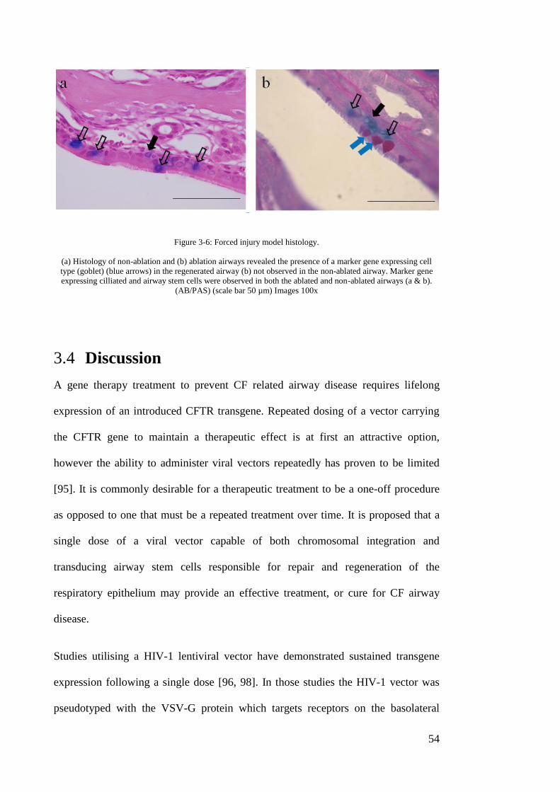
54
Figure 3-6: Forced injury model histology.
(a) Histology of non-ablation and (b) ablation airways revealed the presence of a marker gene expressing cell
type (goblet) (blue arrows) in the regenerated airway (b) not observed in the non-ablated airway. Marker gene
expressing cilliated and airway stem cells were observed in both the ablated and non-ablated airways (a & b).
(AB/PAS) (scale bar 50 µm) Images 100x
3.4 Discussion
A gene therapy treatment to prevent CF related airway disease requires lifelong
expression of an introduced CFTR transgene. Repeated dosing of a vector carrying
the CFTR gene to maintain a therapeutic effect is at first an attractive option,
however the ability to administer viral vectors repeatedly has proven to be limited
[95]. It is commonly desirable for a therapeutic treatment to be a one-off procedure
as opposed to one that must be a repeated treatment over time. It is proposed that a
single dose of a viral vector capable of both chromosomal integration and
transducing airway stem cells responsible for repair and regeneration of the
respiratory epithelium may provide an effective treatment, or cure for CF airway
disease.
Studies utilising a HIV-1 lentiviral vector have demonstrated sustained transgene
expression following a single dose [96, 98]. In those studies the HIV-1 vector was
pseudotyped with the VSV-G protein which targets receptors on the basolateral

55
surface of airway epithelial cells [103]. As these VSV-G receptors reside on the
basolateral surface of epithelial cells, access to the HIV-1 VSV-G vector to the
receptors is restricted. To allow access to the basolateral surface, Limberis et al and
Stocker et al utilised a pre-treatment of LPC to transiently open the tight junctions
present between cells allowing the vector access to the basolateral cell surface [96,
98]. The ability to access the basolateral cell surface following a pre-treatment with
LPC also provides the opportunity to gain access to the airway stem cells that reside
on the epithelial basement membrane. Taken together, access to airway stem cells
following pre-treatment with LPC and the ability of the HIV-1 VSV-G vector to
transduce dividing cells (airway stem cells) may be responsible for the sustained
gene expression reported in both of these studies.
By using a PDOC forced injury model in the nasal and tracheal airways the airway
epithelium can be ablated to assess the fate of potentially transduced airway stem
cells.
Cells expressing LacZ were present following one ablation and two ablations and the
subsequent epithelium regeneration in the nasal and tracheal airways. By comparing
the pattern of LacZ expression to that present in the 7 days controls following pre-
treatment and gene transduction, a different pattern of expression was observed,
providing clues to the underlying mechanisms of sustained gene expression. The
speckled pattern of LacZ expressing cells seen in the 7 day control was clearly
different to the clustering of LacZ expressing cells in the epithelial forced injury and
regeneration animals. The clustered appearance of LacZ expressing cells is
consistent with a pattern of clonal expansion whereby transduced airway stem cells
have passed the marker gene onto their progeny as they have differentiated during

56
the epithelium repair process.
Importantly, clusters of LacZ expressing cells were also observed following two
PDOC treatments of the epithelium demonstrating that the clusters observed from
the single ablation were unlikely to be a consequence from incomplete stripping of
the epithelium during the ablation treatment
The pattern of LacZ expression observed in the long term (14 week) control group
that did not receive PDOC, displayed both speckled and clustered LacZ- expressing
cells. This finding is consistent with the speckled LacZ positive cells representing
terminally differentiated cells that had been transduced at the time of vector
treatment and the clustering of LacZ positive cells being progeny of airway stem
cells that were transduced at the time of vector treatment.
Further evidence supporting airway stem cells passing the transgene on to their
progeny can be seen in Figure 3-6 which shows that in the no ablation treatment
group the presence of both LacZ expressing airway stem cells and ciliated cells.
Following the regeneration of the airway epithelium after the ablation treatment the
same LacZ expressing cells types were present; however, there was also the presence
of mucin secretory cells expressing the marker gene. This suggests that transduced
nasal airway stem cells have passed the marker gene to all their progeny upon
differentiation, including those not targeted by the cell specific VSV-G pseudotype.
Clonal expansion from airway stem cells transduced with the LacZ marker gene was
predicted to produce symmetrical LacZ positive clusters of cells upon epithelial
repair and regeneration. These clusters were anticipated as the newly differentiated
cells expanded out from the transduced parent airway stem cell (Figure 3-7) as
previously reported [95].

57
Figure 3-7: Pattern of clonal cluster.
Illustration demonstrating the outgrowth of differentiated cells from a transduced stem cell forming a
symmetrical cluster. Adapted from Mitomo et al [95]
In the single and double ablation groups in both the nasal and tracheal airways, the
clustering of LacZ positive cells was observed and confirmed. Unexpectedly though,
in conjunction with the observation of symmetrical clustering of LacZ positive cells
there were also linear clusters of cells present. There are several potential causes for
the two distinct patterns of clonal expansion observed following repair and
regeneration. Firstly, the pattern may have been a result from the fluid dynamics of
the introduced doses during the instillation of the pre-treatment and vector. It is
feasible that the nature of fluid delivery caused the vector to transduce airway stem
cells along a linear track resulting in the pattern observed. However, if true the
clusters within the linear expansion should be of approximately the same size as
those in the symmetrical clusters, not smaller as observed. Secondly, epithelial
disturbance caused by the ablation treatment could result in an altered airway
cytokine expression leading to an abnormal linear outgrowth of cells from the airway
stem cell parent.

58
Alternatively the two distinct patterns may be a result of the natural process of
epithelial repair/regeneration. To the authors’ knowledge these distinctively different
clonal patterns have not been reported in airway stem cell literature and further
studies are warranted to elucidate the mechanism(s) behind the differences, as this
may gain further insight into the nature of airway stem cell repair and regeneration.
This demonstration of airway stem cell transduction in the airway epithelium
provides a foundation that may be built on with further studies. Targeting the airway
stem cell compartment may be a viable and promising option for elevating the level
of sustained CFTR expression. One option is to consider the integrin profile of the
airway stem cell, specifically integrins αvβ5 and α5β1 to target binding by the
lentiviral vector and thereby enhancing transduction events. The integrins αvβ5 and
α5β1 act as receptors for proteins that contain the Arg-Gly-Asp (RGD) attachment
site [104].
Studies incorporating the integrin-targeting peptide RGD into vector envelopes have
shown encouraging results. Morizono et al demonstrated increased transduction of a
lentiviral vector in human umbilical vein endothelial cells following insertion of the
RGD peptide into envelope proteins [105]. In addition they demonstrated that the
RGD peptide could be incorporated into the lentivirus envelope without interfering
with the VSV-G pseudotype [105], a possibility suited to the LV vector used in these
thesis studies. Another group have used nanocomplexes containing the RGD peptide
to target tumours, resulting in a 2 fold increase in transduction and also achieving
delivery into the lungs [106]. Studies investigating the use of the RGD peptide to
target specific integrins are encouraging. A detailed study designed to assess the
ability of the HIV-1 VSV-G pseudotyped lentiviral vector to target airway stem cells

59
using this approach is an encouraging option for future studies.
While the proof-of-principle for sustained transgene expression has been presented
in this chapter, there are a number of considerations to address. There has been
substantial progress in the understanding of airway stem cell development, repair,
and regeneration; however, there still remains an unknown factor involved that may
impact the targeting of airway stem cells in a gene therapy setting.
The notion of targeting airway stem cells for sustained gene expression relies on the
premise that a transduced airway stem cell will remain continuously throughout the
lifetime of the patient, constantly passing on the inserted gene to its progeny upon
epithelial repair. There are two models of tissue self-renewal that have been
proposed that have yet to be resolved in the airways and depending on which model
is correct will have an impact on the targeting of airway stem cells. The two
proposed models are the division asymmetry model and the population asymmetry
model. The division asymmetry model proposes that as stem cells divide they give
rise to one stem cell (self-renewal) and one differentiated cell which may be
terminally differentiated such as ciliated cell, secretory cell or a differentiated
progenitor cell further down the stem cell hierarchy such as a Club cell. The
important feature of this model is that a transduced stem cell would pass on its
transgene through self-renewal; hence a transduced stem cell would potentially be
present for life. The population asymmetry model proposes that a portion of stem
cells in a niche will differentiate without self-renewal and the remainder will divide
symmetrically to renew those that are lost. In this model, there is a chance that a
transduced stem cell is lost through neutral drift and is replaced with a stem cell not
carrying the transgene of interest.
60
To determine if the division population asymmetries model and neutral drift is a
limiting factor in airway stem cell targeting for sustained gene expression, further
studies are warranted. It is proposed that by transducing airway stem cells with the
LV vector carrying a fluorescent gene the flow cytometric airway stem cell isolation
technique presented in Chapter 4 may be used to provide insight into the question of
neutral drift. Flow cytometry could be used to ascertain and quantify the number of
airway stem cells expressing the fluorescent protein compared to the total number of
airway stem cells sorted. This procedure could be performed over time points
representing the life span of mice, to determine if the proportion of transduced stem
cells changes over time. If the proportion remains stable over time it would suggest
that neutral drift is not occurring, and that the division asymmetry model is more
likely to explain the dynamics of airway stem cell activity.
3.5 Conclusion
The observations reported in this chapter support the notion that a HIV-1 VSV-G
pseudotyped lentiviral vector when delivered with a pre-treatment of LPC can
transduce airway stem cells. By utilising the ability of airway stem cells to pass on
the transgene to their progeny upon differentiation, sustained transgene expression
can then be produced without further airway gene transfer events.
.

61
4 Airway stem cells and epithelial
remodelling in the conducting
airways of CF mice
4.1 Introduction
Epithelial remodelling of the conductive airways in those afflicted with CF is poorly
understood and a better understanding of the effect of CF on the epithelial phenotype
is warranted. A more concise understanding of epithelial remodelling will potentially
allow for the further development of approaches such as gene therapy to correct
airway disease associated with CF.
Airway stem cell hyper proliferation in the CF airway has been reported [107, 108],
Leigh et al showed the presence of cell hyper proliferation in CF compared to
normal airways [107] and Voynow et al extended this finding to propose that airway
stem cells were the responsible proliferating cell population [108]. However, the
methods used to ascertain the state of cell proliferation in the CF airways have been
restricted to antigen labelling, such as a proliferating cell nuclear antigen (PCNA)
[107, 108]. Similarly, identification of proliferating cell types has been restricted to
morphological and histological analysis [108]. There remains uncertainty as to
whether an increase in respiratory cell proliferation is due to; (1) an increase in
proliferation of the normal airway stem cell population, (2) is reflective of
hyperplasia of the airway stem cell population, or (3) a combination of both.
The recent development of definitive cell surface markers now enables isolation of
respiratory airway stem cells via flow cytometry, and an ability to subsequently
measure their proliferative potential of specific subsets via a clonogenic colony

62
forming assay [101]. This approach provides for the first time a method to quantify
and compare the airway stem cell compartment in CF and normal mice.
The pseudostratified epithelium of the murine tracheobronchial region is not fully
reflective of that observed in humans: while both are comprised of airway stem cells,
secretory cells and ciliated cells [109], goblet cells in mice are confined to the most
proximal laryngeal region of the trachea, and to the nasal airways [110].
Nevertheless there remains sufficient similarity in cell types in humans and mice to
allow the mouse animal model to assist in elucidating the potential presence of
airway stem cell hyperplasia in the conducting airways of CF mice.
In addition to potential airway stem cell hyperplasia in the epithelium of CF airways,
goblet cell hyperplasia has also been previously studied [32, 111]. In vitro studies
assessing goblet cell hyperplasia in the differentiated epithelium of CF versus non
CF airways have concluded there was no significant difference in goblet cell
numbers [112, 113]. In contrast, in vivo studies have suggested goblet cell
hyperplasia is present in the nasal airways of CF mice [32, 111].
It is currently unknown if the goblet cell hyperplasia in the airway of CF mice is
present in conjunction with airway stem cell hyper proliferation and potential
hyperplasia. Gaining a better insight into the possible dual presence of hyperplasic
goblet cells and stem cells in the CF airways will provide a better understanding of
the targeted epithelial environment which is the target of CF gene therapy.
In this chapter the remodelling of the CF respiratory epithelium was observed,
quantified and compared across CF knockout mice and normal mice, using cell
counts, histochemical analysis, and AB/PAS staining. An airway stem cell
proliferation index was calculated using the staining of an antigen (KI-67) that is

63
only present in dividing cells. The number of airway stem cells and their
proliferative capacity in CF knockout mice was compared to that in normal mice
using flow cytometry and clonogenic assays.
4.2 Material and Methods
Three genotypes of mice from 2 colonies of CF mice were used in this study: (1)
Cftrtm 1 unc-/ -
and their litter mate controls cftrtm 1 unc+/-
(C57BL/6 background [114],
termed CF UNC and UNC HET respectively). (2) Cftrtm 1 unc
Tg(FABPCFTR)
-/-
(mixed
background [115], termed CF FABP) mice were bred from (-,-)homozygote’s mating
and therefore no littermates controls are produced. When comparison between CF
FABP mice and non CF controls was required cftrtm 1 unc+/-
mice (UNC het) were
used. All animals were anaesthetised using a mixture of 10 μl/g body weight of
Domitor, 0.1 mg/ml, (Orion Corporation, Finland) and Ketamine, 7.6 mg/ml,
(Parnell Laboratories Aust Pty Ltd), given as a single dose.
Animals were humanely killed via CO2 inhalation. Tracheal tissue used for
histochemistry and immuno-histochemistry were removed and fixed in 10% neutral
buffered formalin (NBF). Mouse heads (for nasal airways) and trachea used to
provide cells for flow cytometric analysis were removed from the body; the fur and
skin was removed from heads and both tissue samples immediately washed with
sterile PBS and placed into PBS with 2% glucose. Nasal septa were dissected free of
the nose while submerged in PBS 2% glucose. Mouse head’s used for counts of
nasal airway goblet cell and airway stem cell numbers via immuno-histochemistry
4.2.1 Mouse models
4.2.2 Processing of tissue

64
were removed as above and placed into 2% (w/v) paraformaldehyde/0.5%
glutaraldehyde (v/v) in PBS on ice for 2 hours, transferred to 10% neutral buffered
formalin (NBF) for 24 hours before being decalcified in 1.5% (w/v) EDTA, 7% (v/v)
HCL for 22 hours. Decalcified tissue was then rinsed in running water for 30 mins
and stored in 70 % (v/v) ethanol. Following initial processing and decalcification of
the head, the jaw was removed exposing the upper palate and gross sections made in
locations at levels 6, 16, 24, and 30 described by Mery et al [116]. The four gross
tissue sections were embedded in paraffin wax and 5 µm sections cut and mounted
on glass microscope slides. A proliferation index was established via Ki-67 immuno-
histochemistry in trachea were dissected from mice immediately following humane
killing and subsequently placed into 10% NBF overnight.
Airway stem cells were FACS sorted from the processed respiratory airway
epithelium of trachea to establish the incidence of this cell type in CF UNC, CF
FABP and CF HET mice as described in Methods section.
Cell culture for clonal assays to gain the growth fraction of airway stem cells were
performed in matrigel as described in Methods section.
To assess airway remodelling: (1) Nasal airway sections were stained with AB/PAS
reagent, to identify goblet cells, quantify their numbers, measure the size of the
mucin-stained cells and assess mucin acidity. (2) To establish a proliferation index
4.2.3 Tracheal and nasal septum cell preparation and flow
cytometry
4.2.4 Cell culture
4.2.5 Histochemistry and Immuno-histochemistry

65
within the tracheal stem cell compartment, a nuclear proliferation antigen Ki-67
(MIB-1 clone) that is only present in cycling cells was employed.
All comparisons between CF and controls were analysed for statistical significance
using an unpaired t test with significance being set at p < 0.05.
4.3 Results
Goblet cell hyperplasia and hypertrophy was assessed through AB/PAS staining in
the nasopharyngeal airways sections from region 30 [116], were the airway is tubular
and lined with ciliated respiratory epithelium. When standardised to goblet cells per
10 µm of nasal respiratory epithelium, CF UNC
(6.6 ± 0.3 goblet cells) (n=5) showed
a statistically significant (p<0.001, t-test) 2.02 fold increase compared to UNC het
controls (3.2 ± 0.2) (n=5) (Figure 4-1). Goblet cell hypertrophy was also
significantly increased (p<0.001, t-test) in CF UNC (3.5 ± 0.548 µm2) (n=5)
compared to controls (1.6 ± 0.115 µm2) (n=5) that resulted in a 2.2 fold increase
(Figure 4-1).
4.2.6 Statistics
4.3.1 Goblet cell hyperplasia and hypertrophy in CF mouse nasal
airways

66
Figure 4-1: Goblet cell hyperplasia and hypertrophy.
(a) Goblet cell hyperplasia and hypertrophy in the non CF airway compared to the (b) CF UNC airway was
statistically significant (****p< 0.0001) and (**p < 0.01) respectively. (AB/PAS) (Scale bar 50µm). Sections
show typical examples from the same upper region of the naso-pharynx in normal and CF mice.
Histological examination of AB/PAS stained section revealed that the non CF mice
had a more acidic mucin content (Figure 4-2 a) than the mucin content of the CF
goblet cells (Figure 4-2 b).
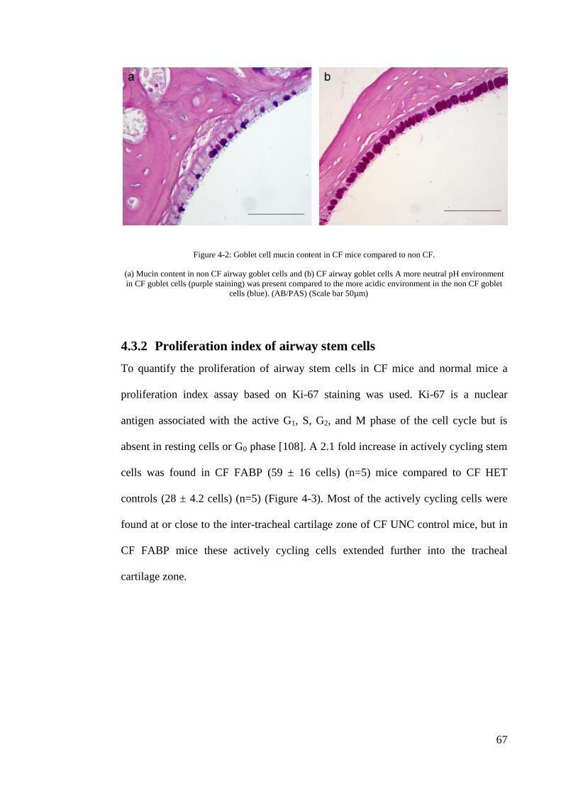
67
Figure 4-2: Goblet cell mucin content in CF mice compared to non CF.
(a) Mucin content in non CF airway goblet cells and (b) CF airway goblet cells A more neutral pH environment
in CF goblet cells (purple staining) was present compared to the more acidic environment in the non CF goblet
cells (blue). (AB/PAS) (Scale bar 50µm)
To quantify the proliferation of airway stem cells in CF mice and normal mice a
proliferation index assay based on Ki-67 staining was used. Ki-67 is a nuclear
antigen associated with the active G1, S, G2, and M phase of the cell cycle but is
absent in resting cells or G0 phase [108]. A 2.1 fold increase in actively cycling stem
cells was found in CF FABP (59 ± 16 cells) (n=5) mice compared to CF HET
controls (28 ± 4.2 cells) (n=5) (Figure 4-3). Most of the actively cycling cells were
found at or close to the inter-tracheal cartilage zone of CF UNC control mice, but in
CF FABP mice these actively cycling cells extended further into the tracheal
cartilage zone.
4.3.2 Proliferation index of airway stem cells

68
Figure 4-3: Ki-67 analysis of CF FABP trachea airway epithelium.
(a) Ki-67 analysis in CF control and (b) CF FABP trachea airway sections (c) showed a statistically greater
number of proliferating cells positive for Ki-67 (black arrows) in the CF FABP mice (*p < 0.05). t test, (scale
bar 50 µm)
To assess and quantify the relative abundance of airway stem cells within the
respiratory stem cell compartment, a twostep flow cytometry cell sorting and
clonogenic assay was employed (see methods section 2.2.7) in 2 separate studies.
To enable comparisons within each study and between the two studies it was
necessary to normalize the data by calculating the relative abundance of airway stem
cells obtained by the colony forming efficiency (Table 2: Study 1, relative
abundance of isolated airway stem cells and their colony forming efficiency
normalized. (Table 3: Study 2, relative abundance of isolated airway stem cells and
their colony forming efficiency normalized.
4.3.3 Stem cell hyperplasia in the respiratory epithelium

69
Table 2: Study 1, relative abundance of isolated airway stem cells and their colony forming efficiency
normalized.
Sample Relative abundance
(stem cells) x
Colony forming
efficiency =
Normalized
UNC HET 36.0 0.38 13.68
CF UNC 24.0 2.30 55.2
CF FABP 27.0 0.97 26.19
Table 3: Study 2, relative abundance of isolated airway stem cells and their colony forming efficiency
normalized.
Sample Relative abundance
(stem cells) x
Colony forming
efficiency =
Normalized
UNC HET 33.6 0.62 20.83
CF UNC 43.5 0.67 29.14
CF FABP 29.4 0.65 19.11
Study one, showed that the level of functional airway stem cells present was
increased in CF mice. CF UNC mice and CF FABP displayed a 4.0 and 1.9 fold
higher levels of respiratory epithelium stem cells respectively when compared to the
controls (Figure 4-4a). In study two, CF UNC mice displayed a 1.4 fold higher level
of respiratory epithelium stem cells whilst CF FABP displayed no increase in airway

70
stem cells when compared to the controls (Figure 4-4 b).
Figure 4-4: Quantification of airway stem cells.
The incidence of colony forming airway stem cells in UNC Het (normal), CF UNC, and CF FABP mice.
Normalised for relative abundance x colony forming efficiency to quantify stem cells.
A retrospective assement of the available CF mice in study1 and study 2 revealed a
statistical significant difference in ages, with the CF mice used in study 1 having an
average of 7 months and study 2 the mean average was 4 ½ months of age (Figure
4-5).
4.3.4 Retrospective assessment of the ages of mice in the airway
stem cell study

71
Figure 4-5: Average age of mice in the two airway stem cell studies
Analysis of the average age of mice in Study 1 compared to Study 2 showed there was a significant difference in
average age between the two groups (**p <0.005, n=??). t test.
4.4 Discussion
There have been few studies characterising the conducting respiratory epithelium of
the nasal and tracheal pathways in CF mice since their creation in the late 1990’s.
This is surprising as the nasal compartment of CF mouse models share an
electrophysiological and histological profile which is similar to the epithelium of the
CF nasal and lower airways in humans [117] and has been the preferred site for
analysis of CFTR dysfunction in CF mice. The tracheobronchial epithelium in mice
also shares some similarities with the same airway region in humans [109] although
it does not share CF pathophysiological characteristics [118]. These similarities in
airway phenotype have allowed researchers to explore and assess potential new
therapies for CF airway disease, such as gene and cell therapies [119, 120].
It is unexpected that there have not been more studies in this area considering the
implications from the findings of the few studies that have been published. Although
goblet cell hyperplasia and airway remodelling in CF mice was first reported in 2001

72
and correlated to inflammation and infection of the respiratory airway [32], it was
not until 2007 that goblet cell hyperplasia was independently reported in a CF animal
model by Hilliard et al [111].
Prior to assessing the level of airway stem cell remodelling in the CF airways in this
project it was important to verify if there was indeed a hyperplasia of airway goblet
cells. This would allow for a more accurate assessment of relative influence of the
stem cell compartment in a model with airway cellular phenotype changes similar to
those seen in human CF airway disease.
In these thesis studies, the goblet cell hyperplasia in CF mice compared to normal
mice (Figure 4-1) which is similar to that reported by Tarran et al who observed by
morphometric comparison a 2-fold change in goblet cell numbers [32]. Although,
Hilliard and colleagues demonstrated that CF goblet cell hyperplasia may occur
independently from infection and inflammation [111], this conflicted with earlier
reports that associated goblet cell hyperplasia in both the human airway [121] and
CF mouse airways [32] with infection and inflammation.
In the current study, goblet cells in CF mice compared to controls displayed a 2.2
fold increase in the mucin-stained area within the goblet cells. This hypertrophy,
together with the 2.0 fold increase in the number of goblet cells, represent a 4.4 fold
increase in overall presence of mucin potentially able to be secreted into the airways.
The mucin within the CF airway goblet cells displayed a neutral pH, compared to the
more acidic pH in non-CF airway goblet cells, as revealed by AB/PAS staining
(Figure 4-2). This finding may reflect differences in key mucins that are found in the
airway. Secretory cells in the mouse airway are known to express the mucin Muc5b
at baseline conditions and these cells are a constitutive element of the homeostatic

73
mucosal defence [122]. Another mucin, Muc5a is up regulated in response to
inflammation [34, 122] providing a possible explanation behind the change in mucin
acidity. To clarify if an up-regulation of Muc5a produces the change in mucin
acidity observed in the airway epithelial cells in CF mice, future studies should
compare the levels of Muc5a mRNA in the goblet cells of the airway epithelium
between non-CF and CF mice.
A different insight into the reasons for goblet cell hyperplasia in the respiratory
epithelium of the CF airway may be provided by analysis of the airway stem cell
compartment.
Studies that have examined the hyperplasia of the airway stem cell compartment in
CF airways are limited. Hajj et al [112] have reported a 1.6 fold increase in
proliferating airway stem cells in an airway epithelia regeneration study, using a
model of humanized airway xenografts in nude mice. In addition, Voynow and
colleagues [108] demonstrated a 5 fold increase in airway stem cells proliferation in
human CF conducting airways compared to the normal airway. However, it is
unclear if these rises in proliferative cells reflected an increase in the cell cycling rate
in a normal airway stem cell population, or an increase in the total number of
resident airway stem cell population undergoing cycling.
Until recently the analysis of airway stem cell pathophysiology in the CF airway
conducting epithelium has lacked definitive assays to isolate, quantify, and index
their proliferative capacity. The development of a robust airway stem cell sorting and
clonal assay strategy to isolate and quantify the sub population of basal cells that act
as stem cells in the respiratory airways of mice [101] was utilised in the studies
presented here.

74
The hyperplasia of airway stem cells present in both CF mouse models at an average
of age 7 months (Figure 4-4) which was not present in the CF FABP mice and
substantially lower in CF UNC mice at an average age of 4.5 months indicates that
the degree of hyperplasia maybe age/development dependent.
These preliminary data suggests that the airway stem cells of the conducting
respiratory epithelium of the two CF mice models may not be hyperplasic at an early
age but develops at a later stage in life. In addition, the statistically greater level of
the airway stem cell hyperplasia observed in the CF FABP mice was correlated to a
statistically different 2.1 fold increase in an actively cycling pool of airway stem
cells. Together, the increase in proliferating cells correlates to the increase in colony
forming airway stem cell numbers in CF FABP mice, suggesting that the increase in
airway stem cell proliferation previously reported may be representative of an
actively cycling hyperplasic stem cell population present in the CF airway.
The etiology of airway stem cell hyperplasia in the CF airway is unknown. The stem
cell hyperplasia found in CF mice may be related to the hyperplasia of goblet cells in
CF airways
It has been proposed that CFTR may function to regulate cell differentiation in the
developing airway [111, 123], and the paradigm these authors proposed of a
regulatory cascade, dependant on CFTR in the development and repair of the
respiratory epithelium is logical. However, no further evidence to support this notion
has appeared and the proposition is not supported by some of the knowledge gained
from studies involving a mouse model that over expresses the epithelium sodium
channel (ENaC). In those mice increased airway epithelium sodium absorption
produces pathological changes that parallel those seen in CF, including severe

75
spontaneous lung disease, goblet cell metaplasia, neutrophilic inflammation and poor
bacterial clearance [124]. One possible effect of this over expression of ENaC in an
environment of an otherwise properly functioning CFTR channel, is that the elevated
absorption of sodium can interrupt development of a normal airway epithelium
and/or the epithelial repair cascade in airways that does not depend on CFTR
dysfunction. It also suggests that elevated sodium absorption is more likely
responsible for the initiation of CF-like lung disease, and importantly for these
studies it may also underlie the ensuing respiratory airway remodelling such as
goblet cell hyperplasia.
This elevated sodium absorption is associated with reduction in ASL depth, leading
to poor mucus clearance and raised concentrations of neutrophil elastase [124] as
occurs in CF. Neutrophil elastase has been linked to the release of pro inflammatory
cytokine interleukin (IL)-8 which can support a self-perpetuating inflammatory
process as the neutrophils present release neutrophil elastase into the airway
epithelium to release IL-8, which then recruits more neutrophils to the airway
surface [125], and so on.
Pro inflammatory cytokines such as IL-8 may also play a role in deregulating the
Notch signalling pathway and its involvement in respiratory airway epithelium
remodelling. The Notch pathway is an evolutionarily conserved signalling pathway
that plays a role in integrating information cues from the extracellular environment
to the cellular interior to enable appropriate developmental and physiological
responses [126].
In the process of differentiation from airway stem cells into specific lineages, the
Notch signalling pathway is important in determination of cell fate [127]. When the

76
Notch pathway is activated in the mature and developing lung epithelium the cell
fate is pushed towards an increase in mucus cell production and a decrease in ciliated
cell production [128] (Figure 4-6). This suggests that the Notch pathway could play
an important part in the modelling of the lung epithelium in development. Similarly
it also plays a role during repair of the mature lung epithelium [128].
Figure 4-6: Schematic model of how signalling can effect airway stem cell (basal cell) behaviour.
A model depicting how signals including IL-8 influence airway stem cells (basal cells) and their progeny through
interaction with the Notch signalling pathway [110].
The Notch pathway is modulated in part by signalling which includes cytokines such
as IL-6 and IL-8 [110]. Expression mediators including cytokine IL-8 are altered in
the CF airway and associated with a delay in repair [112]. As the Notch pathway
plays a part in cell fate upon differentiation from airway stem cells, and activation of
the Notch pathway suppresses the differentiation into ciliated cells [127], it may also
lead to augmented self proliferation of airway stem cells.
An observation made during this study was that there was a statistically significant
higher number of tracheal cartilage rings over a given distance in the proximal

77
trachea for normal mice, compared to CF UNC mice (see Appendix A). Whilst the
tracheal ring observations were not the main focus of this study, it is already a
known that tracheal ring remodelling is associated with decreased contractile
responses from the smooth muscle surrounding the proximal trachea in both adult
and new born CF mice [36]. Those structural and functional abnormalities could be
viewed as a congenital malformation related to CFTR channel dysfunction during
tracheal development. That is, the lack of properly functioning CFTR in
development may contribute to intrinsic defects in the epithelial progenitor cell pool.
If so, this could lead to dysregulation affecting cross-talk between the progenitors
and the mesenchymal compartment during development, resulting in the changes
observed in this study.
4.5 Conclusion
This study showed the presence of goblet cell hyperplasia in the respiratory
epithelium of the CF mouse nasopharyngeal airway. Additionally, remodelling of the
respiratory epithelium of the CF airway was found to extend to the airway stem cell
compartment, with an increased proportion of actively-cycling hyperplasic airway
stem cells present in CF mice at 7 months of age. These initial findings support the
need for further studies able to elucidate the origins of pathogenic remodelling of the
respiratory epithelium of the CF airway.

78

79
5 Airway gene transfer and stem cell
transduction by a VSV-G
pseudotyped HIV-1 lentiviral vector
in the Ferret (Mustela putorius furo)
and common Marmoset (Callithrix
jacchus)
5.1 Preface
The development of new therapeutic approaches, including gene therapy provide an
exciting new modality which holds great promise for a novel treatment for CF
airway disease in the future. However, prior to embarking on clinical trials the safety
and efficacy of gene therapy must be explored in pre clinical animal models. To this
end the CF ferret provides an animal model which far better recapitulates the
respiratory phenotype than that of the transgenic CF knockout mouse models widely
used in CF studies to date. Additionally, the common marmoset provides a primate
animal model which is evolutionarily close to humans, allowing for a transitional
step between mouse models and human clinical trials. This chapter is presented in
two parts: Part A details a peer reviewed published study [129] on LV transduction
in the airway of normal ferrets in preparation for future CF ferret trials and Part B
presents findings from a peer reviewed published study [130] demonstrating the
success of LV transduction in the airways of the common marmoset.

80
5.2 PART A: The Ferret (Mustela putorius furo)
The CF mouse has provided an animal model which has been useful in establishing
proof of principle and a solid foundation for gene therapy development for CF.
However, the absence of CF disease pathophysiology in the tracheobronchial region
and distal parts of the lung in mice has restricted the value of this animal model. In
recent years animal models have been developed that more accurately reproduce the
CF disease phenotype in the respiratory airways observed in humans.
Amongst the new generation of CF animal models is the CFTR knockout ferret
which exhibits many characteristics homologous to the human CF disease
phenotype, including pancreatic, liver, and vas deferens disease; variably-penetrant
meconium ileus; defective chloride transport, submucosal gland fluid secretion, and
a propensity towards lung infection in the early post natal period [131]. The
similarities between CF humans and CF ferrets in their respiratory pathophysiology
provides a new animal model to build upon the foundation work achieved to date in
the nasal conductive airways of CF mice. At present there is no CF ferret colony in
Australia or the Southern Hemisphere and prior to embarking on the lengthy process
of gaining permission to import the CF ferrets and establishing a colony, a pilot
study was performed to assess delivery parameters and the effectiveness of an LPC
pre-treatment and HIV-1 VSV-G pseudotyped lentiviral vector gene transfer
protocol in the airways of normal ferrets.
Part A of this chapter presents the results of a pilot study in which normal ferrets
were pre-treatmed with LPC followed by dosing of the HIV-1 VSV-G pseudotyped
lentiviral vector using the LacZ marker gene to reveal the resulting gene expression.

81
This study is presented as published in BMC Pulmonary Medicine:
Cmielewski, P*., Farrow, N*., et al., Transduction of ferret airway epithelia using a
pre-treatment and lentiviral gene vector. BMC Pulm Med, 2014. 14(1): p. 183. [129]
* Cmielewski, P. and Farrow, N. Were equal first author contributors.


84
Transduction of ferret airway epithelia using a pre-
treatment and lentiviral gene vector
Patricia Cmielewski1,2,*,†
Email: [email protected]
Nigel Farrow1,2,3,†
Email: [email protected]
Martin Donnelley1,2,3
Email: [email protected]
Chantelle McIntyre1,3
Email: [email protected]
Jahan Penny-Dimri3
Email: [email protected]
Tim Kuchel4
Email: [email protected]
David Parsons1,2,3
Email: [email protected]
1 Respiratory and Sleep Medicine, Women’s and Children’s Hospital, 72 King
William Road, North Adelaide, SA 5006, Australia
2 Centre for Stem Cell Research, University of Adelaide, Adelaide, SA 5001,
Australia
3 School of Paediatrics and Reproductive Health, University of Adelaide, Adelaide,
SA 5001, Australia
4 South Australian Health and Medical Research Institute, Gilles Plains, SA 5086,
Australia
* Corresponding author.
† Equal contributors.

85
5.3 Abstract
The safety and efficiency of gene therapies for CF need to be assessed in pre-clinical
models. Using the normal ferret, this study sought to determine whether ferret
airway epithelia could be transduced with a lysophosphatidylcholine (LPC) pre-
treatment followed by a VSV-G pseudotyped HIV-1 based lentiviral (LV) vector, in
preparation for future studies in CF ferrets.
Six normal ferrets (7 -8 weeks old) were treated with a 150 μL LPC pre-treatment,
followed one hour later by a 500 μL LV vector dose containing the LacZ transgene.
LacZ gene expression in the conducting airways and lung was assessed by X-gal
staining after 7 days. The presence of transduction in the lung, as well as off-target
transduction in the liver, spleen and gonads, were assessed by qPCR. The levels of
LV vector p24 protein bio-distribution in blood sera were assessed by ELISA at 0, 1,
3, 5 and 7 days.
The dosing protocol was well tolerated. LacZ gene expression was observed en face
in the trachea of all animals. Histology showed that ciliated and airway stem cells
were transduced in the trachea, with rare LacZ transduced single cells noted in lung.
p24 levels was not detectable in the sera of 5 of the 6 animals. The LacZ gene was
not detected in the lung tissue and no off-target transduction was detected by qPCR.
5.3.1 Background
5.3.2 Methods
5.3.3 Results
5.3.4 Conclusions

86
This study shows that ferret airway epithelia are transducible using our unique two-
step pre-treatment and LV vector dosing protocol. We have identified a number of
unusual anatomical factors that are likely to influence the level of transduction that
can be achieved in ferret airways. The ability to transduce ferret airway epithelium is
a promising step towards therapeutic LV-CFTR testing in a CF ferret model.
Ferret, Lung, Lentivirus, Cystic fibrosis, Gene therapy
5.4 Background
Cystic Fibrosis (CF) is an autosomal recessive disorder caused by a mutational error
in the Cystic Fibrosis Transmembrane Conductance Regulator (CFTR) gene and its
associated protein [132]. The CFTR channel is used in multiple body systems that
utilise epithelial ion gradients, including but not limited to the pancreas, sweat
glands, gastrointestinal tract and most crucially the lungs [132]. The resultant
dehydration of the airway surface, loss of cilia movement, and accumulation of
highly viscous mucous obstructs the airways and hinders clearance, thereby
promoting bacterial infection [133]. CF airway disease currently limits the life
expectancy of a CF patient to a predicted median survival estimate of approximately
37 years [134]. Therefore, the successful treatment of CF airway disease, especially
from the earliest stages of life, is imperative in improving short term and long term
survival of those afflicted with CF.
Gene therapy for a loss-of-function disease like CF involves delivering the
appropriate corrective DNA into the cells of an organism to produce adequate
functional protein to ameliorate the symptoms of that disease. Lentiviral gene
vectors have a number of potential benefits, including the ability to transduce both
5.3.5 Keywords

87
dividing and non-dividing cells, be pseudotyped with an envelope glycoprotein to
allow for broad tissue tropism [135] and have the ability to carry large genes such as
CFTR [136].
Amongst the new generation of CF animal models, the CFTR knockout ferret
exhibits many characteristics homologous to human CF, including disease of the
pancreas, liver, vas deferens; and variably-penetrant meconium ileus . The
similarities between CF humans and CF ferrets in their respiratory pathophysiology
include defective chloride transport, submucosal gland fluid secretion, and a
propensity towards lung infection in the early post natal period [131].
The aim of this study was to expand work done in the mouse, sheep and marmoset
[99, 130, 137] and test the ability of our two-step LPC pre-treatment and lentiviral
vector delivery system to transduce airway cells of the adolescent normal ferret. The
rationale for this work was to prepare for the use of CF ferrets, by establishing
dosing, technical, and analytical procedures to be applicable for future use in these
expensive and husbandry-intensive disease-specific CF animals.
5.5 Methods
This study was approved by the Animal Ethics committees of the Women’s and
Children’s Health Network, South Australia; SA Pathology, South Australia; and the
University of Adelaide, South Australia. Six 7-8 week old adolescent ferrets Mustela
spp (2 male and 4 female, weight 335 to 460 g) were sourced from a commercial
ferret supplier. Experiments and subsequent monitoring were performed under
specialist veterinarian supervision at the Gilles Plains South Australian Medical
Research Facility.

88
The nuclear-localised LacZ (LacZnlsco) VSV-G pseudotyped HIV-1 based LV
vector was produced by transient transfection of 293T cells using a five plasmid
system according to previously published protocols [99, 138]. The ratio of plasmids
used for all virus preparation (per 245 mm square plate) was 170 μg of the transgene
1SDmMPSV LacZnlsco, 3.16 μg of pcDNA3 Tat, 3.16 μg of pHCMVRev, 1 μg
pHCMVgagpol, 7.9 μg pHCMV-G.
For purification approximately 1 L of vector supernatant was collected, and passed
through a 0.45 μm filter (Whatman Polydisc AS, GE Healthcare, PA, USA) and then
two MustangQ Acrodiscs (Pall Corporation, NY, USA) connected in series, at a flow
rate of 10 ml/min. After loading, the Acrodiscs were immediately flushed with 30 ml
of PBS. The viral particles were then eluted with 4 ml of 1.5 M NaCl into a sterile
tube containing an equal volume of 2% (v/v) mouse serum in H2O. The virus was
concentrated by ultracentrifugation at 20,000 rpm (53,750 x g) for 90 minutes at 4°C
and then resuspended in 6×100 μl aliquots of 0.9% (w/v) NaCl /0.1% (w/v) mouse
serum albumin and stored at -80 °C. A second batch was made in the same manner
and added to the first without thawing.
Virus titre was 4.7×10⁸ TU/ml as assayed by qPCR [97]. The LV vector volume
(500 μl) was established based on body weight linear scale-up from vector deliveries
in mice [137] and comparable dosage protocols in other animal models, including
marmoset and sheep[130, 137].
5.5.1 Gene vector

89
Pre-treatment and vector were delivered using an ~15 cm PE delivery cannula
(Sterihealth, VIC, Australia) attached to a syringe, to reach the distal portion of the
very long ferret trachea (~9 cm with 60-70 cartilage rings [139]). To minimise
delivery losses using the long cannula we tested several vector delivery methods in
cell culture. Vector delivery was tested in a 10:1 scale system on CHO cells: (a) as a
bolus delivered using a standard 200 μl lab pipette (control); (b) using the cannula
and syringe setup; (c) via the cannula setup with a 20 μl air chaser [140]; (d) via the
cannula with a 20 μl primer of 0.3% Bovine Serum Albumin (BSA, Sigma Aldrich,
Cat # A7906), a known LV vector stabiliser. Note that only the 50 μl LV vector was
delivered to the cells, leaving the 20 μl BSA in the syringe.
CHO cells were seeded at 0.5×10⁶ cells/ml in 12 well plates and incubated at 37°C,
5% CO2 for 3 hours in F12 Hams Media (SAFC Biosciences (USA) Cat # 51655) /
10% (v/v) Fetal Calf Serum (FCS, JRH Biosciences (USA) Cat # 12103)/ 2 mM
glutamine (SAFC Biosciences (USA) Cat # 59202C) and 1:1000 penicillin /
streptomycin (pen-strep, Sigma Aldrich (USA) Cat # P4458). Media was aspirated,
replaced with F12/10% FCS/glutamine/pen-strep supplemented with 4 μg/ml
polybrene and 2 μg/ml gentamycin and cells were transduced with the LV-LacZ
vector diluted 1:200. A 50 μl aliquot of the LV vector was added to wells in
triplicate, using the four groups described above. Plates were incubated at 37°C and
media was changed at 24 hours and replaced with F12 / 10% FCS / glutamine for a
further 48 hours. Media was aspirated and cells were rinsed with PBS and fixed with
0.1% glutaraldehyde in PBS for 15 minutes on a rocking platform at room
temperature. Cells were washed 3 times in 1 mM MgCl2 / PBS for 10 minutes each
5.5.2 In vitro assessment of vector delivery methods

90
and were incubated overnight with 1:40 dilution of Pre-Xgal (35 mM K3Fe(CN)6, 35
mM K4Fe(CN)6 1 M MgCl2 : Xgal (40 mg/ml in dimethylformamamide)) solution at
37°C. The Xgal solution was aspirated, rinsed with PBS twice, and cells were stored
in 80% glycerol. LacZ gene expression was quantified as the number of blue stained
cells averaged from 3 fields of 3.83 cm2/well using light microscopy.
Anaesthesia was induced in each of the ferrets with a s.c. injection of medetomidine
(Domitor, 0.15 mg/kg, Orion Corporation, Finland) and ketamine (12 mg/kg, Parnell
Laboratories, Australia). Animals were intubated with an endotracheal tube (14 Ga.
BD Insyte i.v. cannula, extended to 15 cm in length with polyethylene tubing) placed
so that the tip was midway between the epiglottis and the carina. All dosing events
were performed with the ferret held in a prone position, with the head raised at a 20
degree angle. The pre-treatment was a 150 μl of LPC (0.1% in PBS, Sigma Aldrich
L4129). The concentration was derived from studies in mouse models [98, 137] and
the volume was scaled up in the same manner as for the gene vector. The airway pre-
treatment was administered through the PE cannula placed to project ~2 mm past the
end of the endotracheal tube proximal to the carina, via a single bolus delivery over
10 seconds. Anaesthesia was maintained with a second s.c. injection of
medetomidine (0.07 mg/kg) and ketamine (6 mg/kg) 30 minutes after pre-treatment.
Lentiviral dosing was performed 1 hour after pre-treatment. Ferrets were kept
intubated and maintained in a supine position for the hour after pre-treatment. A
single priming dose of 200 μl BSA was drawn into the cannula, followed by the 500
μl LV-LacZ (200 μl diluted with 300 μl PBS) vector to be delivered. The 500 μl
gene vector dose was administered through a cannula inserted through the
5.5.3 Ferret in vivo pre-treatment and LV dosing

91
endotracheal tube using a single 500 μl bolus delivery over 15 seconds. Animals
were kept anaesthetised and prone for a further 20 minutes following dosing.
Anaesthetic was reversed using an s.c. injection of atipamezole (Antisedan, 1.5
mg/kg, Orion Corporation, Finland). Vital signs were monitored during pre-
treatment and dosing as well as in the post-operative period.
Body weight and general behaviour were monitored daily, and blood samples were
taken at baseline, 1, 3, 5 and 7 days after LV dosing. Samples were centrifuged at
13,000 rpm, with sera stored at -80°C. Animals were humanely killed one week after
dosing by i.p. nembutal overdose (Lethabarb, >100mg/kg). The right-most rostral
lobe of the lungs was ligated and the remainder of the lungs was inflation-fixed in
situ for 15 minutes in 2% paraformaldehyde / 0.5% glutaraldehyde (PFA / Glut) in
PBS at 4 °C and a pressure of 30 cmH2O. The right-most rostral lobe of the lung was
snap-frozen (dry ice), along with samples of liver, spleen and gonads.
The inflation fixed lung was then excised and submerged in fresh chilled PFA/Glut
overnight, and were processed for LacZ expression by X-gal staining [137]. The
extent of LacZ gene expression throughout the airways of the lung was assessed
prima facie though examination of gross transversely-sectioned portions of trachea
and lung. Portions that had indications of blue LacZ cell staining were prepared for
routine histological sectioning and stained with a light eosin counterstain using
standard methods.
5.5.4 Monitoring and tissue harvesting
5.5.5 LacZ gene expression: histology

92
Blood sera was analysed using an HIV-1 p24 ELISA kit (Perkin Elmer Life Sciences
USA) performed as per manufacturer instructions.
Tissue samples were processed to extract DNA via the Wizard SV Genomic DNA
Purification System (Promega, USA, Cat. # A2361) as per manufacturer instructions.
Qualitative PCR (CFX Connect Real-Time PCR, Bio-Rad) was used to identify the
presence of integrated NLS-LacZ gene, compared to the ferret GAPDH
housekeeping gene [141]. PCR was performed in 8 well strips and specific
amplification was detected using a TaqMan probe master mix according to the
manufacturer’s standard protocol. All samples were performed in triplicate including
a non-template control under the following cycles: 50°C for 2 min, 95°C for 10 min,
40 cycles of 95°C for 15 sec and 60°C for 1 min. Cycle thresholds (Ct) for the LacZ
gene were normalised with respect to the housekeeping gene (∆Ct) and copy
numbers per cell of the gene were determined from ∆Ct [8]. The following primers
were used: NLS-LacZ forward GCC ACT TCT TGA TGG ACC ACT T, NLS-LacZ
reverse CCG CCA CCG ACA TCA TCT, NLS-LacZ probe FAM-CAC GCG GGC
GTA CAT-NFQ, GAPDH forward CAT CCG GTG TAC CTT TCC TT, GAPDH
reverse CCA GGA AGA CAG GGA GAG TG, and GAPDH probe GCA CTG CTG
CCA TGC (GeneWorks, Australia).
Results are represented as a mean and standard error of the mean. Statistical analyses
were performed using GraphPad Prism 6. Statistical significance was set at p = 0.05.
5.5.6 LV vector presence: p24 ELISA analysis of sera
5.5.7 LacZ gene presence: qPCR
5.5.8 Statistical analysis

93
Multiple treatment groups were analyzed by one way analysis of variance (ANOVA)
with Dunnett’s multiple comparisons.
5.6 Results
Figure 1 shows that direct bolus delivery through the 15 cm cannula produced
significant loss of vector titre compared to a bolus control (p <0.01, ANOVA).
Furthermore, using an air chaser to maximise volume delivery also resulted in a
significantly reduced titre. However, using a liquid BSA primer was not significantly
different to the bolus delivery, so this method was used for all subsequent animal
studies.
Figure 5-1: In vitro assessment of vector delivery methods.
In vitro testing showed significant reductions in titre when the vector was delivered via the 15 cm PE cannula,
with or without an air chaser (**p <0.01, ANOVA compared to bolus, mean ± SE, n = 3). The addition of a BSA
primer maintained a titre that was not significantly different to the bolus delivery, so this delivery method was
chosen for all in vivo experiments.
5.6.1 In vitro assessment of vector delivery methods
5.6.2 Animal health

94
Based on vital signs monitoring and behavioural observations throughout the study
the airway gene delivery procedure was well tolerated in all animals.
The level of LacZ gene transfer was assessed en face and in cross-sections of ferret
trachea and lung. All six ferrets showed small amounts of LacZ gene expression in
the trachea, evident as blue-stained cells, with some stained cells present in the
lobular regions of some animals. Figures 2, 3, 4 show typical examples of the
patterns and extent of gene expression in the trachea, including both ciliated cells
and stem cells. Areas of irregular lines or intense patches of blue-stained cell clusters
were apparent. The majority of the LacZ expression was found in the trachea with
very little extending into the lower lung airways and alveolar regions. Rare scattered
LacZ transduced cells were observed in the lung (see Figure 5), but these were
localised within the upper lobes rather than spread evenly across all lobes.
Figure 5-2: En face tracheal sections.
X-Gal stained en face sections of trachea (distal to the cannula tip) from all six ferrets (A-F), following intra-
tracheal delivery of 150 μl of 0.1% LPC followed one hour later by 500 μl LV-LacZ, demonstrate low levels of
5.6.3 LacZ gene expression: Histology

95
patchy LacZ gene transduction at 1 week (20× magnification).
Figure 5-3: Tracheal histology.
Light eosin-stained tracheal cross section of trachea shows patchy LacZ transduction. Scale bar 100 μm. Arrows
mark LacZ transduced tracheal cells.

96
Figure 5-4: High-power tracheal histology.
Light eosin-stained high-power sections of trachea show LacZ transduction of ciliated and airway stem cells (A-
C). Arrow in C points to an example of a transduced airway stem cell. Scale bar 10 μm.

97
Figure 5-5: Lung histology.
In the lung only rare LacZ-stained alveolar macrophages (arrow) were detected in one or two lobes, in some
animals (right). Scale bar 10 μm.
The presence of the p24 protein in the blood is an indication of vector or vector
constituents that have passed from the airway into the vascular space after dosing.
p24 protein in serum was above the threshold of detection in one animal at day 1.
This animal also had the highest level of LacZ transduction, as assessed subjectively
during en face examination.
5.6.4 LV vector presence: p24 ELISA analysis of sera

98
The presence of the LacZ gene in the lung, liver, spleen and gonads was assessed
with qualitative PCR (qPCR). The LacZ gene was not detected in any of the animal
tissues assayed.
5.7 Discussion
Our in vitro findings were surprising, firstly due to the magnitude of the losses when
using the long cannula, and secondly that the losses could be prevented using a BSA
primer. Use of an air chaser – as used by other lung gene vector dosing studies to
ensure complete volume delivery and wide distribution [140] may not be an
appropriate delivery strategy for use with LV vectors.
This study was designed to determine if the established LPC and gene vector
delivery system we have developed in mice [96-98, 129, 137] and has been utilised
by others in normal pigs [142] and rabbits [143] would also transduce the conducting
airway tissues of the normal ferret. The results show that the tracheal airway in
normal ferrets is transducible using our lentiviral gene transfer system employing an
LPC pre-treatment. While total LacZ gene expression was low (but greater than that
achieved in sheep [137]) the patchy pattern of LacZ marker gene expression is
similar to that observed in other animal models [98, 130, 137].
Importantly, histological examination showed transduction of both ciliated cells and
airway stem cells. Transduction of these cell types is important, firstly because
airway ciliated cells are a primary target for a CF gene therapy, with the aim to
restore the ASL depth and allow normal mucocilliary clearance to be driven by
properly functioning ciliated cells. Secondly, the ability to transduce respiratory
5.6.5 LacZ gene presence: qPCR

99
airway stem cells enables the corrected CF gene to be passed on to all of its progeny
during normal epithelial cell turnover, resulting in sustained gene expression.
There are a number of possible reasons why lower than expected levels of transgene
expression were observed in this study: (1) the viral titre was a approximately 10-
fold lower than previously used; (2) the lung volume of the ferret is not proportional
to body size and exceeds the predicted value by 297% [144], so a linear volume
scale up by weight from mouse may not be appropriate; (3) the trachea is longer than
other similarly-sized animals (9 cm with 60-70 C-shaped tracheal rings [139]).
The upscaling required to produce the large vector volumes used in this study
resulted in a vector titre that was 10-fold lower than previously used in mouse
studies. Furthermore, the long tracheal length and the considerably larger than
expected ratio between lung volume and body weight/size meant that animals did not
receive sufficient gene vector. Overall, this suggests that a linear scale up by weight
from previous mouse studies was insufficient. Results from the analysis of p24
presence in the blood serum also support the notion that the vector dose was
insufficient.
The intubation tube was placed with the opening mid-way between the epiglottis and
the carina so that the bulk of the pre-treatment and vector dose would be delivered to
the distal portion of the trachea and lung. However, histological examination showed
that there was LacZ expression along the length of the trachea, indicating significant
retrograde transduction had occurred (as observed in mouse fluid-dosing studies
[145]). Moreover, this would affect the volume reaching the distal trachea, lower
conducting airways, and lung parenchyma.

100
Since the ferret trachea is very long (9 cm) and the lung volume is approximately
three-fold larger than predicted allometrically [139, 144], it is likely that the
delivered pre-treatment and vector doses were not sufficient to produce robust gene
expression in the airways. The pre-treatment and vector volumes may not have acted
on the same regions of airway epithelium, further reducing the effectiveness of the
vector.
Although the bulk of the delivered doses may have been retained in the trachea, the
level of tracheal transduction was still lower than expected. It is conceivable that the
prone positioning resulted in retention of LPC within the trachea that may have
subsequently reduced the viability of the LV vector. Furthermore, in this orientation
vector dose could be more readily lost via fluid clearance and swallowing.
5.8 Conclusion
Despite the low levels of gene expression the outcomes confirm that the airways of
the ferret can be transduced utilizing our gene delivery protocol employing LPC pre-
treatment and a HIV-1 VSV-G pseudotyped lentiviral vector. Ciliated and airway
stem cells types can be transduced, potentially providing a basis for both immediate
and sustained transgene expression, respectively. This study has also revealed unique
aspects of ferret conducting airways and alveolar space anatomy that may have
affected the level of gene expression achieved. Ensuring adequate pre-treatment and
vector volume, maximising the vector titre, attention to animal orientation during
dosing, and determining the influence of LPC are specific factors to be addressed
when embarking on future studies in normal and CF ferrets. With optimal dose
volumes and vector titre for the ferret lung anatomy the levels of gene transfer are

101
likely to approach those in previous mouse studies and provide effective protocols
for future use of lentiviral CFTR gene transfer studies in CF ferrets.
Abbreviations
CF, Cystic fibrosis; CFTR, Cystic fibrosis transmembrane conductance regulator; LPC, Lysophosphatidylcholine; LV, Lentiviral; PBS, Phosphate buffered saline
5.9 Competing interests
The authors declare they have no competing interests.
PC, MD and DP designed the study. PC and NF performed the animal experiments.
NF and CM performed the molecular and histological analyses. TK provided ferret-
specific veterinary advice and support, and JPD performed initial experiment
planning including setup of animal procurement. NF, MD, PC, and DP drafted the
manuscript. All authors read and approved the final manuscript.
Studies supported by the Women’s and Children’s Hospital Foundation and
philanthropic donors via the Cure4CF Foundation (www.cure4cf.org). We thank
Harshavardini Padmanabhan for her assistance with LV vector preparation, and
Professor John F. Englehardt from the University of Iowa for providing the primers
for the ferret GADPH housekeeping gene. MD is supported by a MS McLeod
Fellowship, and NF by a MS McLeod PhD Scholarship.
5.9.1 Authors’ contributions
5.9.2 Acknowledgements

102
5.10 PART B: The common Marmoset (Callithrix
jacchus)
5.11 Introduction
The study outlined in this chapter was designed to test the efficacy of gene
transduction and expression in the lung of a non-human primate, the marmoset
(Callithrix jacchus).
Prior to clinical application of gene therapy for CF utilizing the HIV-1 VSV-G
pseudotyped lentiviral vector the safety and efficacy of the vector must be assessed
in valid and practical animal models. Non human primates (NHP) are animals which
are evolutionarily far closer to humans than the inbred mouse animal models initially
used for establishing proof of principle evidence for an effective gene therapy.
Focusing on the aim to establish a safe and effective gene therapy preventative
treatment for CF-associated lung disease, the marmoset (Callithrix jacchus), a small
new world monkey, provides a suitable model. The marmoset shares a close
homology to humans, which has already allowed it to be used in preclinical and
safety toxicology studies [146]. Additionally, the marmoset has already been used
for the purpose of testing a variety of gene therapy treatments and preclinical gene
vector safety studies for a variety of diseases [147-149]. Furthermore, the marmoset
has been used to assess gene therapy aimed at hematopoietic stem cells providing
valuable insight into potential hurdles when moving from mouse models to human
clinical application[150]. Importantly, Hanazono et al [150] reported that in initial
human studies following transplantation of gene corrected stem cells there were
lower levels of gene modified cells detected compared to mouse studies. This finding
again confirms that findings from mouse studies are not always directly translatable
to human clinical use, and that the use of a NHP can provide a substantially stronger

103
understanding of the efficacy and safety of a gene therapy treatment prior to human
clinical trials beginning.
The use of a NHP will also potentially provide valuable safety information,
regardless of the transducibility of cell types in a required anatomical region. When
utilizing a lentiviral vector such as the HIV-1 VSV-G pseudotyped vector used in
this study, it is important to consider the effect of the vector on the overall health of
the recipient. Through the monitoring of body weight, general behaviour, post
mortem organ examination and histological analysis this health effect can be
assessed. Additionally, through the collection of pre and post treatment serum,
samples can be used to monitor the dissemination and dynamics of the vector both
intact and as non viable viral particles.
This study is presented as published in Scientific Reports:
Farrow, N., et al., Airway gene transfer in a non-human primate: lentiviral gene
expression in marmoset lungs. Sci Rep, 2013. 3: p. 1287. [130]


106
Airway gene transfer in a non-human primate:
Lentiviral gene expression in marmoset lungs
N. Farrow1,2,3 *
, D. Miller1,4 *
, P. Cmielewski1,2,3
, M. Donnelley1,2,3
, R. Bright1,4
,
D.W. *Parsons1,2,3,4
1Respiratory and Sleep Medicine, Women’s and Children’s Health Network, 72 King
William Rd., North Adelaide, South Australia, 5006,
2Centre for Stem Cell Research, University of Adelaide, South Australia, 5000
3Department of Paediatrics and Reproductive Health, University of Adelaide, South
Australia, 5000
4Women's and Children's Health Research Institute, 72 King William Rd., North
Adelaide, South Australia, 5006
* Denotes equal first author contributions
*Correspondence to: [email protected]
Lung; Airway; Gene Transfer; Cystic Fibrosis; Lentivirus; Marmoset; Non-Human
Primate
Keywords:

107
5.12 Abstract
Genetic therapies for cystic fibrosis (CF) must be assessed for safety and efficacy, so
testing in a non-human primate (NHP) model is invaluable. In this pilot study we
determined if the conducting airways of marmosets (n=2) could be transduced using
an airway pre-treatment followed by an intratracheal bolus dose of a VSV-G
pseudotyped HIV-1 based lentiviral (LV) vector (LacZ reporter).
LacZ gene expression (X-gal) was assessed after 7 days and found primarily in
conducting airway epithelia as well as in alveolar regions. The LacZ gene was not
detected in liver or spleen via qPCR. Vector p24 protein bio-distribution into blood
was transient. Dosing was well tolerated.
This preliminary study confirmed the transducibility of CF-relevant airway cell
types. The marmoset is a promising NHP model for testing and translating genetic
treatments for CF airway disease towards clinical trials.
5.13 Introduction
The relentless progression of cystic fibrosis (CF) airway disease remains an
unresolved problem although the rate of decline has been slowed by the range of
treatments now available. The majority of morbidity and mortality relates to the
chronic airway infection and inflammation that commences in early infancy and
leads to premature death from lung failure.
Gene-based correction of defective airway epithelial cell function, by introduction of
the functional CF transmembrane conductance regulator (CFTR) gene, is a rational
approach designed to produce lasting therapeutic benefit and could be the basis for a
cure. Practical success would restore CFTR ion-channel function, producing a

108
physiologically-balanced treatment for CF airway disease. Additionally, CFTR gene
transduction of airway stem / progenitor cells should produce extended correction
because the daughter cells that re-populate the airway epithelium contain the
corrected CFTR gene [151]. Early correction, when CF lungs are considered to be
functionally unaffected, has the potential to prevent the initiation of CF lung disease
[152].
In mice our single-dose lentiviral (LV) airway gene transfer technique produces
immediate as well as long-term airway reporter gene transfer [96], extending up to a
mouse lifetime. Use of a lysophosphatidylcholine (LPC) airway pre-treatment
improves gene transfer and may enable access to airway stem/progenitor cells able to
support long term gene expression [99]. Importantly, this method has already shown
its utility with the therapeutic CFTR gene; we have reported more than 12 months of
functional CFTR gene expression in CF mouse airways [153]. Furthermore, we have
demonstrated robust reporter gene transfer in mouse lung and produced low-level
proof-of-principle expression in sheep lung [137].
The aim of this pilot study was to test the potential of this method in the lungs of a
non-human primate (NHP), the marmoset (Callithrix jacchus). The marmoset has a
number of advantages as a NHP animal model for studying lung gene transfer. In
particular, due to its small size, similar in body size to a very large rat, the modest
LV volumes required are within the capabilities of current vector production
methods
5.14 Results
This preliminary study was designed to establish if our LV vector delivery system
could transduce NHP conducting airway tissue prior to proposing and designing

109
more extensive NHP gene transfer studies. For that reason in this short-term study
only two animals (one male and one female) were used. No experimental controls
were employed since published [96, 97, 137, 153] data and our previous studies in
mice and sheep strongly indicate that none of these controls would elicit gene
expression.
LPC and LV dose deliveries were uneventful. General behaviour (demeanour,
feeding, and respiration) and auscultation findings were within normal limits over
the post-treatment study period. In both animals the gross appearance of the lungs
was unremarkable when the chest was opened. Histological examination of lung
tissue by a veterinary pathologist showed no abnormalities (including cellular
infiltration) of consequence.
One week post-dosing LacZ gene expression was visible in excised lung tissue from
both marmosets, although the female showed a higher level of transduction. Figure
5-6 shows an en face example of LacZ gene expression in lung tissue. LacZ gene
expression was present primarily in surface and basal epithelial cells in the
conducting airways. The patchy blue cell staining had a generally peribronchiolar
distribution along conducting airways in several lobes (Figure 5-6). In cross-sections
of the trachea (Figure 5-7) patchy transduction was observed across the full
thickness of the epithelium.
In the alveolar tissue (Figure 5-8) there was substantially less cell transduction
present compared to the conducting airways. The transduced cells in the distal lung
airways included alveolar Type 1 and Type II cells (Figure 5-8 b-c) and
inflammatory cells such as alveolar macrophages (Figure 5-8 d-e), and these were
5.14.1 LacZ gene expression: Histology

110
distributed across the lobes, again in a patchy pattern. The macrophages displayed
blue LacZ staining indicating that both direct transduction and phagocytic capture of
transduced cells could occur.
Histological analysis of the liver and spleen samples showed no LacZ gene
expression.
Figure 5-6: LacZ gene expression (blue stained cells) in marmoset lung one week after gene transfer.
Transduction across several lobes occurred in an airway-associated and a patchy-diffuse pattern. (a) Gene
expression is apparent in the intact trachea above the carina and adjacent to the upper lobes seen here in the un-
dissected lung after X-gal processing. (b) shows an en face gross lobe section with blue stained cell patches
distributed around the lobe periphery. (c) In another thick en face section the patchy peri-bronchioar distribution
is evident, with the boxed region enlarged for clarity in (d). Scale bars 2.5 mm.

111
Figure 5-7: LacZ histology:
(a) Cross-sections show regions of patchy LacZ gene expression along the trachea. The boxed areas in (a) are
magnified in (b) and (c) to show the nature of transduction of the conducting airway epithelium. Examples of full
thickness staining that includes airway stem cells (arrow heads), surface and individual-cell gene expression are
shown. In (d) the next serial section adjacent to that shown in (c) is counterstained with Safranin-O and more
clearly displays the epithelial layer staining. H&E stain in (a), (b), and (c). Scale bars 100 µm.

112
Figure 5-8: 7-day LacZ gene expression in the alveolar region.
(a) A grouping of LacZ-expressing alveolar cells are present together with an alveolar macrophage
(arrow). Examples of (b) Type I pneumocyte transduction, and (c) Type II pneumocyte transduction of
several adjacent cells with strong nuclear LacZ staining that has bled into the cytoplasm; (d) and (e)
show examples of alveolar macrophage staining showing evidence of phagocytic LacZ staining (blue
granules within the cytoplasm) and direct cellular transduction (blue nuclear staining; arrow),
respectively. Scale bar 50 µm.
LacZ gene presence in the lung, liver and spleen was assessed by quantitative PCR
(qPCR). The LacZ gene was detected in the lung tissue from the female marmoset
(the ΔCt values for the three tissue samples, performed in triplicate, were 11.91, 6.57
and 5.89), but not in the sampled lung tissue from the male. The LacZ gene was not
detected in the liver or spleen tissue from either marmoset, nor in scavenged
(untreated colony cull) marmoset control tissue.
5.14.2 LacZ gene presence: PCR

113
The protein p24 is a component of the HIV-based LV vector virion, and its presence
in blood was used to determine the extent of non-target distribution of vector or
vector constituents after vector delivery into the airway. The levels of serum p24 in
both animals (Figure 5-9) show that although the protein is found in serum one day
after dosing the levels declined therafter and returned to baseline by day six.
Figure 5-9: p24 assay on marmoset serum demonstrates that the vector components are present in the blood
shortly after dosing, but are cleared prior to day 7.
5.15 Discussion
To our knowledge these are the first studies to test and show that airway gene
transfer can be achieved in vivo using a VSV-G pseudotyped HIV-based LV vector
in an adult NHP model. Our findings extend earlier work in human airway cell
cultures and tracheal explants showing efficient transduction with an Ebola-
pseudotyped HIV vector [154], and airway transduction in infant macaques after in
utero parenchymal lung injection of a VSV-G pseudotyped HIV-based vector [155].
5.14.3 Vector particle dissemination

114
The primary finding was that the marmoset lung can be transduced using our LV
vector delivery system. Based on our behavioural assessments, gross post mortem
organ examination, and histological analysis, no adverse effects of either LPC or the
LV vector were apparent. As this was a limited observational pilot-study design
there were insufficient numbers of animals to provide detailed quantitative
comparisons.
The patchy gene expression observed in both marmosets has also been observed in
mice and sheep [137] when fluid-based gene vector dosing was used. This may be
caused by multiple factors including, but not limited to, incomplete coverage of the
large surface area of the lung airways by the relatively small dose volume, specificity
of the vector for particular cell types, and a mismatch in the areas treated by the LPC
and LV vector doses. Importantly, the patchiness that would accompany LV-CFTR
vector dosed in the same way does not prevent physiologically functional CFTR
gene expression occurring in our CF mouse nasal airway studies [96, 153].
Ciliated airway surface epithelial cells are a primary target of CF gene therapies, but
are terminally-differentiated [156]. Gene expression will be lost when those cells
undergo apoptosis as part of normal lung cell turnover processes. The lifespan of
these epithelial cells in the marmoset is unknown, but transduction of
stem/progenitor cells may be important for producing long-term gene expression.
Studies in mice suggest that the epithelial stem/progenitor cells responsible for
regular regeneration of the conducting airway epithelium reside, in part, in the
epithelial basal-cell compartment [110]. The transient permeabilisation that LPC
imposes on epithelial cell tight junctions [99] can provide access to those deep lying
airway stem cells. Importantly, we detected LacZ gene expression in these deep

115
lying epithelial cells at 7 days, suggesting our LV vector can reach regions where
some niches of adult stem/progenitor cells are expected to reside in mammalian
airways [157]. Similarly, alveolar epithelial cells and macrophages [158] were also
transduced, with the latter finding extending the results in rodents to a NHP model.
LacZ gene expression was not detected via X-gal staining or qPCR in any sub-
epithelial tissues or in the liver or spleen. Although LacZ positive cells were visible
in the lungs of both animals, the negative qPCR finding in the male may have been
the result of the randomly selected tissue samples not containing any LacZ
transduced cells. The finding of a transient presence of the vector protein component
p24 in serum (Figure 5-9) confirms that although LV vector or vector components
can reach the circulation they are rapidly removed. Although even conventional
lipid-based lung gene transfer events can deliver vector DNA to non-target organs
[159], further studies are required to elucidate the mechanisms and implications of
any off-target organ transduction.
While the finding of gene transfer and expression in airway epithelial cells is clear,
there are obvious limitations. The very small group size and absence of control
animals (other than scavenged tissue for qPCR analyses) were deliberately chosen to
prevent unwarranted use of additional NHPs should reporter gene transfer had not
proven effective. With the success of this pilot study, future studies now warrant the
use of larger group sizes including control animals. Studies should be designed to
assess the longevity of gene expression, and should include a LV-only treatment
group to directly determine the requirement for LPC in the production of both short
and long-term lung gene expression. Additional detailed biosafety assays and
physiological, behavioural, immunological and histological monitoring must also be

116
included.
In summary, this pilot study demonstrated the successful extension of our LV
delivery technique into a NHP lung. In the translation of basic and preclinical
science into useful human therapies this initial success validates the use of LV gene
delivery into a NHP lung and suggests marmosets could serve as a valuable pre-
clinical model for translational studies in respiratory medicine.
5.16 Materials and Methods
This multi-institutional study was approved by the Animal Ethics committees of the
Women’s and Children’s Health Network, South Australia; and Monash University,
Victoria. Two marmosets, one female (F: ~275 g) and one male (M: ~300 g) were
sourced from the marmoset colony of the NH&MRC National Primate Colony,
Monash University. Animal husbandry, experiments and subsequent monitoring
were performed under specialist NHP veterinarian supervision and care provided by
Monash Animal Services, Victoria, Australia.
Airway pre-treatment with LPC (Sigma Aldrich L4129, prepared w/v in PBS) was
given prior to a single airway gene transfer event. The LPC concentration (0.1%
w/v) was derived from mouse studies [137, 153] where effectiveness and tolerability
were previously established. LPC dose volumes (F: 200 µl and M: 350 µl) were
calculated by scaling-up our successful mouse lung gene transfer doses [137].
The nuclear-localised LacZ (NLS-LacZ) VSV-G pseudotyped HIV-1 based LV
5.16.1 Airway pre-treatment
5.16.2 Gene vector

117
vector was a 5-plasmid non-replicating SIN vector produced according to previously
published methods [73, 153]. Virus titre was 1.2 x 10⁹ viral genome equivalents per
ml as assayed by quantitative PCR [97]. The LV vector volumes (F: 350 µl and M:
500 µl) were also estimated by scale-up from our mouse deliveries.
Anaesthetic induction was with 10mg/ml alfaxalone (Jurox Rutherford, NSW,
Australia) and animals were intubated with an endotracheal tube (Sheridan Uncuffed
2.0, Hudson RCI, USA) placed with the tip midway between the epiglottis and the
carina. Anaesthesia was maintained using 1.25% - 1.5% isofluorane. Dosing was
performed in a Class II biosafety cabinet with animals held supine throughout. The
LV vector was delivered as a single fluid bolus to the trachea via the endotracheal
tube over ten seconds, one hour after the LPC pre-treatment. Animals were held
anaesthetised and supine in the cabinet for an additional hour after LV dosing before
being extubated and placed in PC-2 caging facilities.
Vital signs were monitored throughout dosing and in the post-operative period. Body
weight and general behaviour were monitored daily along with blood sampling
(centrifuged at 13,000 rpm, with sera stored at -80°C until required).
Animals were humanely killed at 7 days using an Alfaxalone induction followed by
pentobarbital overdose. The lungs were inflation-fixed in situ in 2%
paraformaldehyde / 0.5% glutaraldehyde (PFA/Glut) in PBS at 4°C and a pressure of
30 cmH2O for 15 mins. Samples of liver and spleen were also harvested and fixed in
5.16.3 Pre-treatment and LV dosing
5.16.4 Monitoring
5.16.5 Tissue harvesting

118
PFA/Glut at 4°C.
The lungs were then excised and submerged in fresh chilled PFA/Glut overnight, and
were subsequently processed for LacZ expression by our standard X-gal staining
method [137]. The extent of LacZ gene expression in airway cells was first assessed
en face in gross transversely-sectioned portions of lung. Blocks that showed blue
LacZ staining were then prepared for routine histological sectioning with
haematoxylin/eosin (H&E) or a Safranin-O counterstain.
Triplicate tissue samples (20 mg) were selected from random locations in the lungs,
liver and spleen. Scavenged tissue from untreated animals was used as a negative
control. All lung samples were then incubated at 55°C overnight in tail lysis buffer
(Viagen Biotech, USA, Cat. # AB102-T) and 20 mg/ml Proteinase K (Promega,
USA, Cat. # V3021) at a 1:50 ratio then heated to 80°C for one hour and crude
lysates were stored at -20°C until analysis. All other samples were processed via the
Wizard SV Genomic DNA Purification System (Promega, USA, Cat. # A2361)
according to manufacturers specifications.
qPCR for the NLS-LacZ gene and marmoset GAPDH housekeeping gene [160] was
performed in a 96 well plate format (CFX Connect Real-Time PCR, Bio-Rad)
according to the manufacturers standard protocol. Specific amplification was
detected using SYBR Green (Fast SYBR Green Master Mix, Applied Biosystems
USA, Cat. # 4381562). Cycle thresholds (Ct) for the LacZ gene were normalised
with respect to the housekeeping gene and presented as ΔCt. The following primers
5.16.6 LacZ gene expression: Histology
5.16.7 LacZ gene presence: qPCR

119
were used: NLS-LacZ forward GCC ACT TCT TGA TGG ACC ACT T, NLS-LacZ
reverse CCG CCA CCG ACA TCA TCT, GAPDH forward AAA GTG GAT GTC
GTC GCC ATC AAT GAT and GAPDH reverse CTG GAA GAT GGT GAT GGG
ATT TCC ATT (GeneWorks, Australia).
Sera were analysed using a HIV-1 p24 ELISA Kit (Perkin Elmer Life Sciences USA
Ca# NEK050) performed as per manufacturer instructions.
5.17 Acknowledgements
Studies were supported by an NHMRC project grant, with supplemental assistance
from the Cure4CF Foundation Ltd. Research veterinarians Dr Anne Gibbon and Dr
Chris Mackay provided expert handling, intubation, anaesthesia, post-operative and
ongoing husbandry for the marmosets. Ruth Williams (Adelaide Microscopy)
performed sectioning and staining. Dr John Finnie (SA Pathology) provided
histological assessment of lung sections. A/Prof Don Anson provided the LV vector.
Shannon LeBlanc (Centre for Stem Cell Research) assisted with qPCR analysis. NF
was supported by the MS McLeod PhD scholarship and CF Australia.
5.18 Author Contribution Statement
DWP conceived and designed the study. DWP, DM and RB performed the gene
transfer protocols; NF, DM, RB and DWP completed tissue harvesting; NF, DM,
PC, RB, and DWP conducted laboratory studies and data analyses; DWP, NF and
MD wrote the manuscript.
All authors reviewed the manuscript.
5.16.8 Vector particle dissemination

120
5.19 Conflict of Interest
All authors declare there are no competing financial interests in relation to the work
described.
5.20 Additional discussion
This additional discussion has been included to consider information from literature
published after the publication of the paper presented in this section of the Chapter.
The distribution of ciliated cells in the conductive airways of the marmoset has now
been shown to be restricted to portions around the circumference of the tracheal
airway. In a study published after the study presented here was published, an
important finding which has an impact on the observations of this study was
reported. Hoffmann et al [161] used light and scanning electron microscopy to
evaluate the morphology of the tracheal epithelium of the common marmoset, after
an inhomogeneous distribution of ciliated cells in the marmoset trachea was
observed in a previous ultrastructural investigation [161, 162]. Hoffmann’s findings
showed that in the marmoset trachea; ciliated cells were virtually absent in the
epithelium over cartilage rings, and that these cells decreased in number in the short
transitional zone; and were concentrated in cartilage-free areas of the respiratory
airway [161] (Figure 5-10). In light of this recent finding the patchy distribution of
LacZ positive cells in the trachea (Figure 5-6 a) and (Figure 5-7 a & b) coincide with
the diminished number and location of ciliated cells in the marmoset trachea. At
present it is unknown if the distribution pattern of cilia in the marmoset trachea,
which is uncommon to other mammals [161] is recapitulated in the more distal parts
of the marmoset conductive airways.

121
Figure 5-10: Schematic drawing of a tracheal ring cross section showing the pattern and distribution of ciliated
cells in the common marmoset. Adapted from Hoffman et al 2014 [161].

122

123
6 Discussion
Gene therapy aimed at correcting the underlying cause of CF associated lung disease
is no longer in its infancy. Since the isolation and discovery of the CFTR gene in
1989 there have been numerous attempts to use the gene therapy approach utilizing
both non viral and viral vectors. To date, non viral vectors have proven to be
unsatisfactory in providing an efficient method of gene delivery to the airways.
While the progress of AdV and AAV vectors have been limited by problems
encountered during clinical trials, LV vectors have shown the potential to provide
successful transgene delivery and sustained transgene expression. Recently, LV
vectors have successfully been used to treat inherited genetic disorders such as
chronic granulomatous disease and ADA-SCID providing a proof of principle for
this vector class and for its potential use as a gene therapy vector for other
monogenetic diseases such as CF.
An effective gene therapy for CF airway disease requires that lifelong correction is
achieved. LV vectors have shown potential at being less immunogenic [95] than
previous vectors also raising the possibility that repeat administration could be
possible, something that cannot be achieved with other viral vectors.
Despite the possibility that repeated dosing may achieve lifelong correction, a single
effective treatment that achieves stable lifelong correction may be more beneficial
for a number of reasons. Firstly, it would remove the need for a patient to be
continually treated at intervals for the remainder of their life. Secondly, it would be
more feasible economically to administer a single treatment than to burden the health
system with a treatment that needs to be repeatedly readministered over the lifetime
of the patient. Thirdly, as a personal belief I feel medicine is morally and ethically
124
obliged to explore potential treatments that provide maximum health benefits to the
patient with minimal disruption to their day to day lives. In short, a single gene
therapy treatment providing lifelong correction to the airways of CF patients is
practically, economically and morally a therapeutic paradigm that requires further
consideration.
The aims of the studies presented in this thesis were: (1) Establish if long term
transgene expression following treatment with the HIV-1 viral vector and LPC pre
treatment is a consequence of transducing airway stem cells. (2) Isolation and
quantification of stem cells and mucin secretory cells in the airways of CF mice to
assess if remodeling of the CF epithelium in terms of cell hyperplasia is present. (3)
Determine if the HIV-1 vector can transduce the relevant cells types including stem
cells in the airways of the normal ferret. (4) Determine if the HIV-1 vector can
transduce the relevant cell types including stem cells in the airways of a non human
primate.
In the first section of this thesis, Aim 1 investigated whether the transduction of
airway stem cells with a HIV-1 VSV-G LV vector following pre-treatment with LPC
would explain the basis behind long term transgene expression. The premise of the
study was that by transducing the airway stem cells the transgene would be passed
on to their progeny during mitosis following the natural turnover process of
terminally differentiated cells of the airway epithelium. To evaluate the premise, the
conductive airways of mice were pre-treated with LPC then followed by delivery of
the LV vector. A forced injury model was subsequently employed to transiently
ablate the airway epithelium to force epithelial regeneration and determine if the
transgene was passed from transduced airway stem cells to the differentiated cells of

125
the new epithelium. At the conclusion of this study, the airway epithelium contained
clusters of LacZ positive cells only in the forced injury model groups.
The presence of LacZ positive cells following ablation and regeneration suggest that
the transgene had been passed on to the progeny of transduced airway stem cells
during the repair process. Furthermore, the clustered patterns of LacZ positive cells
observed in the forced injury model groups upon epithelial regeneration compared to
the speckled pattern in the terminally differentiated cells of the short term controls is
supportive of transduced airway stem cells.
The clusters of LacZ positive cells present following regeneration occurred in two
distinct patterns: a rounded symmetrical shape and a linear shape (Figure 3-7). The
first pattern of a rounded symmetrical shape was as predicted [95], as the progeny
grow out uniformly from a central transduced airway stem cell. The linear pattern
however, is suggestive of a subset of transduced airway stem cells that have renewed
and /or differentiated spreading along an axis to cover the wide area that the injury
model has ablated. However, the two different patterns of clonal regrowth may be a
consequence of the experimental design, i.e. specifically a result of the forced injury
method, and may not reflect the usual nature of clonal expansion patterns during the
normal cellular turnover process. It is also possible the forced injury model has
changed the natural expression of cytokines and/or other cellular factors influencing
the expansion of clonal colonies in the manner observed. Further work is required to
properly understand these factors.
The evidence of airway stem cell transduction and subsequent dissemination of the
transgene to its differentiated progeny in both nasal and tracheal airways provides
proof of principle to the proposition of transducing airway stem cells to obtain

126
sustained transgene expression. With this foundation future studies can be proposed
to help explore methods to enhance the targeting of airway stem cells and
subsequently improve overall differentiated cell transgene expression to the
therapeutic level required for the correction of the CF airway phenotype. As
discussed in Chapter 3, one avenue to explore is the addition of the RGD motif on
the viral vector envelope in conjunction with the VSV-G pseudotype to target
specific integrins on the surface of airway stem cells.
While effective targeting of airway stem cells for sustained transgene expression has
been demonstrated in this thesis, the theoretical possibility of neutral drift of airway
stem cells needs to be clarified to ascertain if it is a limiting factor. As explained in
the discussion section of Chapter 3, there are currently two proposed models of
tissue self-renewal: the division asymmetry model, and the population asymmetry
model. The division symmetry model proposes that each airway stem cell will self-
renew as well as differentiate; hence a transduced stem cell would potentially be
present for life. The population asymmetry model proposes that a proportion of the
airway stem cells will differentiate without self-renewal, leading to the gradual
potential loss of transduced stem cells through neutral drift. A long term study
quantifying the percentage of transduced airway stem cells at different time point
throughout the life span of mice would potentially clarify if neutral drift is a limiting
factor
The second aim of this research was to investigate and clarify epithelia remodelling
of airway stem cells and airway goblet cells in CF mice. Airway stem cell
hyperplasia was detected in the CF mouse airway epithelium; however the data
quantifying airway stem cells in the CF mouse airways revealed other potential

127
influences that were not considered in the original study design. The data presented
in Chapter 4 showed a hyperplasia of airway stem cells in both CF mouse models in
three of the four assays. However, further analysis of the data showed that although
airway stem cell hyperplasia was present in both CF mouse models approximately 7
months of age, there was a substantially lower level of hyperplasia in CF UNC mice
and it was absent in CF FABP mice at an average age of 4.5 months. These findings
suggest the possibility that the degree of hyperplasia maybe age/development
dependent. These observations of a potential age or development influence on
airway stem cell hyperplasia warrants further investigation.
While goblet cell hyperplasia in CF airways has been previously reported [32] [111]
it was important to confirm its presence in the airways of CF mice used in the
investigation of CF related airway stem cell hyperplasia.
Both goblet cell hyperplasia and hypertrophy were present, and when taken together
there was an overall 4.4 fold increase in the mucin load for release into the airway.
Such an increase in available mucins would exacerbate the effects of a reduced ASL
present in CF as the airway may be inundated with a heavy mucin load from the
hyperplasic and hypertrophic goblet cells. It is acknowledged that while there is a
significant increase in available mucins for secretion, whether these mucins are
actually released into the airway, as well as the mechanisms governing mucin release
are poorly understood and need to be addressed in future studies.
The presence of hyperplastic airway stem cells and goblet cells existing together in
the CF airway epithelium as shown here may be the result of activity in the Notch
signalling pathway. This pathway plays a role in determining cell fate in both the
developing and regenerating airway epithelium [127], and may provide one

128
mechanism to produce the dual hyperplasic cell types in the CF airway. That is the
raised pro inflammatory cytokines such as IL-8 in the CF airways up regulate the
Notch signalling pathway and direct cell fate towards the production of goblet cells
and potentially airway stem cells [110].
The third and fourth aims explored the potential of the LPC pre-treatment, HIV-1
VSV-G pseudotyped LV vector delivery and its potential to transduce the desired
ciliated cells and airway stem cells in additional animal models. Such studies can
ensure that observed gene transfer affects are not restricted to a particular species
and can provide supportive evidence of likely applications in humans.
To bridge the gap between proof of principle studies in mice and potential use in
humans, additional animal models including non-human primates that have closer
relevance to humans were trialled. The same established protocol and LV vector
used in the airway stem cell transduction studies with relevant changes in vector
volume to accommodate animal size were utilised to test transducion in the ferret
and in a non-human primate, the common marmoset. In both studies LV vector
mediated transgene expression was observed in both ciliated cells and airway stem
cells. These observations of successful transduction of airway stem cells in these
very different animal models provides further support for the premise of effective
airway gene transfer and for the ability to access and transduce airway stem cells for
sustained gene correction in CF.
Additionally, the transduction of desired cell types in the ferret airway provides a
foundation from which to explore testing of the therapeutic CFTR gene in the
emerging CF ferret model which displays a human like CF airway phenotype that is
not present in any of the CF mouse models.

129
A limiting factor for a CFTR gene therapy aimed at targeting airway stem cells in the
upper airways of CF animal models such as the CF ferret and CF pig is the lack of
functional in vivo assays or means to quantify the desired changes such as a restored
ASL depth and MCT. In Appendix B, evidence is presented in 3 published papers I
co-investigated/authored during my candidature which demonstrate a new means to
assess and quantify changes in the ASL and MCT potentially providing a direction
that could be developed to assess the effects of instilling a therapeutic CFTR gene
into a CF airway.

130

131
7 Conclusion
The work presented in this thesis has shown that transduction of airway stem cells,
and their subsequent dissemination of the transgene into the differentiated cell
progeny can be successfully achieved in both the nasal and tracheal regions of the
airways. Airway epithelial goblet cell hyperplasia and hypertrophy in the CF mouse
compared to normal mice was also described. The hyperplasia in airway stem cells
found here in CF mice may be age or development dependent; and this finding
warrants follow up studies. The transduction of relevant airway cell types, including
airway stem cells, was demonstrated in two further animal models, the ferret and the
marmoset. This indicates a general ability of the LPC/LV vector gene delivery
protocol to transduce the airway epithelium, in situ, in mammals.
The evidence presented in this thesis provides support for the continued investigation
of targeting airway stem cells for long term transgene expression in an effort to
provide an effective protocol to correct the CF airway disease phenotype over an
extended period.

132

133
8 Appendix
8.1 Appendix A
There have been reports of tracheal cartilage ring remodelling in CF knockout mice,
however while there is a consensus in some research areas of cartilage ring
remodelling there is also a discrepancy. The discrepancy reported is that Bonvin et al
observed no change in cartilage ring numbers between CF knockout mice and
controls while in contrast Wallace et al observed there was a statistical difference
[36, 37].
Trachea cartilage rings, 5 µm sections of trachea, mounted on glass microscope
slides were stained with H&E, inspected microscopically and scored as distance
between first five cartilage rings immediately following the cricoid cartilage and
number of cartilage rings over a distance of 2mm in the same area.
8.1.3.1 Tracheal cartilage ring remodelling
Tracheal remodelling of the cartilage rings was confirmed in CF FABP mice (6.2 ±
0.2 cartilage rings) (n=5) compared to controls (7.2 ± 0.3 cartilage rings) (n=5) with
a 1.16 fold decrease over a distance of 2 mm (Figure 8-1 a). The CF mice also
showed on average a 310 µm increase in distance over five tracheal rings (1627 ±
69.1 µm) compared to the control (1317 ± 73.7 µm) (Figure 8-1 b)
8.1.1 Tracheal ring remodelling in CF mice
8.1.2 Methods
8.1.3 Results

134
Figure 8-1: Remodelling of the tracheal cartilage in CF mice.
Analysis of cartilage rings in CF FABP mice showed a statistically significant reduction of the number of rings
over 2mm (a) and an increase in the distance of 5 cartilage rings (b).* p = < 0.05 unpaired t test, n=5.
Given the disparity in reporting of potential trachea cartilage ring remodelling [36,
37] it was important to assess and clarify this discrepancy. Tracheal ring remodelling
observed in this study (Figure 8-1) is not consistent with that reported by Bonvin et
al [36], however in another study tracheal cartilage rings were found to be
diminished in number in CF knockout mice compared to controls [37] which is
consistent with the finding in this study. The proximal location was chosen as it is
this area that a narrowing of the trachea in CF knockout mice has been reported [36,
37]. The observation that there is evidence to support tracheal cartilage ring
remodelling with fewer rings in CF mice compared to controls provides independent
supportive data potentially clearing up the discrepancy between the two previous
findings by Bonvin et al and Wallace et al.
8.1.4 Discussion

135
8.2 Appendix B
The primary goal of gene therapy for CF airway disease is to correct the ion channel
defect, thereby restoring the ASL depth and MCT to that observed in the normal
airways, eradicating an environment susceptible to bacterial colonisation. A limiting
factor in assessing the potential for gene therapy (and indeed other pharmaceutical
therapies) in the airways is the inability to accurately measure the ASL depth and
MCT changes in vivo. CF drug development studies in animal models and humans
are currently restricted to choices such as lung function testing, structural lung
imaging and quality of life assessments, which are all indirect and delayed measures
of airway hydration. The synchrotron X-ray based studies presented in this Appendix
use the recently developed techniques of phase contrast X-ray imaging (PCXI) [163]
and single-grid-based phased contrast x-ray imaging (SGB-PCXI) [164, 165] to
rapidly and non-invasively visualise and measure MCT and ASL depth respectively
in vivo.
A synchrotron is a large particle accelerator that is specifically designed to produce
extremely bright light by deflecting a beam of electrons through a magnetic field.
The synchrotron light can range from visible to hard x-ray wavelengths, and is
highly coherent. When x-rays pass through an object some are absorbed (this is how
normal x-ray images are formed) and some are refracted at the interfaces between
materials of different x-ray index (more correctly, the x-rays change phase). Using a
highly coherent x-ray source and introducing a spacing between the object and
detector allows this phase information to be recorded, and this is the basis of PCXI.
8.2.1 Synchrotron based studies: development of new methods for
assessing CF disease and treatment

136
The boundaries between tissue and air produce large phase differences, making
PCXI ideal for imaging the respiratory system. The high flux and collimation
available at long beamlines, such as the BL20XU biomedical imaging beamline at
the SPring-8 synchrotron in Japan are ideal for imaging small regions of interest
(typically ~2 x 1 mm) at high resolution and high frame rates. We have been
developing PCXI techniques that utilise synchrotron x-rays to visualise the airway
surfaces at high magnification to assess changes in ASL and MCT.
The first paper in this appendix “In Vivo X-Ray Imaging Improves Airway Surface
Hydration after Therapy Designed for Cystic Fibrosis” utilised the SGB-PCXI
technique to non-invasively assess the effects of a two-agent rehydrating treatment
on the ASL. Briefly, SGB-PCXI uses a uniform absorption or phase grid in front of
the sample to produce a high contrast reference pattern. Subtle changes in the x-ray
phase at the tissue to ASL interface alter this pattern, allowing the tissue to ASL
interface to be detected, and the depth measurement to be made. In this experiment a
long-acting sodium channel blocker (P308; Parion Sciences, Durham, NC) (1mM) in
7% hypertonic saline (the latter an established beneficial treatment in CF children
and adults) was aerosolized onto the trachea of live anaesthetised mice. The airway
was imaged at high magnification and the ASL depth was measured, showing that
the average ASL depth increased by 10-15µm. The development and demonstration
of this new non-invasive technique allows repeated measures to be performed on the
same animal at multiple time points which may have an application in assessing
CFTR gene therapy and its effects on ASL in CFTR animal models.
8.2.2 Airway surface liquid depth assessment, non-invasively

137
The last two papers in the Appendix “Non-invasive airway health assessment:
Synchrotron imaging reveals effects of rehydrating treatments on mucociliary transit
in-vivo” and “Tracking extended mucociliary transport activity of individual
deposited particles: longitudinal synchrotron X-ray imaging in live mice” utilised the
same PCXI technique to track the effect of ASL rehydration on MCT. Briefly, the
same two-agent rehydrating treatment used in the SGB-PCXI studies was applied
after micron-sized marker particles were delivered to the airways. The micron-sized
particles were then tracked via the PCXI X-ray images obtained at regular intervals
over time allowing for the change in MCT that was produced by ASL rehydration to
be quantified. As the final paper demonstrates, these measurements were
successfully performed over staggered repeat time points allowing for greater insight
to be gained on the effects of ASL rehydrating agents on the airways.
8.2.3 Non-invasive measurement of mucociliary transit activity on
live and intact airway surfaces

Statement of Authorship Title of Paper
Publication Status Published, Accepted for Publication, Submitted for Publication, Publication style
Publication Details
Author Contributions
By signing the Statement of Authorship, each author certifies that their stated contribution to the publication is accurate and that
permission is granted for the publication to be included in the candidate’s thesis.
Name of Principal Author (Candidate)
Contribution to the Paper
Signature Date
Name of Co-Author
Contribution to the Paper
Signature Date
Name of Co-Author
Contribution to the Paper
Signature Date
Name of Co-Author
Contribution to the Paper
Signature Date
In vivo X-ray Imaging Reveals Improved Airway Surface Hydration after a Therapy Designed for Cystic Fibrosis
●
K. Morgan, M. Donnelley, N. Farrow, A. Fouras, R. Boucher, N. Yagi, Y. Suzuki, A. Takeuchi, K. Uesugi, K. Siu, D. Parsons, In vivo X-ray Imaging Reveals Improved Airway Surface Hydration after a Therapy Designed for Cystic Fibrosis, American Journal of Respiratory and Critical Care Medicine, vol. 190, pp 469-472, 2014.
Kaye Morgan
Designed the grid imaging methodImage analysisPaper writing
Martin Donnelley
Performed experimentsPaper writing
Nigel Farrow
Performed all animal work, including intubation, animal preparation, etc in JapanContributed to paper writing
Andreas Fouras
Provided technical expertise

Statement of Authorship Title of Paper
Publication Status Published, Accepted for Publication, Submitted for Publication, Publication style
Publication Details
Author Contributions
By signing the Statement of Authorship, each author certifies that their stated contribution to the publication is accurate and that
permission is granted for the publication to be included in the candidate’s thesis.
Name of Principal Author (Candidate)
Contribution to the Paper
Signature Date
Name of Co-Author
Contribution to the Paper
Signature Date
Name of Co-Author
Contribution to the Paper
Signature Date
Name of Co-Author
Contribution to the Paper
Signature Date
In vivo X-ray Imaging Reveals Improved Airway Surface Hydration after a Therapy Designed for Cystic Fibrosis
●
K. Morgan, M. Donnelley, N. Farrow, A. Fouras, R. Boucher, N. Yagi, Y. Suzuki, A. Takeuchi, K. Uesugi, K. Siu, D. Parsons, In vivo X-ray Imaging Reveals Improved Airway Surface Hydration after a Therapy Designed for Cystic Fibrosis, American Journal of Respiratory and Critical Care Medicine, vol. 190, pp 469-472, 2014.
Richard Boucher
Provided CF related biological expertise
Naoto Yagi
Chief beamline scientist at SPring-8, Japan
Yoshio Suzuki
Beamline scientist at SPring-8, Japan
Akahisa Takeuchi
Beamline scientist at SPring-8, Japan

Statement of Authorship Title of Paper
Publication Status Published, Accepted for Publication, Submitted for Publication, Publication style
Publication Details
Author Contributions
By signing the Statement of Authorship, each author certifies that their stated contribution to the publication is accurate and that
permission is granted for the publication to be included in the candidate’s thesis.
Name of Principal Author (Candidate)
Contribution to the Paper
Signature Date
Name of Co-Author
Contribution to the Paper
Signature Date
Name of Co-Author
Contribution to the Paper
Signature Date
Name of Co-Author
Contribution to the Paper
Signature Date
In vivo X-ray Imaging Reveals Improved Airway Surface Hydration after a Therapy Designed for Cystic Fibrosis
●
K. Morgan, M. Donnelley, N. Farrow, A. Fouras, R. Boucher, N. Yagi, Y. Suzuki, A. Takeuchi, K. Uesugi, K. Siu, D. Parsons, In vivo X-ray Imaging Reveals Improved Airway Surface Hydration after a Therapy Designed for Cystic Fibrosis, American Journal of Respiratory and Critical Care Medicine, vol. 190, pp 469-472, 2014.
Kentaro Uesugi
Beamline scientist at SPring-8, Japan
Karen Siu
Experimental designAssisted with development of grid-based imaging method
David Parsons
Experimental design"MacGyver'ed" bits and pieces togetherPaper writing

141
American Journal of Respiratory and Critical Care Medicine, Vol. 190, No. 4(2014)
8.2.4 In Vivo X-Ray Imaging Reveals Improved Airway Surface
Hydration after a Therapy Designed for Cystic Fibrosis.

142

143

Statement of Authorship Title of Paper
Publication Status Published, Accepted for Publication, Submitted for Publication, Publication style
Publication Details
Author Contributions
By signing the Statement of Authorship, each author certifies that their stated contribution to the publication is accurate and that
permission is granted for the publication to be included in the candidate’s thesis.
Name of Principal Author (Candidate)
Contribution to the Paper
Signature Date
Name of Co-Author
Contribution to the Paper
Signature Date
Name of Co-Author
Contribution to the Paper
Signature Date
Name of Co-Author
Contribution to the Paper
Signature Date
Tracking extended mucociliary transport activity of individual deposited particles: Longitudinal synchrotron imaging in live mice
●
M. Donnelley, K. Morgan, K. Siu, A. Fouras, N. Farrow, R. Carnibella, D. Parsons, Tracking extended mucociliary transport activity of individual deposted particles: Longitudinal synchrotron imaging in live mice, Journal of Synchrotron Radiation, vol. 21, 2014
Martin Donnelley
Experimental designPerformed experimentsPaper writing
Kaye Morgan
Performed experimentsPaper writing
Karen Siu
Performed experimentsProvided synchrotron related technical advice
Andreas Fouras
Provided technical advice

Statement of Authorship Title of Paper
Publication Status Published, Accepted for Publication, Submitted for Publication, Publication style
Publication Details
Author Contributions
By signing the Statement of Authorship, each author certifies that their stated contribution to the publication is accurate and that
permission is granted for the publication to be included in the candidate’s thesis.
Name of Principal Author (Candidate)
Contribution to the Paper
Signature Date
Name of Co-Author
Contribution to the Paper
Signature Date
Name of Co-Author
Contribution to the Paper
Signature Date
Name of Co-Author
Contribution to the Paper
Signature Date
Tracking extended mucociliary transport activity of individual deposited particles: Longitudinal synchrotron imaging in live mice
●
M. Donnelley, K. Morgan, K. Siu, A. Fouras, N. Farrow, R. Carnibella, D. Parsons, Tracking extended mucociliary transport activity of individual deposted particles: Longitudinal synchrotron imaging in live mice, Journal of Synchrotron Radiation, vol. 21, 2014
Nigel Farrow
Performed all animal preparation at SPring-8 in JapanPaper writing
Richard Carnibella
Assisted with experiments
David Parsons
Experiment designAssisted with experimentsPaper writing

146
8.2.5 Tracking extended mucociliary transport activity of
individual particles: longitudinal synchrotron X-ray imaging
in live mice.

147

148

149

150

151

Statement of Authorship Title of Paper
Publication Status Published, Accepted for Publication, Submitted for Publication, Publication style
Publication Details
Author Contributions
By signing the Statement of Authorship, each author certifies that their stated contribution to the publication is accurate and that
permission is granted for the publication to be included in the candidate’s thesis.
Name of Principal Author (Candidate)
Contribution to the Paper
Signature Date
Name of Co-Author
Contribution to the Paper
Signature Date
Name of Co-Author
Contribution to the Paper
Signature Date
Name of Co-Author
Contribution to the Paper
Signature Date
Non-invasive airway health assessment: Synchrotron imaging reveals effects of rehydrating treatments on mucociliary transit in-vivo
●
M. Donnelley, K. Morgan, K. Siu, N. Farrow, C. Stahr, R. Boucher, A. Fouras, D. Parsons, Non-invasive airway health assessment: Synchrotron imaging reveals effects of rehydrating treatments on mucociliary transit in-vivo, Scientific Reports, vol. 4(3689), 2014
Martin Donnelley
Experiment designPerformed experimentsData analysisPaper writing
Kaye Morgan
Performed experimentsPaper writing
Karen Siu
Assisted with experimentsProvided synchrotron related advice
Nigel Farrow
Performed all animal work at SPring-8 in JapanPaper writing

Statement of Authorship Title of Paper
Publication Status Published, Accepted for Publication, Submitted for Publication, Publication style
Publication Details
Author Contributions
By signing the Statement of Authorship, each author certifies that their stated contribution to the publication is accurate and that
permission is granted for the publication to be included in the candidate’s thesis.
Name of Principal Author (Candidate)
Contribution to the Paper
Signature Date
Name of Co-Author
Contribution to the Paper
Signature Date
Name of Co-Author
Contribution to the Paper
Signature Date
Name of Co-Author
Contribution to the Paper
Signature Date
Non-invasive airway health assessment: Synchrotron imaging reveals effects of rehydrating treatments on mucociliary transit in-vivo
●
M. Donnelley, K. Morgan, K. Siu, N. Farrow, C. Stahr, R. Boucher, A. Fouras, D. Parsons, Non-invasive airway health assessment: Synchrotron imaging reveals effects of rehydrating treatments on mucociliary transit in-vivo, Scientific Reports, vol. 4(3689), 2014
Charlene Stahr
Assisted with animal experiments
Richard Boucher
Provided expert CF-related biological advicePaper writing
Andreas Fouras
Provided technical advice
David Parsons
Experiment designAssisted with experimentsPaper writing

154
8.2.6 Non-invasive health assessment: Synchrotron imaging reveals
effects of rehydrating treatments on mucociliary transit in-
vivo.

155

156

157

158

159

160

161
9 References
1. Amaral, M.D., Processing of CFTR: traversing the cellular maze--how much
CFTR needs to go through to avoid cystic fibrosis? Pediatr Pulmonol, 2005.
39(6): p. 479-91.
2. Fanconi G, U.E., Knauer C., Das Coeliakie-syndrom bei angeborener zystischer
Pankreasfibromatose und Bronchiektasien (Celiac syndrome with congenital
cystic fibromatosis of the pancreas and bronchiectasis) Wiener Medizinische
Wochenschrift, 1936. 86: p. 753-756.
3. Busch, R., On the history of cystic fibrosis. Acta Univ Carol Med (Praha), 1990.
36(1-4): p. 13-5.
4. Schmidt, J.G., Book of Folk Philosophy. 1779.
5. Garrod, A.E.a.H., W. H, Congenital familial steatorrhoea. The Quarterly Journal
of Medicine, 1912. 6: p. 242-258.
6. Andersen, D.H., Cystic fibrosis of the pancreas and its relation to celiac disease:
a clinical and pathological study. The American Journal of disease of
children, 1938. 56: p. 344-399.
7. Farber, S., Some organic digestive disturbances in early life. The Journal of the
Michigan State medical Society., 1945. 44(587-594).
8. Andersen, D.H.a.H., R. C, Celiac syndrome V. Genetics of cystic fibrosis of the
pancreas with consideration of the etiology. The american Journal of Disease
in Children, 1946. 72: p. 62-80.
9. Kessler, W.R. and D.H. Andersen, Heat prostration in fibrocystic disease of the
pancreas and other conditions. Pediatrics, 1951. 8(5): p. 648-56.
10. Di Sant'Agnese, P.A., et al., Abnormal electrolyte composition of sweat in cystic
fibrosis of the pancreas; clinical significance and relationship to the disease.
Pediatrics, 1953. 12(5): p. 549-63.
11. LeGrys, V.A. and R.W. Burnett, Current status of sweat testing in North
America. Results of the College of American Pathologists needs assessment
survey. Arch Pathol Lab Med, 1994. 118(9): p. 865-7.
12. Zuelzer, W.W. and W.A. Newton, Jr., The pathogenesis of fibrocystic disease of
the pancreas; a study of 36 cases with special reference to the pulmonary
lesions. Pediatrics, 1949. 4(1): p. 53-69.
13. Di, S.A.P.E. and D.H. Andersen, Celiac syndrome; chemotherapy in infections of
the respiratory tract associated with cystic fibrosis of the pancreas;
observations with penicillin and drugs of the sulfonamide group, with special
reference to penicillin aerosol. Am J Dis Child, 1946. 72: p. 17-61.
14. Stoppelman, M.R. and H. Shwachman, Effect of antibiotic therapy on
mucoviscidosis; a bacteriologic study. N Engl J Med, 1954. 251(19): p. 759-
63.
15. West, J.R., S.M. Levin, and P.A. Di Sant'Agnese, Pulmonary function in cystic
fibrosis of the pancreas. Pediatrics, 1954. 13(2): p. 155-64.
16. Tsui, L.C., et al., Cystic fibrosis locus defined by a genetically linked
polymorphic DNA marker. Science, 1985. 230(4729): p. 1054-7.
17. Wainwright, B.J., et al., Localization of cystic fibrosis locus to human
chromosome 7cen-q22. Nature, 1985. 318(6044): p. 384-5.
18. Kerem, B., et al., Identification of the cystic fibrosis gene: genetic analysis.
Science, 1989. 245(4922): p. 1073-80.

162
19. Riordan, J.R., et al., Identification of the cystic fibrosis gene: cloning and
characterization of complementary DNA. Science, 1989. 245(4922): p. 1066-
73.
20. Rommens, J.M., et al., Identification of the cystic fibrosis gene: chromosome
walking and jumping. Science, 1989. 245(4922): p. 1059-65.
21. Rowe, S.M., S. Miller, and E.J. Sorscher, Cystic fibrosis. N Engl J Med, 2005.
352(19): p. 1992-2001.
22. Gregory, R.J., et al., Expression and characterization of the cystic fibrosis
transmembrane conductance regulator. Nature, 1990. 347(6291): p. 382-6.
23. Sheppard, D.N. and M.J. Welsh, Structure and function of the CFTR chloride
channel. Physiol Rev, 1999. 79(1 Suppl): p. S23-45.
24. Rosenstein, B.J. and P.L. Zeitlin, Cystic fibrosis. Lancet, 1998. 351(9098): p.
277-82.
25. Boyle, M.P. and K. De Boeck, A new era in the treatment of cystic fibrosis:
correction of the underlying CFTR defect. Lancet Respir Med, 2013. 1(2): p.
158-63.
26. Dorwart, M., P. Thibodeau, and P. Thomas, Cystic fibrosis: recent structural
insights. J Cyst Fibros, 2004. 3 Suppl 2: p. 91-4.
27. Davies, J.C., D.M. Geddes, and E.W. Alton, Gene therapy for cystic fibrosis. J
Gene Med, 2001. 3(5): p. 409-17.
28. Orenstein, D., B. Rosenstein, and R. Stern, Cystic Fibrosis Medical Care. 2000,
USA: Lippincott Williams & Wilkins.
29. De Braekeleer, M., et al., Genotype-phenotype correlation in cystic fibrosis
patients compound heterozygous for the A455E mutation. Hum Genet, 1997.
101(2): p. 208-11.
30. De Braekeleer, M., et al., Genotype-phenotype correlation in five cystic fibrosis
patients homozygous for the 621 + 1G-->T mutation. J Med Genet, 1997.
34(9): p. 788-9.
31. Quinton, P.M., Too much salt, too little soda: cystic fibrosis. Sheng Li Xue Bao,
2007. 59(4): p. 397-415.
32. Tarran, R., et al., The CF salt controversy: in vivo observations and therapeutic
approaches. Mol Cell, 2001. 8(1): p. 149-58.
33. Hays, S.R. and J.V. Fahy, Characterizing mucous cell remodeling in cystic
fibrosis: relationship to neutrophils. Am J Respir Crit Care Med, 2006.
174(9): p. 1018-24.
34. Voynow, J.A., et al., Neutrophil elastase increases MUC5AC mRNA and protein
expression in respiratory epithelial cells. Am J Physiol, 1999. 276(5 Pt 1): p.
L835-43.
35. Boucher, R.C., An overview of the pathogenesis of cystic fibrosis lung disease.
Adv Drug Deliv Rev, 2002. 54(11): p. 1359-71.
36. Bonvin, E., et al., Congenital tracheal malformation in cystic fibrosis
transmembrane conductance regulator-deficient mice. J Physiol, 2008.
586(13): p. 3231-43.
37. Wallace, H.L., et al., Abnormal tracheal smooth muscle function in the CF
mouse. Physiological Reports, 2013. 1(6): p. n/a-n/a.
38. Davies, J.C. and E.W. Alton, Gene therapy for cystic fibrosis. Proc Am Thorac
Soc, 2010. 7(6): p. 408-14.
39. Hansen, C.R., et al., Long-term azitromycin treatment of cystic fibrosis patients
with chronic Pseudomonas aeruginosa infection; an observational cohort
study. J Cyst Fibros, 2005. 4(1): p. 35-40.

163
40. Eckford, P.D., et al., Cystic fibrosis transmembrane conductance regulator
(CFTR) potentiator VX-770 (ivacaftor) opens the defective channel gate of
mutant CFTR in a phosphorylation-dependent but ATP-independent manner.
J Biol Chem, 2012. 287(44): p. 36639-49.
41. Rowe, S.M. and A.S. Verkman, Cystic fibrosis transmembrane regulator
correctors and potentiators. Cold Spring Harb Perspect Med, 2013. 3(7).
42. Dalemans, W., et al., Altered chloride ion channel kinetics associated with the
delta F508 cystic fibrosis mutation. Nature, 1991. 354(6354): p. 526-8.
43. Denning, G.M., et al., Processing of mutant cystic fibrosis transmembrane
conductance regulator is temperature-sensitive. Nature, 1992. 358(6389): p.
761-4.
44. Du, K., M. Sharma, and G.L. Lukacs, The DeltaF508 cystic fibrosis mutation
impairs domain-domain interactions and arrests post-translational folding of
CFTR. Nat Struct Mol Biol, 2005. 12(1): p. 17-25.
45. Lin, S., et al., Identification of synergistic combinations of F508del cystic fibrosis
transmembrane conductance regulator (CFTR) modulators. Assay Drug Dev
Technol, 2010. 8(6): p. 669-84.
46. Kerr, W.G. and J.J. Mule, Gene therapy: current status and future prospects. J
Leukoc Biol, 1994. 56(2): p. 210-4.
47. Driskell, R.A. and J.F. Engelhardt, Current status of gene therapy for inherited
lung diseases. Annu Rev Physiol, 2003. 65: p. 585-612.
48. O'Connor, T.P. and R.G. Crystal, Genetic medicines: treatment strategies for
hereditary disorders. Nat Rev Genet, 2006. 7(4): p. 261-76.
49. Coutelle, C. and R. Williamson, Liposomes and viruses for gene therapy of cystic
fibrosis. J Aerosol Med, 1996. 9(1): p. 79-88.
50. Gao, X. and L. Huang, Cationic liposome-mediated gene transfer. Gene Ther,
1995. 2(10): p. 710-22.
51. Avalosse, B., et al., Method for concentrating and purifying recombinant
autonomous parvovirus vectors designed for tumour-cell-targeted gene
therapy. J Virol Methods, 1996. 62(2): p. 179-83.
52. Ziady, A.G. and P.B. Davis, Current prospects for gene therapy of cystic
fibrosis. Curr Opin Pharmacol, 2006. 6(5): p. 515-21.
53. Palu, G., R. Bonaguro, and A. Marcello, In pursuit of new developments for gene
therapy of human diseases. J Biotechnol, 1999. 68(1): p. 1-13.
54. Pickles, R.J., et al., Limited entry of adenovirus vectors into well-differentiated
airway epithelium is responsible for inefficient gene transfer. J Virol, 1998.
72(7): p. 6014-23.
55. Liu, X. and J.F. Engelhardt, The glandular stem/progenitor cell niche in airway
development and repair. Proc Am Thorac Soc, 2008. 5(6): p. 682-8.
56. Escors, D. and K. Breckpot, Lentiviral vectors in gene therapy: their current
status and future potential. Arch Immunol Ther Exp (Warsz), 2010. 58(2): p.
107-19.
57. Mann, R., R.C. Mulligan, and D. Baltimore, Construction of a retrovirus
packaging mutant and its use to produce helper-free defective retrovirus.
Cell, 1983. 33(1): p. 153-9.
58. Ryser, M.F., et al., Gene therapy for chronic granulomatous disease. Expert
Opin Biol Ther, 2007. 7(12): p. 1799-809.
59. Kim, S.H., S. Kim, and P.D. Robbins, Retroviral vectors. Adv Virus Res, 2000.
55: p. 545-63.
60. Bukrinsky, M.I., et al., A nuclear localization signal within HIV-1 matrix protein

164
that governs infection of non-dividing cells. Nature, 1993. 365(6447): p. 666-
9.
61. Gallay, P., et al., HIV-1 infection of nondividing cells through the recognition of
integrase by the importin/karyopherin pathway. Proc Natl Acad Sci U S A,
1997. 94(18): p. 9825-30.
62. Gallay, P., et al., HIV-1 infection of nondividing cells: C-terminal tyrosine
phosphorylation of the viral matrix protein is a key regulator. Cell, 1995.
80(3): p. 379-88.
63. Frankel, A.D. and J.A. Young, HIV-1: fifteen proteins and an RNA. Annu Rev
Biochem, 1998. 67: p. 1-25.
64. Freed, E.O., HIV-1 replication. Somat Cell Mol Genet, 2001. 26(1-6): p. 13-33.
65. Landau, N.R., K.A. Page, and D.R. Littman, Pseudotyping with human T-cell
leukemia virus type I broadens the human immunodeficiency virus host
range. J Virol, 1991. 65(1): p. 162-9.
66. Naldini, L., et al., In vivo gene delivery and stable transduction of nondividing
cells by a lentiviral vector. Science, 1996. 272(5259): p. 263-7.
67. Ramezani, A., T.S. Hawley, and R.G. Hawley, Lentiviral vectors for enhanced
gene expression in human hematopoietic cells. Mol Ther, 2000. 2(5): p. 458-
69.
68. Zufferey, R., et al., Multiply attenuated lentiviral vector achieves efficient gene
delivery in vivo. Nat Biotechnol, 1997. 15(9): p. 871-5.
69. Dull, T., et al., A third-generation lentivirus vector with a conditional packaging
system. J Virol, 1998. 72(11): p. 8463-71.
70. Miyoshi, H., et al., Development of a self-inactivating lentivirus vector. J Virol,
1998. 72(10): p. 8150-7.
71. Zufferey, R., et al., Self-inactivating lentivirus vector for safe and efficient in
vivo gene delivery. J Virol, 1998. 72(12): p. 9873-80.
72. Fuller, M. and D.S. Anson, Helper plasmids for production of HIV-1-derived
vectors. Hum Gene Ther, 2001. 12(17): p. 2081-93.
73. Koldej, R., et al., Optimisation of a multipartite human immunodeficiency virus
based vector system; control of virus infectivity and large-scale production. J
Gene Med, 2005. 7(11): p. 1390-9.
74. Zufferey, R., et al., Woodchuck hepatitis virus posttranscriptional regulatory
element enhances expression of transgenes delivered by retroviral vectors. J
Virol, 1999. 73(4): p. 2886-92.
75. Schlegel, R., et al., Inhibition of VSV binding and infectivity by
phosphatidylserine: is phosphatidylserine a VSV-binding site? Cell, 1983.
32(2): p. 639-46.
76. Hamaguchi, I., et al., Lentivirus vector gene expression during ES cell-derived
hematopoietic development in vitro. J Virol, 2000. 74(22): p. 10778-84.
77. May, C., et al., Therapeutic haemoglobin synthesis in beta-thalassaemic mice
expressing lentivirus-encoded human beta-globin. Nature, 2000. 406(6791):
p. 82-6.
78. Till, J.E. and C.E. Mc, A direct measurement of the radiation sensitivity of
normal mouse bone marrow cells. Radiat Res, 1961. 14: p. 213-22.
79. Korbling, M. and Z. Estrov, Adult stem cells for tissue repair - a new therapeutic
concept? N Engl J Med, 2003. 349(6): p. 570-82.
80. Weissman, I.L., Stem cells: units of development, units of regeneration, and units
in evolution. Cell, 2000. 100(1): p. 157-68.
81. Weissman, I.L., D.J. Anderson, and F. Gage, Stem and progenitor cells: origins,

165
phenotypes, lineage commitments, and transdifferentiations. Annu Rev Cell
Dev Biol, 2001. 17: p. 387-403.
82. O'Dea, S. and D.J. Harrison, CFTR gene transfer to lung epithelium--on the trail
of a target cell. Curr Gene Ther, 2002. 2(2): p. 173-81.
83. Engelhardt, J.F., Stem cell niches in the mouse airway. Am J Respir Cell Mol
Biol, 2001. 24(6): p. 649-52.
84. Borthwick, D.W., et al., Evidence for stem-cell niches in the tracheal epithelium.
Am J Respir Cell Mol Biol, 2001. 24(6): p. 662-70.
85. Strebhardt, K. and A. Ullrich, Paul Ehrlich's magic bullet concept: 100 years of
progress. Nat Rev Cancer, 2008. 8(6): p. 473-80.
86. Rando, T.A., Stem cells, ageing and the quest for immortality. Nature, 2006.
441(7097): p. 1080-6.
87. Stripp, B.R., Hierarchical organization of lung progenitor cells: is there an adult
lung tissue stem cell? Proc Am Thorac Soc, 2008. 5(6): p. 695-8.
88. Rawlins, E.L. and B.L. Hogan, Epithelial stem cells of the lung: privileged few
or opportunities for many? Development, 2006. 133(13): p. 2455-65.
89. Reynolds, S.D. and A.M. Malkinson, Clara cell: progenitor for the bronchiolar
epithelium. Int J Biochem Cell Biol, 2010. 42(1): p. 1-4.
90. Boers, J.E., A.W. Ambergen, and F.B. Thunnissen, Number and proliferation of
basal and parabasal cells in normal human airway epithelium. Am J Respir
Crit Care Med, 1998. 157(6 Pt 1): p. 2000-6.
91. Ghosh, M., et al., A Single Cell Functions as a Tissue-Specific Stem Cell and the
In Vitro Niche-Forming Cell. Am J Respir Cell Mol Biol, 2010.
92. Hong, K.U., et al., Basal cells are a multipotent progenitor capable of renewing
the bronchial epithelium. Am J Pathol, 2004. 164(2): p. 577-88.
93. Reynolds, S.D., et al., Lung epithelial healing: a modified seed and soil concept.
Proc Am Thorac Soc, 2012. 9(2): p. 27-37.
94. Ghosh, M., et al., Context-Dependent Differentiation of Multipotential Keratin
14-Expressing Tracheal Basal Cells. Am J Respir Cell Mol Biol, 2010.
95. Mitomo, K., et al., Toward gene therapy for cystic fibrosis using a lentivirus
pseudotyped with sendai virus envelopes. Mol Ther. 18(6): p. 1173-82.
96. Limberis, M., et al., Recovery of airway cystic fibrosis transmembrane
conductance regulator function in mice with cystic fibrosis after single-dose
lentivirus-mediated gene transfer. Hum Gene Ther, 2002. 13(16): p. 1961-70.
97. Kremer, K.L., et al., Gene delivery to airway epithelial cells in vivo: a direct
comparison of apical and basolateral transduction strategies using
pseudotyped lentivirus vectors. J Gene Med, 2007. 9(5): p. 362-8.
98. Stocker, A.G., et al., Single-dose lentiviral gene transfer for lifetime airway gene
expression. J Gene Med, 2009. 11(10): p. 861-7.
99. Cmielewski, P., D.S. Anson, and D.W. Parsons, Lysophosphatidylcholine as an
adjuvant for lentiviral vector mediated gene transfer to airway epithelium:
effect of acyl chain length. Respir Res, 2010. 11: p. 84.
100. Farrow, N., et al., Airway gene transfer in a non-human primate: Lentiviral
gene expression in marmoset lungs. Sci Rep, 2013. 3: p. 4.
101. McQualter, J.L., et al., Evidence of an epithelial stem/progenitor cell
hierarchy in the adult mouse lung. Proc Natl Acad Sci U S A, 2010. 107(4):
p. 1414-9.
102. Sinn, P.L., E.R. Burnight, and P.B. McCray, Jr., Progress and prospects:
prospects of repeated pulmonary administration of viral vectors. Gene Ther,
2009. 16(9): p. 1059-65.

166
103. Johnson, L.G., et al., Pseudotyped human lentiviral vector-mediated gene
transfer to airway epithelia in vivo. Gene Ther, 2000. 7(7): p. 568-74.
104. Ruoslahti, E., RGD and other recognition sequences for integrins. Annu Rev
Cell Dev Biol, 1996. 12: p. 697-715.
105. Morizono, K., et al., Redirecting lentiviral vectors by insertion of integrin-
tageting peptides into envelope proteins. J Gene Med, 2009. 11(7): p. 549-58.
106. Tagalakis, A.D., et al., Integrin-targeted nanocomplexes for tumour specific
delivery and therapy by systemic administration. Biomaterials, 2011. 32(5):
p. 1370-6.
107. Leigh, M.W., et al., Cell proliferation in bronchial epithelium and
submucosal glands of cystic fibrosis patients. Am J Respir Cell Mol Biol,
1995. 12(6): p. 605-12.
108. Voynow, J.A., et al., Basal-like cells constitute the proliferating cell
population in cystic fibrosis airways. American journal of respiratory and
critical care medicine, 2005. 172(8): p. 1013-8.
109. Ghosh, M., et al., Context-dependent differentiation of multipotential keratin
14-expressing tracheal basal cells. Am J Respir Cell Mol Biol, 2011. 45(2):
p. 403-10.
110. Rock, J.R., S.H. Randell, and B.L. Hogan, Airway basal stem cells: a
perspective on their roles in epithelial homeostasis and remodeling. Dis
Model Mech, 2010. 3(9-10): p. 545-56.
111. Hilliard, T.N., et al., Nasal abnormalities in cystic fibrosis mice independent
of infection and inflammation. Am J Respir Cell Mol Biol, 2008. 39(1): p.
19-25.
112. Hajj, R., et al., Human airway surface epithelial regeneration is delayed and
abnormal in cystic fibrosis. J Pathol, 2007. 211(3): p. 340-50.
113. Danel, C., et al., Quantitative assessment of the epithelial and inflammatory
cell populations in large airways of normals and individuals with cystic
fibrosis. Am J Respir Crit Care Med, 1996. 153(1): p. 362-8.
114. Snouwaert, J.N., et al., An animal model for cystic fibrosis made by gene
targeting. Science, 1992. 257(5073): p. 1083-8.
115. Zhou, L., et al., Correction of lethal intestinal defect in a mouse model of
cystic fibrosis by human CFTR. Science, 1994. 266(5191): p. 1705-8.
116. Mery, S., et al., Nasal diagrams: a tool for recording the distribution of nasal
lesions in rats and mice. Toxicol Pathol, 1994. 22(4): p. 353-72.
117. Grubb, B.R., R.N. Vick, and R.C. Boucher, Hyperabsorption of Na+ and
raised Ca(2+)-mediated Cl- secretion in nasal epithelia of CF mice. Am J
Physiol, 1994. 266(5 Pt 1): p. C1478-83.
118. Grubb, B.R. and R.C. Boucher, Pathophysiology of gene-targeted mouse
models for cystic fibrosis. Physiol Rev, 1999. 79(1 Suppl): p. S193-214.
119. Hyde, S.C., et al., Correction of the ion transport defect in cystic fibrosis
transgenic mice by gene therapy. Nature, 1993. 362(6417): p. 250-5.
120. MacVinish, L.J., et al., Normalization of ion transport in murine cystic
fibrosis nasal epithelium using gene transfer. Am J Physiol, 1997. 273(2 Pt
1): p. C734-40.
121. Rogers, D.F., Airway goblet cells: responsive and adaptable front-line
defenders. Eur Respir J, 1994. 7(9): p. 1690-706.
122. Kreda, S.M., C.W. Davis, and M.C. Rose, CFTR, mucins, and mucus
obstruction in cystic fibrosis. Cold Spring Harb Perspect Med, 2012. 2(9): p.
a009589.

167
123. Larson, J.E. and J.C. Cohen, Developmental paradigm for early features of
cystic fibrosis. Pediatr Pulmonol, 2005. 40(5): p. 371-7.
124. Mall, M., et al., Increased airway epithelial Na+ absorption produces cystic
fibrosis-like lung disease in mice. Nat Med, 2004. 10(5): p. 487-93.
125. Nakamura, H., et al., Neutrophil elastase in respiratory epithelial lining fluid
of individuals with cystic fibrosis induces interleukin-8 gene expression in a
human bronchial epithelial cell line. J Clin Invest, 1992. 89(5): p. 1478-84.
126. Andersson, E.R., R. Sandberg, and U. Lendahl, Notch signaling: simplicity in
design, versatility in function. Development, 2011. 138(17): p. 3593-612.
127. Tsao, P.N., et al., Notch signaling controls the balance of ciliated and
secretory cell fates in developing airways. Development, 2009. 136(13): p.
2297-307.
128. Guseh, J.S., et al., Notch signaling promotes airway mucous metaplasia and
inhibits alveolar development. Development, 2009. 136(10): p. 1751-9.
129. Cmielewski, P., et al., Transduction of ferret airway epithelia using a pre-
treatment and lentiviral gene vector. BMC Pulm Med, 2014. 14(1): p. 183.
130. Farrow, N., et al., Airway gene transfer in a non-human primate: lentiviral
gene expression in marmoset lungs. Sci Rep, 2013. 3: p. 1287.
131. Sun, X., et al., Disease phenotype of a ferret CFTR-knockout model of cystic
fibrosis. J Clin Invest, 2010. 120(9): p. 3149-60.
132. Thomson, A. and A. Harris, Cystic Fibrosis. 4th edition ed. 2008, New York:
Oxford University Press.
133. Goor, F., S. Hadida, and P. Grootenhuis, Pharmacological rescue of mutant
CFTR function for the treatment of cystic fibrosis
Curr Top Med Chem, 2008. 3(19).
134. Jackson, A.D., et al., Validation and use of a parametric model for projecting
cystic fibrosis survivorship beyond observed data: a birth cohort analysis.
Thorax, 2011. 66(8): p. 674-9.
135. Potash, A.E., et al., Adenoviral gene transfer corrects the ion transport defect
in the sinus epithelia of a porcine CF model. Mol Ther, 2013. 21(5): p. 947-
53.
136. Crystal, R.G., et al., Administration of an adenovirus containing the human
CFTR cDNA to the respiratory tract of individuals with cystic fibrosis. Nat
Genet, 1994. 8(1): p. 42-51.
137. Liu, C., et al., Lentiviral airway gene transfer in lungs of mice and sheep:
successes and challenges. J Gene Med, 2010. 12(8): p. 647-58.
138. Wong, E.S., et al., Correction of methylmalonic aciduria in vivo using a
codon-optimized lentiviral vector. Hum Gene Ther, 2014. 25(6): p. 529-38.
139. Lloyd, M., Health, Husbandry and Diseases. 1st ed. 1999, Oxford:
WileyBlackwell.
140. Gau, P., et al., Air-assisted intranasal instillation enhances adenoviral
delivery to the olfactory epithelium and respiratory tract. Gene Ther, 2011.
18(5): p. 432-6.
141. Nakata, M., T. Itou, and T. Sakai, Quantitative analysis of inflammatory
cytokines expression in peripheral blood mononuclear cells of the ferret
(Mustela putorius furo) using real-time PCR. Vet Immunol Immunopathol,
2009. 130(1-2): p. 88-91.
142. Cao, H., et al., Efficient gene delivery to pig airway epithelia and submucosal
glands using helper-dependent adenoviral vectors. Mol Ther Nucleic Acids,

168
2013. 2: p. e127.
143. Koehler, D.R., et al., Aerosol delivery of an enhanced helper-dependent
adenovirus formulation to rabbit lung using an intratracheal catheter. J Gene
Med, 2005. 7(11): p. 1409-20.
144. Johnson-Delaney, C.A. and S.E. Orosz, Ferret respiratory system: clinical
anatomy, physiology, and disease. Vet Clin North Am Exot Anim Pract,
2011. 14(2): p. 357-67, vii.
145. Donnelley, M., et al., Synchrotron phase-contrast X-ray imaging reveals
fluid dosing dynamics for gene transfer into mouse airways. Gene Ther,
2012. 19(1): p. 8-14.
146. Smith, D., et al., The selection of marmoset monkeys (Callithrix jacchus) in
pharmaceutical toxicology. Lab Anim, 2001. 35(2): p. 117-30.
147. Hibino, H., et al., The common marmoset as a target preclinical primate
model for cytokine and gene therapy studies. Blood, 1999. 93(9): p. 2839-48.
148. t'Hart, B.A., et al., Gene therapy in nonhuman primate models of human
autoimmune disease. Gene Ther, 2003. 10(10): p. 890-901.
149. Deisboeck, T.S., et al., Development of a novel non-human primate model for
preclinical gene vector safety studies. Determining the effects of
intracerebral HSV-1 inoculation in the common marmoset: a comparative
study. Gene Ther, 2003. 10(15): p. 1225-33.
150. Hanazono, Y., K. Terao, and K. Ozawa, Gene transfer into nonhuman
primate hematopoietic stem cells: implications for gene therapy. Stem Cells,
2001. 19(1): p. 12-23.
151. Parsons, D.W., Airway gene therapy and cystic fibrosis. J Paediatr Child
Health, 2005. 41(3): p. 94-6.
152. Stick, S.M. and P.D. Sly, Exciting new clinical trials in cystic fibrosis:
infants need not apply. Am J Respir Crit Care Med, 2011. 183(12): p. 1577-
8.
153. Stocker, A.G., et al., Single-dose lentiviral gene transfer for lifetime airway
gene expression. J Gene Med, 2009. 11(10): p. 861-7.
154. Kobinger, G.P., et al., Filovirus-pseudotyped lentiviral vector can efficiently
and stably transduce airway epithelia in vivo. Nat Biotechnol, 2001. 19(3): p.
225-30.
155. Tarantal, A.F., et al., Lentiviral vector gene transfer into fetal rhesus
monkeys (Macaca mulatta): lung-targeting approaches. Mol Ther, 2001.
4(6): p. 614-21.
156. Rawlins, E.L. and B.L. Hogan, Ciliated epithelial cell lifespan in the mouse
trachea and lung. Am J Physiol Lung Cell Mol Physiol, 2008. 295(1): p.
L231-4.
157. Rock, J.R., et al., Basal cells as stem cells of the mouse trachea and human
airway epithelium. Proc Natl Acad Sci U S A, 2009. 106(31): p. 12771-
12775.
158. Wilson, A.A., et al., Amelioration of emphysema in mice through lentiviral
transduction of long-lived pulmonary alveolar macrophages. Journal of
Clinical Investigation, 2010. 120(1): p. 379-389.
159. Hyde, S., et al., Multiple Doses Of Lipid-Mediated Gene Therapy Nebulised
To The Mouse Lung Show Robust And Sustained Cftr Expression. Pediatric
Pulmonology 2011. 46(S34): p. 281.
160. Lundwall, A., et al., Rapidly evolving marmoset MSMB genes are differently
expressed in the male genital tract. Reprod Biol Endocrinol, 2009. 7: p. 96.

169
161. Hoffmann, R., et al., Distribution of ciliated epithelial cells in the trachea of
common marmosets (Callithrix jacchus). J Med Primatol, 2014. 43(1): p. 55-
8.
162. Hoffmann, R.M., F.J. Kaup, and M. Bleyer, Atypical cilia in the respiratory
tract of common marmosets (Callithrix jacchus) with and without concurrent
lung disease. Exp Lung Res, 2013. 39(9): p. 410-4.
163. Donnelley, M., et al., A new technique to examine individual pollutant
particle and fibre deposition and transit behaviour in live mouse trachea. J
Synchrotron Radiat, 2010. 17(6): p. 719-29.
164. Morgan, K.S., D.M. Paganin, and K.K. Siu, Quantitative x-ray phase-
contrast imaging using a single grating of comparable pitch to sample
feature size. Opt Lett, 2011. 36(1): p. 55-7.
165. Morgan, K.S., D.M. Paganin, and K.K. Siu, Quantitative single-exposure x-
ray phase contrast imaging using a single attenuation grid. Opt Express,
2011. 19(20): p. 19781-9.