cvi accepts, published online ahead of print on 10 august 2011
TRANSCRIPT

1
A recombinant antigen-based enzyme-linked immunosorbent assay for specific diagnosis of 1
Baylisascaris procyonis larva migrans 2
Sriveny Dangoudoubiyam*, Ramesh Vemulapalli, Momar Ndao
1 and Kevin R. Kazacos 3
Department of Comparative Pathobiology, School of Veterinary Medicine, Purdue University, 4
West Lafayette, IN 47907, 1Department of Medicine, McGill University Health Centre, 5
Montreal, Quebec H3G 1A4 6
Running title: B. procyonis RAG1 ELISA 7
8
*Correspondence 9
Department of Comparative Pathobiology 10
Purdue University 11
Veterinary Pathology Building 12
725 Harrison Street 13
West Lafayette, IN 47907, USA 14
Tel: 765-494-7556 15
Fax: 765-494-9830 16
Email: [email protected] 17
*Current address: Gluck Equine Research Center, University of Kentucky, Lexington, KY 18
40546-0099, USA 19
Copyright © 2011, American Society for Microbiology and/or the Listed Authors/Institutions. All Rights Reserved.Clin. Vaccine Immunol. doi:10.1128/CVI.00083-11 CVI Accepts, published online ahead of print on 10 August 2011

2
ABSTRACT 20
Baylisascaris larva migrans is an important zoonotic disease caused by Baylisascaris procyonis, 21
the raccoon roundworm, and is being increasingly considered in the differential diagnosis of 22
eosinophilic meningoencephalitis in children and young adults. Although B. procyonis excretory-23
secretory (BPES) antigen-based ELISA and Western blot assays are useful in the 24
immunodiagnosis of this infection, cross-reactivity remains a major problem. Recently, a 25
recombinant B. procyonis antigen, BpRAG1, was reported for use in development of improved 26
serological assays for the diagnosis of Baylisascaris larva migrans. In this study, we tested a total 27
of 384 human patient serum samples in a BpRAG1 ELISA, including 20 patients with clinical 28
Baylisascaris larva migrans, 137 patients with other parasitic infections (8 helminth and 4 29
protozoan), and 227 with unknown/suspected parasitic infections. A sensitivity of 85% and 30
specificity of 86.9% was observed with the BpRAG1 ELISA, compared to only 39.4% 31
specificity with the BPES ELISA. In addition, the BpRAG1 ELISA had a low degree of cross-32
reactivity with antibodies to Toxocara spp. infection (25%), while the BPES antigen showed 33
90.6% cross-reactivity. Based on these results, BpRAG1 antigen has a high degree of sensitivity 34
and specificity and should be very useful and reliable in the diagnosis and seroepidemiology of 35
Baylisascaris larva migrans by ELISA. 36
INTRODUCTION 37
Baylisascaris larva migrans is an important zoonotic disease caused by the raccoon roundworm, 38
Baylisascaris procyonis (17, 22). The disease manifests as visceral (VLM), ocular (OLM), and/ 39
or neural larva migrans (NLM), related to tissue damage and inflammation caused by aggressive 40
migration of B. procyonis larvae within the human host (1, 14, 16). Similar to other geohelminth 41
zoonoses, the infection occurs following accidental ingestion of infective B. procyonis eggs from 42

3
areas contaminated with raccoon feces (14). Raccoons typically defecate in preferred sites called 43
latrines. These latrine sites and their surrounding areas become heavily contaminated with 44
infective B. procyonis eggs and pose a significant risk of infection to small mammals, birds, and 45
humans. Raccoon latrines are commonly found on logs, at the base of trees, on large tree limbs 46
or rocks, but may also occur on rooftops, in playgrounds, recreational parks, sandboxes, and 47
other areas (23). Children have a higher risk of infection due to their inquisitive nature, 48
exploration of their surroundings and a tendency to put contaminated materials in their mouth. 49
Although this infection is known to occur in Europe and parts of Asia, most reported cases are 50
from North America (United States and Canada), where raccoons are both common and widely 51
distributed (10, 22). 52
Clinical signs and symptoms associated with migrating B. procyonis larvae are often non-53
specific, although there is a greater association with the production of eosinophilic 54
meningoencephalitis (24). Current diagnosis of Baylisascaris larva migrans is based on a 55
combination of criteria, including the patient’s history of exposure to raccoons or raccoon feces, 56
clinical signs consistent with larva migrans (particularly NLM) and results of clinical laboratory 57
tests. These include eosinophilia in peripheral blood and cerebrospinal fluid, and positive 58
serology (anti-B. procyonis IgG detection, performed at Purdue University) using B. procyonis 59
larval excretory-secretory (BPES) antigen-based ELISA and Western blot assays (6). Although 60
larval recovery and identification is the gold standard for diagnosis, there is a low probability of 61
detecting larvae in brain biopsy samples, and fewer of these invasive procedures are being done 62
with the availability of serologic testing (10, 22). In conjunction with other criteria listed above, 63
serologic tests are performed to assist the diagnosis of clinical Baylisascaris larva migrans, 64
especially NLM and OLM. However, covert infections with B. procyonis, showing mild or no 65

4
symptoms, can also be expected in relatively large numbers based on the widespread distribution 66
of raccoons in North America, the high prevalence of this parasite (68->90%) in raccoon 67
populations, and the level of human exposure to B. procyonis eggs (14, 17). Prior to the 68
knowledge of cross-reactivity in the BPES ELISA, an 8% seroprevalence of B. procyonis 69
infection was reported in children in the Chicago area (3), and may have been affected somewhat 70
by concomitant Toxocara infections in the population. 71
Studies of serologic diagnosis of Baylisascaris larva migrans, using BPES antigen-based 72
ELISA and Western blots, have shown that cross-reactivity occurs with Toxocara spp. and other 73
ascarid infections (2, 6). Cross-reactivity is a common hurdle in the development of 74
serodiagnostic tests with higher specificity. Serodiagnostic tests developed for various 75
nematodes (including Toxocara spp.), using both crude somatic and excretory/secretory (ES) 76
antigens, have demonstrated high sensitivity but often show lower specificity, related to varied 77
levels of cross-reactivity (8, 9, 11, 13). Western blot assays have some advantage over ELISA in 78
separating cross-reacting versus parasite-specific antigens (11, 20), but are logistically more 79
difficult and time consuming to perform. Currently available serodiagnostic tests for 80
Baylisascaris larva migrans include a combination of highly sensitive BPES antigen-based 81
ELISA and Western blot assays, the latter in which Baylisascaris-specific 30-45 kDa ES 82
antigens are recognized by serum from B. procyonis infected individuals (6). Serodiagnostic tests 83
using recombinant antigens have shown increased specificity in the diagnosis of different 84
parasitic infections, including Toxocara larva migrans (21, 26). In addition to possessing high 85
specificity, these recombinant antigens overcome the various limitations involved in the 86
preparation of ES antigens, and obviate the possible infection risk to those involved in generating 87
this material. 88
5
Toxocara spp. larva migrans is known to occur commonly in the United States, where the 89
national seroprevalence is currently 14% (25). Toxocariasis is the most important parasitic 90
infection that needs to be serologically differentiated from B. procyonis, because both parasites 91
overlap with a similar epidemiology in temperate regions, and both infections show similar non-92
specific as well as clinical symptoms. Recently, a recombinant B. procyonis larval excretory-93
secretory antigen, RAG1 (rRAG1), with considerable diagnostic potential was reported for use in 94
the development of improved serological assays for diagnosis of Baylisascaris larva migrans (7). 95
This BpRAG1antigen did not cross-react with anti-Toxocara canis or anti-Ascaris suum 96
antibodies raised in rabbits, and showed great potential for use in ELISA testing. Since this 97
BpRAG1 antigen does not cross-react against antibodies to Toxocara spp. infection, it will also 98
overcome the problem of one-way cross-reactivity observed with BPES antigen and should be of 99
great utility in the diagnosis of Baylisascaris larva migrans. 100
In the present study, we examined the use of this BpRAG1 antigen in a diagnostic ELISA 101
for Baylisascaris larva migrans. We determined the diagnostic sensitivity and specificity of this 102
BpRAG1 ELISA, based on the reactivity of serum samples from patients with Baylisascaris 103
larva migrans, Toxocara larva migrans, and a variety of other parasitic infections. In addition, we 104
report the results of testing 227 serum samples from patients with unknown or suspected 105
parasitic infections. 106
107
MATERIALS AND METHODS 108
Preparation of BPES and BpRAG1 antigens 109
Collection, preservation and in vitro embryonation of B. procyonis eggs were performed 110
as per Kazacos et al. (18). Second stage larvae (L2) were hatched aseptically from in vitro-111

6
embryonated eggs, and larval cultures established and processed at weekly intervals (2, 6). 112
Briefly, the culture medium containing the ES antigen of B. procyonis larvae was collected and 113
dialyzed against 0.1M ammonium bicarbonate solution. The dialyzed antigen was concentrated 114
by lyophilization, aliquoted and stored at -20o C until use. 115
The BpRAG1 antigen was prepared as per the protocol described previously (7). Briefly, 116
the polyhistidine-tagged BpRAG1 protein was expressed in BL -21(DE3) pLysS E.coli cells and 117
purified under denaturing conditions. The eluted protein fractions were extensively dialyzed 118
against phosphate buffered saline at 4oC, aliquoted and stored at -80
oC. 119
Serum samples 120
(i) Positive and negative control sera 121
Positive control serum was obtained from the Division of Parasitic Diseases, CDC, 122
Atlanta GA, and consisted of serum from an experimentally infected baboon that developed 123
severe NLM following infection with B. procyonis embryonated eggs (6). Negative control 124
serum was from a healthy adult human with no history of exposure to raccoons or any clinical 125
signs of infection. 126
(ii) Human serum samples 127
a) Sera from patients with clinical Baylisascaris larva migrans 128
Serum samples from 20 individuals who were diagnosed with clinical Baylisascaris larva 129
migrans and were determined to be seropositive by BPES Western blot assay (6) were used as 130
Baylisascaris-specific human sera to evaluate the sensitivity of the BpRAG1 ELISA. The criteria 131
upon which these sera were considered as Baylisascaris-specific have been described previously 132
(6). 133
b) Sera from patients with other parasitic infections 134

7
115 serum samples from patients with 12 different parasitic infections, viz, Toxocara, 135
Strongyloides, Trichinella, filariasis, Schistosoma, Fasciola, Taenia, Echinococcus, 136
Trypanosoma, Entamoeba, Leishmania, and Plasmodium were obtained from the National 137
Reference Centre for Parasitology, McGill University Health Centre, Montreal, Quebec and used 138
to assess the specificity of both the BpRAG1 and BPES ELISAs. Cross-reactivity of BPES 139
antigen to different parasitic diseases (except Toxocara spp.) is not known and therefore was 140
evaluated during this study. In addition to these 115 samples, 22 serum samples from patients 141
positive for Toxocara larva migrans (identified by testing in the Toxocara EIA), previously 142
obtained from the CDC and tested in the BPES Western blot assay (6), were also used in this 143
study. 144
c) Unknown/suspected parasite serum samples submitted for serology 145
227 serum samples from human patients (either sex and different age groups) primarily 146
from the United States and Canada were submitted to the Parasitology Laboratory, Purdue 147
University, West Lafayette, IN during the period 1986 to 2008 to test for Baylisascaris procyonis 148
infection/antibodies using BPES antigen-based ELISA or immunofluorescence. These patients 149
had a history of exposure to raccoons or raccoon feces and /or symptoms possibly associated 150
with larva migrans or a clinical laboratory test indicating blood or CSF eosinophilia. Neither 151
exposure history nor clinical symptoms necessarily meant they had Baylisascaris infection. 152
Hence, all samples were treated as coming from patients with unknown or suspected parasite 153
infection. These samples were tested in the BpRAG1 ELISA and compared to the BPES ELISA 154
results. 155
Enzyme-linked immunosorbent assay and Western blot 156

8
Checkerboard titrations were done to determine optimum well-coating amounts of 157
antigen, blocking agent, and dilutions of primary and secondary antibodies in the ELISAs (5). 158
Baylisascaris procyonis ES antigen and BpRAG1 antigen at concentrations of 0.1 µg and 0.125 159
µg per well, respectively, were used to coat wells of Immulon2HB flat-bottom microtiter plates 160
(Thermo Scientific, Asheville, North Carolina), and ELISA was performed as described 161
previously (7) with a few modifications. Primary antibody (human patient sera) was used at 162
1:200 dilution in the BPES ELISA and at 1:100 dilution in the BpRAG1 ELISA, respectively. 163
Alkaline phosphatase-conjugated goat anti-human IgG (H+L) (Bethyl Laboratories, Inc., 164
Montgomery, Texas) was used as the secondary antibody and para-nitrophenyl phosphate 165
(Sigma-Aldrich, St. Louis, Missouri) as the substrate. Microtiter plates were read in a 166
THERMOmax absorbance microplate reader (Molecular Devices, Sunnyvale, California) at 405 167
nm. All sera were run in duplicate and results averaged. 168
Western blot assays using BPES antigen were performed on representative patient sera 169
that tested positive in the BPES ELISA, according to the protocol described previously (6), to 170
further determine the specificity of BPES antigen for diagnosis of Baylisascaris larva migrans as 171
well as to identify cross-reacting ES components. 172
Determination of cutoff values and parameters 173
Cutoff values for the BPES ELISA were based on an analysis of multiple sets of sera 174
obtained from children and adults, including (1.) 251 random sera collected from 5-7-year old 175
children in Chicago in 2001; (2.) 84 sera primarily from children in southern California in 2002, 176
associated with a case of NLM; and (3.) 201 sera from all age groups submitted to our laboratory 177
in the 1990s-early 2000s for testing purposes, which included some of the 227 sera mentioned 178
above. Using calculated means and standard deviations (SD) for negative to low OD groupings 179

9
(in increments up to OD <0.250) as “negative” populations, and the customary practice of setting 180
cutoffs for a 99% confidence interval at the Mean+3SDs (5), a cutoff value of approximately OD 181
0.200 was determined for all three sera groups, respectively. Since a true cutoff is never known, 182
a prudent and conservative practice involves bracketing the calculated cutoff as a suspect or 183
indeterminate reactor group (5), which was done with +/- OD 0.050. The following parameters 184
were thus set for the BPES ELISA for use in this study, and until such time as it is in more 185
routine use, better negative population sera is obtained, and/or other analyses such as Receiver 186
Operating Characteristic (ROC) curves or J-index analysis (27) can be done: Serum samples with 187
OD <0.100 were considered negative, those 0.100-0.150 probable negative, those >0.150-0.250 188
suspect reactors, and those >0.250 were considered positive. Similar determinations were done 189
for the rRAG1 ELISA, using 207 sera submitted to our laboratory, which included the 201 sera 190
of group 3 above. Based on its greater sensitivity and specificity, average mean+3SD values 191
were lower for this ELISA, and a cutoff value of approx. OD 0.175 determined for incremental 192
OD groupings <0.250. Taking this into account, and setting a slightly narrower suspect reactor 193
bracket (+/- OD 0.025) (5), the following parameters were set for the rRAG1 ELISA: Samples 194
with OD <0.100 were considered negative, those 0.100-0.150 probable negative, those >0.150-195
0.200 suspect reactors, and those >0.200 were considered positive. 196
RESULTS 197
Diagnostic sensitivity of BpRAG1 ELISA 198
All 20 B. procyonis-specific sera reacted with moderate to high OD values in the BPES 199
ELISA, with an average OD of 1.811 (range 0.326 to 3.132) and 16 samples >1.000. When these 200
20 sera were tested in the BpRAG1 ELISA, 17 samples reacted positive or suspect resulting in 201
85% sensitivity. One of the 20 samples was determined negative in the BpRAG1 ELISA and two 202

10
samples were considered probable negatives. Of the 17 samples that reacted strongly, a majority 203
of 6 samples had ODs between 0.500 and 1.000, while 5 samples had ODs above 1.000 (Table 1, 204
Fig1). 205
Diagnostic specificity comparison of BpRAG1 and BPES ELISAs 206
A high diagnostic specificity of 86.9% was obtained for the BpRAG1 ELISA compared 207
to a 39.4% specificity of the BPES ELISA, when serum samples from individuals positive for 208
different helminth and protozoan diseases were examined in the two ELISAs. Cross-reactions in 209
the BpRAG1 ELISA were mainly observed with other nematode [19.4% (12 of 62 samples)] and 210
trematode [15% (3 of 20 samples)] infections (Table 2). On the other hand, although cross-211
reactions observed in the BPES ELISA were also mainly due to nematode and trematode 212
infections, the degree of cross-reactivity was much higher, with 77.4 % (48 of 62 samples) and 213
85% (17 of 20 samples) positive reactions with different nematode and trematode infections, 214
respectively (Table 2). The BPES ELISA also showed cross-reactivity (45%) with serum from 215
patients with cestode infections, while no cross-reactivity was observed in the BpRAG1 ELISA. 216
Although only 25.7% cross-reactivity was observed in the BPES ELISA with serum from 217
patients with protozoan infections, the BpRAG1 ELISA was comparatively more specific with 218
only 8.6% cross-reactivity. BPES Western blot assays were done on representative serum 219
samples from patients with different parasitic infections that showed high ODs on the BPES 220
ELISA, and confirmed that their reactions were due to cross-reactivity (data not shown). 221
Cross-reactivity with Toxocara spp. infection in BpRAG1 and BPES ELISAs 222
Among the different nematode infections tested in the two ELISAs, a high degree of 223
cross-reactivity occurred with Toxocara spp. infections. Cross-reactivity in the BPES ELISA 224
with Toxocara spp. infections was previously known, and the BpRAG1 ELISA showed much 225

11
lower cross-reactivity by comparison. Of the 32 toxocariasis samples tested in the two ELISAs, 226
the rRAG1 ELISA showed only 25% (8/32 samples) cross-reactivity as opposed to 90.6% (29/32 227
samples) cross-reactivity for the BPES ELISA, and almost all of the OD values were low or 228
borderline positive (Table 2, Fig 1). A single sample showed a very high OD (2.821) on 229
BpRAG1 ELISA, and since this sample also recognized 30-45 kDa proteins on a BPES Western 230
blot (6), it represented a patient with a dual infection with both parasites. 231
Reactivity of unknown/suspected serum samples in BpRAG1 and BPES ELISAs 232
A total of 227 unknown/suspected serum samples were run in the BpRAG1 and BPES 233
ELISAs. Based on the cutoffs that were set for these two ELISAs, a large proportion (89%) of 234
samples were negative in the BpRAG1 ELISA, compared to 59.5% samples being negative in 235
the BPES ELISA. Sixty-three samples with an OD > 0.250 were determined as positive in the 236
BPES ELISA, with a mean OD of 0.693, while only nine samples with an OD > 0.200 were 237
considered positive in the BpRAG1 ELISA, with a mean OD of 0.235. 29 samples were 238
considered suspect reactors in the BPES ELISA, with ODs from 0.150-0.250, whereas 16 239
samples were considered suspect reactors in the BpRAG1 ELISA, with ODs from 0.150-0.200 240
(Table 3). 241
DISCUSSION 242
Cross-reactivity is a major hurdle in the development of serological tests with high specificity for 243
the diagnosis of parasitic diseases. In this study, we have demonstrated the high sensitivity and 244
very low cross-reactivity of a recombinant Baylisascaris antigen, BpRAG1, and its utility in the 245
diagnosis of Baylisascaris larva migrans in human patients. The BpRAG1 antigen showed a 246
sensitivity of 85%, with 17 of 20 samples reacting in this ELISA. Obtaining gold-standard 247
parasite-specific human sera is difficult; however, great efforts were made in defining 248

12
Baylisascaris-specific human sera used in this study. Although multiple parameters, such as 249
exposure history, clinical symptoms, autopsy or biopsy findings, epidemiology, and positive 250
serology in the BPES ELISA were used to define the samples as true positives, a high positive 251
reaction in the BPES ELISA but not in the BpRAG1 ELISA is attributed to cross-reactivity of 252
BPES antigen with antigens of other co-infecting geohelminths as well as some other parasites. 253
Larval ES antigen is a heterogeneous mixture of glycoproteins released by metabolically active 254
larvae. Protein sharing, epitope sharing, and/or the presence of similar sugar moieties on the 255
proteins are some reasons for the cross-reactivity seen with the use of ES antigens (19). 256
Recombinant antigen, on the other hand, is a single protein that is non-glycosylated when 257
produced in E. coli, resulting in less or no cross-reactivity. 258
The BpRAG1 antigen had minimal reactivity with sera from patients with other parasitic 259
diseases and demonstrated a high degree of specificity (86.9%) compared to the BPES antigen 260
(39.4%) in the respective ELISAs. A similar study evaluating a recombinant Toxocara antigen 261
(26) demonstrated 44.4% specificity using Toxocara ES antigen while use of the recombinant 262
antigen at the same concentration showed almost no cross-reactivity. The BpRAG1 antigen still 263
needs to be evaluated for potential cross-reactivity against Ascaris lumbricoides, anisakid 264
infections and some others; however, since BpRAG1 did not cross-react with anti-Ascaris suum 265
antibodies raised in rabbits (7), we would expect it to show minimal cross-reactivity to these 266
other parasites. One drawback of using recombinant antigens for serodiagnostic assays could be 267
lowered sensitivity when compared to ES antigen because the recombinant antigen is a single 268
protein. However, combinations of recombinant antigens are being successfully used to improve 269
the sensitivity of recombinant antigens for serodiagnosis of parasitic infections (21). High cross-270
reactivity using BPES antigen might not be of much concern considering the absence or very low 271
13
prevalence of other helminth infections in the United States, Canada, Europe, etc. However, 272
there is a possibility of background titers to other geohelminth infections being present in 273
immigrant populations and travelers, and in areas of the world where Ascaris and/or other 274
geohelminths are prevalent. Therefore, the BpRAG1 ELISA would have greater utility than the 275
BPES antigen ELISA in any geographical region where serodiagnosis of baylisascariasis is 276
sought. 277
Some of the parasite infection serum samples used for specificity testing in the BpRAG1 278
ELISA were thought to be cross-reacting with the BpRAG1 antigen, however, we speculate that 279
these could be false positive reactions to co-purified E.coli antigens in the purified fraction of 280
BpRAG1 antigen. Similar false positive reactions were observed in our previous study (7) 281
involving sera raised against different ascarid species in experimentally infected rabbits. 282
Adsorbing these sera with E.coli antigens prior to their use in ELISA testing eliminated these 283
reactions. Owing to the large number of sera involved, this wasn’t done in this study. In the long 284
term, this issue can be overcome by either using improved purification techniques or perhaps by 285
using RAG1-based peptide antigens in the ELISA. Occasional false positive reactions in the 286
BpRAG1 ELISA were also evident by the fact that the same serum samples were not 287
simultaneously positive in both BpRAG1 and BPES ELISA specificity testing, indicating a 288
discrepancy. In addition, when unknown or suspected serum samples were run in the BpRAG1 289
ELISA, as expected a fairly large percentage of these samples (89%) were negative compared to 290
the BPES ELISA (59.5%). Similarly, 92 of the unknown/suspected samples tested in the BPES 291
ELISA were positive compared to only 25 samples in the BpRAG1 ELISA. Although there is a 292
low prevalence of other helminth infections in the U.S. population, recently a 14% national 293
seroprevalence of Toxocara spp. was documented (25). Considering the facts that the 294

14
unknown/suspected samples used in this study were submitted to our laboratory from across the 295
United States, that background Toxocara titers are common in the population, and that there is a 296
known cross-reactivity of BPES antigen with Toxocara antibodies, then the observed 40.5% 297
positive reactors in the BPES ELISA (Table 3) should be interpreted with caution. The 298
percentage of positive reactors observed in the BpRAG1 antigen ELISA was 11%, which is 299
much more in line with what would be expected, and similar to what is known for Toxocara spp. 300
(25), which has a similar level of exposure. 301
In endemic areas, there is the real possibility of exposure of people to infective eggs of 302
both Baylisascaris and Toxocara from the same environments, due to the commonality of their 303
respective hosts and their close association with humans (10, 22). In addition, dogs sometimes 304
develop patent Baylisascaris infections and could contaminate domestic environments and 305
neighborhoods with the eggs of both parasites (12, 14, 15). People also could be exposed from 306
infected kinkajous, which are related procyonids sometimes kept as exotic pets (4, 14). Luckily, 307
the prevalence of patent Baylisascaris in dogs appears to be low and pet kinkajous relatively 308
uncommon, so despite the possibility of occurrence, the main concern will continue to be 309
contamination from peridomestic or pet raccoons. The BpRAG1 ELISA showed high specificity 310
and little cross-reactivity to Toxocara spp. infection in humans and therefore can be used in the 311
differential serodiagnosis of larva migrans caused by these two parasites. The BpRAG1 ELISA 312
will be a superior test in cases of larva migrans caused by concurrent infection with these two 313
parasites, as compared to the combination of BPES ELISA, Toxocara ELISA and Western blot 314
assay recommended previously (6). In a previous study, a high-titered Toxocara ELISA-positive 315
serum sample was suspected of dual infection with Baylisascaris and Toxocara, based on its 316
recognition of 30-45 kDa BPES antigen components in a Western blot assay (6). Given the high 317

15
cross-reactivity observed with the BPES ELISA, it was not unexpected that we obtained a strong 318
reaction with this same sample (OD 2.249); however, this particular serum sample also showed 319
strong reactivity in the BpRAG1 ELISA (OD 2.821; outlying sample #1 on Fig 1), confirming 320
dual infection in this patient and the utility of the BpRAG1 ELISA. 321
In conclusion, this study clearly showed a high sensitivity and specificity of the BpRAG1 322
antigen for the serodiagnosis of Baylisascaris larva migrans, including low or no cross-reactivity 323
to other parasites, including Toxocara spp. In endemic areas, all patients suspected of larva 324
migrans should be tested for antibodies against both Baylisascaris and Toxocara spp. Testing for 325
anti-B. procyonis antibodies using the BpRAG1 antigen ELISA is much easier than performing a 326
combination of BPES ELISA and Western blotting, and the BpRAG1 antigen has great promise 327
for use in diagnostic applications and seroepidemiological investigations. Finally, we wish to 328
inform the scientific community that ELISA testing for Baylisascaris has been discontinued by 329
our laboratory at Purdue University, and that in the public interest such testing including the 330
BpRAG1 antigen ELISA has been transferred to the U.S. Centers for Disease Control and 331
Prevention in Atlanta, GA and the Canadian National Reference Centre for Parasitology in 332
Montreal, QC, both of which will undertake serologic testing for Baylisascaris in the near future. 333
REFERENCES 334
1. Beaver, P. C. 1969. The nature of visceral larva migrans. J. Parasitol. 55:3-12. 335
2. Boyce, W. M., B. A. Branstetter, and K. R. Kazacos. 1988. Comparative analysis of 336
larval excretory-secretory antigens of Baylisascaris procyonis, Toxocara canis and 337
Ascaris suum by Western blotting and enzyme immunoassay. Int. J. Parasitol. 18:109-338
113. 339

16
3. Brinkman, W. B., K. R. Kazacos, P. J. Gavin, H. J. Binns, J. D.Robichaud, M. 340
O'Gorman, and S. T. Shulman. 2003. Seroprevalence of Baylisascaris procyonis 341
(raccoon roundworm) in Chicago area children, abstr. 1872. Proc.Ann.Meet.Pediatr. 342
Academic Societies, Seattle, WA, May 3-6. 343
4. Centers for Disease Control. 2011. Raccoon roundworms in pet kinkajous - Three 344
states, 1999 and 2010. Morbid. Mortal. Wkly. Rep. 60:302-305. 345
5. Crowther, J. R. 2001. The ELISA guidebook. Humana Press, Totowa, NJ, 421 pp. 346
6. Dangoudoubiyam, S., and K. R. Kazacos. 2009. Differentiation of larva migrans 347
caused by Baylisascaris procyonis and Toxocara species by Western blotting. Clin. 348
Vaccine Immunol. 16:1563-1568. 349
7. Dangoudoubiyam, S., R. Vemulapalli, K. Hancock, and K. R. Kazacos. 2010. 350
Molecular cloning of an immunogenic protein of Baylisascaris procyonis and expression 351
in Escherichia coli for use in developing improved serodiagnostic assays. Clin. Vaccine 352
Immunol. 17:1933-1939. 353
8. de Savigny, D. H., A. Voller, and A. W. Woodruff. 1979. Toxocariasis: serological 354
diagnosis by enzyme immunoassay. J. Clin. Pathol. 32:284-288. 355
9. Fan, C. K., and K. E. Su. 2004. Cross-reactions with Ascaris suum antigens of sera from 356
mice infected with A. suum, Toxocara canis, and Angiostrongylus cantonensis. Parasitol. 357
Int. 53:263-271. 358
10. Gavin, P. J., K. R. Kazacos, and S. T. Shulman. 2005. Baylisascariasis. Clin. 359
Microbiol. Rev. 18:703-718. 360

17
11. Gomez-Morales, M. A., A. Ludovisi, M. Amati, S. Cherchi, P. Pezzotti, and E. Pozio. 361
2008. Validation of an enzyme-linked immunosorbent assay for diagnosis of human 362
trichinellosis. Clin. Vaccine Immunol. 15:1723-1729. 363
12. Greve, J. H., and S. E. O'Brien. 1989. Adult Baylisascaris infections in two dogs. 364
Comp. Anim. Pract. 19:41-43. 365
13. Jacquier, P., B. Gottstein, Y. Stingelin, and J. Eckert. 1991. Immunodiagnosis of 366
toxocarosis in humans: evaluation of a new enzyme-linked immunosorbent assay kit. J. 367
Clin. Microbiol. 29:1831-1835. 368
14. Kazacos, K. R. 2001. Baylisascaris procyonis and related species, p. 301-341. In W. M. 369
Samuel, M. J. Pybus, and A. A. Kocan (ed.), Parasitic diseases of wild mammals, 2nd ed. 370
Iowa State University Press, Ames, IA. 371
15. Kazacos, K. R. 2006. Unusual fecal parasite in a dog. NAVC Clinician's Brief 4:37-39. 372
16. Kazacos, K. R. 1997. Visceral, ocular, and neural larva migrans, p. 1459-1473. In D. H. 373
Connor, F. W. Chandler, D. A. Schwartz, H. J. Manz, and E. E. Lack (ed.), Pathology of 374
infectious diseases, vol. 2. Appleton and Lange, Stamford, CT. 375
17. Kazacos, K. R., and W. M. Boyce. 1989. Zoonosis update: Baylisascaris larva migrans. 376
J. Am. Vet. Med. Assoc. 195:894-903. 377
18. Kazacos, K. R., W. L. Wirtz, P. P. Burger, and C. S. Christmas. 1981. Raccoon 378
ascarid larvae as a cause of fatal central nervous system disease in subhuman primates. J. 379
Am. Vet. Med. Assoc. 179:1089-1094. 380
19. Lightowlers, M. W., and M. D. Rickard. 1988. Excretory-secretory products of 381
helminth parasites: effects on host immune responses. Parasitology 96 Suppl:S123-166. 382
18
20. Magnaval, J. F., L. Malard, B. Morassin, and R. Fabre. 2002. Immunodiagnosis of 383
ocular toxocariasis using Western-blot for the detection of specific anti-Toxocara IgG 384
and CAP for the measurement of specific anti-Toxocara IgE. J. Helminthol. 76:335-339. 385
21. Mohamad, S., N. C. Azmi, and R. Noordin. 2009. Development and evaluation of a 386
sensitive and specific assay for diagnosis of human toxocariasis by use of three 387
recombinant antigens (TES-26, TES-30USM, and TES-120). J. Clin. Microbiol. 47:1712-388
1717. 389
22. Murray, W. J., and K. R. Kazacos. 2004. Raccoon roundworm encephalitis. Clin. 390
Infect. Dis. 39:1484-1492. 391
23. Roussere, G. P., W. J. Murray, C. B. Raudenbush, M. J. Kutilek, D. J. Levee, and K. 392
R. Kazacos. 2003. Raccoon roundworm eggs near homes and risk for larva migrans 393
disease, California communities. Emerg. Infect. Dis. 9:1516-1522. 394
24. Rowley, H. A., R. M. Uht, K. R. Kazacos, J. Sakanari, W. V. Wheaton, A. J. 395
Barkovich, and A. W. Bollen. 2000. Radiologic-pathologic findings in raccoon 396
roundworm (Baylisascaris procyonis) encephalitis. Am. J. Neuroradiol. 21:415-420. 397
25. Won, K. Y., D. Kruszon-Moran, P. M. Schantz, and J. L. Jones. 2008. National 398
seroprevalence and risk factors for zoonotic Toxocara spp. infection. Am. J. Trop. Med. 399
Hyg. 79:552-557. 400
26. Yamasaki, H., K. Araki, P. K. Lim, N. Zasmy, J. W. Mak, R. Taib, and T. Aoki. 401
2000. Development of a highly specific recombinant Toxocara canis second-stage larva 402
excretory-secretory antigen for immunodiagnosis of human toxocariasis. J. Clin. 403
Microbiol. 38:1409-1413. 404

19
27. Zweig, M. H., and E. A. Robertson. 1987. Clinical validation of immunoassays: A well-405
designed approach to a clinical study, p. 97-127. In D. S. Chan (ed.), Immunoassay. A 406
practical guide. Academic Press, New York, NY.407

20
408
FIGURE LEGENDS 409
FIG 1. Reactivity of serum samples from patients with B. procyonis and Toxocara spp. larva 410
migrans on B. procyonis ES antigen and BpRAG1 ELISAs. Serum from patients with Toxocara 411
infection showed strong reactivity on the BPES ELISA (0.250 OD cutoff) indicating cross-412
reactivity, but few of them reacted on the BpRAG1 ELISA indicating low cross-reactivity on 413
that assay (0.200 OD cutoff). The single very strong reactor is considered a dual infection ( ) 414
with both parasites, which was also confirmed by Western blotting. 415
416

TABLE 1. Sensitivity of BpRAG1 ELISA for diagnosis of Baylisascaris larva migrans. 1
2
Category
Baylisascaris procyonis recombinant
RAG1 antigen ELISA
Optical Density
at 405nm
No. of samples
(n=20)
Negative < 0.100 1
Probable negative 0.100 - 0.150 2
Suspect reactor >0.150 - 0.200 2
Positive >0.200 15
0.200-0.500
0.500-1.000
1.000-1.500
>1.500
4
6
2
3
3
4
5
6
7
8
9
10
11
12
13

TABLE 2. Specificity comparison of BPES and BpRAG1 ELISAs for serodiagnosis of 14
Baylisascaris larva migrans 15
BPES ELISA results BpRAG1 ELISA results Infection group Number of
serum
samples
tested
No.
negative
No.
positive
percent
positive
reactions
No.
negative
No.
positive
percent
positive
reactions
Nematode
Strongyloidiasis 10 4 6 60 7 3 30
Filariasis 10 4 6 60 9 1 10
Trichinellosis 10 3 7 70 10 0 0
Toxocariasis 32 3 29 90.6 24 8 25
Subtotal 62 14 48 77.4 50 12 19.4
Trematode
Fascioliasis 10 0 10 100 9 1 10
Schistosomiasis 10 3 7 70 8 2 20
Subtotal 20 3 17 85 17 3 15
Cestode
Cysticercosis 10 7 3 30 10 0 0
Echinococcosis 10 4 6 60 10 0 0
Subtotal 20 11 9 45 20 0 0
Protozoa
Amebiasis 10 6 4 40 10 0 0
Leishmaniasis 5 5 0 0 5 0 0
Malaria 10 8 2 20 10 0 0
Chagas 10 7 3 30 7 3 30
Subtotal 35 26 9 25.7 32 3 8.6
Total 137 54 83 60.6 119 18 13.1
16
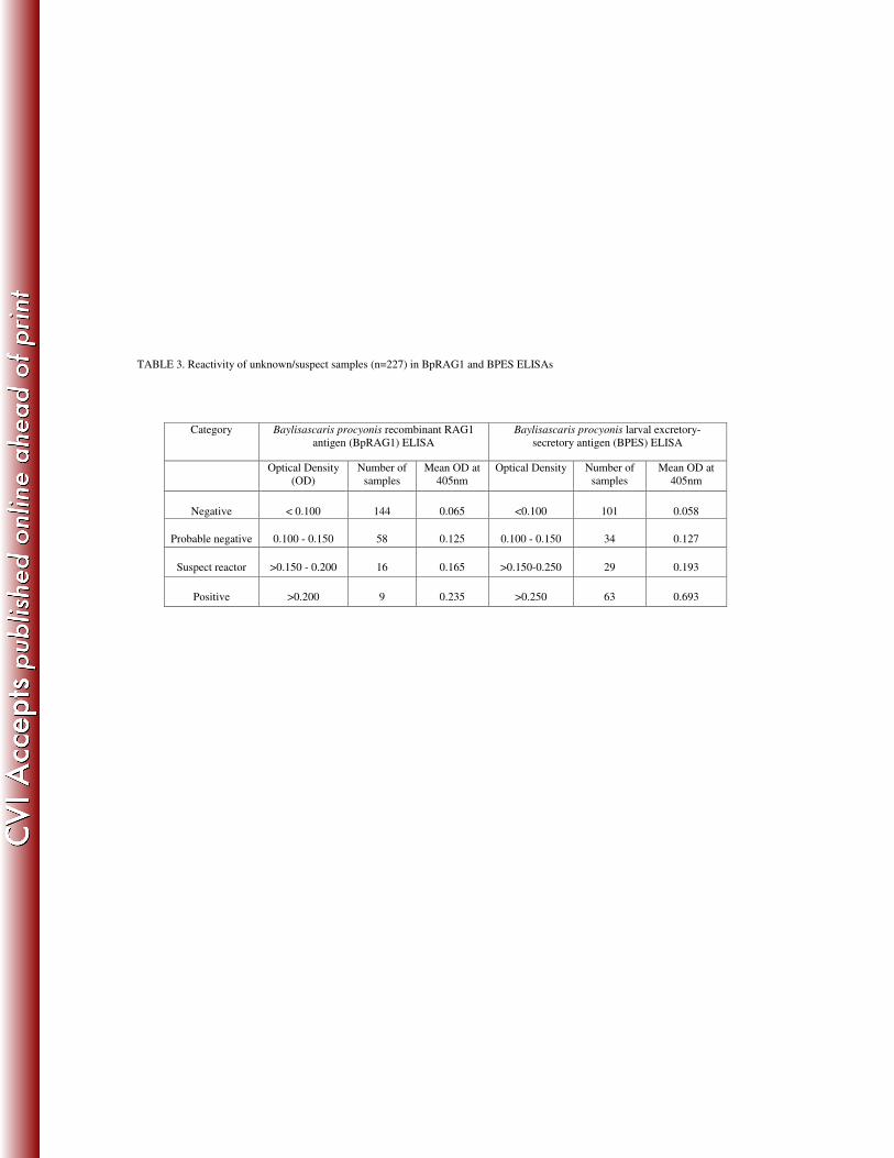
TABLE 3. Reactivity of unknown/suspect samples (n=227) in BpRAG1 and BPES ELISAs
Category Baylisascaris procyonis recombinant RAG1
antigen (BpRAG1) ELISA
Baylisascaris procyonis larval excretory-
secretory antigen (BPES) ELISA
Optical Density
(OD)
Number of
samples
Mean OD at
405nm
Optical Density Number of
samples
Mean OD at
405nm
Negative
< 0.100
144
0.065
<0.100
101
0.058
Probable negative
0.100 - 0.150
58
0.125
0.100 - 0.150
34
0.127
Suspect reactor
>0.150 - 0.200
16
0.165
>0.150-0.250
29
0.193
Positive
>0.200
9
0.235
>0.250
63
0.693
