cuzd1 and anti-cuzd1 antibodies as markers of cancer and
TRANSCRIPT

Hindawi Publishing CorporationClinical and Developmental ImmunologyVolume 2013, Article ID 968041, 11 pageshttp://dx.doi.org/10.1155/2013/968041
Review ArticleCUZD1 and Anti-CUZD1 Antibodies as Markers of Cancer andInflammatory Bowel Diseases
Christos Liaskos,1 Eirini I. Rigopoulou,2 Timoklia Orfanidou,1
Dimitrios P. Bogdanos,1,2,3 and Christos N. Papandreou4
1 Cellular Immunotherapy and Molecular Immunodiagnostics, Institute of Research and Technology Thessaly,41222 Larissa, Greece
2 Department of Medicine, Faculty of Medicine, School of Health Sciences, University of Thessaly, Biopolis,41110 Larissa, Greece
3 Division of Transplantation Immunology and Mucosal Biology, King’s College London School of Medicine,King’s College Hospital, Denmark Hill Campus, London SE5 9RS, UK
4Department of Medical Oncology, University Hospital of Larissa, Faculty of Medicine, School of Health Sciences,University of Thessaly, Biopolis, 41110 Larissa, Greece
Correspondence should be addressed to Dimitrios P. Bogdanos; [email protected]
Received 16 March 2013; Accepted 29 March 2013
Academic Editor: Yehuda Shoenfeld
Copyright © 2013 Christos Liaskos et al.This is an open access article distributed under theCreativeCommonsAttributionLicense,which permits unrestricted use, distribution, and reproduction in any medium, provided the original work is properly cited.
CUZD1, the CUB, and zona pellucida-like domains-containing protein 1, is a newly identified antigen of pancreatic autoantibodies(PAB) giving a reticulogranular pattern in patients with inflammatory bowel diseases, and in particular Crohn’s disease. Theexact mechanisms by which this pancreatic antigen becomes the target of IBD-specific pancreatic autoantibodies are unclear. Atthe same time, evolving data strongly support a role for CUZD1 in carcinogenesis. Human CUZD1 is mapped at chromosome10q26.13 and the loss of this region is a frequent event in various malignant tumours. mRNA overexpression of CUZD1 hasbeen noted in ovarian cancer and serum levels of CUZD1 are elevated in women with ovarian cancer and patients sufferingfrom pancreatic cancer. CUZD1 appears to be one of the relatively few biomarkers that serve as both cancer biomarker andautoantigen of autoantibodies in an autoimmune disease unrelated to cancerous organs. This review discusses the role ofCUZD1 in cancer and autoimmunity. We anticipate that a better understanding of the function of CUZD1 will help us tounderstand how it becomes the focus of an autoimmune attack specifically targeting the intestine and its enigmatic role incarcinogenesis.
1. Introduction
In recent years, the identification and validation of novelbiomarkers has become the focus of intense research in boththe laboratory and clinic. Biomarkers now have several appli-cations and their role has been extended from “diagnostics,”to include “prognostics” and more recently “theranostics.”Theranostics describes a wide range of applications includingthe identification of a novel diagnostic marker that is used inorder to identify patients for whom a newly developed drugwill work.
2. Cancer Biomarkers andAutoantibody Markers
Cancer biomarkers have changed the way we detect andtreat tumors. In recent years, the elucidation of severalcarcinogenic pathways, and a better understanding of tumorprogression, has led to the identification of numerous tumormarkers. The list of markers that have been identified isextensive and includes secreted proteins, transcription fac-tors, and cell surface receptors. For example, 𝛼-fetoprotein(AFP), a member of the albuminoid superfamily, is a cancer
2 Clinical and Developmental Immunology
biomarker used for the monitoring of hepatoblastoma andhepatocellular carcinoma, as well as certain gastrointestinalcancers [1, 2]. However, an increase of AFP is not alwayspathognomonic for liver cancer, as elevated AFP levels canbe found in end-stage liver disease (cirrhosis) unrelatedto tumor development [2, 3]. In fact, virtually all cancerbiomarkers lack specificity to a particular tumor type andmay be found in a variety of cancerous and noncancerousconditions. Hence, tumor markers alone are not diagnosticfor cancer, and at present relatively few tumor markers arewidely used by practicing physicians.
Autoantibodies targeting cellular constituents that act asautoantigens are useful serological markers for the diagnosisof autoimmune diseases and are also used in aiding can-cer diagnosis [4–7]. Most of them are used for diagnosticpurposes, and several of them show very good sensitivitiesand specificities for particular autoimmune diseases [4, 8–14].Autoantibodies may also have prognostic significance, beingable to identify individuals that will develop overt disease orpatients at advanced stages of the disease and also those whowill progress in a fast pace. The presence of autoantibodiesmay also identify patients who may have a better or poorerresponse to treatment and may also be used to monitortreatment response.
The great majority of tumor markers are not potent auto-antibodymarkers and vice versa [15].However, several studieshave addressed the role of autoantibodies and, in particular,tumor-associated antigens (TAA), as targets of humoral andcellular immune responses [15–20]. Various autoantibodyspecificities have been described in patients with malignancyand some of those appear to be of diagnostic and prognosticsignificance, being able to allow early diagnosis or stratifica-tion of patients according to clinical phenotypes and diseaseoutcome [18]. Investigations are being carried out for the deli-neation of autoantibody markers useful for the identificationof individuals at risk for cancer [20]. Of particular interest areautoantibody markers of paraneoplastic neurological disor-ders characterised by highly specific autoantibodies directedagainst onconeuronal antigens [21–27]. Autoantibody pan-els and signature profiling with diagnostic or prognosticsignificance for various malignancies are under validationand may prove to be useful in the clinical setting [28]. Theprevailing notion has been that antigens overexpressed in astate of a tumour act as cryptic antigens or neoantigens thatare perceived as foreign from the immune system [15, 18].TAA are therefore capable of priming the immune system torecognize TAA and indeed tumor cells expressing them [15,18]. On the other hand in conditions such as lymphomas, thedegenerated B cells produce large amounts of autoantibodiesthat are insufficiently controlled by the peripheral regulatorymachinery of the immune system.
Themagnitude, duration, and efficacy of the TAA-specificCD4 andCD8 immune responses depend on several intrinsicfactors [29]. An increasing number of studies are investi-gating ways to manipulate the efficacy of antigen-specificimmune responses in a manner that can facilitate the erad-ication of antigen-expressing tissue-specific target cells [29].
The investigation of the fine specificity of the immuneresponses against specific TAA has led to the appreciation
that some autoantibody specificities related to TAAmay beardiagnostic and prognostic significance [17, 20]. An increasingnumber of studies have obtained data to suggest that severalTAA can be potential immunotherapeutic targets, in additionto aiding in the monitoring of disease progression andresponse to treatment [29–36]. There is no doubt that thestudy of humoral autoreactivity to TAA has helped investi-gators to estimate the sensitivity and specificity of individualanti-TAA antibodies. The role of TAA autoantibodies withinthe processes of carcinogenesis has been a topic of ongoingresearch. A systematic search of the literature published in2009 has revealed more than 107 different TAA identified sofar, most of which are of limited diagnostic relevance [37].The majority of these TAA correspond to mutated or overex-pressed antigens and were cytoplasmic proteins (42%), while26% corresponded to nuclear antigens and 21% tomembrane-bound proteins [37]. Of interest, only 10% of the reportedTAA corresponded to extracellular proteins such as secretedor extracellular matrix proteins [37].
In this review, we discuss the clinical significance ofCUZD1, a novel biomarker with a dual role as a cancer and anautoantibody marker.
3. CUZD1: Introduction to the Gene
3.1. Terminology. CUZD1 stands forCUBand zona pellucida-like domains-containing protein 1. The CUZD1 gene is alsoknown as the uterine-ovarian-specific gene 44 (UO-44) andthe estrogen-regulated gene 1 (ERG1).
3.2. Genomic Location. Human CUZD1 is mapped at chro-mosome 10q26.13 and contains nine exons.The loss of 10q is afrequent event in the development and progression of variousmalignant tumours including prostate adenocarcinoma,endometrial cancers, glioblastoma multiforme, and smallcell lung cancer [38–42], suggesting the presence of severaltumor suppressor genes in this chromosomal region, whichare important for suppression of tumorigenesis and cancerprogression.
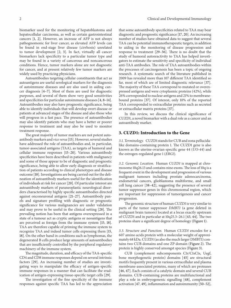
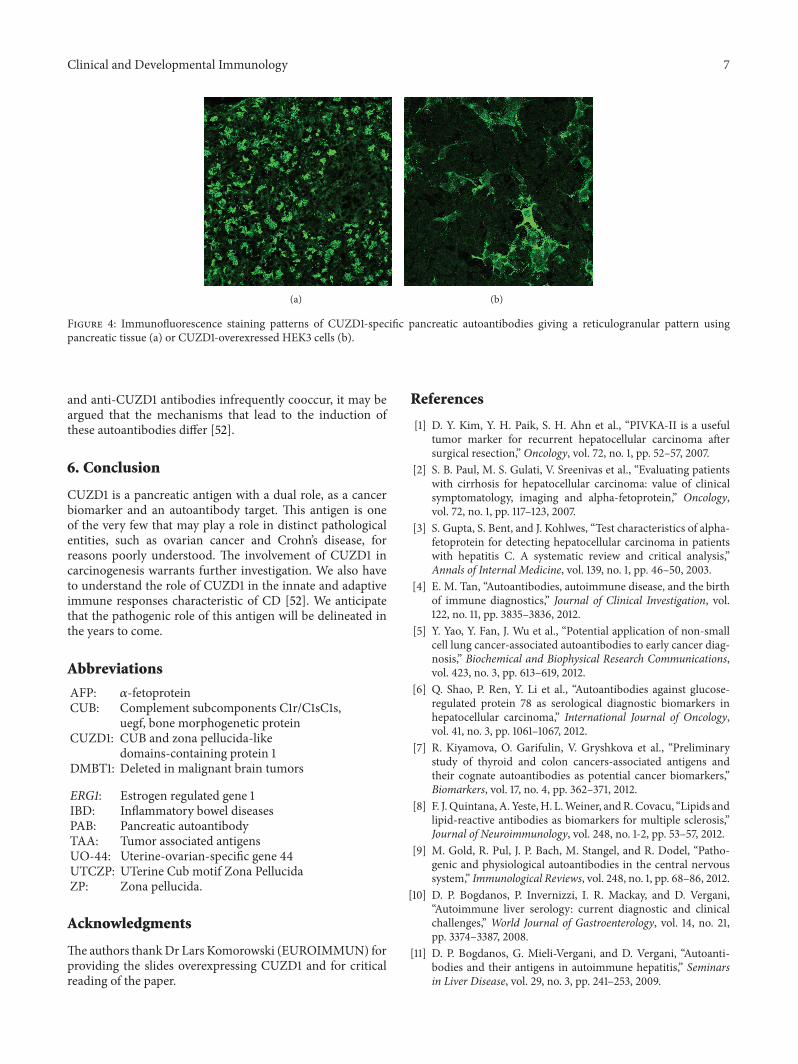
The genomic structure of humanCUZD1 is very similar toparts of the tumor suppressor DMBT1 (a gene deleted inmalignant brain tumors) located at a locus exactly upstreamof CUZD1 and in particular at 10q25.3–26.1 [43, 44]. The twoproteins share a significant degree of homology (Figure 1).
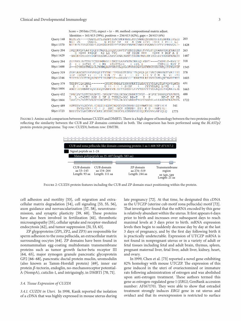
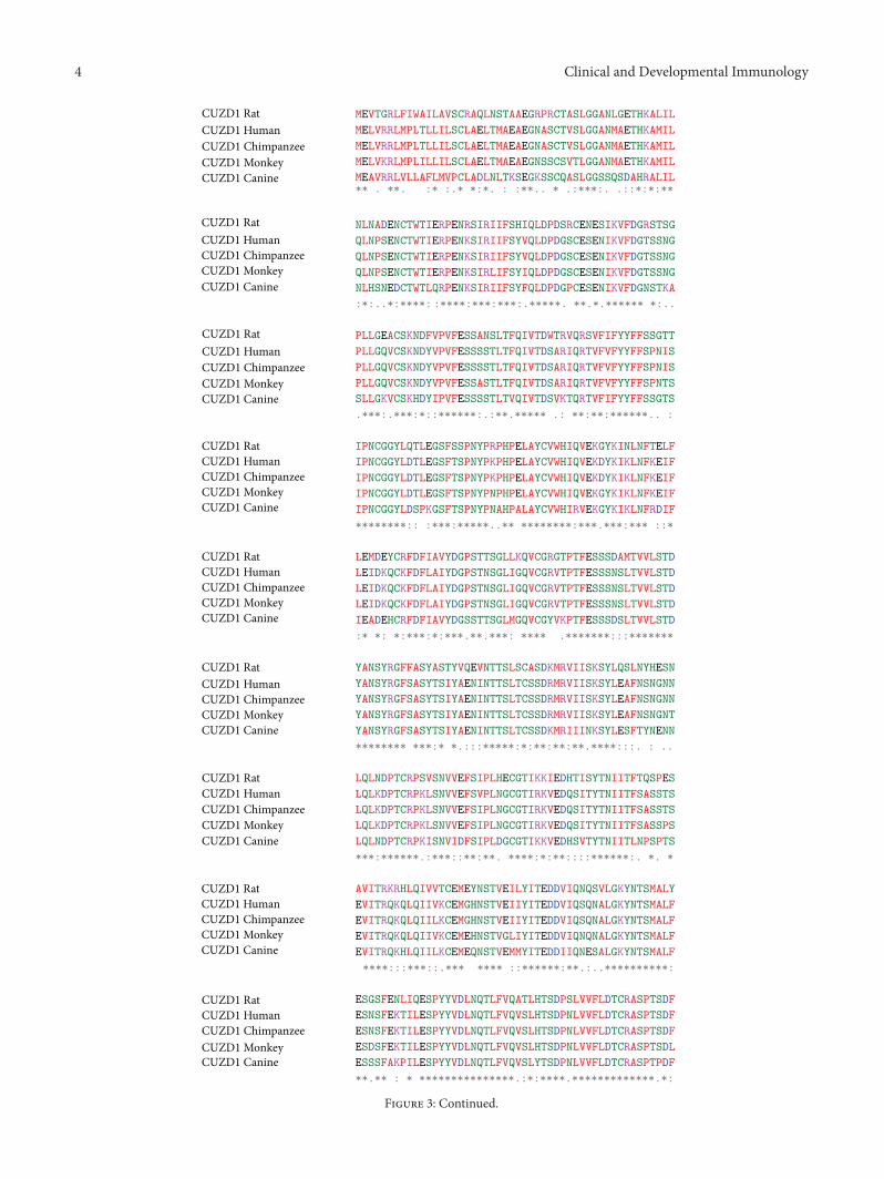
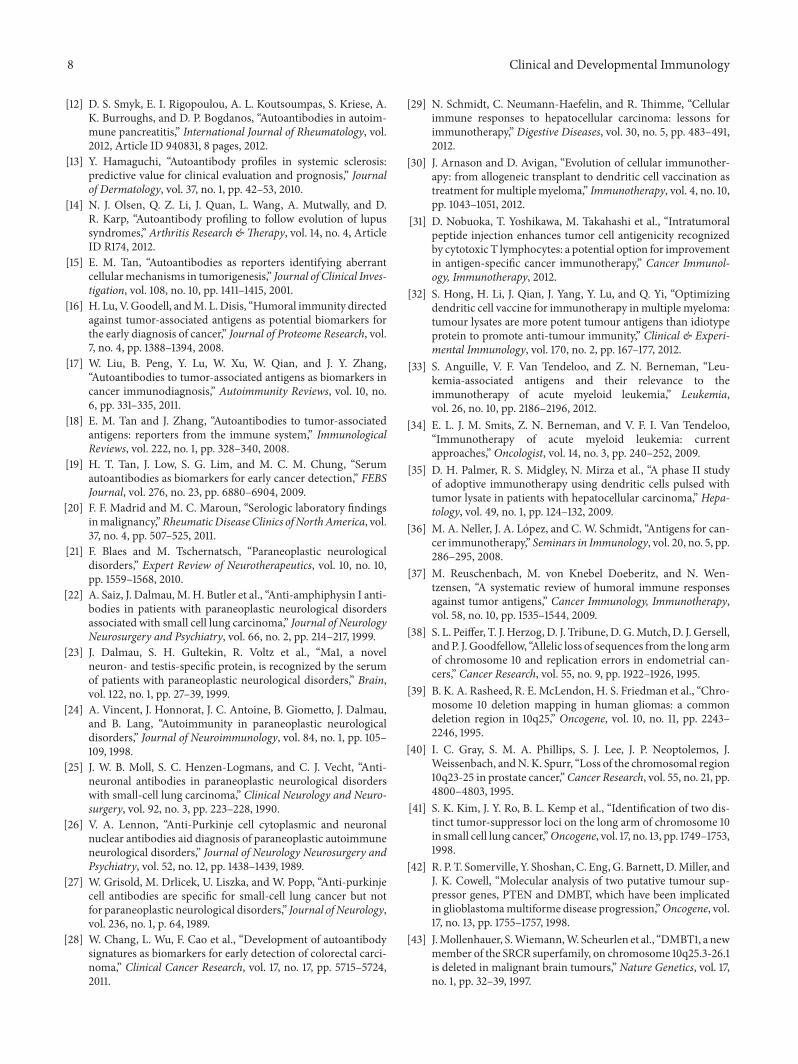
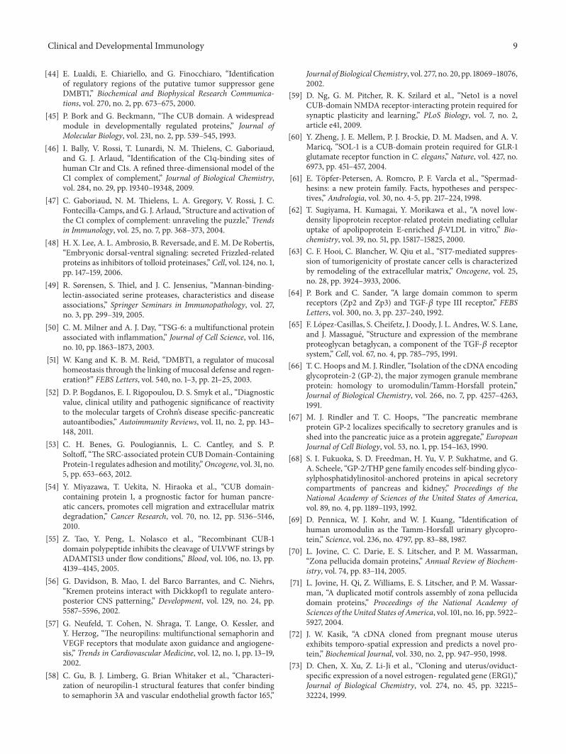
3.3. Structure and Function. Human CUZD1 encodes for a607 amino acids protein with a molecular weight of approxi-mately 68 kDa. CUZD1 (as also themuch largerDMBT1) con-tains two CUB domains and one ZP domain (Figure 2). Theprotein is highly conserved amongst species (Figure 3).
CUB (complement subcomponents C1r/C1sC1s, Uegf,bone morphogenetic protein) domains [45] are structuralmotifs frequently present in various extracellular and plasmamembrane-associated proteins, many of which are proteases[46, 47]. Each consists of a catalytic domain and several CUBdomains. CUB-containing proteins are multifunctional andplay a role in embryogenenic signalling [48], complementactivation [47, 49], inflammation and autoimmunity [50–52],
Clinical and Developmental Immunology 3
1775
Query 148
Sbjct 1370
Query 204Sbjct 1429
Query 264Sbjct 1488
Query 319Sbjct 1546
Query 379Sbjct 1604
Query 432
Sbjct 1664
Query 489Sbjct 1723
203
1428
2631487318
1545378
1603431
1663488
1722
541
Score = 295 bits (755), expect =1𝑒 − 89, method: compositional matrix adjust.Identities = 161/413 (39%), positives = 256/413 (62%), gaps = 26/413 (6%)
Figure 1: Amino acid comparison between humanCUZD1 andDMBT1.There is a high degree of homology between the two proteins possiblyreflecting the similarity between the CUB and ZP domains contained in both. The comparison has been performed using the BLASTp2protein-protein programme. Top row: CUZD1; bottom row: DMTB1.
CUB and zona pellucida-like domain containing protein-1 aa 1-608 NP 071317.2
Signal peptide aaMature polypeptide aa (length: 583 aa)
CUB domainaa
Length: 93 aa
CUB domainaa
Length: 111 aa
ZP domainaa
Length: 244 aa
Transmembraneregion
aaLength: 21 aa
1–24
53–145 154–264 276–519569–589
25–607
Figure 2: CUZD1 protein features including the CUB and ZP domain exact positioning within the protein.
cell adhesion and motility [53], cell migration and extra-cellular matrix degradation [54], cell signaling [53, 55, 56],axon guidance and neovascularization [57, 58], neurotrans-mission, and synaptic plasticity [59, 60]. These proteinshave also been involved in fertilization [61], thromboticmicroangiopathy [55], cellular uptake and receptor-mediatedendocytosis [62], and tumor suppression [51, 53, 63].
ZP glygoproteins (ZP1, ZP2, and ZP3) are responsible forsperm adhesion to the zona pellucida, an extracellular matrixsurrounding oocytes [64]. ZP domains have been found innonmammalian egg-coating multidomain transmembraneproteins such as tumor growth factor-beta receptor III[64, 65], major zymogen granule pancreatic glycoproteinGP2 [66–68], pancreatic ductal protein muclin, uromodulin(also known as Tamm-Horsfall protein) [69], inner earprotein 𝛽-tectorin, endoglin, no-mechanoreceptor potential-A (NompA), cuticlin-1, and intriguingly, in DMBT1 [70, 71].
3.4. Tissue Expression of CUZD1
3.4.1. CUZD1 in Uteri. In 1998, Kasik reported the isolationof a cDNA that was highly expressed in mouse uterus during
late pregnancy [72]. At that time, he designated this cDNAas the UTCZP (uterine cub motif zona pellucida) motif [72].That investigator found that the mRNA encoded by this geneis relatively abundant within the uterus. It first appears 6 daysprior to birth and increases over subsequent days to reachmaximal levels at 3 days prior to birth. mRNA expressionlevels then begin to suddenly decrease day by day at the last3 days of pregnancy, and by the first day following birth itis practically undetectable. Expression of UTCZP mRNA isnot found in nonpregnant uterus or in a variety of adult orfetal tissues including fetal and adult brain, thymus, spleen,pregnant maternal liver, fetal liver, adult liver, kidney, heart,and ovary.
In 1999, Chen et al. [73] reported a novel gene exhibiting82% homology with mouse UTCZP. The expression of thisgene induced in the uteri of ovariectomized or immaturerats following administration of estrogen and was abolishedupon anti-estrogen treatment. These authors termed thisgene as estrogen-regulated gene 1 (ERG1; GenBank accessionnumber: AF167170). They were able to show that estradioltreatment strongly induces ERG1 gene in rat uterus andoviduct and that its overexpression is restricted to surface
4 Clinical and Developmental Immunology
CUZD1 RatCUZD1 HumanCUZD1 ChimpanzeeCUZD1 MonkeyCUZD1 Canine
CUZD1 RatCUZD1 HumanCUZD1 ChimpanzeeCUZD1 MonkeyCUZD1 Canine
CUZD1 RatCUZD1 HumanCUZD1 ChimpanzeeCUZD1 MonkeyCUZD1 Canine
CUZD1 RatCUZD1 HumanCUZD1 ChimpanzeeCUZD1 MonkeyCUZD1 Canine
CUZD1 RatCUZD1 HumanCUZD1 ChimpanzeeCUZD1 MonkeyCUZD1 Canine
CUZD1 RatCUZD1 HumanCUZD1 ChimpanzeeCUZD1 MonkeyCUZD1 Canine
CUZD1 RatCUZD1 HumanCUZD1 ChimpanzeeCUZD1 MonkeyCUZD1 Canine
CUZD1 RatCUZD1 HumanCUZD1 ChimpanzeeCUZD1 MonkeyCUZD1 Canine
CUZD1 RatCUZD1 HumanCUZD1 ChimpanzeeCUZD1 MonkeyCUZD1 Canine
MEVTGRLFIWAILAVSCRAQLNSTAAEGRPRCTASLGGANLGETHKALIL
MELVRRLMPLTLLILSCLAELTMAEAEGNASCTVSLGGANMAETHKAMIL
MELVRRLMPLTLLILSCLAELTMAEAEGNASCTVSLGGANMAETHKAMIL
MELVKRLMPLILLILSCLAELTMAEAEGNSSCSVTLGGANMAETHKAMIL
MEAVRRLVLLAFLMVPCLADLNLTKSEGKSSCQASLGGSSQSDAHRALIL
NLNADENCTWTIERPENRSIRIIFSHIQLDPDSRCENESIKVFDGRSTSG
QLNPSENCTWTIERPENKSIRIIFSYVQLDPDGSCESENIKVFDGTSSNG
QLNPSENCTWTIERPENKSIRIIFSYVQLDPDGSCESENIKVFDGTSSNG
QLNPSENCTWTIERPENKSIRLIFSYIQLDPDGSCESENIKVFDGTSSNG
NLHSNEDCTWTLQRPENKSIRIIFSYFQLDPDGPCESENIKVFDGNSTKA
:*:..*:****::****:***:***:.*****. **.*.****** *:..
PLLGEACSKNDFVPVFESSANSLTFQIVTDWTRVQRSVFIFYYFFSSGTT
PLLGQVCSKNDYVPVFESSSSTLTFQIVTDSARIQRTVFVFYYFFSPNIS
PLLGQVCSKNDYVPVFESSSSTLTFQIVTDSARIQRTVFVFYYFFSPNIS
PLLGQVCSKNDYVPVFESSASTLTFQIVTDSARIQRTVFVFYYFFSPNTS
SLLGKVCSKHDYIPVFESSSSTLTVQIVTDSVKTQRTVFIFYYFFSSGTS
.***:.***:*::******:.:**.***** .: **:**:******.. :
IPNCGGYLQTLEGSFSSPNYPRPHPELAYCVWHIQVEKGYKINLNFTELF
IPNCGGYLDTLEGSFTSPNYPKPHPELAYCVWHIQVEKDYKIKLNFKEIF
IPNCGGYLDTLEGSFTSPNYPKPHPELAYCVWHIQVEKDYKIKLNFKEIF
IPNCGGYLDTLEGSFTSPNYPNPHPELAYCVWHIQVEKGYKIKLNFKEIF
IPNCGGYLDSPKGSFTSPNYPNAHPALAYCVWHIRVEKGYKIKLNFRDIF
********:: :***:*****..** ********:***.***:*** ::*
LEMDEYCRFDFIAVYDGPSTTSGLLKQVCGRGTPTFESSSDAMTVVLSTD
LEIDKQCKFDFLAIYDGPSTNSGLIGQVCGRVTPTFESSSNSLTVVLSTD
LEIDKQCKFDFLAIYDGPSTNSGLIGQVCGRVTPTFESSSNSLTVVLSTD
LEIDKQCKFDFLAIYDGPSTNSGLIGQVCGRVTPTFESSSNSLTVVLSTD
IEADEHCRFDFIAVYDGSSTTSGLMGQVCGYVKPTFESSSDSLTVVLSTD
:* *: *:***:*:***.**.***: **** .*******:::*******
YANSYRGFFASYASTYVQEVNTTSLSCASDKMRVIISKSYLQSLNYHESN
YANSYRGFSASYTSIYAENINTTSLTCSSDRMRVIISKSYLEAFNSNGNN
YANSYRGFSASYTSIYAENINTTSLTCSSDRMRVIISKSYLEAFNSNGNN
YANSYRGFSASYTSIYAENINTTSLTCSSDRMRVIISKSYLEAFNSNGNT
YANSYRGFSASYTSIYAENINTTSLTCSSDKMRIIINKSYLESFTYNENN
******** ***:* *.:::*****:*:**:**:**.****:::. : ..
LQLNDPTCRPSVSNVVEFSIPLHECGTIKKIEDHTISYTNIITFTQSPES
LQLKDPTCRPKLSNVVEFSVPLNGCGTIRKVEDQSITYTNIITFSASSTS
LQLKDPTCRPKLSNVVEFSIPLNGCGTIRKVEDQSITYTNIITFSASSTS
LQLKDPTCRPKLSNVVEFSIPLNGCGTIRKVEDQSITYTNIITFSASSPS
LQLNDPTCRPKISNVIDFSIPLDGCGTIKKVEDHSVTYTNIITLNPSPTS
***:******.:***::**:**. ****:*:**::::******:. *. *
AVITRKRHLQIVVTCEMEYNSTVEILYITEDDVIQNQSVLGKYNTSMALY
EVITRQKQLQIIVKCEMGHNSTVEIIYITEDDVIQSQNALGKYNTSMALF
EVITRQKQLQIILKCEMGHNSTVEIIYITEDDVIQSQNALGKYNTSMALF
EVITRQKQLQIIVKCEMEHNSTVGLIYITEDDVIQNQNALGKYNTSMALF
EVITRQKHLQIILKCEMEQNSTVEMMYITEDDIIQNESALGKYNTSMALF
****:::***::.*** **** ::******:**.:..**********:
ESGSFENLIQESPYYVDLNQTLFVQATLHTSDPSLVVFLDTCRASPTSDF
ESNSFEKTILESPYYVDLNQTLFVQVSLHTSDPNLVVFLDTCRASPTSDF
ESNSFEKTILESPYYVDLNQTLFVQVSLHTSDPNLVVFLDTCRASPTSDF
ESDSFEKTILESPYYVDLNQTLFVQVSLHTSDPNLVVFLDTCRASPTSDL
ESSSFAKPILESPYYVDLNQTLFVQVSLYTSDPNLVVFLDTCRASPTPDF
**.** : * ***************.:*:****.*************.*:
** . **. :* :.* *:*. : :**.. * .:***:. .::*:*:**
Figure 3: Continued.
Clinical and Developmental Immunology 5
CUZD1 RatCUZD1 HumanCUZD1 ChimpanzeeCUZD1 MonkeyCUZD1 Canine
CUZD1 RatCUZD1 HumanCUZD1 ChimpanzeeCUZD1 MonkeyCUZD1 Canine
CUZD1 RatCUZD1 HumanCUZD1 Chimpanzee
CUZD1 Canine
CUZD1 RatCUZD1 HumanCUZD1 ChimpanzeeCUZD1 MonkeyCUZD1 Canine
ASPTYDLISSGCSRDETCKVYPLFGHYGRFQFNAFKFLRHLSSVYLKCKI
ASPTYDLIKSGCSRDETCKVYPLFGHYGRFQFNAFKFLRSMSSVYLQCKV
ASPTYDLIKSGCSRDETCKVYPLFGHYGRFQFNAFKFLRSMSSVYLQCKV
ASPTYDLIKSGCSRDETCKVYPLFGHYGRFQFNAFKFLRSLSSVYLQCKV
TSPTYDLIKSGCNQDETCKVYPLSKHYGRFQFNAFKFLNSLGSVYLQCKI
:*******.***.:********* *************. :.****:**:
LICDTSDHTSRCNQGCVSRRKRDIPSYKWKTDSVIGPIRLKRDRLANGDS
LICDSSDHQSRCNQGCVSRSKRDISSYKWKTDSIIGPIRLKRDRSASGNS
LICDSSDHQSRCNQGCVSRSKRDISSYKWKTDSIIGPIRLKRDRSASGNS
LICDSSDHQSRCSQGCVSRSKRDISSYKWKTDSIIGPIRLKRDRSASGNS
LICDSGDHQSRCSQGCVSRTKRDLSSYKWKTDAVIGPIRLKRDRSASGNT
****:.** ***.****** ***:.*******::********** *.*::
GLLPQTHEAEISKQPLSHLHLFSFMVLALNVVIVVTATVRHFLNRWKDHG
GFQHETHAEETPNQPFNSVHLFSFMVLALNVVTVATITVRHFVNQRADYK
GFQHETHAEETPNQPFNSLHLFSFMVLALNVVIVATITVRHFVNQRADYK
GFQHETHAEETPNQPFNSLHLFSFMVLVLNVVIVATITVRHFVNQRAYYK
GFQDQIHKEETQSQPFSSLHLFSFTVLALNLVIVAVIITRHFVNQRTDYK
*: : * * .**:. :***** **.**:* *.. .***:*: :
YQKLQV-Y
YQKLQN-Y
YQKLQN-Y
YQKLQN-Y
YQKLQNMN
*****
CUZD1 Monkey
Figure 3: Multiple amino acid sequence alignment of CUZD1 of various species shows striking conservation. Sequences aligned includeRattus norvegicus (EDM 11686.1), Homo sapiens (NP 071317.2), Pan troglodytes (XP 001160209.2), Macaca fascicularis (EHH 65083.1), andCanis lupus familiaris (XP 544054.2). Asterisk indicates identities and semicolon indicates conserved or semiconserved substitutions. Thealignment has been performed using the CLUSTALW (1.83) multiple sequence alignment tool.
epithelium [73]. The expression level of ERG1mRNA is highon day 1 of pregnancy, declined on day 2, and was practicallyundetectable from days 3 to 6 of gestation. ERG1 expressionwas maximal at the proestrus stage of the ovarian cycle,coinciding with the estrogen-induced uterine cell prolifera-tion, and clearly indicating that stage-specific manner of itsexpression during the ovarian cycle.
Two years later, Huynh et al. [74] reported the isolationof a uterine-ovarian-specific gene 44 (UO-44; GenBankaccession number: AF022147) from a tamoxifen-induced ratuterine complementary DNA library in 2001. The UO-44gene, currently known as CUZD1, was specifically expressedin the uterus and ovary of rats [74]. The rat UO-44 cDNAshowed 99% homology with the rat ERG1 cDNAs; thus thetwo genes were practically identical. In their study, Huynh etal. have shown that UO-44 mRNA was undetectable in uteriderived from OVX rats and was expressed after tamoxifentreatment [74]. In a set of in situ hybridization experimentson sections from uteri originated from control OVX andOVX-tamoxifen-treated rats, the same group of investiga-tors have shown that while there was no UO-44 signal inuterus of OVX rats, high levels of UO-44 were detectablein the luminal epithelial cells and glandular populationupon treatment with tamoxifen. Intriguingly, treatment withthe pure antiestrogen ICI 182780 abrogated the effects oftamoxifen on the expression of UO-44, further suggestingthat tamoxifen functions as an estrogen and induces UO-44expression.
3.4.2. CUZD1 in Ovaries (and in Pancreas). Huynh et al.have obtained data clearly demonstrating that UO-44mRNAis detectable in granulosa cells of ovaries [74]. They havealso found varying amounts of UO-44 mRNA in granulosacells of a mixed population of follicles. In particular, highlevels of UO-44 expression were noted in the granulosacells of medium-size follicles, while low-to-moderate UO-44 expression was observed in granulosa cells of smalland large follicles. Of interest, the UO-44 mRNA amonggranulosa cells within the same follicle was not uniformand greatly CUZD1 [75]. In a murine model of necrotizingpancreatitis, UTCZP- (CUZD1-) deficient mice developedmore severe pancreatitis suggesting that CUZD1 may play animportant role in trypsinogen activation and in the severityof pancreatitis [75].
4. CUZD1: As a Cancer Biomarker
One key experiment that suggested an important role forCUZD1 in carcinogenesis demonstrated that human UO-44-specific antisera strikingly inhibit cell attachment andproliferation of NIH-OVCAR3 ovarian cancer cells [76].Thisobservation has led the authors to postulate that human UO-44may promote cell growth and facilitate invasion in ovariancancer. The same group of investigators has shown thatcisplatin treatment leads to the downregulation of humanUO-44 expression and that silencing of human UO-44 basedon sequence-specific siRNAs confers an enhanced sensitivity
6 Clinical and Developmental Immunology
in cisplatin treatment of human ovarian cancer cells [77]. Italso appears that UO-44 in ovarian cancer cells with overex-pressed human UO-44 are also resistant to cisplatin [77]. Ina recent study, Leung et al. [78] have measured CUZD1 levelsin the serum of patients with various types of malignanciesand healthy normal controls. Serum samples from patientswith ovarian, breast, lung, colorectal, prostate, and testicularcancer were tested. Elevated levels of CUZD1 were found inpatients with ovarian cancer, but also in breast and lung can-cer [78]. However, this study was conducted in a very smallnumber of sera and the diagnostic significance of CUZD1 asa cancer serum biomarker remains to be assessed in largercohorts. Nevertheless, the latter study has shown that CUZD1performed equally well as CA125 in two independent cohortsof samples consisting of healthy controls and ovarian cancercases, and this may further suggest its potential role as a spe-cific marker of ovarian cancer [78].The authors reported thatCUZD1 is a novel pancreatic cancer serum biomarker as well,but they did not present data to support this statement [78].
5. Anti-CUZD1 Antibodies inInflammatory Bowel Diseases
Patients with Crohn’s disease are characterised by the pres-ence of organ-specific and nonorgan-specific autoantibodies[79–89]. Up to 30% of patients with Crohn’s disease (CD)contain detectable pancreatic autoantibodies (PAB), givingeither a droplet-like or a reticulogranular, cytoplasmic pat-tern [52, 84–87, 90–93]. The recognition of GP2 as the targetof the droplet-like PABs in 2009 [94] has been followed bythe recent identification of CUZD1 as the sole autoantigenof PABs giving the reticulogranular, cytoplasmic pattern byindirect immunofluorescence [91–93]. Several studies haveattempted to delineate the diagnostic and clinical significanceof anti-GP2 PAB [52, 90, 95–99], and the immunopatho-genetic role of GP2 in CD has started to emerge [100–102].However, the biological and clinical significance of humoraland cellular immune responses against CUZD1 remainspoorly understood [91–93]. It has become apparent that thetwo autoreactivities rarely coexist and that the GP2 andCUZD1 are unlikely targets of cross-reactive autoantibodies.
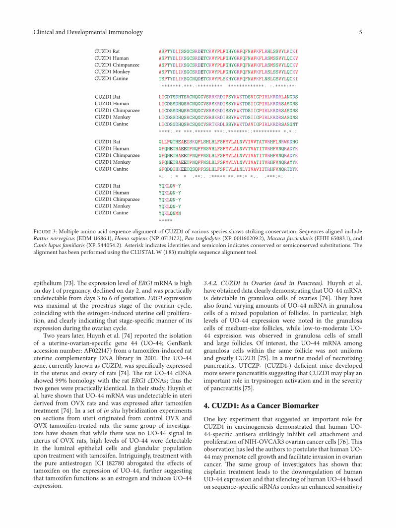
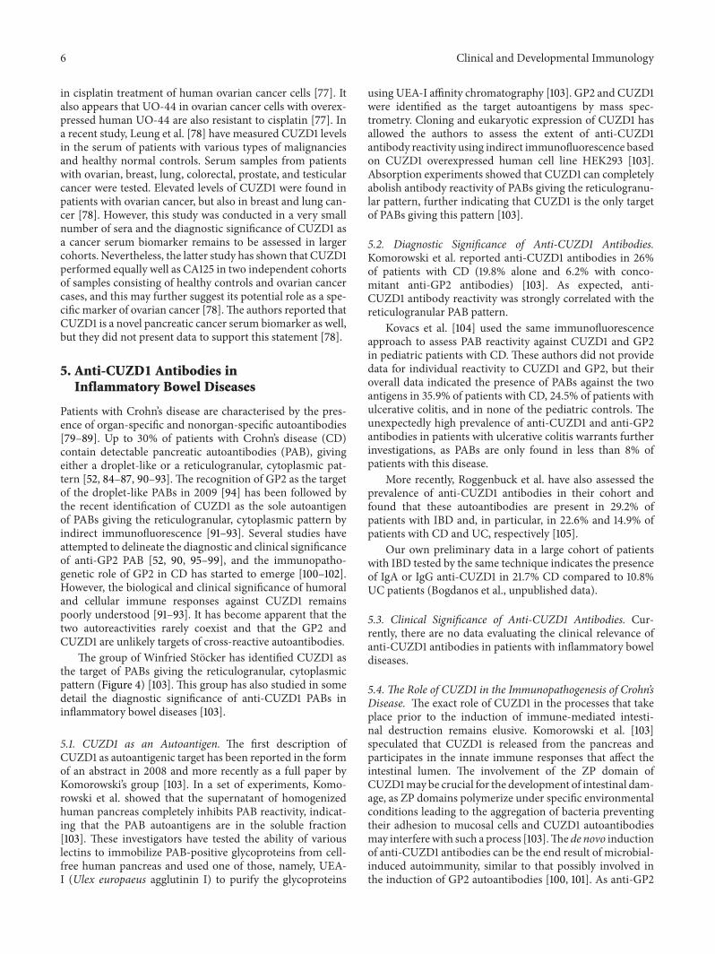
The group of Winfried Stocker has identified CUZD1 asthe target of PABs giving the reticulogranular, cytoplasmicpattern (Figure 4) [103]. This group has also studied in somedetail the diagnostic significance of anti-CUZD1 PABs ininflammatory bowel diseases [103].
5.1. CUZD1 as an Autoantigen. The first description ofCUZD1 as autoantigenic target has been reported in the formof an abstract in 2008 and more recently as a full paper byKomorowski’s group [103]. In a set of experiments, Komo-rowski et al. showed that the supernatant of homogenizedhuman pancreas completely inhibits PAB reactivity, indicat-ing that the PAB autoantigens are in the soluble fraction[103]. These investigators have tested the ability of variouslectins to immobilize PAB-positive glycoproteins from cell-free human pancreas and used one of those, namely, UEA-I (Ulex europaeus agglutinin I) to purify the glycoproteins
using UEA-I affinity chromatography [103]. GP2 and CUZD1were identified as the target autoantigens by mass spec-trometry. Cloning and eukaryotic expression of CUZD1 hasallowed the authors to assess the extent of anti-CUZD1antibody reactivity using indirect immunofluorescence basedon CUZD1 overexpressed human cell line HEK293 [103].Absorption experiments showed that CUZD1 can completelyabolish antibody reactivity of PABs giving the reticulogranu-lar pattern, further indicating that CUZD1 is the only targetof PABs giving this pattern [103].
5.2. Diagnostic Significance of Anti-CUZD1 Antibodies.Komorowski et al. reported anti-CUZD1 antibodies in 26%of patients with CD (19.8% alone and 6.2% with conco-mitant anti-GP2 antibodies) [103]. As expected, anti-CUZD1 antibody reactivity was strongly correlated with thereticulogranular PAB pattern.
Kovacs et al. [104] used the same immunofluorescenceapproach to assess PAB reactivity against CUZD1 and GP2in pediatric patients with CD. These authors did not providedata for individual reactivity to CUZD1 and GP2, but theiroverall data indicated the presence of PABs against the twoantigens in 35.9% of patients with CD, 24.5% of patients withulcerative colitis, and in none of the pediatric controls. Theunexpectedly high prevalence of anti-CUZD1 and anti-GP2antibodies in patients with ulcerative colitis warrants furtherinvestigations, as PABs are only found in less than 8% ofpatients with this disease.
More recently, Roggenbuck et al. have also assessed theprevalence of anti-CUZD1 antibodies in their cohort andfound that these autoantibodies are present in 29.2% ofpatients with IBD and, in particular, in 22.6% and 14.9% ofpatients with CD and UC, respectively [105].
Our own preliminary data in a large cohort of patientswith IBD tested by the same technique indicates the presenceof IgA or IgG anti-CUZD1 in 21.7% CD compared to 10.8%UC patients (Bogdanos et al., unpublished data).
5.3. Clinical Significance of Anti-CUZD1 Antibodies. Cur-rently, there are no data evaluating the clinical relevance ofanti-CUZD1 antibodies in patients with inflammatory boweldiseases.
5.4. The Role of CUZD1 in the Immunopathogenesis of Crohn’sDisease. The exact role of CUZD1 in the processes that takeplace prior to the induction of immune-mediated intesti-nal destruction remains elusive. Komorowski et al. [103]speculated that CUZD1 is released from the pancreas andparticipates in the innate immune responses that affect theintestinal lumen. The involvement of the ZP domain ofCUZD1may be crucial for the development of intestinal dam-age, as ZP domains polymerize under specific environmentalconditions leading to the aggregation of bacteria preventingtheir adhesion to mucosal cells and CUZD1 autoantibodiesmay interferewith such a process [103].The de novo inductionof anti-CUZD1 antibodies can be the end result of microbial-induced autoimmunity, similar to that possibly involved inthe induction of GP2 autoantibodies [100, 101]. As anti-GP2
Clinical and Developmental Immunology 7
(a) (b)
Figure 4: Immunofluorescence staining patterns of CUZD1-specific pancreatic autoantibodies giving a reticulogranular pattern usingpancreatic tissue (a) or CUZD1-overexressed HEK3 cells (b).
and anti-CUZD1 antibodies infrequently cooccur, it may beargued that the mechanisms that lead to the induction ofthese autoantibodies differ [52].
6. Conclusion
CUZD1 is a pancreatic antigen with a dual role, as a cancerbiomarker and an autoantibody target. This antigen is oneof the very few that may play a role in distinct pathologicalentities, such as ovarian cancer and Crohn’s disease, forreasons poorly understood. The involvement of CUZD1 incarcinogenesis warrants further investigation. We also haveto understand the role of CUZD1 in the innate and adaptiveimmune responses characteristic of CD [52]. We anticipatethat the pathogenic role of this antigen will be delineated inthe years to come.
Abbreviations
AFP: 𝛼-fetoproteinCUB: Complement subcomponents C1r/C1sC1s,
uegf, bone morphogenetic proteinCUZD1: CUB and zona pellucida-like
domains-containing protein 1DMBT1: Deleted in malignant brain tumors
ERG1: Estrogen regulated gene 1IBD: Inflammatory bowel diseasesPAB: Pancreatic autoantibodyTAA: Tumor associated antigensUO-44: Uterine-ovarian-specific gene 44UTCZP: UTerine Cub motif Zona PellucidaZP: Zona pellucida.
Acknowledgments
Theauthors thankDr Lars Komorowski (EUROIMMUN) forproviding the slides overexpressing CUZD1 and for criticalreading of the paper.
References
[1] D. Y. Kim, Y. H. Paik, S. H. Ahn et al., “PIVKA-II is a usefultumor marker for recurrent hepatocellular carcinoma aftersurgical resection,” Oncology, vol. 72, no. 1, pp. 52–57, 2007.
[2] S. B. Paul, M. S. Gulati, V. Sreenivas et al., “Evaluating patientswith cirrhosis for hepatocellular carcinoma: value of clinicalsymptomatology, imaging and alpha-fetoprotein,” Oncology,vol. 72, no. 1, pp. 117–123, 2007.
[3] S. Gupta, S. Bent, and J. Kohlwes, “Test characteristics of alpha-fetoprotein for detecting hepatocellular carcinoma in patientswith hepatitis C. A systematic review and critical analysis,”Annals of Internal Medicine, vol. 139, no. 1, pp. 46–50, 2003.
[4] E. M. Tan, “Autoantibodies, autoimmune disease, and the birthof immune diagnostics,” Journal of Clinical Investigation, vol.122, no. 11, pp. 3835–3836, 2012.
[5] Y. Yao, Y. Fan, J. Wu et al., “Potential application of non-smallcell lung cancer-associated autoantibodies to early cancer diag-nosis,” Biochemical and Biophysical Research Communications,vol. 423, no. 3, pp. 613–619, 2012.
[6] Q. Shao, P. Ren, Y. Li et al., “Autoantibodies against glucose-regulated protein 78 as serological diagnostic biomarkers inhepatocellular carcinoma,” International Journal of Oncology,vol. 41, no. 3, pp. 1061–1067, 2012.
[7] R. Kiyamova, O. Garifulin, V. Gryshkova et al., “Preliminarystudy of thyroid and colon cancers-associated antigens andtheir cognate autoantibodies as potential cancer biomarkers,”Biomarkers, vol. 17, no. 4, pp. 362–371, 2012.
[8] F. J.Quintana,A. Yeste,H. L.Weiner, andR.Covacu, “Lipids andlipid-reactive antibodies as biomarkers for multiple sclerosis,”Journal of Neuroimmunology, vol. 248, no. 1-2, pp. 53–57, 2012.
[9] M. Gold, R. Pul, J. P. Bach, M. Stangel, and R. Dodel, “Patho-genic and physiological autoantibodies in the central nervoussystem,” Immunological Reviews, vol. 248, no. 1, pp. 68–86, 2012.
[10] D. P. Bogdanos, P. Invernizzi, I. R. Mackay, and D. Vergani,“Autoimmune liver serology: current diagnostic and clinicalchallenges,” World Journal of Gastroenterology, vol. 14, no. 21,pp. 3374–3387, 2008.
[11] D. P. Bogdanos, G. Mieli-Vergani, and D. Vergani, “Autoanti-bodies and their antigens in autoimmune hepatitis,” Seminarsin Liver Disease, vol. 29, no. 3, pp. 241–253, 2009.
8 Clinical and Developmental Immunology
[12] D. S. Smyk, E. I. Rigopoulou, A. L. Koutsoumpas, S. Kriese, A.K. Burroughs, and D. P. Bogdanos, “Autoantibodies in autoim-mune pancreatitis,” International Journal of Rheumatology, vol.2012, Article ID 940831, 8 pages, 2012.
[13] Y. Hamaguchi, “Autoantibody profiles in systemic sclerosis:predictive value for clinical evaluation and prognosis,” Journalof Dermatology, vol. 37, no. 1, pp. 42–53, 2010.
[14] N. J. Olsen, Q. Z. Li, J. Quan, L. Wang, A. Mutwally, and D.R. Karp, “Autoantibody profiling to follow evolution of lupussyndromes,” Arthritis Research &Therapy, vol. 14, no. 4, ArticleID R174, 2012.
[15] E. M. Tan, “Autoantibodies as reporters identifying aberrantcellularmechanisms in tumorigenesis,” Journal of Clinical Inves-tigation, vol. 108, no. 10, pp. 1411–1415, 2001.
[16] H. Lu, V.Goodell, andM. L.Disis, “Humoral immunity directedagainst tumor-associated antigens as potential biomarkers forthe early diagnosis of cancer,” Journal of Proteome Research, vol.7, no. 4, pp. 1388–1394, 2008.
[17] W. Liu, B. Peng, Y. Lu, W. Xu, W. Qian, and J. Y. Zhang,“Autoantibodies to tumor-associated antigens as biomarkers incancer immunodiagnosis,” Autoimmunity Reviews, vol. 10, no.6, pp. 331–335, 2011.
[18] E. M. Tan and J. Zhang, “Autoantibodies to tumor-associatedantigens: reporters from the immune system,” ImmunologicalReviews, vol. 222, no. 1, pp. 328–340, 2008.
[19] H. T. Tan, J. Low, S. G. Lim, and M. C. M. Chung, “Serumautoantibodies as biomarkers for early cancer detection,” FEBSJournal, vol. 276, no. 23, pp. 6880–6904, 2009.
[20] F. F. Madrid and M. C. Maroun, “Serologic laboratory findingsinmalignancy,”RheumaticDisease Clinics ofNorthAmerica, vol.37, no. 4, pp. 507–525, 2011.
[21] F. Blaes and M. Tschernatsch, “Paraneoplastic neurologicaldisorders,” Expert Review of Neurotherapeutics, vol. 10, no. 10,pp. 1559–1568, 2010.
[22] A. Saiz, J. Dalmau,M. H. Butler et al., “Anti-amphiphysin I anti-bodies in patients with paraneoplastic neurological disordersassociated with small cell lung carcinoma,” Journal of NeurologyNeurosurgery and Psychiatry, vol. 66, no. 2, pp. 214–217, 1999.
[23] J. Dalmau, S. H. Gultekin, R. Voltz et al., “Ma1, a novelneuron- and testis-specific protein, is recognized by the serumof patients with paraneoplastic neurological disorders,” Brain,vol. 122, no. 1, pp. 27–39, 1999.
[24] A. Vincent, J. Honnorat, J. C. Antoine, B. Giometto, J. Dalmau,and B. Lang, “Autoimmunity in paraneoplastic neurologicaldisorders,” Journal of Neuroimmunology, vol. 84, no. 1, pp. 105–109, 1998.
[25] J. W. B. Moll, S. C. Henzen-Logmans, and C. J. Vecht, “Anti-neuronal antibodies in paraneoplastic neurological disorderswith small-cell lung carcinoma,” Clinical Neurology and Neuro-surgery, vol. 92, no. 3, pp. 223–228, 1990.
[26] V. A. Lennon, “Anti-Purkinje cell cytoplasmic and neuronalnuclear antibodies aid diagnosis of paraneoplastic autoimmuneneurological disorders,” Journal of Neurology Neurosurgery andPsychiatry, vol. 52, no. 12, pp. 1438–1439, 1989.
[27] W. Grisold, M. Drlicek, U. Liszka, and W. Popp, “Anti-purkinjecell antibodies are specific for small-cell lung cancer but notfor paraneoplastic neurological disorders,” Journal of Neurology,vol. 236, no. 1, p. 64, 1989.
[28] W. Chang, L. Wu, F. Cao et al., “Development of autoantibodysignatures as biomarkers for early detection of colorectal carci-noma,” Clinical Cancer Research, vol. 17, no. 17, pp. 5715–5724,2011.
[29] N. Schmidt, C. Neumann-Haefelin, and R. Thimme, “Cellularimmune responses to hepatocellular carcinoma: lessons forimmunotherapy,” Digestive Diseases, vol. 30, no. 5, pp. 483–491,2012.
[30] J. Arnason and D. Avigan, “Evolution of cellular immunother-apy: from allogeneic transplant to dendritic cell vaccination astreatment formultiple myeloma,” Immunotherapy, vol. 4, no. 10,pp. 1043–1051, 2012.
[31] D. Nobuoka, T. Yoshikawa, M. Takahashi et al., “Intratumoralpeptide injection enhances tumor cell antigenicity recognizedby cytotoxic T lymphocytes: a potential option for improvementin antigen-specific cancer immunotherapy,” Cancer Immunol-ogy, Immunotherapy, 2012.
[32] S. Hong, H. Li, J. Qian, J. Yang, Y. Lu, and Q. Yi, “Optimizingdendritic cell vaccine for immunotherapy inmultiple myeloma:tumour lysates are more potent tumour antigens than idiotypeprotein to promote anti-tumour immunity,” Clinical & Experi-mental Immunology, vol. 170, no. 2, pp. 167–177, 2012.
[33] S. Anguille, V. F. Van Tendeloo, and Z. N. Berneman, “Leu-kemia-associated antigens and their relevance to theimmunotherapy of acute myeloid leukemia,” Leukemia,vol. 26, no. 10, pp. 2186–2196, 2012.
[34] E. L. J. M. Smits, Z. N. Berneman, and V. F. I. Van Tendeloo,“Immunotherapy of acute myeloid leukemia: currentapproaches,” Oncologist, vol. 14, no. 3, pp. 240–252, 2009.
[35] D. H. Palmer, R. S. Midgley, N. Mirza et al., “A phase II studyof adoptive immunotherapy using dendritic cells pulsed withtumor lysate in patients with hepatocellular carcinoma,” Hepa-tology, vol. 49, no. 1, pp. 124–132, 2009.
[36] M. A. Neller, J. A. Lopez, and C. W. Schmidt, “Antigens for can-cer immunotherapy,” Seminars in Immunology, vol. 20, no. 5, pp.286–295, 2008.
[37] M. Reuschenbach, M. von Knebel Doeberitz, and N. Wen-tzensen, “A systematic review of humoral immune responsesagainst tumor antigens,” Cancer Immunology, Immunotherapy,vol. 58, no. 10, pp. 1535–1544, 2009.
[38] S. L. Peiffer, T. J. Herzog, D. J. Tribune, D.G.Mutch, D. J. Gersell,andP. J. Goodfellow, “Allelic loss of sequences from the long armof chromosome 10 and replication errors in endometrial can-cers,” Cancer Research, vol. 55, no. 9, pp. 1922–1926, 1995.
[39] B. K. A. Rasheed, R. E. McLendon, H. S. Friedman et al., “Chro-mosome 10 deletion mapping in human gliomas: a commondeletion region in 10q25,” Oncogene, vol. 10, no. 11, pp. 2243–2246, 1995.
[40] I. C. Gray, S. M. A. Phillips, S. J. Lee, J. P. Neoptolemos, J.Weissenbach, andN. K. Spurr, “Loss of the chromosomal region10q23-25 in prostate cancer,”Cancer Research, vol. 55, no. 21, pp.4800–4803, 1995.
[41] S. K. Kim, J. Y. Ro, B. L. Kemp et al., “Identification of two dis-tinct tumor-suppressor loci on the long arm of chromosome 10in small cell lung cancer,”Oncogene, vol. 17, no. 13, pp. 1749–1753,1998.
[42] R. P. T. Somerville, Y. Shoshan, C. Eng, G. Barnett, D.Miller, andJ. K. Cowell, “Molecular analysis of two putative tumour sup-pressor genes, PTEN and DMBT, which have been implicatedin glioblastomamultiforme disease progression,”Oncogene, vol.17, no. 13, pp. 1755–1757, 1998.
[43] J.Mollenhauer, S.Wiemann,W. Scheurlen et al., “DMBT1, a newmember of the SRCR superfamily, on chromosome 10q25.3-26.1is deleted in malignant brain tumours,” Nature Genetics, vol. 17,no. 1, pp. 32–39, 1997.
Clinical and Developmental Immunology 9
[44] E. Lualdi, E. Chiariello, and G. Finocchiaro, “Identificationof regulatory regions of the putative tumor suppressor geneDMBT1,” Biochemical and Biophysical Research Communica-tions, vol. 270, no. 2, pp. 673–675, 2000.
[45] P. Bork and G. Beckmann, “The CUB domain. A widespreadmodule in developmentally regulated proteins,” Journal ofMolecular Biology, vol. 231, no. 2, pp. 539–545, 1993.
[46] I. Bally, V. Rossi, T. Lunardi, N. M. Thielens, C. Gaboriaud,and G. J. Arlaud, “Identification of the C1q-binding sites ofhuman C1r and C1s. A refined three-dimensional model of theC1 complex of complement,” Journal of Biological Chemistry,vol. 284, no. 29, pp. 19340–19348, 2009.
[47] C. Gaboriaud, N. M. Thielens, L. A. Gregory, V. Rossi, J. C.Fontecilla-Camps, andG. J. Arlaud, “Structure and activation ofthe C1 complex of complement: unraveling the puzzle,” Trendsin Immunology, vol. 25, no. 7, pp. 368–373, 2004.
[48] H. X. Lee, A. L. Ambrosio, B. Reversade, and E. M. De Robertis,“Embryonic dorsal-ventral signaling: secreted Frizzled-relatedproteins as inhibitors of tolloid proteinases,” Cell, vol. 124, no. 1,pp. 147–159, 2006.
[49] R. Sørensen, S. Thiel, and J. C. Jensenius, “Mannan-binding-lectin-associated serine proteases, characteristics and diseaseassociations,” Springer Seminars in Immunopathology, vol. 27,no. 3, pp. 299–319, 2005.
[50] C. M. Milner and A. J. Day, “TSG-6: a multifunctional proteinassociated with inflammation,” Journal of Cell Science, vol. 116,no. 10, pp. 1863–1873, 2003.
[51] W. Kang and K. B. M. Reid, “DMBT1, a regulator of mucosalhomeostasis through the linking of mucosal defense and regen-eration?” FEBS Letters, vol. 540, no. 1–3, pp. 21–25, 2003.
[52] D. P. Bogdanos, E. I. Rigopoulou, D. S. Smyk et al., “Diagnosticvalue, clinical utility and pathogenic significance of reactivityto the molecular targets of Crohn’s disease specific-pancreaticautoantibodies,” Autoimmunity Reviews, vol. 11, no. 2, pp. 143–148, 2011.
[53] C. H. Benes, G. Poulogiannis, L. C. Cantley, and S. P.Soltoff, “The SRC-associated protein CUB Domain-ContainingProtein-1 regulates adhesion andmotility,”Oncogene, vol. 31, no.5, pp. 653–663, 2012.
[54] Y. Miyazawa, T. Uekita, N. Hiraoka et al., “CUB domain-containing protein 1, a prognostic factor for human pancre-atic cancers, promotes cell migration and extracellular matrixdegradation,” Cancer Research, vol. 70, no. 12, pp. 5136–5146,2010.
[55] Z. Tao, Y. Peng, L. Nolasco et al., “Recombinant CUB-1domain polypeptide inhibits the cleavage of ULVWF strings byADAMTS13 under flow conditions,” Blood, vol. 106, no. 13, pp.4139–4145, 2005.
[56] G. Davidson, B. Mao, I. del Barco Barrantes, and C. Niehrs,“Kremen proteins interact with Dickkopf1 to regulate antero-posterior CNS patterning,” Development, vol. 129, no. 24, pp.5587–5596, 2002.
[57] G. Neufeld, T. Cohen, N. Shraga, T. Lange, O. Kessler, andY. Herzog, “The neuropilins: multifunctional semaphorin andVEGF receptors that modulate axon guidance and angiogene-sis,” Trends in Cardiovascular Medicine, vol. 12, no. 1, pp. 13–19,2002.
[58] C. Gu, B. J. Limberg, G. Brian Whitaker et al., “Characteri-zation of neuropilin-1 structural features that confer bindingto semaphorin 3A and vascular endothelial growth factor 165,”
Journal of Biological Chemistry, vol. 277, no. 20, pp. 18069–18076,2002.
[59] D. Ng, G. M. Pitcher, R. K. Szilard et al., “Neto1 is a novelCUB-domain NMDA receptor-interacting protein required forsynaptic plasticity and learning,” PLoS Biology, vol. 7, no. 2,article e41, 2009.
[60] Y. Zheng, J. E. Mellem, P. J. Brockie, D. M. Madsen, and A. V.Maricq, “SOL-1 is a CUB-domain protein required for GLR-1glutamate receptor function in C. elegans,” Nature, vol. 427, no.6973, pp. 451–457, 2004.
[61] E. Topfer-Petersen, A. Romcro, P. F. Varcla et al., “Spermad-hesins: a new protein family. Facts, hypotheses and perspec-tives,” Andrologia, vol. 30, no. 4-5, pp. 217–224, 1998.
[62] T. Sugiyama, H. Kumagai, Y. Morikawa et al., “A novel low-density lipoprotein receptor-related protein mediating cellularuptake of apolipoprotein E-enriched 𝛽-VLDL in vitro,” Bio-chemistry, vol. 39, no. 51, pp. 15817–15825, 2000.
[63] C. F. Hooi, C. Blancher, W. Qiu et al., “ST7-mediated suppres-sion of tumorigenicity of prostate cancer cells is characterizedby remodeling of the extracellular matrix,” Oncogene, vol. 25,no. 28, pp. 3924–3933, 2006.
[64] P. Bork and C. Sander, “A large domain common to spermreceptors (Zp2 and Zp3) and TGF-𝛽 type III receptor,” FEBSLetters, vol. 300, no. 3, pp. 237–240, 1992.
[65] F. Lopez-Casillas, S. Cheifetz, J. Doody, J. L. Andres, W. S. Lane,and J. Massague, “Structure and expression of the membraneproteoglycan betaglycan, a component of the TGF-𝛽 receptorsystem,” Cell, vol. 67, no. 4, pp. 785–795, 1991.
[66] T. C. Hoops andM. J. Rindler, “Isolation of the cDNA encodingglycoprotein-2 (GP-2), the major zymogen granule membraneprotein: homology to uromodulin/Tamm-Horsfall protein,”Journal of Biological Chemistry, vol. 266, no. 7, pp. 4257–4263,1991.
[67] M. J. Rindler and T. C. Hoops, “The pancreatic membraneprotein GP-2 localizes specifically to secretory granules and isshed into the pancreatic juice as a protein aggregate,” EuropeanJournal of Cell Biology, vol. 53, no. 1, pp. 154–163, 1990.
[68] S. I. Fukuoka, S. D. Freedman, H. Yu, V. P. Sukhatme, and G.A. Scheele, “GP-2/THP gene family encodes self-binding glyco-sylphosphatidylinositol-anchored proteins in apical secretorycompartments of pancreas and kidney,” Proceedings of theNational Academy of Sciences of the United States of America,vol. 89, no. 4, pp. 1189–1193, 1992.
[69] D. Pennica, W. J. Kohr, and W. J. Kuang, “Identification ofhuman uromodulin as the Tamm-Horsfall urinary glycopro-tein,” Science, vol. 236, no. 4797, pp. 83–88, 1987.
[70] L. Jovine, C. C. Darie, E. S. Litscher, and P. M. Wassarman,“Zona pellucida domain proteins,” Annual Review of Biochem-istry, vol. 74, pp. 83–114, 2005.
[71] L. Jovine, H. Qi, Z. Williams, E. S. Litscher, and P. M. Wassar-man, “A duplicated motif controls assembly of zona pellucidadomain proteins,” Proceedings of the National Academy ofSciences of theUnited States of America, vol. 101, no. 16, pp. 5922–5927, 2004.
[72] J. W. Kasik, “A cDNA cloned from pregnant mouse uterusexhibits temporo-spatial expression and predicts a novel pro-tein,” Biochemical Journal, vol. 330, no. 2, pp. 947–950, 1998.
[73] D. Chen, X. Xu, Z. Li-Ji et al., “Cloning and uterus/oviduct-specific expression of a novel estrogen- regulated gene (ERG1),”Journal of Biological Chemistry, vol. 274, no. 45, pp. 32215–32224, 1999.
10 Clinical and Developmental Immunology
[74] H. Huynh, C. Y. Ng, K. B. Lim et al., “Induction of UO-44gene expression by tamoxifen in the rat uterus and ovary,” Endo-crinology, vol. 142, no. 7, pp. 2985–2995, 2001.
[75] T. Imamura, M. Asada, S. K. Vogt, D. A. Rudnick, M. E. Lowe,and L. J. Muglia, “Protection from pancreatitis by the zymo-gen granule membrane protein integral membrane-associatedprotein-1,” Journal of Biological Chemistry, vol. 277, no. 52, pp.50725–50733, 2002.
[76] C. T. C. Leong, Y. N. Chuan, P. N. Chee et al., “Molecular clo-ning, characterization and isolation of novel spliced variantsof the human ortholog of a rat estrogen-regulated membrane-associated protein, UO-44,”Oncogene, vol. 23, no. 33, pp. 5707–5718, 2004.
[77] C. T. C. Leong, C. K. Ong, S. K. Tay, and H. Huynh, “Silencingexpression of UO-44 (CUZD1) using small interfering RNAsensitizes human ovarian cancer cells to cisplatin in vitro,”Oncogene, vol. 26, no. 6, pp. 870–880, 2007.
[78] F. Leung, A. Soosaipillai, V. Kulasingam, and E. P. Diamandis,“CUB and zona pellucida-like domain-containing protein 1(CUZD1): a novel serological biomarker for ovarian cancer,”Clinical Biochemistry, vol. 45, no. 18, pp. 1543–1546, 2012.
[79] X. Bossuyt, “Serologic markers in inflammatory bowel disease,”Clinical Chemistry, vol. 52, no. 2, pp. 171–181, 2006.
[80] K. Conrad, H. Schmechta, A. Klafki et al., “Serological differ-entiation of inflammatory bowel diseases,” European Journalof Gastroenterology and Hepatology, vol. 14, no. 2, pp. 129–135,2002.
[81] B. Desir, D. K. Amre, S. E. Lu et al., “Utility of serum antibodiesin determining clinical course in pediatric Crohn’s disease,”Clinical Gastroenterology and Hepatology, vol. 2, no. 2, pp. 139–146, 2004.
[82] H. Fricke, A. Birkhofer, C. Folwaczny, W. Meister, and P. C.Scriba, “Characterization of antigens from the human exo-crine pancreatic tissue (Pag) relevant as target antigens forautoantibodies in Crohn’s disease,” European Journal of ClinicalInvestigation, vol. 29, no. 1, pp. 41–45, 1999.
[83] S. Joossens, W. Reinisch, S. Vermeire et al., “The value of sero-logic markers in indeterminate colitis: a prospective follow-upstudy,” Gastroenterology, vol. 122, no. 5, pp. 1242–1247, 2002.
[84] S. Joossens, S. Vermeire, K. Van Steen et al., “Pancreatic auto-antibodies in inflammatory bowel disease,” Inflammatory BowelDiseases, vol. 10, no. 6, pp. 771–777, 2004.
[85] F. H. Klebl, F. Bataille, C. Huy, F. Hofstadter, J. Scholmerich, andG. Rogler, “Association of antibodies to exocrine pancreas withsubtypes of Crohn’s disease,” European Journal of Gastroenterol-ogy and Hepatology, vol. 17, no. 1, pp. 73–77, 2005.
[86] P. L. Lakatos, I. Aitorjay, T. Szamosi et al., “Pancreatic autoan-tibodies are associated with reactivity to microbial antibodies,penetrating disease behavior, perianal disease, and extrain-testinal manifestations, but not with NOD2/CARD15 or TLR4genotype in a Hungarian IBD cohort,” Inflammatory BowelDiseases, vol. 15, no. 3, pp. 365–374, 2009.
[87] S. Desplat-Jego, C. Johanet, A. Escande et al., “Update in Anti-Saccharomyces cerevisiae antibodies, anti-nuclear associatedanti-neutrophil antibodies and antibodies to exocrine pancreasdetected by indirect immunofluorescence as biomarkers inchronic inflammatory bowel diseases: results of a multicenterstudy,” World Journal of Gastroenterology, vol. 13, no. 16, pp.2312–2318, 2007.
[88] D. Roggenbuck, D. Reinhold, T. Wex et al., “Letter: the authors’reply,” Gut, vol. 61, no. 1, pp. 164–165, 2012.
[89] N. Vermeulen, K. O. De Beeck, S. Vermeire et al., “Identificationof a novel autoantigen in inflammatory bowel disease by proteinmicroarray,” Inflammatory Bowel Diseases, vol. 17, no. 6, pp.1291–1300, 2011.
[90] K. Op De Beeck, S. Vermeire, P. Rutgeerts, and X. Bossuyt,“Antibodies to GP2, the major zymogen granule membraneglycoprotein, in inflammatory bowel diseases,” Gut, vol. 61, pp.162–164, 2012.
[91] W. Stocker, M. Otte, S. Ulrich et al., “Autoimmunity to pan-creatic juice in Crohn’s disease. Results of an autoantibodyscreening in patients with chronic inflammatory bowel disease,”Scandinavian Journal of Gastroenterology, vol. 139, pp. 41–52,1987.
[92] W. Stocker, M. Otte, S. Ulrich, D. Normann, K. Stocker, and G.Jantschek, “Autoantibodies against the exocrine pancreas andagainst intestinal goblet cells in the diagnosis of Crohn’s diseaseand ulcerative colitis,” Deutsche Medizinische Wochenschrift,vol. 109, pp. 1963–1969, 1984.
[93] W. Stocker, B. Teegen, C. Probst et al., “CUZD1 and GP2 arethe exocrine pancreas autoantigens in Crohn’s disease,” in FromPathogenesis to Therapy of Autoimmune Diseases, Pabst SciencePublishers, Lengerich, Germany, 2009.
[94] D. Roggenbuck, G.Hausdorf, L.Martinez-Gamboa et al., “Iden-tification of GP2, the major zymogen granule membrane glyco-protein, as the autoantigen of pancreatic antibodies in Crohn’sdisease,” Gut, vol. 58, no. 12, pp. 1620–1628, 2009.
[95] P. Pavlidis, O. Romanidou, D. Roggenbuck et al., “Ileal inflam-mationmay trigger the development of GP2-specific pancreaticautoantibodies in patients with Crohn’s disease,” Clinical andDevelopmental Immunology, vol. 2012, Article ID 640835, 8pages, 2012.
[96] P. Pavlidis, A. Forbes, and D. P. Bogdanos, “Antibodies to glyco-protein 2 (GP2) in patients with inflammatory bowel diseasesfrom UK,” Clinica Chimica Acta, vol. 412, no. 11-12, pp. 1163–1164, 2011.
[97] D. Roggenbuck, D. Reinhold, T. Wex et al., “Autoantibodies toGP2, the major zymogen granule membrane glycoprotein, arenewmarkers in Crohn’s disease,” Clinica Chimica Acta, vol. 412,no. 9-10, pp. 718–724, 2011.
[98] D. Roggenbuck, R. L. Humbel, D. Reinhold, D. P. Bogdanos, K.Conrad, andM.W. Laass, “Glycoprotein 2 antibodies in inflam-matory bowel disease—no association with disease pheno-type?” Journal of Pediatric Gastroenterology and Nutrition, vol.56, no. 1, article e5, 2013.
[99] D. P. Bogdanos, D. Roggenbuck, D. Reinhold et al., “Pancreatic-specific autoantibodies to glycoprotein 2mirror disease locationand behaviour in younger patients with Crohn’s disease,” BMCGastroenterology, vol. 12, article 102, 2012.
[100] K. Hase, K. Kawano, T. Nochi et al., “Uptake through glycopro-tein 2 of FimH + bacteria by M cells initiates mucosal immuneresponse,” Nature, vol. 462, no. 7270, pp. 226–230, 2009.
[101] H. Ohno and K. Hase, “Glycoprotein 2 (GP2) grabbing thefimH+ bacteria into m cells for mucosal immunity,” GutMicrobes, vol. 1, no. 6, pp. 407–410, 2010.
[102] M. A. Holzl, J. Hofer, J. J. Kovarik et al., “The zymogen granuleprotein 2 (GP2) binds to scavenger receptor expressed on endo-thelial cells I (SREC-I),”Cellular Immunology, vol. 267, no. 2, pp.88–93, 2011.
[103] L. Komorowski, B. Teegen, C. Probst et al., “Autoantibodiesagainst exocrine pancreas inCrohn’s disease are directed against

Clinical and Developmental Immunology 11
two antigens: the glycoproteins CUZD1 and GP2,” Journal ofCrohn’s and Colitis, 2012.
[104] M. Kovacs, P. L. Lakatos, M. Papp et al., “Pancreatic autoan-tibodies and autoantibodies against goblet cells in pediatricpatients with inflammatory bowel disease,” Journal of PediatricGastroenterology andNutrition, vol. 55, no. 4, pp. 429–435, 2012.
[105] D. Roggenbuck,D. Bogdanos, andK.Conrad, “Loss of toleranceto one or two major targets in Crohn’s disease or just cross-reactivity?” Journal of Crohn’s and Colitis, 2012.

Submit your manuscripts athttp://www.hindawi.com
Stem CellsInternational
Hindawi Publishing Corporationhttp://www.hindawi.com Volume 2014
Hindawi Publishing Corporationhttp://www.hindawi.com Volume 2014
MEDIATORSINFLAMMATION
of
Hindawi Publishing Corporationhttp://www.hindawi.com Volume 2014
Behavioural Neurology
EndocrinologyInternational Journal of
Hindawi Publishing Corporationhttp://www.hindawi.com Volume 2014
Hindawi Publishing Corporationhttp://www.hindawi.com Volume 2014
Disease Markers
Hindawi Publishing Corporationhttp://www.hindawi.com Volume 2014
BioMed Research International
OncologyJournal of
Hindawi Publishing Corporationhttp://www.hindawi.com Volume 2014
Hindawi Publishing Corporationhttp://www.hindawi.com Volume 2014
Oxidative Medicine and Cellular Longevity
Hindawi Publishing Corporationhttp://www.hindawi.com Volume 2014
PPAR Research
The Scientific World JournalHindawi Publishing Corporation http://www.hindawi.com Volume 2014
Immunology ResearchHindawi Publishing Corporationhttp://www.hindawi.com Volume 2014
Journal of
ObesityJournal of
Hindawi Publishing Corporationhttp://www.hindawi.com Volume 2014
Hindawi Publishing Corporationhttp://www.hindawi.com Volume 2014
Computational and Mathematical Methods in Medicine
OphthalmologyJournal of
Hindawi Publishing Corporationhttp://www.hindawi.com Volume 2014
Diabetes ResearchJournal of
Hindawi Publishing Corporationhttp://www.hindawi.com Volume 2014
Hindawi Publishing Corporationhttp://www.hindawi.com Volume 2014
Research and TreatmentAIDS
Hindawi Publishing Corporationhttp://www.hindawi.com Volume 2014
Gastroenterology Research and Practice
Hindawi Publishing Corporationhttp://www.hindawi.com Volume 2014
Parkinson’s Disease
Evidence-Based Complementary and Alternative Medicine
Volume 2014Hindawi Publishing Corporationhttp://www.hindawi.com