cutaneous neuro-myofibroblastic sarcoma induced by avian ... · iranian ournal of veterinary...
TRANSCRIPT
Iranian Journal of Veterinary Medicine
331IJVM (2016), 10 (4):
Cutaneous neuro-myofibroblastic sarcoma induced by avian leukosis virus subgroup J in a rooster (Gallus gallus domesticus)Norouzian, H.1*, Dezfoulian, O.2, Hosseini, H.3
1Department of Clinical Sciences, School of Veterinary Medicine, Lorestan University, Khorramabad, Iran2Department of Pathobiology, School of Veterinary Medicine, Lorestan University, Khorramabad, Iran3Department of Clinical Sciences, School of Veterinary Medicine, Islamic Azad University, Karaj, Iran
Abstract:An adult native cock (Gallus gallus domesticus) referred to
the aviary clinic with multiple different sizes of round der-mal nodules. The bird died few days later, and was then sub-mitted for further evaluation. Macroscopic and microscopic examinations as well as a PCR test were done to identify type and cause of the tumor. In histopathological assessment of biopsy specimen, it consisted of interlacing bundles of fibroblasts that orientated in different directions with plump or elongated spindle shaped nuclei and fairly abundant cy-toplasm. At necropsy several large white nodules were im-planted in lung and liver. Microscopically the proliferated fibroblastic cells were invaded to both organs, and were sim-ilar to those described for skin lesion. The tumor cells had immunoreaction for alpha smooth muscle actin, vimentin and S100 protein, whereas they were negative for desmin and pancytokeratin, suggesting a diagnosis of metastatic neuro-myofibroblastic sarcoma. A PCR test specific for avi-an leukosis virus subgroup J (ALV-J) confirmed the presence of that virus in tumor specimens. Sequencing and phyloge-netic analysis showed a relatively low similarity in the LTR segment (90%) of the studied virus with other ALV-J strains. It might be the first report of cutaneous neuro-myofibroblas-tic sarcoma, potentiated to metastasis to other organs in-duced by ALV-J.
Key words:avian leukosis, cutaneous sar-coma, Gallus gallus, myofibro-blastic tumor, rooster
CorrespondenceNorouzian, H.Department of Clinical Sci-ences, School of Veterinary Medicine, Lorestan University, Khorramabad, IranTel: +98(66) 33120109Fax: +98(66) 33120109Email: [email protected]
Received: 23 May 2016Accepted: 15 August 2016
Case History
The etiology of neoplastic diseases, spe-cifically those appearing in exotic or wild birds is not well-defined, conversely the oncogenesis in poultry is more commonly identified, in which the most documented causes are infective. Avian leukosis/sarco-ma viruses (ALSVs; subgroups A-E and J) related to the oncogenic retroviruses, are involved in the pathogenesis of numerous
classified tumors originated from either mesenchymal or epithelial cells (Hafner et al. 1998; Ono et al. 2004; Ochi et al. 2012; Wang et al. 2013).
Among the ALV-encoded proteins, the envelope glycoprotein (env) on the surface of retroviral particles is the major determi-nant of the subgroup phenotype, host range and antigenicity. The env gene sequence of the prototype virus of subgroup J, HPRS-103, differs extremely from other ALV
331-340
332 IJVM (2016), 10 (4):
Cutaneous sarcoma in rooster Norouzian, H.
subgroups and has high identity (75%) to env-like sequences of some members of the EAV family of the endogenous avian retro-viruses. Moreover, a novel EAV related se-quence, designated EAV-HP, demonstrated in the chicken genome, has revealed about 97% homology to that of HPRS-103 env gene (Bai et al., 1995). Multiple copies of EAV-HP elements were present in the ge-nome of all the lines of chickens and the an-cestral jungle fowl (Smith et al., 1999). The above mentioned researches suggest that the emergence of ALV subgroup J might be the result of recombination with env-like sequences of avian leukosis E family. In the present study, the first report of neuromyo-fibroblastic sarcoma in chicken induced by ALV subgroup J in Iran has been described.
Clinical Presentation
A 2-year-old cock (Gallus domesticus) referred to birds’ clinic in the School of Vet-erinary Medicine of Loretan university was examined with raised dermal multicentric nodules, locating on both left and right knee region, left lateral shank region, and right ra-dial region of wing from single to multiple, clustered together, but the largest singular mass was 3.5 cm in diameter. The bird was being kept in a free-range grazing environ-ment with several poultry, however, others had no similar signs. The firm nodules had no attaching to their underlying tissue, and it was possible they originated from the skin.
The biopsy sample obtained from knee was presented for pathologic examination. It was fixed in 10% neutral buffered forma-lin, routinely processed and stained with he-matoxylin and eosin (HE).
Three weeks later the general condition of bird deteriorated progressively and it
died before returning to clinic.
Diagnostic Testing
Macroscopic examination: In addition to nodules which were described previous-ly, a few small skin nodules were also un-veiled in post mortem evaluation, deeply embedded and firmly adherent to their adja-cent subcutaneous fascia and muscles. The nodules were well-circumscribed, firm with gray/white color and smooth on cut surface. Of the internal organs, only liver and lung were affected with white spherical nodular growths, fairly invariable in size (Fig 1 and 2).
Microscopic examination (Biopsy specimen histopathology): The lesion was well-circumscribed but it had no true capsule. The cells were either arranged in broad interwoven bundles of highly cellular fibroblast-type cells, with moderately col-lagenous fibrous tissue, or to a lesser ex-tent were present in loose-ground substance like- whorl pattern. Most of them had oval to elongated vesicular nuclei which con-tained one or two prominent nucleoli. Al-though many were markedly pleomorphic, nuclear hyperchromatism was occasionally present. The cells had considerable amounts of cytoplasm, but their boundaries were ill-defined. Moreover, moderate population of cells with typical eosinophilic granules were dispersed among the neoplastic cells.
Mild acanthosis, but extensive hyperkera-tosis were striking features of epidermal lay-er. It was characterized by the formation of intraepidermal spongiosis of keratinocytes which differed from micro-size to moder-ate. Basophilic stained colonies of bacteria were striking in epidermal layer; however, inflammatory cell did not increase.
331-340

Iranian Journal of Veterinary Medicine
333IJVM (2016), 10 (4):
Norouzian, H.
Post mortem histopathology: Micro-scopic examination in liver and lung was
exactly the same as the description stated for subcutaneous mass, in which tumor cells invaded their parenchyma.
Immunohistochemistry: The following antibodies were applied in appropriate di-lutions on tissue targets: vimentin (Dako, Glostrup, Denmark), S-100 protein (Dako), neuron-specific enolase {NSE (Dako)}, pan-cytokeratin (Dako), alpha-smooth mus-cle actin (Dako) and desmin (Dako) by use of Envision kit (Dako). Moreover, S-100 and alpha-SMA were also used on normal skin of chicken. Neoplastic cells were strongly immunoreacted for alpha-smooth muscle actin, vimentin, S100 and NSE (Fig.3-6), but they had no reaction to desmin and pan-cytokeratin. The smooth muscle cells of blood vessel walls were considered as inter-nal positive controls for actin and vimentin, but control skin of chicken was not immu-nolabeled at all to alpha-SMA and S100. S100 and NSE antibodieshad cross reaction with intestine and brain of chicken respec-tively and for pituitary adenocarcinoma of budgie. Based on histopathology results, the type of tumor was neuro-myofibroblastic sarcoma.
Polymerase chain reaction. RNA was ex-tracted from tumor tissues (liver, lung and skin) by using RNeasy Mini Kit, according to the manufacturer’s instructions (Qiagen, Germany), and was stored at -80 C.
Specific reverse transcription Polymerase chain reaction (RT-PCR) for subgroup J of ALV was performed directly on tumor tis-sues. RT-PCR analysis with the primers, forward primer DU5F 5’-GGGCGGG-GCTTCGGTTGTA-3’ and reverse primer DU5R 5’-TCGCTCATGCAGGTGCTC-GTAGTT-3’ was performed (Zavala et al. 2002). These primer sets for ALV-J ampli-fied the 517 base pair (bp) segment specific
Figure 1. Multiple white nodules are implanted in the lung parenchyma.
Figure 3. Pulmonary positive immunoreaction of neoplas-tic cells to alpha smooth muscle actin. The sm ooth muscle cells in the vascular wall as well as in the bronchus (arrow) are also immunolabled consistently as internal positive control. (200µm).
Figure 2. Multiple white nodules are implanted in the liver.
331-340

334 IJVM (2016), 10 (4):
for the LTR region of ALV subgroup J. For
positive control, positive DNA was kindly provided by Dr. K. Venugopal and for neg-ative control DEPC treated water was used. The PCR mixture was heated at 93C for 3 minutes, then subjected to 35 cycles of PCR amplification (93C for 10 seconds, 54C for 10 seconds, and 72C for 20 seconds) and fi-nally at 72C for 5 minutes using a T100 TM Thermal Cycler (Bio-Rad, USA).
In another PCR for ALV, the primers’ H5 (5-GGATGAGGTGACTAAGAAAG-3) and AJ1 (5-ATGAACGGCCCATTC(T/C)CCTATTCC- 3) amplified the hyper vari-able region in the env gene of ALV sub-groups A to E (Venugopal et al. 2008).The PCR mixture was heated to 94 C for 1 min-ute and then subjected to 30 cycles of PCR amplification (94C for 60 seconds, 60C for 60 seconds, decreasing to 48c during 30 cy-cles, and 72C for 2 minutes, followed by an extra 10 minutes at the end of the last cycle), which produced 1758 bp DNA fragments from the tumors of the affected chicken. To determine any role of Marek’s disease virus 1 (MDV1) in tumor lesions, PCR was per-formed on the same materials (Handberg et al. 2001). The positive control was positive DNA, obtained from Dr. K. Venugopal.
PCR products were detected after 1.5% agarose gel electrophoresis and were pu-rified by the Gene JET Gel Extraction Kit (Fermentas, Canada), according to the man-ufacturer’s instructions. The purified DNA fragments were cloned into the pTZ57R/T cloning vector (Fermentas, Canada). Three clones of each fragment were sequenced using M13 forward and M13 reverse prim-ers (Promega, USA) at Sequetech Co. Ltd., USA. Nucleotide sequence alignment and phylogenetic analysis were carried out by the Clustal W method in MegAlign of DNASTAR program (Madison, WI). The
Figure 5. expression of S100 protein in Schwann tumor cells, those positive cells are more dispersed than al-pha-sma and vimentin. The red blood cells are also false positive stained.
Figure 6. immunopositive neoplastic cells to NSE, are more condensed around parabronchus. (E-J; 200µm).
Figure 4. The tumor cells in lung are displaying positive staining for vimentin. The rim of large vein is unevenly stained (arrow). (200µm).
Cutaneous sarcoma in rooster Norouzian, H.
331-340
Iranian Journal of Veterinary Medicine
335IJVM (2016), 10 (4):
sequences determined in this study are available in GenBank (accession numbers KT326192 and KT326193).
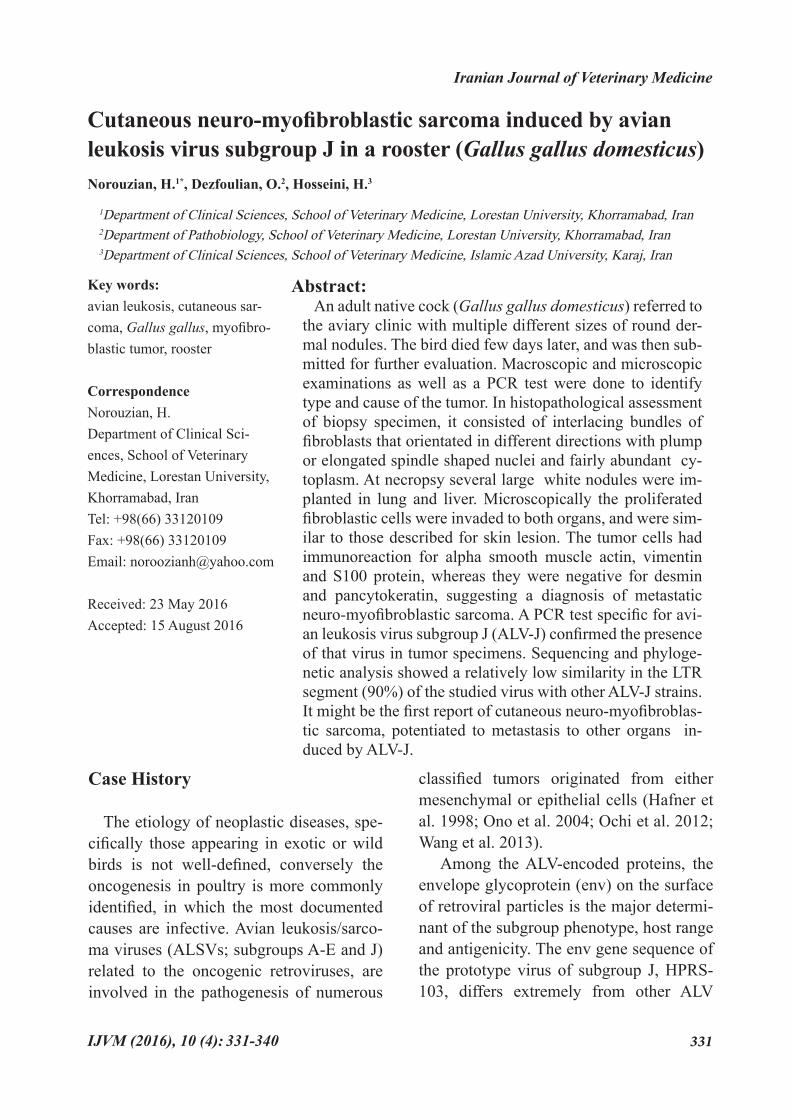
Using primers specific for ALV subgroup J (DU5F and DU5R), a positive result could be detected in all examined tumor tissues (Fig. 7), although the MDV-1 specific se-quence was detected in positive control but not in the tissues. A segment of about 1.8 bp of env gene was detected in the other PCR test.
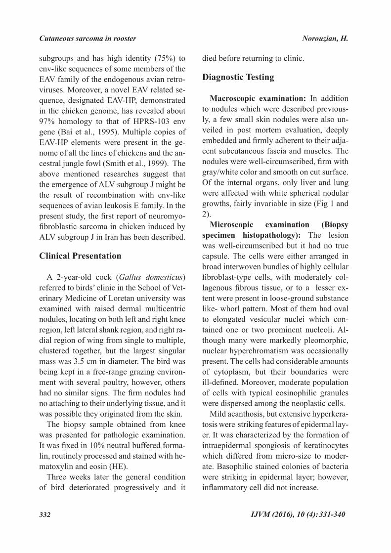
Molecular characterization of LTR. The nucleotide sequence of the LTR region of the studied ALV subgroup J was compared with strains ADOL-7501, HPRS-103 and WN100401 (Fig. 8). A total number of 26 base substitutions were found in the LTR re-gion of the studied ALV subgroup J in com-parison to the prototype strain HPRS-103.
The sequence analysis of LTR of the studied virus revealed that its U3 region has only 215 bp, because of an 11 bp deletion. The R and U5 regions of the studied ALV subgroup J wereconserved compared with other ALV subgroup J isolates.
Some sequences regulating transcrip-tion of the ALV have been identified in the U3 region of the LTR, including CArG, Y, and PRE, C/EBP, CAAT, and TATA boxes (Gao et al. 2012). In most ALV subgroup J isolates (including HPRS-103 and ADOL-7501), there are two PRE boxes (GGTGG motif), flanking the nucleotide sequence AAGTAA. One of the PRE boxes was omit-ted at position 171 to 174, for the deletion (Fig. 8).
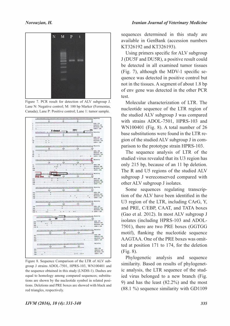
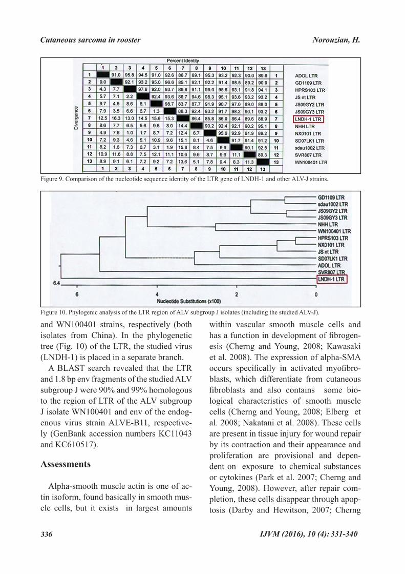
Phylogenetic analysis and sequence similarity. Based on results of phylogenet-ic analysis, the LTR sequence of the stud-ied virus belonged to a new branch (Fig. 9) and has the least (82.2%) and the most (88.1 %) sequence similarity with GD1109
Figure 7. PCR result for detection of ALV subgroup J. Lane N: Negative control; M: 100 bp Marker (Fermentas, Canada); Lane P: Positive control; Lane 1: tumor sample.
Figure 8. Sequence Comparison of the LTR of ALV sub-group J strains ADOL-7501, HPRS-103, WN100401 and the sequence obtained in this study (LNDH-1). Dashes are equal to homology among compared sequences; substitu-tions are shown by the nucleotide symbol in related posi-tions. Deletions and PRE boxes are showed with black and red triangles, respectively.
Norouzian, H.
331-340
336 IJVM (2016), 10 (4):
and WN100401 strains, respectively (both isolates from China). In the phylogenetic tree (Fig. 10) of the LTR, the studied virus (LNDH-1) is placed in a separate branch.
A BLAST search revealed that the LTR and 1.8 bp env fragments of the studied ALV subgroup J were 90% and 99% homologous to the region of LTR of the ALV subgroup J isolate WN100401 and env of the endog-enous virus strain ALVE-B11, respective-ly (GenBank accession numbers KC11043 and KC610517).
Assessments
Alpha-smooth muscle actin is one of ac-tin isoform, found basically in smooth mus-cle cells, but it exists in largest amounts
within vascular smooth muscle cells and has a function in development of fibrogen-esis (Cherng and Young, 2008; Kawasaki et al. 2008). The expression of alpha-SMA occurs specifically in activated myofibro-blasts, which differentiate from cutaneous fibroblasts and also contains some bio-logical characteristics of smooth muscle cells (Cherng and Young, 2008; Elberg et al. 2008; Nakatani et al. 2008). These cells are present in tissue injury for wound repair by its contraction and their appearance and proliferation are provisional and depen-dent on exposure to chemical substances or cytokines (Park et al. 2007; Cherng and Young, 2008). However, after repair com-pletion, these cells disappear through apop-tosis (Darby and Hewitson, 2007; Cherng
Figure 9. Comparison of the nucleotide sequence identity of the LTR gene of LNDH-1 and other ALV-J strains.
Figure 10. Phylogenic analysis of the LTR region of ALV subgroup J isolates (including the studied ALV-J).
Cutaneous sarcoma in rooster Norouzian, H.
331-340

Iranian Journal of Veterinary Medicine
337IJVM (2016), 10 (4):
and Young, 2008). Alpha-SMA is present in mouse subcutaneous fibroblasts (Storch et al. 2007; Cherng and Young, 2008), but in present study it was absent in normal skin tissue of chicken. Therefore, it might be concluded that presence of neoplastic myofibroblasts is a process in response to permanent mutated fibroblasts which are motivated by ALV-subgroup j.
Myofibroblastic tumors which are also designated as myofibrosarcoma and myo-fibroblastic sarcoma are documented in a few reports in veterinary literature (Bell et al. 2011, Newman et al., 1999, Silva et al., 2012).
According to the case, in one study the authors concluded that positive labeling of alpha-smooth muscle actin and vimentin and negative immunoreactions for desmin and S100 protein were suggestive of myo-fibroblastic sarcoma in the limb of a horse (Silva et al . 2012). In another report in a horse, desmin was also positive, similar to alpha-SMA and vimentin but negative for S-100 which the authors believed that the originated tumor based on diagnostic cri-teria of human being was myofibroblastic sarcoma and such tumors are consistently negative to S-100 (Newman et al. 1999).
According to the investigation of 10 fe-line orbital tumors, Bell et al. (2011) real-ized that all myofibroblastic sarcomas im-munostained with smooth muscle actin and vimentin, whereas interestingly, in contrast to previous reports all tumors reacted to S-100 which is probably related to orig-ination of cells from neural crest in facial mesenchyme. As mentioned, other tumors of myofibroblastic sarcomas whether in animal or human being were negative for S-100, therefore, presumably its expression in rooster skin tumor pertains to two distinct
lines of tumor cells. Among tumor types the prevalence of fi-
brosarcoma in chickens is significantly less-er than other tumors which were exposed by infected ALV-J( Cheng et al. 2010). The low incidence of connective tissue neoplasms in chickens indicates that the origin and his-togenesis of such lesions are not defined exactly and their appearance sporadic (Stra-fuss and Ladds, 1970; Reece, 1996).
Based on the PCR results, genome of the studied rooster has been affected by an ALV of subgroup J. In the tumor tissues, MDV proviral DNA was not detected using PCR, but a 1.8- kbp fragment of the env region was amplified and it was most similar to ALVE-B11. ev loci are inheritable provi-ral elements and ubiquitously identified in chicken cells. Normally, few or no endog-enous ALVs (EAV) are expressed from ev loci in chickens. The reason behind this inefficient viral particle production is that the locus has a (+1) frame shift mutation in the pol RT-β region, which results in a truncated RT-β subunit and integrase (John-son and Heneine, 2001). Hatai et al. (2008) isolated a strain of ALV (TymS_90) from layer chickens with fowl glioma that was a recombinant ALV derived from ev-1 and other strains of avian leukemia and sarcoma viruses. Although such hypothesis could be assumed for the ALV subgroup J sequenced in the present study, it could not be clarified whether the detected fragment was derived from an EAV or such a recombinant ALV. More detailed study, using primers specific to ALV subgroup J should be performed in the future.
The pathogenicity and oncogenesis po-tential of ALVs are not merely concerned with changes in the env gene, although they are the most important known factors. Two
Norouzian, H.
331-340

338 IJVM (2016), 10 (4):
non-coding segments in the genome of ALV subgroup J, including U3 region of LTR and E element, also have a significant role in the pathogenicity of the virus (Hue et al. 2006; Gao et al. 2012). The LTR sequences of the ALV subgroup J strains have been previ-ously compared (Gao et al. 2012). The phy-logenetic analysis indicated that the LTR sequence of the studied isolate has approxi-mately low homology with each of the other strains presented in the phylogenetic anal-ysis. Twenty-six nucleotide substitutions in the U3 region were found in the studied isolate. Other scientists also mentioned that the U3 region of LTR evolves rapidly (Gao et al. 2012). A deletion of 11 bp was seen in our studied ALV subgroup J. Such a dele-tion is relatively rare among ALVs of sub-group J and is seen in only some Chinese strains, e.g. WN100401 (accession number HQ271447). The U3 region of the LTR con-tains transcriptional regulating elements and determines both the level of viral transcrip-tion and the oncogenic potential of ALVs (Gao et al. 2012).The nucleotide deletions in the U3 region of LTR in our studied virus caused omission of PRE box (Fig. 8).
This report is the first of ALV subgroup J, obtained for a chicken’s tumor in Iran. The LTR region of the ALV subgroup J isolate, studied in this article, formed a separate branch from the prototype strain HPRS-103, a strain from the US (ADOL-7501) and other Chinese ALV subgroup J strains, including those isolated from broiler or lay-er chickens (Fig. 10). According to the se-quence identity, the homology among LTR region of the studied ALV subgroup J and other ALV-J strains is relatively low (maxi-mum 88.1%) (Fig. 9). Although it is not de-termined whether this information is relat-ed to changes in functional characteristics
of LTR gene, some interesting points on the molecular characteristics of the studied ALV subgroup J isolate were revealed. Such information together with more comprehen-sive future studies on the virus will contrib-ute to a better understanding of the genetic differences of the studied ALV subgroup J with other countries’ isolates.
Acknowledgements
We gratefully acknowledge the Deputy of Research Office of Lorestan University for their financial support.
Cutaneous sarcoma in rooster Norouzian, H.
Bai, J., Payne, L.N., Skinner, M.A. (1995) HPRS-103 (exogenous avian leukosis virus, subgroup J) has an env gene related to those of endogenous elements EAV-0 and E51 and an E element found previously only in sar-coma viruses. J Virol. 69: 779-784. Bell, C.M., Schwarz, T., Dubielzig, R.R. (2011) Diagnostic features of feline restric-tive orbital myofibroblastic sarcoma. Vet Pathol. 48: 742-750.Cheng, Z., Liu, J., Cui, Z., Zhang, L. (2010) Tumors associated with avian leukosis vi-rus subgroup J in layer hens during 2007 to 2009 in China. J Vet Med Sci. 72: 1027-1033.Cherng, S., Young, J., Ma, H. (2008) Alpha smooth muscle actin (alpha-SMA). J Am Sci. 4: 7-9.Darby, I.A., Hewitson, T.D. (2007) Fibro-blast differentiation in wound healing and fibrosis. Int Rev Cytol. 257: 143-179.Elberg, G., Chen, L., Elberg, D., Chan, M.D., Logan, C.J., Turman, M.A. (2008) MKL1 mediates TGF-{beta} 1- induced {alpha}- smooth muscle actin expression in human renal epithelial cells. American J Renal Physiol. 294: 1116-1128.
1.
2.
3.
4.
5.
6.
References
331-340

Iranian Journal of Veterinary Medicine
339IJVM (2016), 10 (4):
Norouzian, H.
Hafner, S., Goodwin, M.A., Smith, E.J., Fad-ly, A., Kelley, L.C. (1998) Pulmonary Sar-comas in a Young Chicken. Avian Dis. 42: 824-826.Handberg, K.J., Nielsen, O.L., Jørgensen, P. H. (2001) The use of serotype 1- and sero-type 3-specific polymerase chain reaction for the detection of Marek’s disease virus in chickens. Avian Pathol. 30: 243-249.Hatai, H., Ochiai, K., Nagakura, K., Iman-ishi, S., Ochi., A., Kozakura, R., Ono, M., Goryo, M., Ohashi, K., Umemura, T. (2008) A recombinant avian leukosis virus associ-ated with fowl glioma in layer chickens in Japan. Avian Pathol. 37: 127-137.Johnson, J.A., Heneine, W. (2001) Charac-terization of endogenous avian leukosis vi-ruses in chicken embryonic fibroblast sub-strates used in production of measles and mumps vaccines. J Virol. 75: 3605-3612.Kawasaki, Y., Imaizumi, T., Matsuura, H., Ohara, S., Takano, K, Suyama, K., Hashimo-to, K., Nozawa, R., Suzuki H., Hosoya, M. (2008) Renal expression of alpha-smooth muscle actin and c-Met in children with Henoch-Schönleinpurpura nephritis. Pedi-atr Nephrol. 23: 913-919.Nakatani, T., Honda, E., Hayakawa, S., Sato, M., Satoh, K, Kudo, M., Munakata, H. (2008) Effects of decorin on the expression of alpha-smooth muscle actin in a human myofibroblast cell line. Mol Cell Biochem, 308: 201-207.Newman, S.J., Cheramie, H., Duniho, S.M., Scarratt, W.K. (1999) Abdominal spindle cell sarcoma of probable myofibroblastic or-igin in a horse. J Vet Diagn Invest. 11: 278-282.Ochi, A., Ochiai, K., Nakamura, S., Koba-ra, A., Sunden, Y., Umemura, T. (2012) Mo-lecular characteristics and pathogenicity of an avian leukosis virus isolated from avian
neurofibrosarcoma. Avian Dis. 56: 35-43.Ono, M., Tsukamoto, K., Tanimura, N., Har-itani, M., Kimura, K.M, Suzuki, G., Okuda, Y., Sato, S. (2004) An epizootic of subcuta-neous tumors associated with subgroup a avian leukosis/sarcoma virus in young layer chickens. Avian Dis. 48: 940-946.Park, S., Bivona, B.J., Harrison-Bernard, L.M (2007) Compromised renal microvas-cular reactivity of angiotensin type 1 double null mice. American J Physiol Renal Physi-ol. 293: 60-67.Reece, R.L (1996) Some observations of naturally occurring neoplasms of domestic fowls I the state of Victoria, Australia (1997-87). Avian Pathol. 25: 407-447.Silva, J.F., Palhares, M.S., Maranhão, R.P.A., Gheller, V.A., Boeloni, J.N., Serakides, R., Ocarino, N.M. (2012) Myofibroblastic Sar-coma in the Limb of a Horse. J Equine Vet Sci. 32: 197-200.Smith, L.M., Toye, A.A., Howes, K, Bum-stead, N., Payne, L.N., Venugopal, K. (1999) Novel endogenous retroviral sequences in the chicken genome closely related to HPRS-103 (subgroup J) avian leukosis virus. J Gen Virol. 80: 261-268.Storch, K.N., Taatjes, D.J., Bouffard, N.A., Locknar, S., Bishop, N.M., Langevin, H.M. (2007) Alpha smooth muscle actin distribu-tion in cytoplasm and nuclear invaginations of connective tissue fibroblasts. Histochem Cell Biol. 127: 523-530.Strafuss, A.C., Ladds, P.W. (1970) Fibrosar-coma in a young chicken. Avian Dis. 14: 406-409.Venugopal, K., Smith, L.M., Howes, K., Payne, L.N. (1998) Antigenic variants of J subgroup avian leukosis virus: sequence analysis reveals multiple changes in the env gene. J Gen Virol. 79: 757-764.Wang, G., Jiang, Y., Yu, L., Wang, Y., Zhao,
7.
8.
9.
10.
11.
12.
13.
14.
15.
16.
17.
18.
19.
20.
21.
22.
23.
331-340

340 IJVM (2016), 10 (4):
Cutaneous sarcoma in rooster Norouzian, H.
X. (2013) Avian leukosis virus subgroup J associated with the outbreak of erythroblas-tosis in chickens in China. Virol J. 10: 92.Zavala, G., Jackwood, M.W., Hilt, D.A. (2002) Polymerase chain reaction for detec-tion of avian leukosis virus subgroup J in feather pulp. Avian Dis. 46: 971-978.
24.
331-340

Abstracts in Persian Language
40
مجله طب دامی ایران، 1395، دوره 10، شماره 4، 331-340
نورومیوفیبروبالستیک سارکومای جلدی متاستاتیک ناشی از لکوز J در یک قطعه خروس بومی: گزارش موردیحسن نوروزیان1* امید دزفولیان2 حسین حسینی3
1( گروه علوم بالینی، دانشکده دامپزشکی دانشگاه لرستان، خرم آباد، ایران2( گروه پاتولوژی، دانشکده دامپزشکی دانشگاه لرستان، لرستان، ایران
3( گروه علوم بالینی، دانشکده دامپزشکی دانشگاه آزاد اسالمی، کرج، ایران
) دریافت مقاله: 3 خرداد ماه 1395، پذیرش نهایی: 25 مرداد ماه 1395(
چکیده تومورهای ناشی از لکوز j در پرندگان فراوان است ولی گزارشات اندکی از آن در ایران ارائه شده است. در این مطالعه یک مورد تومور پوستی ناشی از لکوز j در یک خروس بومی بررسی شده است. یک خروس نژاد الری ارجاع شده به کلینیک دامپزشکی دچار تومورهای گرد پوستی در قسمت های مختلف بدن شده بود. پرنده پس از چند روز تلف شد و برای بررسی بیشتر مورد کالبد گشایی قرار گرفت. در بررسی هیستوپاتولوژی نمونه بیوپسی دسته های فیبروبالست با جهت گیری های مختلف مشاهده شد که هسته آنها حالت کشیده چیدا کرده و حاوی مقدار نسبتًا زیادی سیتوپالسم بود. در کالبد گشایی ندول های بزرگ سفید رنگ در ریه و کبد مشاهده گردید. در بررســی میکروسکوپی سلول های فیبروبالست تکثیر شده مشاهده شــده در این دو اندام مشابه ضایعات پوست بودند. در ایمونوهیستوشیمی بافت های تومورد با اکتین عضله صاف آلفا و پروتیین S100 واکنش نشان دادند ولی با دسمین و پانسیتوکراتین واکنش نشان ندادند که حاکی از سارکومای نورومیو فیبروزی متاستاتیک بود. نتایج آزمون PCR برای بیماری مارک منفی و برای لکوز j مثبت شد. آنالیز فیلوژنیک حاکی از شباهت نسبتًا کم قطعه LTR )90%( ویروس مطالعه شده با دیگر سویه های لکوز J بود. این
مورد اولین گزارش تومور از نوع مذکور ناشی از لکوز j در ایران است.
واژه های کلیدی: لکوز j، سارکومای جلدی، گالوس گالوس، تومور نورومیوفیبروبالستیک، خروس بومی ________________________________________________________________________________________________
Email: [email protected] +98)66( 33120109 :98+ نمابر)( نویسنده مسؤول: تلفن: 33120109 )66*