curriculum / statutes & regulations for 5 years …
TRANSCRIPT

CURRICULUM / STATUTES & REGULATIONS
FOR
5 YEARS DEGREE PROGRAMME IN
ORTHOPAEDICS (MS Orthopaedics)

UNIVERSITY OF HEALTH SCIENCES,
LAHORE
STATUTES
Nomenclature Of The Proposed Course
The name of degree programme shall be MS Orthopaedics. This name is well
recognized and established for the last many decades worldwide.
Course Title:
MS Orthopaedics
Training Centers
Departments of Orthopaedics (accredited by UHS) in affiliated institutes of University
of Health Sciences Lahore.
Duration of Course

The duration of MS Orthopaedics course shall be five (5) years with structured
training in a recognized department under the guidance of an approved
supervisor.
After admission in MS Orthopaedics Programme the resident will spend first 6
Months in the relevant Department of Orthopaedics as Induction period during
which resident will get orientation about the chosen discipline and will also
participate in the mandatory workshops (Appendix E). The research project
shall be designed and the synopsis be prepared during this period
On completion of Induction period the resident shall start training to learn Basic
Principles of General Surgery for 18 Months.
During this period the Research Synopsis shall be got approved by the AS&RB of
the university. At the end of 2nd Calendar year the candidate shall take up
Intermediate Examination.
During 3rd, 4th & 5th years, of the Program, there shall be two components of the
training.
1) Clinical Training in Orthopaedics
2) Research and Thesis writing
The candidate will undergo clinical training in the discipline to achieve the
educational objectives (knowledge & Skills) alongwith rotation in the relevant

fields during the 4th & 5th years of the programme. The clinical training shall be
competency based. There shall generic and specialty specific competencies and
shall be assessed by continuous Internal Assessment. (Appendix F&G).
The Research & Thesis Component shall be completed over the five years
duration of the course. The Candidate will spend total time equivalent to one
calendar year on research during the training. Research can be done as one
block or it can be done as regular periodic rotations over five years as long as
total research time is equivalent to one calendar year.
Admission Criteria
Applications for admission to MS Training Programs will be invited through
advertisement in print and electronic media mentioning closing date of
applications and date of Entry Examination.
Eligibility: The applicant on the last date of submission of applications for
admission must possess the:
i) Basic Medical Qualification of MBBS or equivalent medical qualification
recognized by Pakistan Medical & Dental Council.
ii) Certificate of one year's House Job experience in institutions recognized by
Pakistan Medical & Dental Council Is essential at the time of interview. The
applicant is required to submit Hope Certificate from the concerned Medical
Superintendent that the House Job shall be completed before the Interview.
iii) Valid certificate of permanent or provisional registration with Pakistan

Medical & Dental Council.
Registration and Enrollment
As per policy of Pakistan Medical & Dental Council the number of PG Trainees/
Students per supervisor shall be maximum 05 per annum for all PG
programmes including minor programmes (if any).
Beds to trainee ratio at the approved teaching site shall be at least 5 beds per
trainee.
The University will approve supervisors for MS courses.
Candidates selected for the courses after their enrollment at the relevant
institutions shall be registered with UHS as per prescribed Registration
Regulation.
Accreditation Related Issues Of The Institution
A). Faculty
Properly qualified teaching staff in accordance with the requirements of
Pakistan Medical and Dental Council (PMDC)
B). Adequate Space
Including class-rooms (with audiovisual aids), demonstration rooms, computer lab
and clinical pathology lab etc.
C). Library

Departmental library should have latest editions of recommended books, reference
books and latest journals (National and International).
Accreditation of Orthopaedics training program can be suspended on temporary
or permanent basis by the University, if the program does not comply with
requirements for residents training as laid out in this curriculum.
Program should be presented to the University along with a plan for
implementation of curriculum for training of residents.
Programs should have documentation of residents training activities and
evaluation on monthly basis.
To ensure a uniform and standardized quality of training and availability of the
training facilities, the University reserves the right to make surprise visits of
the training program for monitoring purposes and may take appropriate action
if deemed necessary.

AIMS AND OBJECTIVES OF THE COURSE
AIM
The aim of five years MS programme in Orthopaedics is to train residents to
acquire the competency of a specialist in the field so that they can become
good teachers, researchers and clinicians in their specialty after completion of
their training.

GENERAL OBJECTIVES
MS Orthopaedics training should enable a student to:
1. Access and apply relevant knowledge to clinical practice:
Maintain currency of knowledge
Apply scientific knowledge in practice
Appropriate to patient need and context
Critically evaluate new technology
2. Safely and effectively performs appropriate surgical procedures:
Consistently demonstrate sound surgical skills
Demonstrate procedural knowledge and technical skill at a level
appropriate to the level of training
Demonstrate manual dexterity required to carry out procedures
Adapt their skills in the context of each patient and procedure
Maintain and acquire new skills
Approach and carries out procedures with due attention to safety of
patient, self and others
Critically analyze their own clinical performance for continuous
improvement
3. Design and implement effective management plans:
Recognize the clinical features, accurately diagnose and manage
Orthopaedic problems
Formulate a well-reasoned provisional diagnosis and management plan
based on a thorough history and examination
Formulate a differential diagnosis based on investigative findings

Manage patients in ways that demonstrate sensitivity to their physical,
social, cultural and psychological needs
Recognize disorders of the nervous system and differentiate those
amenable to surgical treatment
Effectively manage the care of patients with Orthopaedic-trauma
including multiple system trauma
Effectively recognize and manage complications
Accurately identify the benefits, risks and mechanisms of action of
current and evolving treatment modalities
Indicate alternatives in the process of interpreting investigations and
in decision-making
Manage complexity and uncertainty
Consider all issues relevant to the patient
Identify risk
Assess and implement a risk management plan
Critically evaluate and integrate new technologies and techniques.
4. Organize diagnostic testing, imaging and consultation as needed:
Select medically appropriate investigative tools and monitoring
techniques in a cost-effective and useful manner
Appraise and interpret appropriate diagnostic imaging and
investigations according to patients' needs
Critically evaluates the advantages and disadvantages of different
investigative modalities
5. Communicate effectively:

Communicate appropriate information to patients (and their family)
about procedures, potentialities and risks associated with surgery in
ways that encourage their participation in informed decision making
Communicate with the patient (and their family) the treatment
options including benefits and risks of each
Communicate with and co-ordinate health management teams to
achieve an optimal surgical environment
Initiate the resolution of misunderstandings or disputes
Modify communication to accommodate cultural and linguistic
sensitivities of the patient
6. Recognize the value of knowledge and research and its application to clinical
practice:
Assume responsibility for self-directed learning
Critically appraise new trends in Orthopaedics
Facilitate the learning of others.
7. Appreciate ethical issues associated with Orthopaedics:
Consistently apply ethical principles
Identify ethical expectations that impact on medico-legal issues
Recognize the current legal aspects of informed consent and
confidentiality
Be accountable for the management of their patients.
8. Professionalism by:
Employing a critically reflective approach to Orthopaedics
Adhering with current regulations concerning workplace harassment

Regularly carrying out self and peer reviewed audit
Acknowledging and have insight into their own limitations
Acknowledging and learning from mistakes
9. Work in collaboration with members of an interdisciplinary team where
appropriate:
Collaborate with other professionals in the selection and use of various
types of treatments assessing and weighing the indications and
contraindications associated with each type
Develop a care plan for a patient in collaboration with members of an
interdisciplinary team
Employ a consultative approach with colleagues and other professionals
Recognize the need to refer patients to other professionals.
10. Management and Leadership
Effective use of resources to balance patient care and system resources
Identify and differentiate between system resources and patient needs
Prioritize needs and demands dealing with limited system resources.
Manage and lead clinical teams
Recognize the importance of different types of expertise which contribute
to the effective functioning of clinical team.
Maintain clinically relevant and accurate contemporaneous records
11. Health advocacy:
Promote health maintenance of patients
Advocate for appropriate health resource allocation

Promote health maintenance of colleagues and self scholar and teacher
SPECIFIC LEARNING OUTCOMES
On completion of the training programme, Orthopaedics trainees pursuing an
academic pathway will be expected to have demonstrated competence in all aspects
of the published syllabus. The specific training component would be targeted for
establishing clearly defined standards of knowledge and skills required to practice
Orthopaedics at secondary and tertiary care level with proficiency in the Basic and
applied clinical sciences, Basic Orthopaedic surgical care, Orthopaedic intensive care,
Emergency medicine and Complementary surgical disciplines. Following competencies
are expected from the resident;
Didactic Education:
Trauma:
1. Diagnose and describe common fractures using terms like comminuted,
open, closed, spiral, impacted, angulated, displaced, segmental, transverse,
or oblique.
2. Describe an open fracture and its management, including the use of
debridement, primary closure, and delayed primary closure.
3. Diagnose common injuries such as Colles' fracture, anterior dislocation of
the shoulder, and hip fractures, given appropriate historical, physical
examination, and radiographic data.
4. Describe the physiologic process of fracture healing and discuss the
importance of factors such as blood supply, contact of fracture fragments,
immobilization, and soft-tissue interposition.

5. Discuss the complications of severe fracture of the spine, pelvis, and major
long bones, including neurologic deficit, genitourinary injury, blood loss,
avascular necrosis, Volkmann's ischemic contracture, and compartment
syndromes.
6. Describe the appropriate diagnostic and first aid measures to be taken
regarding the musculoskeletal system in cases of trauma.
7. Define various modes of treatment for closed fractures, including closed
reduction, open reduction, traction, splinting, circumferential casting,
external fixation, and internal fixation.
8. Describe the differences in treatment, complications, anatomy, and
physiology in children's fractures compared to adults.
Spinal Disorders:
1. Discuss the management of low-back pain, degenerative joint disease of the
spine, and the syndrome of the herniated lumbar or cervical disc.
2. Diagnose acute low-back strain, herniated cervical or lumbar disc with nerve
root irritation (specific roots and disc levels), and degenerative joint disease
of the spine, given appropriate historical, physical, radiographic, and
laboratory data.
Infections of Bones and Joints:
1. Discuss the pathology, physiology and natural history of hematogenous
osteomyelitis, chronic osteomyelitis and septic arthritis.

2. Discuss the therapeutic management of hematogenous osteomyelitis,
chronic osteomyelitis and septic arthritis and the complications of these
diseases.
Pediatric Orthopaedics:
1. Define the terms varus, valgus, equinus, calcaneus, genu, pes, talus,
dislocation, subluxation, and anteversion.
2. Diagnose common mal-alignments of the lower extremities in children,
including increased femoral anteversion, tibial torsion, metatarsus
adductus, and clubfoot, given necessary historical, physical, and
radiographic data.
3. Diagnose congenital dislocation of the hip, Legg-Calvé-Perthes disease,
slipped capital femoral epiphysis, and idiopathic scoliosis, given necessary
historical, physical, and radiographic data.
Inflammatory, Degenerative, and Traumatic Disorders of
Musculoskeletal Soft Tissue:
1. Diagnose common disorders of soft tissue, including tendinitis and bursitis
of the shoulder, lateral humeral epicondylitis, and bursitis
about the greater trochanter and knee, given the appropriate historical,
physical, radiographic and laboratory data.
2. Discuss the diagnostic criteria differentiating degenerative arthritis from
rheumatoid arthritis.
3. Discuss osteotomy, arthrodesis and arthroplasty as they apply to the
treatment of arthritis.
Clinical Knowledge:
1. Cognitive knowledge: Describe embryology, applied anatomy, physiology,
pathology, clinical features, diagnostic procedures and the therapeutics
including preventive methods, (medical/surgical) pertaining to musculo-
skeletal system.
2. Clinical decision making ability & management expertise: Diagnose
conditions from history taking, clinical evaluation and investigations and
develop expertise to manage medically as well as surgically the commonly
encountered, disorders and disease in different areas as follows:
a. Pediatric orthopaedics- The student should be exposed to all aspects of
congenital and developmental disorders such as CTEV (club-Foot),
developmental dysplasia of hip, congenital deficiency of limbs, Perthe’s
disease and infections, and also to acquire adequate knowledge about the
principles of management of these disorders in 6 months rotation.
b. Orthopaedic oncology- The resident is expected to be familiar with the
tumours encountered in orthopaedic practice. The recent trends towards limb
salvage procedures and the advances in chemotherapy need to be familiar to
him.
c. Management of Trauma- Trauma in this country is one of the main causes
of morbidity and mortality in our demographic statistics. The student is
expected to be fully conversant with trauma in its entirety. In any type of
posting after qualification the orthopaedic surgeon would be exposed to all

varieties of acute trauma. Hence, it is his responsibility to be able to
recognize, assess and manage it including the medico legal aspects.
d. Sports Medicine- A lot of importance is being given to sports medicine
especially in view of the susceptibility of the athlete to injury and his failure to
tide over them. Sports medicine not only encompasses diagnostic and
therapeutic aspects of athletic injuries but also their prevention, training
schedules of personnel & their selection.
e. Physical Medicine and Rehabilitation- The student is expected to be
familiar with this in all its aspects. Adequate exposure in the workshop
manufacturing orthotics and prosthetics is mandatory, as is the assessment of
the orthopedically handicapped.
f. Orthopedic Neurology- The student should be exposed to all kinds of nerve
injuries as regards their recognition & management cerebral palsy and
acquired neurologic conditions such as post polio residual paralysis also need
to be emphasized in their entirety.
g. Spine Surgery- The student is expected to be familiar with various kinds of
spinal disorders such as scoliosis, kypho-scoliosis, spinal trauma, PIVD,
infections (tuberculosis and pyogenic), & tumours as regards their clinical
presentations and management.
h. Basic sciences in Orthopaedics- This deals with some of the fundamentals
in orthopaedics such as the structure and function of bone cartilage etc, and
their metabolic process. In addition the student learns about implants in
orthopaedics and their metallurgy.

i. Radiology- Acquire knowledge about radiology/imaging and to interpret
different radiological procedures and imaging in musculo-skeletal disorders.
There should be collaboration with Radiology department for such activities.
j. Psychologic and social aspect- Some elementary knowledge in clinical
Psychology and social, work management is to be acquired for management
of patients, especially those terminally ill and disabled-persons and interacting
with their relatives.
3. Teaching: Acquire ability to teach an MBBS student in simple and
straightforward language about the common orthopaedic ailment/disorders
especially about their signs/symptoms for diagnosis with their general
principles of therapy.
4. Preventive Aspect: Acquire knowledge about prevention of some conditions
especially in children such as poliomyelitis, congenital deformities, cerebral
palsy and common orthopaedic malignancies.
5. Identification of a special areas within the subject: To further develop
higher skills within the specialty in a specialized area such as Arthroplasty,
Neurology, Arthroscopy oncology, spine surgery, hand surgery and
Rheumatology, identify some area of interest during the residency and do
fellowship/ senior residency programme in one of such areas.
6. Research Experience:
All residents in the categorical program are required to complete an academic
outcomes-based research project during their training. This project can consist

of original bench top laboratory research, clinical research or a combination of
both. The research work shall be compiled in the form of a thesis which is to be
submitted for evaluation by each resident before end of the training. The
designated Faculty will organize and mentor the residents through the process,
as well as journal clubs to teach critical appraisal of the literature
Clinical Skills to be Learnt
Demonstrate the following clinical skills with proficiency:
History of Orthopaedics
Professional Values, Student – Teacher relationship.
Laws of Biomechanics
Orientation of Out-patient, In patient, Accident & Emergency, Operation
Theatre and learning resources.
History taking, date extraction and recording, documentation and
presentation.
Examination of swelling, wounds, deformities.
Examination of Joints with special reference to measurement of range of
movements.
Examination of Muscle power, tone and function.
Examination of sensory and motor nerves.
Interpretation of X-ray, CT scan, Ultra sound, MRI and Bone Scans.
Application and removal of Splints and POP casts.
Application of Tractions.
Administration of Injections / Aspirations.
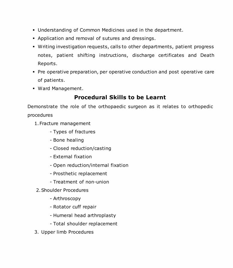
Understanding of Common Medicines used in the department.
Application and removal of sutures and dressings.
Writing investigation requests, calls to other departments, patient progress
notes, patient shifting instructions, discharge certificates and Death
Reports.
Pre operative preparation, per operative conduction and post operative care
of patients.
Ward Management.
Procedural Skills to be Learnt
Demonstrate the role of the orthopaedic surgeon as it relates to orthopedic
procedures
1. Fracture management
- Types of fractures
- Bone healing
- Closed reduction/casting
- External fixation
- Open reduction/internal fixation
- Prosthetic replacement
- Treatment of non-union
2. Shoulder Procedures
- Arthroscopy
- Rotator cuff repair
- Humeral head arthroplasty
- Total shoulder replacement
3. Upper limb Procedures
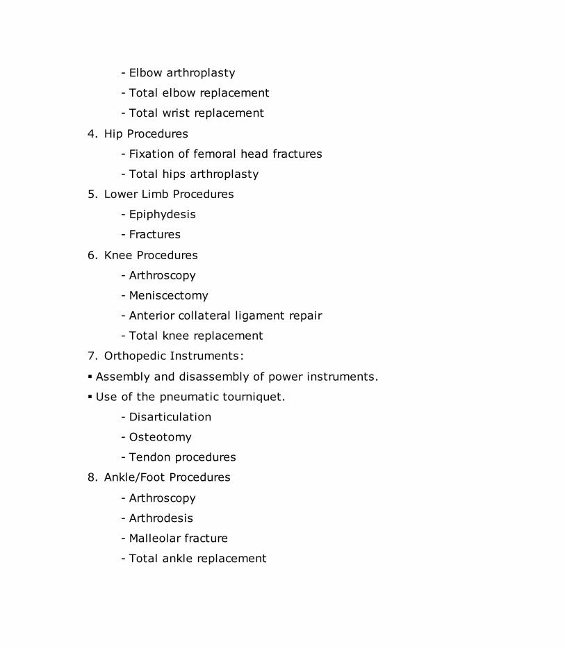
- Elbow arthroplasty
- Total elbow replacement
- Total wrist replacement
4. Hip Procedures
- Fixation of femoral head fractures
- Total hips arthroplasty
5. Lower Limb Procedures
- Epiphydesis
- Fractures
6. Knee Procedures
- Arthroscopy
- Meniscectomy
- Anterior collateral ligament repair
- Total knee replacement
7. Orthopedic Instruments:
Assembly and disassembly of power instruments.
Use of the pneumatic tourniquet.
- Disarticulation
- Osteotomy
- Tendon procedures
8. Ankle/Foot Procedures
- Arthroscopy
- Arthrodesis
- Malleolar fracture
- Total ankle replacement

- Triple arthrodesis
- Bunionectomy
- Correction of hammer toe
9. Miscellaneous
- Amputation
- Disarticulation
- Osteotomy
- Tendon procedures
A Resident, pursuing MS Orthopaedics Degree course is expected to perform
major and minor surgical procedures independently as well as under
supervision of a faculty member/senior resident.
1. Resident should be able to perform many major procedures
independently such as (few examples):
Closed reduction of fractures
External fixation of compound fractures
Debridement of crush injuries
Amputations
Internal fixation of common simple fractures
Polio surgery such as TA lengthening, Steindler’s procedure etc
Intra-articular injections
Steroid injections for various painful conditions
Sequestrectomy in chronic osteomyelitis
Corrective plaster of Paris (POP) casts for club foot & other congenital
deformities
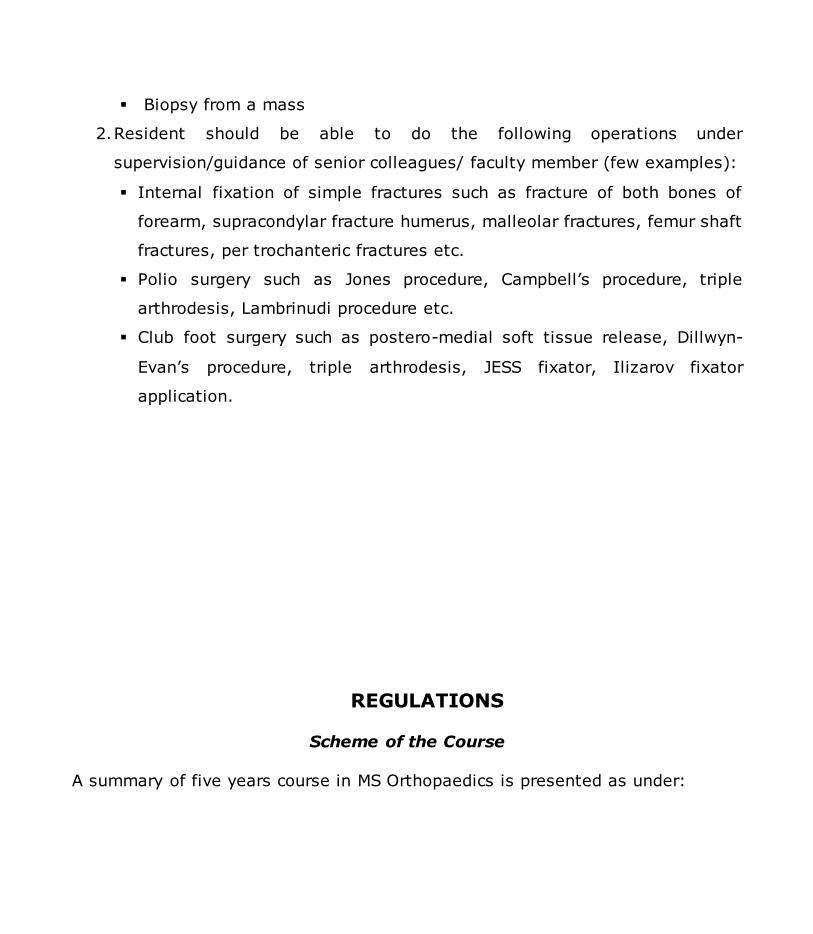
Biopsy from a mass
2. Resident should be able to do the following operations under
supervision/guidance of senior colleagues/ faculty member (few examples):
Internal fixation of simple fractures such as fracture of both bones of
forearm, supracondylar fracture humerus, malleolar fractures, femur shaft
fractures, per trochanteric fractures etc.
Polio surgery such as Jones procedure, Campbell’s procedure, triple
arthrodesis, Lambrinudi procedure etc.
Club foot surgery such as postero-medial soft tissue release, Dillwyn-
Evan’s procedure, triple arthrodesis, JESS fixator, Ilizarov fixator
application.
REGULATIONS
Scheme of the Course
A summary of five years course in MS Orthopaedics is presented as under:

Course Structure Components Examination
At the
End of
2nd
year MS Orthopa
edics
Program
me
Principles of General Surgery
Relevant Basic Science (Anatomy,
Physiology, Pharmacology & Pathology)
Intermediate Examination at the
end of 2nd Year of M.S. Orthopaedics
Programme
Written MCQs = 300 Marks Clinical, TOACS/OSCE & ORAL = 200 Marks
Total = 500 Marks
At the
end of
5th year
MS Orthopa
edics
Program
me
Clinical component
Training in Orthopaedics with rotations in the
relevant fields.
Research component
Research work / Thesis writing must be
completed and thesis be submitted
atleast 6 months before the end of final
year of the programme.
Final Examination at the end of 5th
year of M.S. Orthopaedics
Programme.
Written = 500 Marks
Clinical, TOACS/OSCE & ORAL = 500 Marks
Contribution of CIS = 100 Marks
Thesis Evaluation = 400 Marks
Total = 1500 Marks
Thesis evaluation and defense at the end
of 5th year of the programme.

Intermediate Examinations M. S. Orthopaedics
All candidates admitted in M.S. Orthopaedics courses shall appear in Intermediate
examination at the end of second calendar year.
Eligibility Criteria:
The candidates appearing in Intermediate Examination of the M.S.
Orthopaedics Programme are required:
a) To have submitted certificate of completion of mandatory workshops.
b) To have submitted certificate of completion of first two years of
training from the supervisor.
c) To have submitted CIS assessment proforma from his/her own
supervisor on 03 monthly basis and also from his/her supervisors
during rotation, achieving a cumulative score of 75%.
d) To have submitted certificate of approval of synopsis or undertaking /
affidavit that if synopsis not approved with 30 days of submission of
application for the Intermediate Examination, the candidate will not be
allowed to take the examinations and shall be removed from the
training programme.
e) To have submitted evidence of payment of examination fee.

Intermediate Examination Schedule and Fee
a) Intermediate Examination at completion of two years training, will be
held twice a year.
b) There will be a minimum period of 30 days between submission of
application for the examination and the conduction of examination.
c) Examination fee will be determined periodically by the University.
d) The examination fee once deposited cannot be refunded / carried over
to the next examination under any circumstances.
e) The Controller of Examinations will issue Roll Number Slips on receipt
of prescribed application form, documents satisfying eligibility criteria
and evidence of payment of examination fee.
All candidates admitted in MS Orthopaedics courses shall appear in Intermediate
examination at the end of second calendar year.
Written Examination = 300 Marks
Clinical, TOACS/OSCE & ORAL = 200 Marks
Total = 500 Marks
Written:
MCQs 100 (2 marks each MCQ)
SEQs 10 (10 Marks each SEQ)
Total = 300 Marks
Components of Theory Paper

Principles of General Surgery = 70 MCQs 7 SEQs
Specialty specific = 10 MCQs 1 SEQs
Basic Sciences = 20 MCQs 2 SEQs
Anatomy = 6 MCQs 1 SEQs
Pharmacology = 2 MCQs -------
Pathology = 6 MCQs 1 SEQ
Physiology = 6 MCQs -------
Clinical, TOACS/OSCE & ORAL
Four Short Cases = 100 Marks
One Long Case = 50 Marks
TOACS/OSCE & ORAL = 50 Marks
Total = 200 Marks
Declaration of Results

The Candidate will have to score 50% marks in written and oral, practical/
clinical component and a cumulative score of 60% to be declared successful
in the Intermediate Examination.
A maximum total of four consecutive attempts (availed or unavailed) will be
allowed in the Intermediate Examination during which the candidate will be
allowed to continue his training program. If the candidate fails to pass his
Intermediate Examination within the above mentioned limit of four attempts,
the candidate shall be removed from the training program, and the seat
would fall vacant, stipend/ scholarship if any would be stopped.
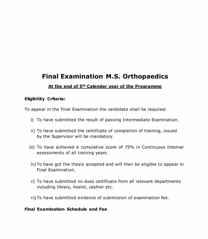
Final Examination M.S. Orthopaedics
At the end of 5th Calendar year of the Programme
Eligibility Criteria:
To appear in the Final Examination the candidate shall be required:
i) To have submitted the result of passing Intermediate Examination.
ii) To have submitted the certificate of completion of training, issued
by the Supervisor will be mandatory.
iii) To have achieved a cumulative score of 75% in Continuous Internal
assessments of all training years.
iv) To have got the thesis accepted and will then be eligible to appear in
Final Examination.
v) To have submitted no dues certificate from all relevant departments
including library, hostel, cashier etc.
vi) To have submitted evidence of submission of examination fee.
Final Examination Schedule and Fee

a) Final examination will be held twice a year.
b) The candidates have to satisfy eligibility criteria before permission is
granted to take the examination.
c) Examination fee will be determined and varied at periodic intervals by
the University.
d) The examination fee once deposited cannot be refunded / carried over
to the next examination under any circumstances.
e) The Controller of Examinations will issue an Admittance Card with a
photograph of the candidate on receipt of prescribed application form,
documents satisfying eligibility criteria and evidence of payment of
examination fee. This card will also show the Roll Number, date / time
and venue of examination.
Written Part = 500 Marks
Clinical, TOACS/OSCE & ORAL = 500 Marks
Contribution Internal Assessment = 100 Marks
Thesis Examination = 400 Marks
Total = 1500 Marks
Written Papers:
Paper 1 = 100 MCQs 5 SEQs
Paper 2 = 100 MCQs 5 SEQs

Clinical, TOACS/OSCE & ORAL
Short Cases = 200 Marks
Long Case = 100 Marks
Toacs/OSCE & Oral = 200Marks
Total = 500 Marks
Declaration of Result
For the declaration of result
I. The candidate must get his/her Thesis accepted.
II. The candidate must have passed the final written examination
with 50 % marks and the clinical & oral examination securing
50% marks. The cumulative passing score from the written and
clinical / oral examination shall be 60%.
III. The MS degree shall be awarded after acceptance of thesis and
success in the final examination.
IV. On completion of stipulated training period, irrespective of the
result (pass or fail) the training slot of the candidate shall be
declared vacant.

Submission / Evaluation of Synopsis
1. The candidates shall prepare their synopsis as per guidelines provided by
the Advanced Studies & Research Board, available on university website.
2. The research topic in clinical subject should have 30% component related
to basic sciences and 70% component related to applied clinical sciences.
The research topic must consist of a reasonable sample size and sufficient
numbers of variables to give training to the candidate to conduct
research, to collect & analyze the data.
3. Synopsis of research project shall be submitted by the end of the 2nd
year of MS program. The synopsis after review by an Institutional Review
Committee shall be submitted to the University for consideration by the
Advanced Studies & Research Board, through the Principal / Dean /Head
of the institution.
Submission of Thesis
1. Thesis shall be submitted by the candidate duly recommended by the
Supervisor.
2. The minimum duration between approval of synopsis and submission of
thesis shall be one year.
3. The research thesis must be compiled and bound in accordance with the
Thesis Format Guidelines approved by the University and available on
website.

4. The research thesis will be submitted along with the fee prescribed by the
University.
Thesis Examination
a) The candidate will submit his/her thesis at least 06 months prior to
completion of training.
b) The Thesis along with a certificate of approval from the supervisory will
be submitted to the Registrar’s office, who would record the date / time
etc. and get received from the Controller of Examinations within 05
working days of receiving.
c) The Controller of Examinations will submit a panel of eight examiners
within 07 days for selection of four examiners by the Vice Chancellor. The
Vice Chancellor shall return the final panel within 05 working days to the
Controller of Examinations for processing and assessment. In case of any
delay the Controller of Examinations would bring the case personally to
the Vice Chancellor.
d) The Supervisor shall not act as an examiner of the candidate and will not
take part in evaluation of thesis.
e) The Controller of Examinations will make sure that the Thesis is
submitted to examiners in appropriate fashion and a reminder is sent
after every ten days.
f) The thesis will be evaluated by the examiners within a period of 06
weeks.
g) In case the examiners fail to complete the task within 06 weeks with 02
fortnightly reminders by the Controller of Examinations, the Controller of
Examinations will bring it to the notice of Vice Chancellor in person.

h) In case of difficulty in find an internal examiner for thesis evaluat ion, the
Vice Chancellor would, in consultation with the concerned Deans, appoint
any relevant person as examiner in supersession of the relevant Clause
University Regulations.
i) There will be two internal and two external examiners. In case of difficulty
in finding examiners, the Vice Chancellor would, in consultation with the
concerned Deans, appoint minimum of three, one internal and two
external examiners.
j) The total marks of thesis evaluation will be 400 and 60% marks will be
required to pass the evaluation.
k) The thesis will be considered / accepted, if the cumulative score of all
the examiners is 60%.
l) The clinical training will end at completion of stipulated training period but
the candidate will become eligible to appear in the Final Examination at
completion of clinical training and after acceptance of thesis. In case
clinical training ends earlier, the slot will fall vacant after stipulated
training period.
Award of MS Orthopaedics Degree
After successful completion of the structured courses of MS Orthopaedics and
Qualifying Intermediate and Final Examinations (written, Clinical, TOACS/OSCE &
ORAL and Thesis) the degree with title MS Orthopaedics shall be awarded.

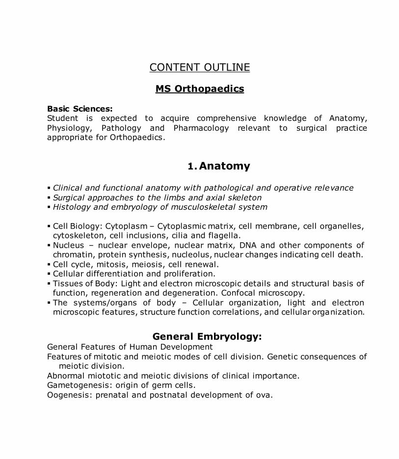
CONTENT OUTLINE
MS Orthopaedics
Basic Sciences: Student is expected to acquire comprehensive knowledge of Anatomy,
Physiology, Pathology and Pharmacology relevant to surgical practice appropriate for Orthopaedics.
1. Anatomy
Clinical and functional anatomy with pathological and operative relevance
Surgical approaches to the limbs and axial skeleton Histology and embryology of musculoskeletal system
Cell Biology: Cytoplasm – Cytoplasmic matrix, cell membrane, cell organelles,
cytoskeleton, cell inclusions, cilia and flagella.
Nucleus – nuclear envelope, nuclear matrix, DNA and other components of chromatin, protein synthesis, nucleolus, nuclear changes indicating cell death.
Cell cycle, mitosis, meiosis, cell renewal. Cellular differentiation and proliferation.
Tissues of Body: Light and electron microscopic details and structural basis of function, regeneration and degeneration. Confocal microscopy.
The systems/organs of body – Cellular organization, light and electron microscopic features, structure function correlations, and cellular organization.
General Embryology: General Features of Human Development
Features of mitotic and meiotic modes of cell division. Genetic consequences of meiotic division.
Abnormal miototic and meiotic divisions of clinical importance. Gametogenesis: origin of germ cells.
Oogenesis: prenatal and postnatal development of ova.

Spermatogenesis: proliferation and maturation of male germ cells. Abnormal gametes, their clinical significance.
Ovulation, fertilization and the consequences of fertilization.
Early Embryonic Development: Cleavage, morula and blastocyst formation and implantation. Formation of the
three primary germ layers. List of the derivatives of the respective germ layers.
Period of the Growing Fetus: Various stages and salient features of the fetus development
Extraembryonic Membranes:
Development, functions and anomalies of yolk sac, amnion, chorion, allantois, umbilical cord and placenta.
Development of the External Body Form:
Shaping of the head, neck, trunk and limbs. Common developmental anomalies associated with this.
The Branchial Apparatus: Development and fate of the bronchial grooves, arches and pouches. Their
derivatives and anomalies.
Teratogenesis: Factors known to be involved in the development of congenital anomalies
especially related to the musculoskeletal system. Concept of critical periods.
General Histology: Structural and Functional Organization of the Tissues of Body
Classification of tissues and identification of various tissues particularly those related to the musculoskeletal system, in routine histological preparations under
the light microscope.
The Epithelial Tissue

General structure, functions and classification of epithelia Their location in the body
General characters of serous and mucous membranes General structural features of exocrine and endocrine glands
The Connective Tissue
Cartilage Structure of bone marrow. Cell lines seen in haemopoiesis.
Factors required for bone growth.
The Muscular Tissue
Structural and functional differences between the smooth skeletal and cardiac types of muscle.
Fine structure of skeletal and cardiac muscle fibers, and its relationship to the mechanism of contraction.
Specialized conducting tissue of the heart.
The Neural Tissue The neuron, morphology of the perikaryon and its processes. Coverings of the axons in the peripheral nerves and the central nervous system.
Types of neuroglia and their functions.
Process of myelination in the peripheral nerves and the central nervous system.
Axon terminals and synapses. Nerve fiber degeneration and regeneration.
Gross Anatomy / Surface Anatomy
1. Musculoskeletal System
Back and Spinal Cord Muscles of the Back Vertebral Canal and Spinal Cord
Upper Limb Scapular Region
Pectoral Region Axillary Region
Arm

Flexor Region of the Forearm Palm of the Hand
Extensor Region of the Forearm and Dorsum of the Hand Joints of the Upper Extremity
Origins and insertions of all muscles associated with the upper extremity. Basic functions of these muscles.
Nerve and general arterial supplies of all muscles associated with the upper extremity.
Structural entities of the upper extremity Structural relationship of a these structures to all neighboring structures. Effect of injury to specific peripheral nerves (i.e. Muscles affected, specific
deformity encountered). Locations for palpation of the pulse in major arteries of the upper limb.
Locations of major nerves of the upper limb dermatomes of the upper limb.
Lower Limb
Superficial Anatomy of the Lower Extremity Anterior and Medial Thigh Region Gluteal Region
Posterior Thigh and Popliteal Fossa Leg
Foot Major Joints of the Lower Extremity
Origins and insertions of all muscles associated with the lower extremity. Basic functions of these muscles.
Nerve and general arterial supplies of all muscles associated with the lower extremity.
Structural entities of the lower extremity and their structural relationship to
all neighboring structures.
Effect of injury to specific peripheral nerves (i.e. Muscles affected, specific deformity encountered).
Locations for compression and palpation of the pulse in major arteries of the lower limb.
Dermatomes of the lower limb. Thorax
Thoracic Wall

Pleural Cavities and Lungs Heart
Mediastinum Bony thorax and relation with the muscles of respiration.
Major vessels and nerves of the thorax and the structures supplied by each.
Organic structural entities of the thorax and the structural relationship of these to all neighboring structures.
Surface projections of the thoracic viscera. Head and Neck
Osteology of the Skull
Superficial Face Posterior Triangle of the Neck
Anterior Triangle of the Neck Cervical Viscera
Infratemporal Fossa Cranial Fossa
Orbits and Eye Temporal Bone and Ear Pharynx
Larynx Nasal Cavity
Oral Cavity Origins and insertions of all muscles associated with the head and neck
Basic functions of these muscles. Nerve supply of all the muscles.
Structural entities of the head and neck and their structural relationship to all neighboring structures.
Locations for palpation of the pulse in major arteries of the head and neck.
Locations of major nerves of the head and neck, along with their cutaneous distributions.
Sensory and/or motor functions, as they apply for the 12 pairs of cranial nerves (including pupillary light and accommodation reflexes, corneal
reflex, and gag reflex). Abdomen:
Anterior Abdominal Wall Inguinal Region, Scrotum and Testes Abdominal Cavity

Stomach, Spleen and Liver Intestines and Pancreas
Posterior Abdominal Wall
Composition of the abdominal wall and identify all structures Significance of the contents of the spermatic cord.
Visceral and peritoneal structures of the abdomen and their structural relationship of a sample structure to all neighboring structures.
Major vessels of the abdominal cavity. General terms the innervation of the GI tract. Surface projections of the abdominal viscera.
Pelvis and Perineum:
Female Perineum Male Perineum and the Penis
Female Pelvis Male Pelvis
Male and female pelvic organs, and external structures.
Bones
Developmental aspects of the skeleton Identification of bony outlines on plain x-ray.
Classification of bones. Bone growth and ossification.
Blood supply of all long and small bones of human body
Axial Skeleton: Skull /
Cranial bones Facial bones
Auditory ossicles Splanchnocranial bones
Vertebral column o Vertebrae
o Cervical o Thoracic o Lumbar

o Sacrum o Coccyx
Curvatures of the spine Primary
Secondary Abnormal spine
o Kyphosis o Lordosis
o Scoliosis Thorax
o Sternum
o Manubrium o Ribs
o Costal cartilages
Appendicular Skeleton
Upper Extremity
Pectoral girdle Scapula
Clavicle Shoulder joint
Brachium [arm] Elbow joint Antebrachium [forearm]
Wrist [carpus] Carpal bones
Hand [manus]
Lower Extremity
Pelvic girdle Hip joint
Thigh Knee Leg
Ankle [tarsus]

Tarsal bones Foot
Joints
Classification of joints Fibrous Joints
Cartilaginous Joints Synovial Joint
Plane Joint Hinge Joint Pivot Joint
Condyloid Joint Saddle Joint
Ball-and-Socket Joint Inflammatory Disorders of Joints
Factors contributing to the stability of joints. Movements of the joints of shoulder, elbow, hip, knee and ankle.
Movements of the shoulder girdle as a whole, supination and pronation of forearm, inversion and aversion of foot and movements of fingers and thumb. Maintenance of normal posture
Muscles and Fasciae
Muscles of the human body General disposition, nerve supply and effects of nerve lesions
Muscle attachments, group actions and nerve supply.
Body Cavities:
Abdominal, thoracic, cranial, pelvic cavity A general description of the boundaries, land marks and surface anatomy of the
internal organs and dermatomes of the body cavities General disposition, morphology, relations, blood and nerve supply, lymph nodes
and areas of drainage of the viscera contained in these cavities. Identification of bony outlines on plain X-ray.

Cross Sectional / Imaging Anatomy of the Musculoskeletal System:
Skull Anterior View
Lateral View Posterior View
Superior View Inferior View Internal View
Mandible Fetal Skull
Vertebral Column Atlas (C1)
Axis (C2) Cervical
Thoracic Lumbar Sacrum
Thoracic Bones Rib & Vertebra Articulated
Sternum Upper Limb Bones
Scapula (Posterior Aspect) Scapula (Lateral Aspect)
Humerus (Proximal End) Humerus (Distal End)
Ulna Radius Hand (Dorsal Surface)
Hand (Palmar Surface) Lower Limb Bones
Os Coxa (Lateral Aspect) Os Coxa (Medial Aspect)
Femur (Proximal End) Femur (Distal End)
Tibia
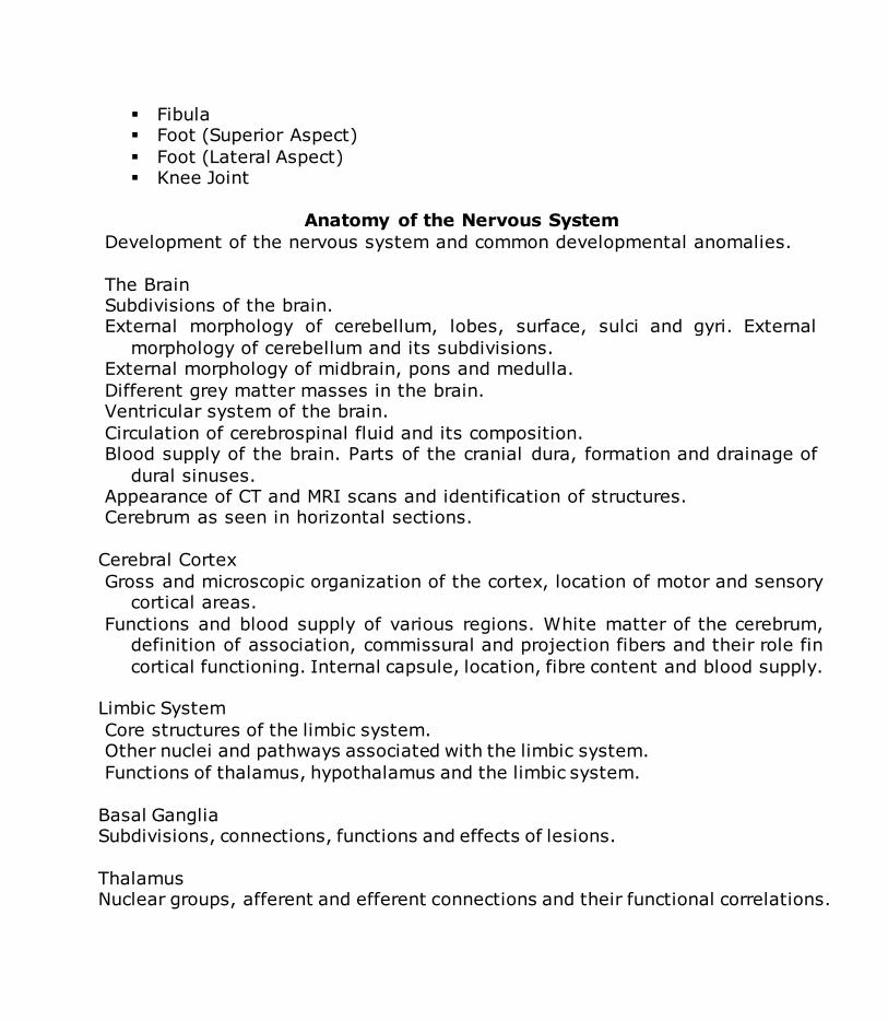
Fibula Foot (Superior Aspect)
Foot (Lateral Aspect) Knee Joint
Anatomy of the Nervous System
Development of the nervous system and common developmental anomalies.
The Brain Subdivisions of the brain. External morphology of cerebellum, lobes, surface, sulci and gyri. External
morphology of cerebellum and its subdivisions. External morphology of midbrain, pons and medulla.
Different grey matter masses in the brain. Ventricular system of the brain.
Circulation of cerebrospinal fluid and its composition. Blood supply of the brain. Parts of the cranial dura, formation and drainage of
dural sinuses. Appearance of CT and MRI scans and identification of structures. Cerebrum as seen in horizontal sections.
Cerebral Cortex
Gross and microscopic organization of the cortex, location of motor and sensory cortical areas.
Functions and blood supply of various regions. White matter of the cerebrum, definition of association, commissural and projection fibers and their role fin
cortical functioning. Internal capsule, location, fibre content and blood supply. Limbic System
Core structures of the limbic system. Other nuclei and pathways associated with the limbic system.
Functions of thalamus, hypothalamus and the limbic system.
Basal Ganglia Subdivisions, connections, functions and effects of lesions.
Thalamus Nuclear groups, afferent and efferent connections and their functional correlations.

Hypothalamus
The nuclei, afferent and efferent connections and their functional correlations. Effects of lesions.
Internal Structure Of Cerebellum
Cerebellar cortex: organization and functions. Cerebellar nuclei: main connections.
Cerebellar peduncles, cerebellar afferent and efferent connections, functional correlations. Effects of lesions.
Spinal Cord
External morphology, meninges and blood supply of the spinal cord. Relationship of the "segments" to vertebrae at different ages.
Internal structure of the spinal cord, organization of the grey and white matter.
Variations sin the structure of the grey matter at different levels and location of the important nuclei. Location of ascending and descending tracts, and their functions.
Effects of injury or disease.
Peripheral Nervous System Anatomy and functions of cranial nerves with their intracranial and extracranial
course and distribution. Location of various cranial nerve nuclei.
Anatomy and functions of spinal nerves. Foundation, course and distribution of a typical nerve. Effects of lesions.
Respiration: Pulmonary ventilation
Mechanics of respiration, pulmonary volumes, capacities and pressures. Transport and exchange of oxygen and carbon dioxide.
Regulation of respiration. (chemical and neural) Physiology of respiratory insufficiencies, hypoxia, dyspnoea, asphyxia
and hypercapnia.
Exercise hypoxia and cyanosis Physiological changes due to altitude and space travel
Principles and methods of artificial respiration. Principles of pulmonary function tests.
Interpretation of data of diagnostic tests. Cardiopulmonary resuscitation.
Patho-physiology of respiratory failure.
Renal function: Renal circulation Glomerular filtration
Tubular function Water excretion
Acidification of urine Regulation of Na + and K + excretion
Regulation of extracellular fluid composition and volume Homeostatic mechanisms to maintain
Tonicity Volume H+ concentration of ECF.
Central Nervous System
Motor cortex corticospinal and corticobulbar system. Basal ganglia
Cerebellum Autonomic Nervous System
Overall functions of sympathetic and parasympathetic nervous systems. Autonomic reflex activity.
Functional Aspects of the Nervous System
Sensory activity: Peripheral sensory receptors, sensory pathways, physiology of pain and disorders of sensations.
Motor activity: corticospinal and extracorticospinal pathways, cerebellum and Vestibular system.
Motor neurons, motor units and neuromuscular junction. Disorders of motor activity.
Muscle and nerve physiology. Reflex activity: Monosynaptic stretch reflexes, polysynaptic withdrawal
reflexes, general characters of reflexes.
Electroencephalogram and its uses. Sleep, types, physiological changes during sleep.
Speech mechanism and its disorders. Cerebrospinal fluid, cerebral circulation, metabolism and functions.
Blood brain and blood CSF barriers.
2. Physiology
Cellular organization, structure function correlations and physiological alterations in the organ systems of body with particular emphasis on the musculoskeletal system
Structural and Functional Organization of the Cells of the Body
Concept of cells as the structural, functional and genetic units of the body. Composition of protoplasm, division into cytoplasm and nucleus.
Role of macromolecules in the structural organization of the cell. Cell components with their role in cell function.
Diversity of cell morphology as related to the varied functional demands. Physical activities of the living cells, intracellular movements, cellular locomotion, endocytosis and exocytosis.
Basic concepts of the principles of transport through cell membrane,
membrane potential and action potential. The cell cycle and cell division.
Energy balance, metabolism & nutrition Uses of cell and tissue cultures.
DNA and RNA structure and protein synthesis. Musculoskeletal Physiology
Introduction Muscle types—skeletal, smooth and cardiac
Shared characteristics Skeletal muscle filaments and associated proteins
Thick filament—myosin Thin filament—actin and tropomyosin/troponin complex
Skeletal muscle fibre types: Type I

Type IIa Type IIx
Contraction cycle Sliding filament hypothesis
Ratchet theory of muscle contraction Biochemical events that occur during a contraction cycle
Mechanical properties of skeletal muscle Length-tension relationship
Force-velocity relationship Motor units The functional unit of a muscle
Motor unit recruitment Muscle contraction, all or non law
Definition, methods of obtaining muscle relaxation Summation and tetanus
Cumulative effect of repeated stimulation of a muscle Group action of muscles
Control of coordination Voluntary movement Pattern of movement
Muscle training and fatigue Tetanus—electrophysiological explanation
Treppe or the staircase effect during repetitive stimulation of muscle contraction
Energy sources for skeletal muscle contraction Short-term regeneration of ATP
Anaerobic—glycolysis Aerobic—Kreb’s Cycle/Oxidative Phosphorylation Fast twitch—Glycolytic (White)
Specialized cellular and subcellular structures Trophic factors that influence neuromuscular junction development
Biosynthesis and metabolism of the neurotransmitter acetylcholine Synthesis—the key metabolic enzyme is choline-O-acetyltransferase
Degradation—acetylcholine esterase terminates the action of released
acetylcholine Neuromuscular transmission Ionic basis of the resting membrane or end-plate potential

Nicotinic acetylcholine receptors Signal initiated by acetylcholine
Excitation-contraction coupling Latent period
Subcellular structures that carry the action potential to sarcoplasmic reticulum
Calcium recycling Miniature end plate potentials
Electrophysiological properties Skin and muscle sensibility Spinal reflexes
Conditioned reflexes Definition, anatomical presentation of correct posture
Reflex regulation of movement and posture Difference between correct and incorrect posture
Strength, power, flexibility and endurance of muscles Changes in neuromuscular transmission following motor nerve section
The reaction of degeneration. Toxins and other pharmacological agents that act on the neuromuscular junction
Myasthenia gravis General adaptations of muscle to increased and decreased activity.
The muscle hypertrophy and atrophy Prevention of muscle atrophy
Skeletal muscle responses to activity Aerobic training
Strength training Disuse or immobilization
Fuel stores in the body
Carbohydrate Fat
Protein Fuel use during exercise
Effects of feeding during exercise Effects of feeding before and after exercise
Recording of muscular contraction EMG

Blood: General properties and composition.
Structure, production, functions and fate of red blood cells, white blood cells and platelets.
Structure, formation, functions, and fate of haemoglobin. Blood volume and principles of its measurement.
Disorders of blood.
Blood groups (ABO, Rh and other systems), blood transfusion and exchange transfusion.
Precautions and hazards of blood transfusion.
Plasma proteins, their production and functions. Diagnosis of various types of anaemias and leukaemias.
Values of various components of blood in different age groups e.g. haemoglobin, WBCs, hormones etc.
Interpretation of complete blood picture, haematological changes in infectious and non infectious diseases
Cardiovascular System:
Cardiac muscle: electrical and mechanical properties.
Metabolism Origin of the heart beat, the electrical activity of the heart
(normal and findings in cardiac and systemic diseases) Mechanism of production of heart sounds, their location, characters and
relationship with the cardiac cycle. The normal electrocardiogram and characters of its various components.
Significance of its parts, voltage and calibration, principles and methods of recording, electrocardiographic leads and general information obtained from ECG.
Physiology and abnormalities of apex beat. Cardiac output, amount, distribution, measurement, control, cardiac index
and cardiac reserve. Echocardiography, exercise tolerance test and the basis of ETT.
Patho-physiology of cardiac failure, valvular heart disease and hypertension. Interpretation of data of diagnostic tests.
Dynamics of blood and lymph flow: biophysics Arterial and arteriolar circulation capillary circulation, lymphatic circulation
and venous circulation

Laws of haemodynamics governing flow, pressure and resistance in blood vessels.
Arterial blood pressure, measurement and regulation. Vasomotor system and control of blood vessels.
Characters of arterial pulse and venous pulse. Significance of central venous pressure.
Mechanism of haemorrhage and shock. Coronary, cutaneous, splanchnic and peripheral circulation.
Its measurement, control and special features, circulatory changes during muscular exercise
Cardiovascular regulatory mechanisms local regulation
Endothelium; systemic regulation by hormones and systemic regulation by nervous system.
Circulation through special organs: organs: coronary circulation, cerebral circulation and pulmonary circulation.
Cardiovascular homeostasis in health and diseases: exercise, gravity, shock, hypertension and heart failure.
Respiration:
Pulmonary ventilation Mechanics of respiration, pulmonary volumes, capacities and pressures.
Transport and exchange of oxygen and carbon dioxide. Regulation of respiration. (chemical and neural)
Physiology of respiratory insufficiencies, hypoxia, dyspnoea, asphyxia and hypercapnia.
Exercise hypoxia and cyanosis Physiological changes due to altitude and space travel Principles and methods of artificial respiration.
Principles of pulmonary function tests. Interpretation of data of diagnostic tests.
Cardiopulmonary resuscitation. Patho-physiology of respiratory failure.
Renal function:
Renal circulation Glomerular filtration Tubular function

Water excretion Acidification of urine
Regulation of Na + and K + excretion Regulation of extracellular fluid composition and volume
Homeostatic mechanisms to maintain Tonicity
Volume H+ concentration of ECF.
Endocrinology:
General concepts of chemical nature, mechanism, site of action and
functions of hormones of the hypothalamus, pituitary, thyroid, adrenal, parathyroid, pancreas, and pineal glands, ovaries and testis.
Comprehensive knowledge of all hormones including their chemistry, biosynthesis, storage, release, transport, mechanism of inactivation mode
and site of action, distribution, physiological and pathological activities and assessment of functions.
Calcium homeostasis Effects of hypo-and hyperactivity of the endocrine glands. Production and functions of hormones related to the sex characters in the
male and female. Endocrinology of the menstrual cycle.
Role of hormones in pregnancy, parturition and lactation. Functions of placenta. Libido, impotence and infertility.
Endocrine function of the kidney, heart, lung and gastrointestinal tract
Gastrointestinal function: Digestion and absorption Regulation of gastrointestinal function
Motility: mastication, swallowing, gastric motility, intestinal motility and gall bladder motility.
Secretary activity: formation, composition, function and control of salvia, gastric, pancreatic, bile and intestinal secretions.
GIT hormones controlling activities: Functions of the stomach, pancreas, gall bladder, liver and large intestine. Formation and composition of faeces,
mechanism of defecation. Circulation of bile. Principles and assessment of liver function tests.
Interpretation of data, diagnostic tests.

Hyperbilirubinaemia and congenital hyperbilirubinaemias. Control of hunger, appetite and its disorders.
Central Nervous System
Motor cortex corticospinal and corticobulbar system. Basal ganglia
Cerebellum Autonomic Nervous System
Overall functions of sympathetic and parasympathetic nervous systems. Autonomic reflex activity.
Functional Aspects of the Nervous System
Sensory activity: Peripheral sensory receptors, sensory pathways, physiology of pain and disorders of sensations.
Motor activity: corticospinal and extracorticospinal pathways, cerebellum and Vestibular system.
Motor neurons, motor units and neuromuscular junction. Disorders of motor activity.
Muscle and nerve physiology. Reflex activity: Monosynaptic stretch reflexes, polysynaptic withdrawal
reflexes, general characters of reflexes.
Electroencephalogram and its uses. Sleep, types, physiological changes during sleep.
Speech mechanism and its disorders. Cerebrospinal fluid, cerebral circulation, metabolism and functions.
Blood brain and blood CSF barriers. embrane biochemistry and signal transduction
Gene expression and the synthesis of proteins Bioenergetics; fuel oxidation and the generation of ATP Enzymes and biologic catalysis
Tissue metabolism Vitamins
Classification, components, sources, absorption and functions (physiological and biochemical role).
Daily requirements, effects of deficiency and hypervitaminosis.
Salient morphologic features of diseases related to deficiency or excess of vitamins.
Minerals

Sources of calcium, phosphorous, iron, iodine, fluorine, magnesium and manganese.
Trace elements and their clinical importance. Absorption and factors required for it.
Functions and fate. Metabolism
Metabolic rate and basal metabolic rate Factors influencing metabolic rate, principles of measurement.
Carbohydrates Classification and dietary sources. Digestion, absorption and utilization of dietary carbohydrates. Glucose
tolerance test. Glycogenesis, glycolysis, gluconeogenesis, glycogenolysis, processes with the
steps involved and effects of hormones. Citric acid cycle, steps involved, its significance and the common final
metabolic pathway. Hexose monophosphate shunt: mechanism and significance.
Lipids Classification of simple, derived and compound lipids. Dietary sources.
Digestion, absorption, utilization and control. Fatty acid oxidation with steps involved.
Ketogenesis and its significance. Lipotropic factors and their actions. Lipoproteins, types and importance.
Proteins and Amino Acids Classification and dietary sources of proteins.
Digestion, absorption, utilization and control. Fate of amino acids. Urea formation with steps involved.
Functions and effects of deficiency. Nucleoproteins:
Structure and metabolism. Pigment Metabolism
Basic concept of endogenous and exogenous pigments. Causes of pigmentation and depigmentation.
Disorders of pigment metabolism, inherited disorders, acquired disorders from deficiency or excess of vitamins, minerals, fats, carbohydrates, proteins etc.

Balanced Diet Requisites of an adequate diet.
Role of carbohydrates, fats, proteins, minerals, vitamins and water in diet.
Principles of nutrition as applied to medical problems Biotechnology and concepts of molecular biology with special emphasis
on use of recombinant DNA techniques in medicine and the molecular biology of cancer
3. Pharmacology
The Evolution of Medical Drugs British Pharmacopia
Introduction to Pharmacology Receptors
Mechanisms of Drug Action Pharmacokinetics
Pharmacokinetic Process Absorption Distribution
Metabolism Desired Plasma Concentration
Volume of Distribution Elimination
Elimination rate constant and half life Creatinine Clearance
Drug Effect Beneficial Responses Harmful Responses
Allergic Responses Drug Dependence, Addiction, Abuse and Tolerance
Drug Interactions Drug use in pregnancy and in children
Autonomic Pharmacology
4. Pathology

Pathological alterations at cellular and structural level along with brief introduction of Basic Microbiology and Haematology
Cell Injury and adaptation
Reversible and Irreversible Injury Fatty change, Pathologic calcification
Necrosis and Gangrene Cellular adaptation
Atrophy, Hypertrophy, Hyperplasia, Metaplasia, Aplasia
Inflammation
Acute inflammation Cellular components and chemical mediators of acute
inflammation Exudates and transudate
Sequelae of acute inflammation Chronic inflammation
Etiological factors and pathogenesis Distinction between acute and chronic (duration) inflammation Histologic hallmarks
Types of chronic inflammation, non-granulomatous and granulomatous, and their causes
Haemodynamic disorders Etiology, pathogenesis, classification and morphological and
clinical manifestations of Edema, Haemorrhage, Thrombosis, Embolism, Infarction & Hyperaemia
Shock; classification etiology, and pathogenesis, manifestations. Describe the compensatory mechanisms involved in shock Describe the pathogenesis and possible consequences of
thrombosis Describe the difference between arterial and venous emboli
Neoplasia Dysplasia and Neoplasia
Benign and malignant neoplasms Etiological factors for neoplasia
Different modes of metastasis Tumor staging system and tumor grade
Immunity and Hypersensitivity

Immunity Immune response
Diagnostic procedures in a clinical microbiology laboratory Protective immunity to microbial diseases
Tumour immunology Immunological tolerance, autoimmunity and autoimmune diseases.
Transplantation immunology Hypersensitivity
Immunodeficiency disorders Immunoprophylaxis & Immunotherapy
Haematopathology Normal blood picture & variation in disease
Related Microbiology
Role of microbes in various central and peripheral nervous system diseases
Infection source Nosocomial infections Bacterial growth and death
Pathogenic bacteria Vegetative organisms
Spores Important viruses
Important parasites Surgically important microorganisms
Sources of infection Asepsis and antisepsis Sterilization and disinfection
Infection prevention Immunization
Personnel protection from communicable diseases Use of investigation and procedures in laboratory
Use Of Investigation And Procedures In Laboratory Sputum, Urine, Stool, Cerebrospinal Fluid(CSF), Pus,
Aspirates
Special Musculoskeletal Pathology:

Bones:
Atrophic and hypertrophic conditions of bones Congenital, developmental and hereditary abnormalities of bone and
cartilage. Traumatic bony lesions leading to osteoporosis, fractures Heading of fracture
Non-union & malunited fracture Pseudoarthrosis
Bone graft Inflammatory and non-inflammatory lesions of bones.
Metabolic Diseases of Bone: Scurvy
Rickets Osteomalacia
Renal dwarfism Skeletal changes due to endocrine dysfunction
Miscellaneous groups of osteopathies Secondary pulmonary hypertrophic osteoarthropathy Bone cyst
Polystatic fibrous dysplasias of bone Paget’s disease of bone
Simple and malignant tumors of bone and cartilage.
Joints: Disease of joints
Infective arthritis; gonococcal, pyogenic, tuberculosis, syphilitic, mycotic etc. Rheumatic arthritis
Degenerative joint disease (osteoarthritis).
Sero-negative and positive polyarthritis Lyme disease (Lyme arthritis)
Bursitis Metabolic arthritis: (a) Due to systemic disorders (b) Due to local disorders.
Muscles
Non-inflammatory myopathies Inflammatory myopathies Metabolic diseases

Denervation, muscular atrophy Muscular dystrophy
Myositis Myasthenia gravis
Torticollis Dypuytern’s contracture
Tendonitis Tumors.
MS Orthopaedics
Basic Principles of Surgery for Intermediate Examination

History of surgery Preparing a patient for surgery
Principles of operative surgery: asepsis, sterilization and antiseptics Surgical infections and antibiotics
Basic principles of anaesthesia and pain management Acute life support and critical care:
Pathophysiology and management of shock Fluids and electrolyte balance/ acid base metabolism
Haemostasis, blood transfusion Trauma: assessment of polytrauma, triage, basic and advanced trauma Accident and emergency surgery
Wound healing and wound management Nutrition and metabolism
Principles of burn management Principles of surgical oncology
Principles of laparoscopy and endoscopy Organ transplantation
Informed consent and medicolegal issues Molecular biology and genetics
Operative procedures for common surgical manifestations e.g cysts, sinuses, fistula, abscess, nodules, basic plastic and reconstructive surgery
Principles of basic diagnostic and interventional radiography Interpretation of conventional and advanced imaging procedures including
Ultrasonography, MRI, CT scan; plain or with contrast, and their correlation with disease
Common Surgical Skills
Incision of skin and subcutaneous tissue: o Langer’s lines
o Healing mechanism o Choice of instrument
o Safe practice Closure of skin and subcutaneous tissue:
o Options for closure o Suture and needle choice o Safe practice

Knot tying: o Choice of material
o Single handed o Double handed
o Superficial o Deep
Tissue retraction: o Choice of instruments
o Placement of wound retractors o Tissue forceps
Use of drains: o Indications
o Types o Insertion
o Fixation o Management/removal
Incision of skin and subcutaneous tissue: o Ability to use scalpel, diathermy and scissors Closure of skin and subcutaneous tissue:
o Accurate and tension free apposition of wound edges Haemostasis:
o Control of bleeding vessel (superficial) o Diathermy
o Suture ligation o Tie ligation
o Clip application o Plan investigations o Clinical decision making
o Case work up and evaluation; risk management
Pre-operative assessment and management: o Orthopaedic respiratory physiology
o Diabetes mellitus o Renal failure
o Pathophysiology of blood loss o Pathophysiology of sepsis o Risk factors for surgery

o Principles of day surgery o Management of comorbidity
Intraoperative care: o Safety in theatre
o Sharps safety o Diathermy, laser use
o Infection risks o Radiation use and risks
o Tourniquets o Principles of local, regional and general anaesthesia Post-operative care:
o Monitoring of postoperative patient o Postoperative analgesia
o Fluid and electrolyte management o Detection of impending organ failure
o Initial management of organ failure o Complications specific to particular operation
o Critical care Blood products: o Components of blood
o Alternatives to use of blood products o Management of the complications of blood product transfusion including
children Antibiotics:
o Common pathogens in surgical patients o Antibiotic sensitivities
o Antibiotic side-effects o Principles of prophylaxis and treatment Safely assess the multiply injured patient:
o History and examination o Investigation
o Resuscitation and early management o Referral to appropriate surgical subspecialties
Technical Skills o Central venous line insertion
o Chest drain insertion o Diagnostic peritoneal lavage o Bleeding diathesis & corrective measures, e.g. warming, packing

o Clotting mechanism; Effect of surgery and trauma on coagulation o Tests for thrombophilia and other disorders of coagulation
o Methods of investigation for suspected thromboembolic disease o Anticoagulation, heparin and warfarin
o Role of V/Q scanning, CT angiography and thrombolysis o Place of pulmonary embolectomy
o Awareness of symptoms and signs associated with pulmonary embolism and DVT
o Role of duplex scanning, venography and d-dimer measurement o Initiate and monitor treatment
Diagnosis and Management of Common Surgical Conditions: Child with abdominal pain
Vomiting child Trauma
Groin conditions o Hernia
o Hydrocoele o Penile inflammatory conditions o Undescended testis
o Acute scrotum Abdominal wall pathologies
Urological conditions Constipation
Head / neck swellings Intussusception
Abscess In growing toenail
In terms of general experience it is expected that trainees would have gained
exposure to the following procedures and to be able to perform those marked (*) under direct supervision.
Elective Procedures Inguinal hernia
(not neo-natal) Orchidopexy
Circumcision* Lymph node biopsy* Abdominal wall herniae

Insertion of CV lines Management of in growing toenails*
EUA rectum* Manual evacuation*
Open rectal biopsy Excision of skin lesions*
Emergency Procedures Appendicectomy
Incision and drainage of abscess* Pyloromyotomy Operation for testicular torsion*
Insertion of pleural drain* Insertion of suprapubic catheter*
Reduction of intussusception

MS Orthopaedics
Specialty Clinical Component for Final Examination
I. Adult Orthopaedics
Trauma
Hand
Neuromuscular
Joints
Tumour
Infection
Spine
Foot and Ankle
Amputation, Prosthetics and Orthotics
Sports Medicine
Pain
II. Paediatric Orthopaedics
Criteria for Acceptable Performance
General Affections of Bones
Infections of Bones and Joints
Affections of Joints
Affections of Nervous System
Affections of Muscle

The Spine
Congenital Disorders of the Upper Limb
The Lower Limb
Traumatic Disorders
Miscellaneous Congenital Disorders
I. Adult Orthopaedics
1. TRAUMA The trainee, upon completion of the rotation indicated, will demonstrate a satisfactory level of knowledge, clinical competence and technical competence as
determined by the Supervisor or designate. This will be done by direct questioning or observation of clinical practice in the following areas:
GENERAL Initial management of major multiple system trauma
Establishment of treatment priorities Systemic effects of trauma
Patterns of injury Major bleeding
Assessment and management of major multiple extremity injuries
FRACTURES Definition, classification Biomechanics and mechanism of production
Principles f management, reduction, maintenance and mobilization, methods, of achieving these principles by closed means and indication for surgical management.
Application of principles of surgical management of simple fractures Surgical management of moderately complex fractures
Applications of external fixation

Surgical management of complex fractures
Healing Histochemical, physical and radiological events
Factors affecting fracture healing Clinical and radiological assessment of union, delayed union and non-union
Complications
Skin Classification of open fracture Assessment and management of Types 1 and 2 open fractures
Assessment and investigation of infection Indication for amputation.
Surgical management of Type III injuries.
Vascular Awareness, assessment and investigative techniques of the ischaemic limb
Impending compartment syndrome, pathophysiology, assessment, initial management, indications for surgery
Fasciotomy
Nerve Injury
Awareness, assessment and investigative techniques
Muscle/Tendon Injury Awareness /assessment
Associated Injuries Awareness of common associated injuries and patterns
Early Fat Embolism Syndrome
Clinical and radiological assessment Differential diagnoses, management including respiratory support
Recognition, assessment, investigation and management of traumatically induced coagulopathies
Deep Venous Thrombosis/ Pulmonary Embolism

Late Delayed Union & Non-Union
Definition, clinical and radiological assessment, classification and non-operative management
Indications for surgery Principles for use of internal fixation
Types and techniques of bone grafting Surgical management using internal fixation, bone grafts
Use of electrical stimulation both internal and external Malunion
Definition, criteria of a acceptable position Factors influencing remodeling and predictability
Biomechanical and pathological effects of malposition Surgical revision of single plane malunion
Surgical revision of complex fractures, malunions and osteotomies Others (eg Joint Stiffness, Muscle/Tendon, Tissue, Osteoporosis, Algodystrophy)
Definition clinical assessment non-operative management Surgical soft tissue releases
Pathologic Fractures Definition, etiology, natural history
Assessment clinically and radiologically, non-operative management Indications for surgery
Adjunctive methods of management of open reduction, internal fixation Management of complex fractures by internal fixation, amputation, excision and
prosthetic replacement. Soft Tissue Injuries
Histological and histochemical events in normal and abnormal healing Assessment of mild, moderately severe and major injuries
Indications for surgery, timing of surgery Selection of incisions, handling of tissues
Indications and methods of wound closure, simple, local flaps and Z-plasties
Principles of methods of closure of soft tissue defects, use of local flaps and free
flaps

Assessment of infected and ischaemic wounds Recognition and assessment of myositis ossificans
Management of wound healing by secondary intention Surgical excision
Joint Injuries
Closed
Classification of ligament injuries, clinical and radiological assessment, non-operative management, indications for surgery
Principles of operative management.
Surgical repair of simple (single) injuries. Surgical management of complex acute injuries and late reconstruction.
Open Assessment and management of simple lacerations into joints, use of suction
irrigation techniques Major surgical joint debridement
Articular Cartilage Classification, assessment, natural history
Use of continuous passive movement devices Arthrotomy for excision of loss bodies or replacement
Internal fixation Surgical management of complex injuries
Upper Limb
Shoulder (Including Scapula, Sternoclavicular Joint, Clavicle and Acromiolavicular Joint)
Clinical and radiological assessment
Classification Complications and associated injuries
Principles of operative management and indications for surgery Operative management including techniques of open reduction
Non-operative management of fractures of the scapula – body, acromion, coracoid, glenoid.
Glenohumeral Joint Clinical and radiological assessment

Classification Complications and associated injuries
Prognostic factors Indications for surgery
Ability to choose appropriately between the various forms of operative management Indications and contraindications
Operative management of traumatic instability Operative management of complex, multi-axial instability
Non-operative management – closed reduction Proximal Humerus
Clinical and radiological assessment Classification (Neer28)
Complications and associated injuries Indications for surgery
Indications for prosthetic replacement Open reduction/internal fixation
Prosthetic replacement Non-operative management
Humeral Shaft Clinical and radiological assessment
Classification Complications and associated injuries
Indications for surgery
Reduction, internal fixation Non-operative management
Distal Humerus Clinical and radiological assessment
Classification and mechanisms Types of complications and their management
Indications for open reduction Techniques of open reduction/internal fixation of simple fractures (unicondylar)
Open reduction/internal fixation of complex supracondylar and T-fractures Closed treatment

Elbow Dislocation Clinical and radiological assessment
Classification Complications and their management
Indications for surgery Open reduction
Closed reduction
Olecranon Clinical and radiological assessment Classification
Indications for surgery Open reduction/internal fixation
Non-operative management
Radial Head and Neck Clinical and radiological assessment
Classification Complications and management of non-operative treatment Indications for surgery
Open reduction/internal fixation Radial head excision
Combined Forearm and Elbow
Clinical and radiological assessment Classification – (Monteggia25)
Surgical management. Radius and Ulna-Shaft
Clinical and radiological assessment Classification
Complications and management Indication and techniques of non-operative treatment
Open reduction/internal fixation Operative management of complex cases
Complications of non-union, malunion, synostosis Distal Radius and Ulna

Clinical and radiological assessment Classification (Colles’9, Smith’s37, Barton’s4)
Complications Indications for surgery
Operative management Surgical management malunion
Non-operative management
Distant Radioulnar Joint Isolated
Clinical and radiological assessment Complications
Indications for surgery Operative treatment
Surgical management of late instability Non-operative management
Combined Clinical and radiological assessment
Classification (Galeazzi15) Complications
Indications for surgery Operative management of simple injuries
Operative management of complex injuries and late instability Non-operative management
Carpal Clinical and radiological assessment
Classification and patterns of injury and late instability Complications and their management
Indications for surgery
Clinical assessment, methods of investigation, non-operative and operative management of late carpal instability
Operative management of simple injuries Operative management of complex injuries Closed treatment

Hand Trauma
Spine And Pelvis
Knowledge of spinal anatomy, including blood supply to the spinal cord Pathology of spinal injury
Complete neurological examination and its interpretation Patterns of neurological deficit
Complications and associated injuries Radiological evaluation including myelography, CT scanning Functional assessment following spinal injury
Anticipated functional capacity Bracing
Team approach to spinal injuries Cervical Spine
Clinical and radiological assessment Classification
Neurological evaluation Patterns of neurological injury Methods of investigation
Non-operative management – orthotics Indications for surgery
Knowledge of anterior approaches & instrumentation (not to include technical ability)
Closed reduction of fractures and fracture dislocations Operative management of acute fractures and fracture dislocation to include
decompression (posterior) Applications of halo and halo traction vest Stabilization of late instability
Thoracic and Lumber
Clinical and radiological assessment Classification and 2 and 3 column concepts and relationship to treatment and
prognosis Neurological evaluation
Patterns and neurological injury Indications for non-operative management, both skeletal and neurological Knowledge of types of surgical approaches and their indications

Knowledge of types of instrumentation and fusion Post-operative care. Rehabilitation
Functional assessment Knowledge of complex an anterior approaches and instrumentation not to include
technical ability Non-operative management
Operative treatment to include laminectomy posterior fusion, posterior
instrumentation Pelvis (General)
Complete knowledge of pelvic anatomy – skeletal, vascular, visceral and neurological
Classification and its relationship to stability, treatment and prognosis Clinical assessment including associated injuries
Complications Radiological assessment, plane films, special views, special techniques (urological
and vascular) Indications for surgery Non-operative management (general, pelvic and complications)
Surgical management of simple fractures. Operative management of moderately complex fractures including internal and
external fixation. Management of complications
Acetabulum
Clinical and radiological assessment Classification (Judet-Letournel21) Complications and associated injuries
Indications for surgery Open reduction – simple dislocations for failed closed reduction
Arthrotomy for osteochondral loose fragments Open reduction for complex dislocation
Late reconstruction Non-operative management (including traction)
Hip Dislocation Clinical and radiological assessment

Classification Complications and associated injuries
Indications for operative management Open reduction of simple dislocations for failed closed reduction
Arthrotomy for osteochondral loose fragments Open reduction of complex dislocations
Late reconstruction Non-operative management including closed reduction
Femur Intracapsular Clinical and radiological assessment
Classification (Pauwels30, Garden16) Complications and associated injuries
Indications and principles for surgical treatment Endoprosthetic replacement
Late reconstruction Non-operative treatment
Exracapsular
Clinical and radiological assessment Classification (Evans12, Boyd & Griffin6, Tronzo43)
Complications and associated injuries Indications for surgical management.
Open reduction/internal fixation of four-part fractures Surgical management of non-union
Late reconstruction and osteotomy Non-Operative management Open reduction/internal fixation of two-part and three-part fractures.
Subtrochanteric
Clinical and radiological assessment Classification (e.g. Fielding13)
Complications and associated injuries Indications for surgery
Open reduction/internal fixation of non-fixation of non-comminuted fractures. Surgical management of complex fractures
Indications for grafting, special devices Non-Operative management
Femur Shaft
Clinical and radiological assessment Classification
Complications and associated injuries Indications for surgery
Indications for grafting Post-operative management Operative management (types of internal fixation)
Operative management of complications, delayed union, malunion, non-union (osteotomy, bone grafting)
Non-Operative management (traction, spica, cast, cast bracing)
Distal Femur Clinical and radiological assessment
Classification (supracondylar, T-condylar and bi-condylar) Complications and associated injuries Indications for surgery
Operative management of simple fractures Operative management of complex fracture, complications, delayed union, non-
union, malunion Non-Operative management
Knee Ligaments & Dislocations
Clinical and radiological assessment Classification Associated injuries and complications
Indications for surgery Recognition and classification of late instability
Operative management of acute injuries
Non-Operative management (therapy and bracing) Early and late ligament reconstruction
Non-Operative management (cast, cast bracing, function bracing) Patella Fractures & Dislocations

Clinical and radiological assessment Operative management of acute injuries (ORIF, patellectomy retinacular repair)
Patella stabilization Non-operative management
Menisci Clinical and radiological assessment
Associated injuries and complications Indications for surgery
Operative management (including operative arthoscopy) Non-Operative management Diagnostic arthroscopy
Tibia Plateau
Clinical and radiological assessment Classification
Complications and associated injuries Indications for surgery
Operative management of simple injuries (ORIF, bone grafting) Complex injuries, late reconstruction and reconstruction for complications Non-Operative management
Tibial Shaft
Clinical and radiological assessment Classification
Complications and associated injuries Indications for surgery
Operative management acute injuries (ORIF) Operative management for complications (delayed union, non-union, malunion,
single plane)
Surgical reconstruction complex osteotomies Non-Operative management
Distal Tibia
Clinical and radiological assessment Classification
Complications and associated injuries Indications for surgery Operative management of acute injuries (simple)

Operative management of acute simple and complex injuries (ORIF) Late reconstruction for complications.
Non-Operative management
Ankle Fractures
Clinical and radiological assessment Classification
Complications and associated injuries Indications for surgery Operative management of complex including trimalleolar
Operative management of late reconstruction for delayed union, non-union, malunion
Non-Operative management Operative management of simple fractures
Ligament Injuries & Dislocations
Clinical and radiological assessment Classification Complications and associated injuries
Indications for surgery Operative management of acute injuries
Reconstruction for late ligament instability Non-Operative management
Foot – Tarsals (Fractures & Dislocations)
Clinical and radiological assessment Classification (e.g. Talus-Hawkins18) Complications and associated injuries
Indications for surgery Operative management of acute injuries
Operative management of acute complex injuries Late reconstruction for complications (non-union, malunion, delayed union and
avascular necrosis) Non-Operative management
Sub-Talar Dislocation Clinical and radiological assessment

Classification Complications and associated injuries
Indications for surgery Operative management of acute dislocations and chronic instability
Operative management of late complications (including triple arthrodesis) Closed treatment
Calcaneus Fractures
Clinical and radiological assessment Classification Complications and associated injuries
Indications for surgery Operative management by manipulation
Operative management by open reduction and internal fixation Non-Operative management not requiring manipulation
Tarsal Fractures Clinical and radiological assessment Classification
Complications and associated injuries Operative management
Non-Operative management
Tarsal Metatarsal Dislocation Clinical and radiological assessment
Classification Complications and associated injuries Indications for surgery
Operative management Non-Operative management
Metatarsal & Phalangeal Fracture
Clinical and radiological assessment Classification
Complications and associated injuries Indications for surgery Operative management

Non-Operative management
2. HAND
The objectives of hand surgery training in a general orthopaedic training programmed are two fold:
The preparation of candidates for specialized training in hand surgery.
Clinical competence in common “minor” hand surgery procedures, traumatic and reconstructive.
Detailed knowledge of anatomy and functional anatomy of hand and wrist Technique of physical examination of the hand to include detailed neurological
examination, long tendon function, deformities and contractures, assessment of functional disability
Knowledge and clinical assessment of the hand and hand function in specific conditions (cerebral palsy, stroke, rheumatoid arthritis, congenital anomalies,
Dypuytern’s contracture)
Trauma Clinical and radiological assessment Classification
Complications and associated injuries Indications for surgery
Detailed knowledge of post-operative care and rehabilitation Knowledge of, but not technical competence for complications, delayed union,
non-union, malunion. Operative management of common, “minor hand injuries, simple fractures
Non-operative management (including the causes of deformity and methods of
splintage)
Technical competence in closed reduction and the use of percutaneous fixation pins
Dislocations & Ligament Injuries
Clinical and radiological assessment Classification

Complications and associated injuries Knowledge of the causes or irreducibility
Indications for surgery Methods of splintage
Rehabilitation and post-operative care Knowledge of but not technical competence, operative management of late
deformity and instability. Operative management of acute injuries
Non-operative management Open Injuries
Clinical and radiological assessment Assessment of tissue viability
Principles of methods of closure Rehabilitation
Operative management of appropriate soft tissue injuries Amputation levels and technique
Technical competence in simple closure, delayed closure, local flaps, Z-plasties Assessment of priorities for repair of associated injuries (nerve, vascular,
tendon, bone)
Tendon
Clinical and radiological assessment of tendon injury Function
Deformity Knowledge of late reconstruction
Indications for operative management of acute injuries Methods of arthrodesis Repairs of extensor tendons and flexor tendons in zones 4,5
Massive Combined Injuries
Clinical and radiological assessment Indications for surgery
Assessment of tissue viability and function Establishment of treatment priorities
Knowledge of principles of advanced reconstruction and rehabilitation Debridement and amputation levels Initial wound management

Rheumatoid Arthritis & Osteoarthritis Clinical and radiological assessment in mechanisms and pathogenesis of hand
deformities Indications for surgery
Assessment of function and disability Principles of hand rehabilitation
Operative management of common soft tissue and bony procedures for the arthritic patient (e.g. synovectomy, wrist fusion, etc)
Non-operative management (including splintage)
Congenital Anomalies
Clinical and radiological assessment Classification
Indications for surgery See also Paediatric Section
Operative management of minor anomalies Paralytic
Clinical and functional assessment Principles and techniques of splintage
Principles of tendon transfer Indications for surgery
Surgical reconstruction of selected common problems (e.g., radial nerve palsy)
Infections Clinical and radiological assessment Non-operative management
Technical competence in the management of paronychia, pulp space infection, septic tenosynovitis
Operative management of infection of fascial spaces (mid-palmar, thenar, hypothenar, subaponeurotic)
Indications and techniques of amputation
Tumours Clinical and radiological assessment Methods of investigation

The assessment and staging of neoplasms Functional assessment
Tecnique of open biopsy Levels and technique of amputation
3. NEUROMUSCULAR
The trainee, upon completion of the rotation indicated, will demonstrate a satisfactory level of knowledge, clinical competence and technical competence as
determined by the Chief of Service, Programme Director or designate. This will be
done by direct questioning, or observation of clinical practice in the following areas:
Brain Knowledge of the anatomy, physiology and pathology of those traumatic,
vascular and degenerative conditions causing, presenting as, or altering musculoskeletal function including control of sensory motor function (head injuries, cerebral palsy, stroke, co-ordination (Fiedreich’s ataxia22)
psychomotor disturbances and psychic regional pain Detailed neurological examination of higher centers as they pertain to
musculoskeletal function Non-operative management of deformity, spasticity by or injection techniques
Principles of surgical management (tendon transfer, osteotomy, arthrodesis) Surgical management of deformity, spasticity by tendon transfer, osteotomy,
arthrodesis, neurectomy) Spinal Cord
Congenital, traumatic, vascular, neoplastic and degenerative conditions including natural history
Methods of presentation, clinical findings, methods of investigation – radiological (myelographic, tomographic, computerized tomography, magnetic
resonance imaging) and electrophysiological (evoked potentials)
Detailed clinical assessment and investigative techniques Principles of surgical management Non-operative management of weakness, deformity or spasticity by orthotics
or injection techniques

Surgical management of tendon transfer, arthrodesis
Peripheral Nervous System Degenerative disorders, neoplasms
Methods of presentation, physical findings Performance of detailed neurological examination on a segmental or peripheral
nerve basis Clinical classification of nerve injuries (according to Seddon34 and
sunderland38) Te use and interpretation of electrophysiological testing Establishment of treatment plan
Indications for surgery Knowledge of peripheral nerve repair (using magnification techniques (loupes,
operating microscope) Operative management neurolysis, epineurolysis, nerve transfer
Non-operative management (orthotics, therapy) Release of simple entrapment syndromes (carpal tunnel)
Phasic activity and synergism, prime movers Retraining and rehabilitation
Muscle
Inflammatory dystrophic, degenerative, neoplastic conditions
Methods of presentation and physical findings, strength grading Clinical assessment, strength grading, long tendon imbalance, secondary
deformities, tenodesis and contractures
Principles of surgical management – pre-requisites for successful tendon transfer
Operative management of simple tendon transfer Operative management of complex neuromuscular disorders
4. JOINTS

The trainee, upon completion of the rotation indicated, will demonstrate a satisfactory level of knowledge, clinical competence and technical competence as
determined by the Chief of Service, Programme Director or designate. This will be done by direct questioning, or observation of the clinical practice in the following
areas:
Response of synovium to injury, effect of joint effusions on cartilage metabolism, supporting capsule and ligaments (joint stability), long tendon
function and balance, (biomechanics of production of deformity) Synovial fluid analysis. Pathophysiology of synovial changes and articular cartilage damage
inflammatory, degenerative, metaplastic, bleeding disorders and villonodular synovitis
History, applied general physical examination, detailed examination of all joints including functional disability rating
Formation of a differential diagnosis of swollen painful joint Principles for non-surgical management.
Investigative Techniques Arthrography, joint aspiration, arthroscopy, radionuclide scans
Synovial fluid analysis
Pharmacology Non-steroidal anti-inflammatory medications, steroids, anti-metabolites,
radioisotopes Including mechanisms of action, indications, complications, side effects,
metabolism and dosage Orthotic Principles
Principles of construction and use of resting and dynamic splints
Role of Surgery Principles of synovectomy, joint realignment, stabilization, balance of long
tendon function, osteotomy, arthrodesis, arthroplasty
Infection

Differential diagnosis (including most likely infective organism), methods of staining and culture
Antibiotic management (both initial blind and specific) Indications for surgery
Investigation, diagnosis and surgery Principles of arthrodesis
Investigation, differential diagnosis and indications for infected arthroplasty or revision surgery for arthroplasty following sepsis
Major joint debridements Arthrodesis of major joints Arthrotomy, arthrocentesis, arthroscopic lavage, establishment of suction
irrigation
Inflammation (Rheumatoid and Sero-negative) Pathology of inflammatory joint disease
Radiological classification of inflammatory joint disease Principles of non-surgical and surgical treatment
Upper Extremities
Hand & Wrist Pathomechanics of joint deformity
Non-operative management (Orthotics, therapy) Indications for surgery
Detailed radiological evaluation (including carpal indices) Arthrodesis
Implant arthroplasty Excision of distal ulna
Elbow Pathomechanics, clinical and radiological evaluation of deformity
Indications for surgery (including debridement, synovectomy, and arthroplasty)
Surgical management including joint debridement, synovectomy, radial head excision
Surgical management of joint arthroplasty Non-operative management Excision rheumatoid nodules

Ulnar nerve transposition
Shoulder Indications for surgery
Indications for arthrodesis and arthroplasty Synovectomy and stabilization
Arthroplasty, non-constrained both hemiarthroplasty and total replacement Arthrodesis
Diagnostic arthroscopy Non-operative management Excision distal clavicle
Spine Awareness of cervical instability, classification
Indications for surgery Knowledge of but not technical competence in surgical stabilization, including
adjunctive methods, methyl methacrylate, internal fixation devices Halo application and management of halo-vest
Non-operative management (types of cervical orthoses and their efficiency, including halo)
Lower Limb
Foot / Ankle Indications for surgery – debridement, synovectomy, arthrodesis
Indications for total ankle arthroplasty Diagnostic arthroscopy
Synovectomy and arthrodesis Non-operative management (orthotics) Knee
Indication of surgery Indications for total joint replacement and arthrodesis
Synovectomy Total joint replacement
Non-operative treatment (Radio-isotopic synovectomy) Hip
Indications for surgery (including cemented vs non-cemented arthroplasty) Total hip arthroplasty Indications for revision surgery for total joint replacement

Technique of revision THR without severe bone loss Non-operative management.
Degenerative (Osteoarthritis)
Upper Limb
Hand / Wrist
Classification of carpal instabilities Indications for surgery – including arthroscopy Indications for arthroplasty (excisional, interpositional)
Detailed clinical and radiological assessment of wrist mechanics Arthrodesis (intercarpal and radiocarpal)
Elbow
Indications for surgery – including arthroscopy, excision of loose bodies, release of contractures
Indications for elbow arthroplasty Indications for release of contractures Joint debridement, excision loose body
Arthrodesis Technique of arthroscopy
Non-operative management Excision of radial head
Shoulder
Indications for surgery Rotator cuff decompression (acromioplasty), repair Unipolar arthroplasty (Neer27)
Arthrodesis Bipolar arthroplasty, non-constrained
Excision distal clavicle Non-operative management (therapy-injection)
Spine (see spine)
Lower Limb

Foot & Ankle (see relevant section)
Knee Assessment of ligamentous instability
Indications for surgery Assessment of biomechanical predisposing factors (clinical, arthroscopic,
radiological) Indications of principles of distal femoral osteotomy
Diagnostic arthroscopy Arthroscopic excision of loose bodies, debridement High tibial osteotomy
Arthrodesis Total joint arthroplasty
Non-operative management Patellectomy
Hip
Indications for surgery Knowledge of biomechanics of proximal femoral and pelvic osteotomy Knowledge and assessment of the painful implant and indications for surgery
Total hip arthroplasty – cemented and non-cemented Revision arthroplasty in the absence of severe bone loss
Non-operative management.
Reactive and Metaplastic Disorders: (Villonodular Synovitis) (Synovial Chondromatosis)
Investigative techniques (arthrography, diagnostic arthroscopy, synovial biopsy)
Excision of pedunculated lesions
Joint debridement, synovectomy, removal of loose bodies
Tumours (Syovioma and Synovial Sarcoma)
Trauma (see relevant section)
5. ONCOLOGY
The trainee, upon completion of the rotation indicated, will demonstrate a
satisfactory level of knowledge, clinical competence and technical competence as determined by the supervisor / co-supervisor. This will be done by direct
questioning, or observation of the clinical practice in the following areas:
Tumours and Reactive Lesions
Primary Definitions an terms (neoplasia, carcinoma, sarcoma, cyst, reactive lesion,
hyperplasia, dysplasia)
Theories of aetiology, pathogenesis, control of grwth, methods of spread of neoplasms in general (local-systemic-privilege of joint) and of sarcomas in
particular Clinical presentation of bone pain, systemic manifestations, limb locations,
spine locations Epidemiology (geographical, social, risk factors)
Methods of investigation (lab-discriminating tests) Radiological, scan, ultrasound, CT, MRI, skeletal survey, xerogram,
tomograms, arteriograms
Classification by cell or origin of primary tumours and reactive lesions of bone and primary soft tissue tumours
Principles of technique of biopsy Principles of action of adjunctive methods of treatment
Local - Cryotherapy
- Caustic agents - Radiotherapy Systemic
- Chemotherapy - Immunotherapy
Regional Techniques and their indications, timing and complications
Clinical pathological and radiological picture of tumours and reactive lesions of bone and soft tissue tumours including incidence, epidemiology natural history
and prognosis Technique of closed and open biopsy (including needle, trephine biopsy, i.e.
spine)

Knowledge of the techniques and significance of staging (Knneking11) Methods of investigation – indications of each study, what each shows and
usefulness Principles of surgical management (margins, types of resection and
compartments in different anatomical locations) Knowledge but not technical competence in the surgical management of major
surgical ablative therapy and reconstruction. Limb sparing surgery, allograft considerations, implant considerations, regional
adjuvant techniques Knowledge of the methods of reconstruction – allograft, autograft, custom
prostheses, rotation plasty, amputations, arthrodesis
Surgical management of intracompartmental soft tissue tumours Surgical management of bone tumours in the distal extremities
Metastatic Tumours to Bone
Knowledge of the most common primary tumours metastasizing to bone Work-up for a tumour of unknown origin
Radiological picture of metastatic bone disease and its differential diagnosis The investigation of a lytic lesion in bone Management of complications of metastatic bone disease (hypercalcaemia)
Knowledge of methods of spine decompression and stabilization Open reduction /internal fixation of pathological fractures
Non-operative management of pathological fracture
6. INFECTIOUS DISORDERS The trainee, upon completion of the rotation indicated, will demonstrate a
satisfactory level of knowledge, clinical competence and technical competence as determined by the supervisor or designate. This will be done by direct
questioning, or observation of the clinical practice in the following areas: Definitions and Terms Pus
Sequestrum Involucrum
Aetiology and Classification
Bacterial (acute, chronic), Tuberculosis

Pathogenesis Hematogenous, inoculation (compound wound), direct spread, septic arthritis,
osteomyelitis (vertebral, girdle and extremities) and periprosthetic (acute and chronic)
Clinical Picture Acute – subacute – chronic
Differential diagnosis Discriminate between bone and joint clinical signs
Investigations Laboratory and Bacteriology
Haematology (CBC, ESR)
Bacteriology, technique of staining and staining characteristics Methods of culture, pathology, gross and microscopic treatment of specimens
Detailed knowledge of the organisms found to produce musculoskeletal infections, their incidence, frequency, methods of spread, methods of culture
microscopic characteristics, predilection for specific sites, tissue and conditions, pathogenesis of infection, mechanisms of spread
Imaging Radiology – early and late changes
Radioisotope – Tc/gallium/indium CT and MRI
Biopsy Principles of biopsy (fine needle, core needle, arthrocentesis, arthroscopy,
open biopsy) Treatment General Principles
Antibiotic selection Mechanism of action
Pharmacology (including complications) Indications for surgery
Pharmacology of Antimicrobial Agents Mechanisms of action, spectrum, dose and administration, metabolism, specific
variations and their use to specific conditions (renal failure), complications Prognosis and Complications Nosocomial infections

Hospital bacteriological environment Altered host resistance
Development of organism resistance, precautions
Osteomyelitis Cognitive
Clinical and radiological assessment Classification
Methods of clinical and radiological investigation Complications Non-operative management
Simple surgical drainage Complex surgical drainage, debridement
Adjunctive methods – management – (Intracavitary antibiotics) Major bone resections and reconstruction
Septic Arthritis
Clinical and radiological assessment Classification Methods of clinical and radiological investigation
Complications Non-operative management
Indications for surgery Complex surgical drainage
Late reconstruction for complications, instability – major joint destruction Simple surgical drainage
Arthrocentesis, arthroscopy Specific
Compound Fractures Clinical and radiological assessment
Assessment and classification of soft tissue wound, associated injuries, bony injuries
Initial non-operative management (culture, wound care, tetanus prophylaxis, antibiotics)
Operative management – Type I and II Operative management of Type III (soft tissue wound, neurovascular injury,
fracture)

Methods of fixation (including internal, external, external fixators) Technique of open cancellous grafting (Papineau29)
Surgical management of established infections and other complications (delay union, non-union)
Soft tissue coverage, local flaps & myocutaneous resection and reconstruction Amputation
Total Joint Replacement
Clinical and radiological evaluation (including radioisotope scans) Differential diagnosis Methods of culture
Antibiotic therapy (including antibiotic cement) Indications for surgery
Operative management (debridement, suction, irrigation) Operative management (implant removal, direct exchange of wound care, late
reconstruction) Aspiration techniques
Trophic Ulcerations (Neurovascular) Prevention (of diabetic foot)
Clinical and radiological assessment Differential diagnosis
Methods of investigation (bone, scan, gallium scan tomography, wound culture, sinogram)
Causative agent Antibiotic management
Operative management – debridement Amputation
Immune Compromised Conditions Clinical and radiological assessment
Differential diagnosis – likely causative organisms Investigative techniques
Methods of wound culture Antibiotic therapy
Indications for surgery Operative management
Tuberculosis
Clinical and radiological assessment Differential diagnosis
Methods of culture Technique of aspiration biopsy of peripheral lesions
Antibiotic therapy Atypical infections
Aspiration spinal biopsy, open biopsy and debridement Resection Amputation
Arthrodesis
REGIONAL DISORDERS
Spine Radiological assessment
Differential diagnosis Methods of spread Pathogenesis
Antibiotic therapy Indications for surgery
Principles of operative management, anterior and anterolateral, decompression, debridement and fusion.
Aspiration biopsy Methods of bracing
Prevention Patient Immune compromise (disease or drugs)
Hospital Ward environment
O.R. environment Skin preparation
Draping technique Surgeon
Communicable disease Gown and glove technique Tissue management

Wound care
7. SPINE
The trainee, upon completion of the rotation indicated, will demonstrate a
satisfactory level of knowledge, clinical competence and technical competence as determined by the supervisor or designate. This will be done by direct
questioning, or observation of the clinical practice in the following areas:
Spinal Disorders: Detailed knowledge of the embryology of the spine especially as it pertains to
pathological processes (deformities, infections, tumours) Gross anatomy of the spinal column, spinal cord, their blood supply, both
extrinsic and extrinsic
Functional and microscopic anatomy of the spinal cord Detailed knowledge of the biomechanics of the spine and its abnormalities
Non-operative management, various types of braces and their efficacy Role of physiotherapy
Fractures
See Adult Trauma - Spine Cervical Spine
Detailed knowledge of the biomechanics of the motion segment (normal and pathological)
Clinical and radiological assessment Classification
Detailed neurological examination Non-operative management (medication, cervical orthoses, physiotherapy,
functional assessment) Indications of surgery Detailed knowledge of pathogenesis, results, complications of management
Methods of investigation Indications for surgery
Laminotomy and disc excision Anterior approaches cervical discectomy and fusion (not necessarily in antero-
lateral approaches – or odontoid)

Degeneration and Instability of the Cervical Spine
Clinical and radiological assessment Classification (cervical segmental instability, cervical myelopathy,
radiculopathy, vertebral foraminosis) Complications
Non-operative management Detailed knowledge of pathogenesis and limitations of methods of investigation
Indications for surgery Posterior and anterior fusion techniques (not in Laminectomy, Laminoplasty or
Extensive anterior decompression
Results and complications
Thoracic Discs Clinical and radiological assessment
Differential diagnosis Classification
Neurological examination Non-operative management (medication, bracing, physiotherapy, other
adjunctive pain treatments)
Indications for surgery Investigation methods (myelography, electrophysiology, tomography and
discography) Knowledge but not technical competence of more complex problems
Results and complications Management of complications
Chymopapain Minimally invasive techniques Foraminotomy
Laminectomy Disc excision
Mechanical Instability (Spondylolytic or Degenerative)
Clinical and radiological assessment Classification
Complications and associated conditions Pathophysiology Non-operative management (spinal orthoses, physiotherapy)

Indications for surgery Methods of investigation
Advanced non-operative management Knowledge of more complex approaches including anterior (but not technical
competence) Results and complications
Basic techniques
Degeneration etc. (Spinal Stenosis, Central and Lateral) Clinical and radiological assessment Classification
Associated conditions Non-operative management
Indications for surgery Detailed knowledge of the pathogenesis
Methods of investigation (myelography, electrophysiology, discography) Indications for surgery
Results and complications) Operative management (foraminotomy, laminectomy, lateral recess release,
fusion)
Infection
Diagnosis Classification
Investigation Basic treatment
Knowledge of operative approaches (but not technical competence in surgical treatment)
Biopsy Awareness
Competence in closed biopsy techniques
Tumours X-ray assessment of disease states
Knowledge of operative approaches Knowledge of methods of surgical treatment (but not technical competence of
surgical treatment)
Deformity
Clinical and radiological assessment Pathogenesis
Natural history Neurological examination
Non-operative management Indications for surgery
Exposure to technique (but not necessarily technical competence of surgical treatment)
8. FOOT AND ANKLE
The trainee, upon completion of the rotation indicated, will demonstrate a satisfactory level of knowledge, clinical competence and technical competence as
determined by the supervisor or designate. This will be done by direct questioning, or observation of the clinical practice in the following areas:
Detailed knowledge of the anatomy of the normal foot (including embryology
and radiological anatomy) Recognition of common foot patterns and their predisposition to symptoms
(variations of normal anatomy)
Biomechanics
Detailed knowledge of the biomechanical analysis of the normal foot and its function.
Predisposing biomechanical factors to symptomatology
Clinical Evaluation The ability to conduct a thorough clinical evaluation (including biomechanical
evaluation, stress mats, neurovascular examination)
Investigative Technique
Detailed knowledge of the normal and radiological appearance, electrophysiological investigation and ankle arthroscopy
Ankle

Trauma
See Trauma – Fractures and Dislocations
Osteochondritis Dissecans Clinical and radiological assessment
Differential diagnosis Classification
Aetiology Non-operative management Indications for surgery
Arthroscopy Operative management (including the indications for arthroplasty, arthrodesis)
Rheumatoid Arthritis
See Rheumatoid Factors
Subtalar Complex Detailed knowledge of the anatomy (including talocalcaneal navicular joint,
both gross and radiological) Biomechanical (long muscle control, function, predisposition to pathological
states) Orthotics – detailed knowledge of the orthotic management of subtalar
conditions.
Hypermobile Pes Planus Clinical and radiological assessment Non-operative treatment
Indications for surgery Operative management (stabilization, osteotomy, arthrodesis)
Spastic Flat Foot
Clinical and radiological assessment Classification
Non-operative management Indications for surgery Operative management

Trauma
See also Trauma
Lateral Process Fracture Clinical and radiological assessment
Non-operative management Indications for surgery
Operative management
Tarsal and Tarsometatarsal
Pes Cavus
Clinical and radiological assessment Aetiology
Associated conditions Non-operative management
Indications for surgery Operative management
Degenerative Clinical and radiological assessment
Non-operative management Indications for surgery
Operative management
Metatarsophalangeal Clinical and radiological assessment Classification
Biomechanical causes Non-operative management
Indications for surgery Technical competence in realignment procedure, exostectomy and
pseudarthrosis (excision arthroplasty) Osteotomy and implant arthroplasty
Sesamoids Clinical and radiological assessment

Non-operative management (orthotics) Indications for surgery
Realignment procedures and excision
Neuroma Assessment and Non-operative management
Indications for surgery Competence in decompression and neurectomy
Toes
Clinical and radiological assessment
Biomechanical forces contributing to deformity Non-operative treatment
Competence in interphalangeal fusion Tendon transfers
Tenotomy Specific Conditions Neuropathic (Diabetic Charcot)
Clinical and radiological assessment Neurological and vascular evaluation
General methods of foot care and precautions Causes of ulceration and deformity
Non-operative management (healing cast technique, orthotics, General foot care of skin lesions)
Indications for antibiotics Special investigative methods (bone scan, tomography) Competence in debridement of calluses and minor ulcerations
Debridement and amputations Arthrodesis
Exostectomies Management of complex combined neuropathic changes
Ankle
Clinical and radiological assessment Biomechanical analysis of the causes of deformity Specific orthotic management

Competence in the operative management
Rheumatoid Clinical and radiological assessment
Differential diagnosis Pathogenesis of deformity
Complications Non-operative management (orthotics, therapy)
Competence in management of minor deformities, nodules Operative management (arthrodesis, arthroplasty – excisional and
interpositional) osteotomies and realignment procedures
9. AMPUTATIONS, PROSTHETICS AND ORTHOTICS The trainee, upon completion of the rotation indicated, will demonstrate a
satisfactory level of knowledge, clinical competence and technical competence as determined by the supervisor or designate. This will be done by direct
questioning, or observation of the clinical practice in the following areas:
Amputations Conditions
Detailed knowledge of the congenital conditions requiring amputation are, ischaemia, due to disease and vascular injuries, tumour, infection, trauma and
the specific indications required for each. Limb Assessment
Knowledge and clinical competence in the assessment of the limb with reference to viability of the stump and successful eradication of disease.
Patient Assessment (Psychological Status) – Social, Psychological and Economics Impact Ability to co-operate with occupational therapist, physiotherapist and
rehabilitation team. Ability to use a prosthetic appliance functionally (intelligence, cardiopulmonary status, motivation).
Team Approach – Contributions and Roles played by the Prosthetist, Physiotherapist, Occupational Therapist, Psychologist and Social Worker.
Surgical Principles

Levels of amputation, optimal stump length, technical considerations for standard amputations, recognition and management of complications, post-
operative care (types of dressings, i.e. immediate vs early fitting), management of stump oedema, haematoma, infection, necrosis, contractures,
neuromas and phantom pain.
Prosthetics
Material Knowledge of the materials used in the formation of the various components of
the prosthesis, their benefits, shortcomings and alternative materials available.
Functional Assessment
Knowledge and functional assessment of the demands placed on the prosthesis related to patient age, size, sex, vocation and the ability to alter various
prosthetic components for best function. Socket
Knowledge of the methods of socket fitting, difficulties with variation in stump size and length, special conditions and the assessment of stump symptoms with their reference to socket fitting.
Suspension
Knowledge of the standard methods of prosthetic suspension, various alternatives available and the ability to choose from these for the most
functional prosthetic fitting.
Joint Knowledge of the standard types of joints used in prosthetic fittings, their
benefits, shortcomings, alternative joints available and the ability to choose
from these alternatives.
Cosmetics Knowledge of the methods of cosmetic covering of prostheses.
Fitting
Knowledge of the methods of prosthetic fitting, balancing, alignment and ability to relieve stump socket symptoms due to these causes.

Knowledge of the above general components to allow correct prosthetic prescription for each of the standard lower extremity amputation levels.
Knowledge of the major contact and pressure areas in the lower extremity stump for each phase of gait.
Upper Extremity
Knowledge of the above general components to allow correct prosthetic prescription for best function prosthesis.
Knowledge and clinical ability to assess the causes of limited function relative to prosthetic fitting or components.
Orthotics Orthotic Principles
Knowledge of the aims of orthotic fitting – stability, control of motion, prevention of deformity, maintenance of alignment and the basic methods by
which these are achieved.
Materials Knowledge of the materials commonly used in orthoses, their benefits,
drawbacks and alternative materials available.
Assessment Knowledge and clinical ability to assess the biomechanical problem at hand,
the function deficiency and apply basic orthotic principles for their correction. Fitting
Knowledge of the techniques of orthotic fitting.
Prescription Knowledge of the above general components to allow correct orthotic fitting
and the assessment of function of the orthoses.
Regional Lower Extremity
Post-operative management including immediate prosthetic fitting Detailed knowledge of prosthetic prescribing and supervision of rehabilitation
Assessment of complications of amputation surgery for toes, transmetatarsal, below knee, above knee amputations, (chronic stump pain, stump ulceration,

neuroma, stump overgrowth, hypertrophic new bone, ankle disarticulation (Syme40 & Boyd5), knee disarticulation and hip disarticulation)
Management of complications Hip disarticulation
Hemipelvectomy Amputations of toes, transmetatarsal, below knee, above knee (including
assessment of neurovascular status, potential viability of the stump, selection of types, location of flaps)
Musculotendinous reinsertion, (“myoplasty, myodesis”)
Upper Extremity Occupational and physiotherapy
{Prescription of prosthetic appliances Post-operative management of the stump
Prescription fitting Shoulder disarticulation and fore-quarter amputation
Amputations of the finger or ray amputation Amputations through the wrist, forearm, elbow disarticulation, upper arm
(including care of the stump)
10. SPORTS MEDICINE The trainee, upon completion of the rotation indicated, will demonstrate a
satisfactory level of knowledge, clinical competence and technical competence as determined by the supervisor or designate. This will be done by direct
questioning, or observation of the clinical practice in the following areas:
Detailed knowledge of applied functional anatomy to athletic performance. Detailed knowledge of the concept of witness, especially with reference to
cardiopulmonary conditioning and exercise physiology. Muscle metabolism, function (slow twitch, fast twitch, neuromuscular conduction, glycogen
metabolism) and training (aerobic and anaerobic). Osteopenia (secondary to amenorrhoea).
Pathophysiology

Inflammatory process (with special reference to repetitive injuries and stresses).
Biomechanics Involving specified function, i.e. walking, running, throwing, swimming.
Development and use of protective equipment and safe surroundings.
Pharmacotherapeutics Use and abuse of drugs in relation to sports (injury management, performance
enhancing). Team Approach
Awareness that the contemporary setting involves a sophisticated team of experts including physicians, physical educationalists, physiotherapists,
nutritionists, physiologists, engineers, nutritionists.
Upper Limb
Shoulder – Impingement Syndromes (Rotator Cuff) Clinical and radiological assessment Classification
Complications and associated conditions
Non-operative management (physiotherapy, injection techniques) Indications for surgery
Investigative techniques (injection, arthrography, arthroscopy) Surgical decompression of impingement
Operative repair of tears, acute and chronic Biceps Tendon
Clinical and radiological examination Non-operative management (injection techniques, physiotherapy)
Indications for surgery Surgical management of instability, chronic inflammation and rupture
Instability (Glenohumeral)
See Adult Trauma

Elbow – Epicondylitis Clinical and radiological assessment
Non-operative management (physiotherapy, mediation, injection therapy) Indications for surgery
Differential diagnosis Investigative techniques (arthrography, electrophysiology)
Surgical management (muscle slide, tendon lengthening and repair)
Instability Medial (Throwing) Clinical and radiological assessment Differential diagnosis
Non-operative management Indications for surgery
Biomechanical analysis Investigative techniques
Operative management
Lower Limb Foot
Clinical and radiological assessment of foot pain (including stress fractures, tendinitis, tibialis posterior)
Differential diagnosis Primary non-operative treatment
Detailed knowledge of the biomechanics of running, normal and pathological (planovalgus, pes cavus concerning pre-disposition)
Non-operative management (footwear, special orthotics, training schedules) Operative management See also Foot and Ankle Objectives
Ankle
See Adult Trauma
Achilles Tendon Assessment (Thompson test41)
Differential diagnosis (“plantaris pop”) Non-operative management (rest, medication, therapy) Indications for surgery

Surgical repair of acute ruptures Surgical reconstruction for late rupture
Lower Leg
Clinical and radiological assessment Differential diagnosis – “shin splints” (compartment syndrome, vascular
insufficiency, stress fracture, tendinitis, periostitis) Techniques of investigation (compartment pressure studies, bone scan,
vascular assessment) Non-operative management Indications for surgery
Operative management Knee
Meniscus and Ligament See Joint and Trauma Objectives
Patella Clinical and radiological assessment (mal-alignment, instability, abnormal
tracking) Non-operative management (activity schedules, bracing, physiotherapy,
medication)
Indications for surgery Others overuse syndrome (patellar tendinitis, iliotibial band friction syndrome)
Biomechanical principles, predisposition Methods of management (including training techniques)
Methods of investigation (including diagnostic arthroscopy) Operative management (arthroscopic shaving, realignment, stabilization,
Maquet procedure23, Elmslie-Trillat10, patellectomy)
Hip
Stress Fractures Clinical and radiological assessment
Differential diagnosis of groin pain (with special reference to overuse syndromes)
Associated conditions Investigative techniques (bone scan, arthrography, arthroscopy)
Non-operative management (medication, physiotherapy, orthotics) Indications for surgery Diagnostic Surgery

Muscle Tendon (adductor Tendinitis, Pubic Symphysitis, Hip Pointer) Clinical and radiological assessment
Differential diagnosis Associated conditions
Non-operative management
Spine (See under Spine) Detailed knowledge of biomechanics of the spine and brachial plexus (with
special reference to sports injuries) Predisposing condition (congenital anomalies) Special precautions
Non-operative management (training techniques, orthotics, physiotherapy) Advice re: risk of injury, participation
11. PAIN
Definition of chronic pain syndromes. Theories of the mechanisms – central (Melzak44) – peripheral.
Role of endogenous opiates (endorphins). Psychological factors.
Clinical picture Investigation
Principles of management.
II. PAEDIATRIC ORTHOPAEDICS
1. GENERAL AFFECTIONS OF BONE
Development Disorders After studying specified material, the trainee, without aid of references, will be
able to: Define
Dysplasia Dystrophy

Dysostosis Classify the epiphyseal, physeal, metaphyseal, and diaphyseal dysplasias,
The trainee should have an understanding of the interpretation of radiographs and knowledge of the reference source to assist in the classification of the
diagnosis. Identify each of the following conditions, pointing out the distinguishing
clinical, radiological and laboratory features giving the prognosis. Acrocephalosyndactyly
Multiple epiphyseal dysplasia Achondroplasia Melorrheostosis
Metaphyseal dysostosis Metaphyseal dysplasia
Osteopetrosis Osteogenesis imperfecta
Englemann’s disease Osteopoikilosis
Diastrophic dwarfism The mucopolysaccaridoses Spondyloepiphyseal dysplasia
Enchondromatosis Hypophosphatasia
Dysplasia eiphysealis hemimelica
Metabolic and Endocrine Disorders Slipped Capital Femoral Epiphysis
After studying specified material, the trainee, without aid of references will be able to: List of predilected age group, sex, body types, and the incidence of bilaterality
in slipped capital femoral epiphysis. Describe the theories of aetiology.
List the expected clinical and roentogenographical features encountered with the following types of slipped epiphysis:
Pre-slip Acute slip of 25%
Acute slip of 50% Acute slip of 75%

Chronic slip of 50% Describe the optimal operative treatment for each of the above slipped capital
femoral epiphyses. Discuss the complications of slipped capital femoral epiphysis and its
treatment. Upon completion of his core curriculum in paediatric orthopaedics, the trainee
will have demonstrated, to the satisfaction of an attending orthopedic surgeon, his ability to:
Plan a corrective osteotomy of the proximal femur. Demonstrate the ability to manage an acute slipped capital femoral epiphysis
by traction and pinning in situ.
Rickets
After studying specified material, the trainee, without aid of references, will be able to
List the types of rickets and the specific aetiology of each. Describe the characteristic clinical, radiological, laboratory, and pathological
findings in each type of rickets. Discuss the management of a given patient with rickets.
Renal Osteodystrophy After studying specified material, the trainee, without aid of references, will be
able to: Describe the initial pathologic lesion in renal osteodystrophy.
List the characteristic clinical, radiological and laboratory features of renal osteodystrophy.
Discuss the management of a given patient with renal osteodystrophy.
Hypophosphatasia
After studying specified material, the trainee, without aid of references will be able to:
Discuss the metabolic defect, genetics, clinical, radiological and laboratory characteristics of hypophosphatasia.
Hypoparathyroidism
After studying specified material, the trainee, without aid of references, will be able to:
Describe the clinical and laboratory findings in idiopathic hypoparathyroidism.

Give the primary feature that distinguishes hypoparathyroidism.
Hypothyroidism After studying specified material, the trainee, without aid of references, will be
able to: Discuss the aetiology and the clinical, radiological and laboratory findings in
Cretinism Discuss the management and prognosis of a given patient with Cretinism.
Circulatory Disorders
Osteonecrosis
After studying specified material, the trainee, without aid of references, will be able to:
Define osteonecrosis as to pathogenesis. Given a specific history, physical examination, and x-rays, identify, outline a
rational plan of management, and give the prognosis for: Osgood-Schlatter’s Disease Köhler’s Disease
Sever’s Disease Freiberg’s Infraction
Scheuermann’s Disease Calve’s disease (not an osteonecrosis)
Panner’s Disease Blount’s Disease
Legg-Calve-Perthes Disease Osteochondritis dissecans
List the causes of aseptic necrosis of the capital femoral epiphysis in children.
Describe the types of Legg-Calve Perthes disease according to Catterall 7 and Herring 19 including at least two characteristics of each type of their
prognostic significance. Describe the pathological stages of coxa plana and correlate each stage with
its radiological appearance. Discuss the operative and non-operative management of Legg-Calve Perthes
Upon completion of his core curriculum in paediatric orthopaedics, the trainee will have demonstrated to the satisfaction of an attending orthopaedic surgeon, his ability to:

Describe the various orthoses available and prescribe and check out an orthoses for use in the ambulatory treatment of unilateral coxa plana.
Perform an arthroscopy or arthrotomy of the knee and remove a loose body.
Sickle Cell Disease After studying specified material, the trainee, without aid of references will be
able to: Classify a given primary tumour (benign or malignant) according to the AFIP
Fascicle in the following series: Osteogenic Chondrogenic
Collagenic Myelogenic
Recognize a given bone or soft somatic tissue tumour by clinical, radiological, and pathological examination.
Stage a tumour. Outline the management and prognosis of the above tumour, and give the
rationale for the plan chosen including indications for limb salvage or amputation.
Demonstrate knowledge of principles of radiotherapy and chemotherapy for
malignant tumours. Describe the clinical, radiological and pathological features, prognosis and
management of : Eosinophilic granuloma
Hand-Schuller-Christian disease Letterer-Siwe disease Gaucher’s disease
Chonodroblastoma Fibrous dysplasia
Unicameral bone cyst Aneurysmal bone cyst
Fibrous cortical defect Non-ossifying fibroma
Chondromyxoid fibroma Enchondroma
Discuss the biopsy techniques and pre-requisites for a satisfactory biopsy.

Discuss the common metastatic bone tumours in children. Upon completion of his core curriculum in paediatric orthopaedics, the trainee
will have demonstrated, to the satisfaction of his supervisor, his ability to: Excise a benign tumour
Inject a bone cyst
2. INFECTIONS OF BONES AND JOINTS After studying specified material, the trainee, without aid of references will be able to:
Discuss the pathogenesis of haematogenous osteomyelitis. List the characteristic clinical, radiological, radio-isotopic, laboratory and
pathological features in acute, subacute, chronic and residual osteomyelitis. Prescribe an appropriate course of antibiotic therapy for a given patient with
acute osteomyelitis. List the indications for operative treatment of acute and chronic osteomyelitis.
Discuss the management of a given patient with one of the following complications of osteomyelitis:
fracture
pyarthrosis growth disturbance
Upon completion of core curriculum in paediatric orthopaedics, the trainee will have demonstrated, to the satisfaction of an attending orthopaedic surgeon,
his ability to : Treat a case of acute osteomyelitis.
Understand the principles and techniques of treatment of chronic osteomyelitis including the Papineaau29 technique.
3. AFFECTIONS OF JOINTS Arthritis
Pyogenic After studying specified material, the trainee, without aid of references will be
able to: Describe the pathogenesis and pathological changes occurring in the joints of
infants and children with acute pyogenic arthritis

Describe the bacteriological, clinical, laboratory, and radiological characteristics of acute septic arthritis.
Describe the management and list possible complications of acute pyogenic arthritis
List factors that may affect the prognosis of acute septic arthritis in children. List the conditions to be considered in the differential diagnosis of a child with
an acutely painful joint and the plan by which you would reach a specific diagnosis.
Discuss the clinical, radiological, laboratory, and bacteriological characteristics of inflammatory conditions of the intervertebral disc.
Discuss the management and prognosis of a given paediatric patient with an
inflammatory condition of the intervertebral disc. Discuss foreign bodies and nail puncture wounds in the aetiology of septic
arthritis. Upon completion of his core curriculum in paediatric orthopaedics, the trainee
will have demonstrated, to the satisfaction of an attending orthopaedic surgeon, his ability to:
Aspirate the hip joint. Incise and drain the affected hip joint of a patient with pyogenic arthritis.
Rheumatoid After studying specified material, the trainee, without aid of references will be
able to: List the clinical, laboratory, radiological and prognostic differences between
rheumatoid arthritis in adults and children. Outline an approach to the management of a given child with monoarticular
rheumatoid arthritis, including the indications for: specific drug therapy
Splints, casts, and traction
Physical therapy Surgery
Ophthalmology consultation Upon completion of his core curriculum in paediatric orthopaedics, the trainee
will have demonstrated, to the satisfaction of an attending orthopaedic surgeon, his ability to:
Make and apply appropriate splints to immobilize specified joints in a patient with arthritis.
Aspirate any joint illustrating anatomical landmarks.

Haemophilia
After studying specified material, the trainee, without aid of references wi ll be able to:
List the congenital disorders of coagulation that may be associated with recurrent haemarthrosis and indicate the distinguishing genetic, laboratory and
clinical characteristics of each. Discuss the management of a given child with an acute hemophilic
haemarthrosis, including: type, amount and duration of replacement therapy use of traction, splints and casts
indications for aspiration Discuss the management of a given child with chronic hemophilic arthropathy,
including the indications for: home maintenance therapy
use of traction, splints, and casts operative treatment
Discuss the specific indications and contraindications for, expected results from, and coagulation management of:
synovectomy of the knee joint
replacement of the knee joint excision of pseudotumour
Discuss the previous high incidence of H.I.V. infection in hemophiliac patients and the precautions to be taken during invasive procedures.
Upon completion of his core curriculum in paediatric orthopaedics, the trainee will have demonstrated, to the satisfaction of an attending orthopaedic
surgeon, his ability to : Demonstrate various traction and casting techniques to correct a knee flexion
contracture in a patient with haemophilia.
Knowledge of orthotics and techniques to arrest a flexion contracture of the knee or elbow.
Set up continuous passive motion equipment for post-haemarthrosis of knee.
Tuberculosis After studying specified material, the trainee, without aid of references will be
able to:

List the distinctive clinical, laboratory (including bacteriology and analysis of the synovial fluid), radiological, and pathological features of tuberculous
arthritis in peripheral joints. Discuss the pathogenesis, clinical, radiological, and pathological findings in
spinal tuberculosis (Pott’s disease) with and without Pott’s paraplegia. Describe the management of a child with tuberculous arthritis of the hip or
spine, including: Type and duration of drug therapy
Indications for casting Types of operative treatment, with indications for each Upon completion of his core curriculum in paediatric orthopaedics, the trainee
will have demonstrated, to the satisfaction of an attending orthopaedic surgeon, his ability to :
Perform a needle biopsy of a vertebral lesion.
Discoid Meniscus After studying specified material, the trainee, without aid of references will be
able to: Discuss the embryology of the knee joint. Describe the altered anatomy in the knee and mechanism by which it produces
a “discoid” lateral meniscus according to Kaplan20. List the clinical and pathological findings encountered in a “discoid” lateral
meniscus. Discuss the operative treatment of a “discoid” lateral meniscus with emphasis
on technical differences between excision of a “non-discoid” and “discoid” menisci, including arthroscopic differences.
Upon completion of his core curriculum in paediatric orthopaedics, the trainee will have demonstrated, to the satisfaction of an attending orthopaedic surgeon, his ability to:
Perform a diagnostic arthroscopy of the knee in a teenage patient.
Popliteal Cyst After studying specified material, the trainee, without aid of references will be
able to: Discuss the aetiology and natural history of popliteal cysts in children (as
opposed to adults). Describe the possible anatomical relationships between popliteal cysts and the
adjacent bursae, muscles and joint.

Discuss the indications for non-operative and operative treatment of a popliteal cyst.
Give a differential diagnosis of swelling behind the knee and how to differentiate.
Upon completion of his core curriculum in paediatric orthopaedics, the trainee will have demonstrated, to the satisfaction of an attending orthopaedic
surgeon, his ability to : Aspirate and inject a popliteal cyst.
Transient Synovitis of the Hip After studying specified material, the trainee, without aid of references will be able to:
Discuss possible aetiologies of transient synovitis of the hip. Describe the clinical, laboratory and radiological findings and differential
diagnosis of transient synovitis of the hip. Discuss the treatment of transient synovitis of the hip.
4. AFFECTIONS OF THE NERVOUS SYSTEM
Cerebral Palsy
After studying specified material, the trainee, without aid of references will be able to: Classify cerebral palsy according to the aetiology and site of neuropathological
changes and discuss the clinical findings in each. Given a patient with cerebral palsy, discuss the principles upon which the total
care of the patient will be based.
Discuss appropriate non-operative and/or operative management of a given patient with cerebral palsy.

List the indications, advantages, disadvantages, post-operative management, and unique complications of each of the following types of surgery in cerebral
palsy: Neurectomy
Tendon or muscle lengthening Tendon transfer
Arthrodesis List the commonly encountered hand, wrist, elbow, spine, hip, knee, ankle and
foot deformities in cerebral palsy and discuss the management of each – alone or in combination.
Discuss the pathogenesis and management of the subluxed hip in the cerebral
palsy child. Understand the progression and management principles of scoliosis in the
cerebral palsy patient. Discuss the principles of seating the non-ambulatory cerebral palsy child.
Discuss the pathogenesis and treatment of pelvic obliquity. Upon completion of his core curriculum in paediatric orthopaedics, the trainee
will have demonstrated, to the satisfaction of an attending orthopaedic surgeon, his ability to :
Perform a tendon transfer or lengthening for correction of a spastic deformity.
Perform a subtalar arthrodesis. Perform a femoral osteotomy.
Spinal Dysraphism
After studying specified material, the trainee, without aid of references will be able to:
Describe the incidence and natural history of myelomeningocele, according to Sharrard 15 and to Smith.
Describe the genetics and list theories of aetiology of spinal dysraphism
indicating the strengths and weaknesses of each. Describe the pathological features of spinal dysraphism.
Describe the most likely deformity of the hip, knee, ankle, foot and toes resulting from neurosegmental lesions at levels L1, L2, L3, L4, L5, S1, S2, S3
and indicate the reasons for reach deformity. List the spinal levels of involvements by myelomeningocele in order of
frequency according to Sharrard.
Describe, for each of the following deformities, the pathogenesis, segmental level, indications, and contraindications to non-operative and operative
management and methods to retain management gains: Equinovarus foot
Equinovalgus foot Planovalgus foot
Hyperextension deformity of the knee Paralytic dislocation of the hip
Knee flexion deformity Pelvic obliquity Lumbar lordosis
Paralytic scoliosis Calcaneus foot
Discuss the incidence, prevention and treatment of each of the following complications of myelomeningocele:
Trophic ulceration Neuropathological arthropathy
Non-union of attempted arthrodesis Fractures and epiphyseal separations Pelvic obliquity
Understand the ambulatory and mobility devices available for the myelomeningocele child.
Upon completion of his core curriculum in paediatric orthopaedics, the trainee will have demonstrated, to the satisfaction of an attending orthopaedic
surgeon, his ability to : Apply a cast to the limb of a given patient with sensory deprivation of the limb.
Prescribe and check an orthoses for deformity in the paraparetic child Diastematomyelia
After studying specified material, the trainee, without aid of references will be able to:
List the pathological, clinical and radiological findings in diastematomyelia. Discuss the management of a given patient with a slowly progressive
neurological deficit in one lower limb caused by diastematomyelia. Discuss tight filum terminale with regards to investigation, differential
diagnosis and management. Friedreich’s Ataxia

After studying specified material, the trainee, without aid of references will be able to:
Give the pathogenesis and mode of inheritance and describe the pathological changes in the spinal cord and cerebellum in Friedreich’s ataxia22,
Describe the clinical findings in classical Friedreich’s ataxia. Discuss the management of the orthopaedic problem in a give patient with
Friedreich’s ataxia.
Peripheral Nerve Disorders After studying specified material, the trainee, without aid of references will be able to:
List the types of inheritance of Charcot-Marie-Tooth disease. Discuss the clinical, electromyographic, and pathological findings in Charcot-
Marie-Tooth disease. Discuss the prognosis and management of a given patient with Charcot-Marie-
Tooth disease. Poliomyelitis
After studying specified material, the trainee, without aid of references will be able to:
List the clinical and laboratory features of poliomyelitis that are important in its differential diagnosis.
Describe the clinical stages of poliomyelitis. Describe the pathological findings in poliomyelitis.
Discuss the non-operative management of a given patient with: Acute, and
Convalescent Poliomyelitis, including indications for the use of bed rest, splints, braces,
muscle strengthening exercises, and timing of ambulation.
List the indications for operative treatment in a patient with residual poliomyelitis.
Describe a rational plan of reconstructive surgery in a given patient with residual poliomyelitis affecting either the limbs or the spine or both.
Neurofibromatosis
After studying specified material, the trainee, without aid of references will be able to:

Describe the clinical manifestations and pathological findings encountered in neurofibromatosis involving the following tissues:
Cutaneous Subcutaneous
Nervous Skeletal
Vascular Lymphatic
Discuss the possible relationships with neurofibromatosis in patients who have as the primary manifestation: Hypertrophy of an upper limb
Leg length inequality Scoliosis
Congenital pseudarthrosis of the tibia
Spinal Atrophy After studying specified material, the trainee, without aid of references will be
able to: Describe the pathological, clinical and laboratory findings encountered in spinal
atrophy.
Discuss the management of a given patient with spinal atrophy.
Discuss the management of spinal curvature in the child with spinal muscular atrophy.
5. AFFECTIONS OF MUSCLE
Muscular Dystrophy Pseudohypetrophic muscular dystrophy. Facioscapulohumeral (Landouzy-Dejerine) muscular dystrophy
Scapulohumeral (Erb) muscular dystrophy Myotonic dystrophy
Congenital myopathies Myotonia Congenita
Myositis Ossificans Progressive Bacterial Myositis
Progressive Systemic Sclerosis (scleroderma)

Dermatomyositis
After studying specified material, the trainee, without aid of references will be able to:
Discuss the mode of inheritance, clinical manifestations, laboratory and electromyographical findings, pathological changes and management of a
given patient with one of the following conditions affecting skeletal muscle: Pseudohypertrophic muscular dystrophy
Facioscapulohumeral (Landouzy-Dejerine) muscular dystrophy Limb girdle muscular dystrophy Myotonia congenita
Myositis ossificans progressiva Bacterial myositis
6. THE SPINE
Scoliosis After studying specified material, the trainee, without aid of references will be able to:
Discuss three possible aetiological factors in “idiopathic” scoliosis. Calssify, list the differences in curve pattern, sex distribution, incidence,
management and prognosis in: Idiopathic scoliosis
Congenital scoliosis Paralytic scoliosis
Demonstrate his ability to measure accurately scoliotic curves according to the methods of Cobb8.
List the factors of prognostic significance in “idiopathic” scoliosis. Discuss the cause(s), significance, and measurement of vertebral rotation in
scoliosis.
List the characteristics of a structural scoliotic curve. List the indications for spinal instrumentation in the treatment of scoliosis.
List the indications for spinal fusion in the treatment of scoliosis. Given a pre-operative patient with scoliosis and appropriate x-rays, select the
proper fusion area, give the optimal method of correction and outline the studies necessary for adequate evaluation of respiratory function.

List the indications and contraindications for use of the Milwaukee Brace and the lower profile brace in the treatment of scoliosis.
List the complications of the non-operative management of scoliosis, and means to avoid them.
Discuss the pseudarthrosis following spinal fusion, including causes, recognition, and management.
Discuss the natural history of the untreated spinal curvature. Upon completion of his core curriculum in paediatric orthopaedics, the trainee
will have demonstrated, to the satisfaction of an attending orthopaedic surgeon, his ability to:
Apply a body cast. Perform a subtalar arthrodesis.
Prescribe and check out an appropriate orthosis for use in the management of a given patient with scoliosis.
After studying specified material, the trainee, without aid of references will be
able to: Discuss the aetiological, clinical and radiological manifestations, and
management of Klippel-Feil syndrome Kyphosis (congenital)
Discuss the possible aetiologies, clinical and pathological findings, and management of muscular torticollis
Scheuermann’s Disease After studying specified material, the trainee, without aid of references will be
able to: Discuss the possible aetiological factors in Scheuermann’s diseases.
List the distinguishing clinical and radiological features encountered in Scheuermann’s disease of: Thoracic spine
Lumber spine Discuss the management and prognosis of a given patient with Scheuermann’s
disease.
7. CONGENITAL DISORDERS
The Upper Limb

After studying specified material, the trainee, without aid of references will be able to:
Classify congenital skeletal limb defects according to Frantz and O’Rahilly14 and according to the New International Classification (Swanson39).
Given one of the following disorders of the upper limb, the trainee will be able to identify and/or classify the defect, describe the clinical and pathological
features, outline a rational plan for management, and write an appropriate prosthetic prescription (in indicated):
Sprengel’s deformity Cleidocranial dysostosis Pseudarthrosis of the clavicle
Ankylosis of the elbow Radioulnar synostosis
Dislocation of the radial head Madelung’s deformity
Polydactyly Syndactyly
Camptodactyly Clasped thumb Macrodactyly
Clinodactyly Brachydactyly
Symphalangism Trigger finger or thumb
Poland’s syndrome Reduction deformities (including club hand, phocomelia, amputations, etc)
Brachial plexus palsy Upon completion of his core curriculum in paediatric orthopaedics, the trainee
will have demonstrated, to the satisfaction of an attending orthopaedic
surgeon, his ability to: Perform the necessary operative procedures for correction of a given patient
with a simple accessory digit, trigger finger or thumb.
The Lower Limb Foot Deformities
Pes Planus After studying specified material, the trainee, without aid of references will be able to:
List the important clinical and radiological features in the differential diagnosis between a relaxed flexible flatfoot deformity secondary to ligamentous laxity
and one associated with a short tendo Achilles. Describe the pathology in one type of flatfoot deformity caused by each of the
following: primary ligamentous laxity
primary osseous abnormality (tarsal coalition) primary joint abnormality (e.g. rheumatoid arthritis)
primary muscle pathology (e.g. cerebral palsy) Describe the differential clinical and radiological features in each of the above
types of flatfoot.
Describe the management (non-operative, operative, or both) of a given patient with one of the above types of flatfoot deformity.
Pes Cavus
After studying specified material, the trainee, without aid of references will be able to:
List the specific neuromuscular lesions that may lead to a cavus deformity of the foot and describe the operative approach to each.
Leg Deformities Torsional
After studying specified material, the trainee, without aid of references will be able to:
Discuss the normal torsional development of the femur and tibia from the pre-natal period until growth is complete.
Describe the postural attitudes associated with the development of abnormal femoral and tibial torsion.
Describe how hip rotation is altered by femoral anteversion and retroversion.
Describe the clinical and radiological techniques for the measurement of femoral torsion.
Describe the clinical techniques for measuring and recording abnormal tibial torsion.
List the acquired conditions attributed to increased femoral anteversion. Discuss the indications for exercises, splinting and osteotomy in the treatment
of abnormal femoral and tibial torsion. Angular

After studying specified material, the trainee, without aid of references will be able to:
Describe the normal variation in the femoral neck shaft angle between birth and old age.
Classify congenital coxa vara according to Amstutz1, describe the pathological, clinical and radiological features of each.
Describe the management of a given child with congenital coxa vara. List the causes of acquired coxa vara in children.
Describe the physiological angulation at the knee (genu varum, valgum) from birth through to adolescence.
Describe the techniques for measurement and recording of angulation at the
knee. Discuss the management of a given patient with genu varum or valgum.
Leg Length Inequality
After studying specified material, the trainee, without aid of references will be able to:
Specify the growth contributions of proximal and distal femur and tibia. List factors to consider in the evaluation of leg length inequality, with the
significance of each.
Discuss the indications, results and complications of : Epiphyseal plate stimulation
Surgical lengthening of a limb Surgical shortening of a limb
Epiphyseal plate arrest Epiphyseal plate stapling
Resection of a bony bridge across the epiphyseal plate Given a patient with leg length inequality:
Make a growth prediction, using the Green-Anderson2 or Moseley26 methods. Read scanograms and skeletal age films, using the Greulich-Pyle17 atlas.
Outline a plan of management of the leg length discrepancy specifying the reasons for the plan chosen.
Understand the principle of lengthening through the callus. Upon completion of his core curriculum in paediatric orthopaedics, the trainee
will have demonstrated, to the satisfaction of an attending orthopaedic surgeon, his ability to:
Perform an epiphyseodesis in the lower limb of a given patient.

Appreciate the concept of limb lengthening using the Ilizarov or Orthofix techniques
Torsional
After studying specified material, the trainee, without aid of references will be able to:
Discuss the pathogenesis of talipes equinovarus. List the pathological findings in ligaments, muscles and bones in talipes
equinovarus. Discuss the aetiology, management and prognosis of talipes calcaneovalgus. List the major elements of the foot deformity in talipes equinovarus.
Describe in detail the management of talipes equinovarus in a neonate by each of the following techniques:
Strapping Splinting
Casting List the characteristic radiological findings of an incompletely corrected talipes
equinovarus in the older child. List the indications for and describe the techniques of operative treatment of
talipes equinovarus in a given patient under the age of six months.
Describe the operative approach to each of the following patients with talipes equinovarus:
Six year old with 150 fixed forefoot adduction Seven year old with fixed heel varus of 100
Sixteen year old male with 100 fixed heel varus and 150 fixed equines List three permanent stigmata of congenital talipes equinovarus.
Upon completion of his core curriculum in paediatric orthopaedics, the trainee will have demonstrated, to the satisfaction of an attending orthopaedic surgeon, his ability to:
Correct the deformities in a given patient with talipes equinovarus by non-operative methods.
Prescribe appropriate orthoses to maintain correction in patients with talipes equinovarus
Metatarsus Varus
After studying specified material, the trainee, without aid of references will be able to:

Describe the types of Metatarsus varus and discuss the management and prognosis of a given patient with either variety.
Upon completion of his core curriculum in paediatric orthopaedics, the trainee will have demonstrated, to the satisfaction of an attending orthopaedic
surgeon, his ability to: Correct a given metatarsus varus deformity by serial cast application.
Tarsal Coalition
After studying specified material, the trainee, without aid of references will be able to: List the most common tarsal coalitions in order of frequency.
Describe the radiological techniques for demonstration of the above coalitions. Describe the usual clinical findings in patients with symptomatic tarsal
coalitions. Discuss the management of a given child with a symptomatic tarsal coalition.
Developmental (Congenital) Dislocation of the Hip
After studying specified material, the trainee, without aid of references will be able to: Discuss the genetic, hormonal, mechanical factors that have a been
incriminated in the aetiology of developmental (congenital) dislocation of the hip.
Demonstrate the clinical findings in development (congenital) dislocation of the hip in the neonate and discuss the relative importance of each in the diagnosis.
List the radiological findings (including arthrography) in development (congenital) dislocation of the hip in the neonate and in the older child.
List the normal value for the acetabular index and C-E angle at a given age. Describe the expected gross pathological findings in development (congenital)
displacement of the hip in a patient:
18 months of age or under over 18 months of age
Discuss the management of unilateral developmental (congenital) displacement of the hip in a given child under the age of 18 months.
Discuss the management of unilateral developmental (congenital) displacement of the hip in a given patient between 18 months and 6 years of
age.

List the complications of development (congenital) displacement of the hip and its treatment and discuss the management of a given patient who has
developed one of the complications. Upon completion of his core curriculum in paediatric orthopaedics, the trainee
will have demonstrated, to the satisfaction of an attending orthopaedic surgeon, his ability to:
Obtain and maintain reduction of a given developmental (congenital) displacement of the hip by non-operative methods.
Congenital Dislocation of the Knee After studying specified material, the trainee, without aid of references will be
able to: List the possible aetiological factors in congenital dislocation of the knee
including genu recurvatum.
Describe the optimum management of a given patient with congenital dislocation of the knee.
Vertical Talus After studying specified material, the trainee, without aid of references will be
able to: Discuss the pathogenesis of congenital vertical talus, and describe the gross
pathological findings. List the clinical and radiological findings that distinguish congenital vertical
talus form the other types of flatfoot. Describe the management of a given child with congenital vertical talus
including: Indications, techniques, and prognosis of non-operative treatment Indications for operative treatment
Type of staging and procedures
Proximal Femoral Focal Deficiency See Leg Deformities – Angular
Reduction Deformities
After studying specified material, the trainee, without aid of references will be able to:

Discuss the principles involved in the selection of the operative procedures for the reconstruction of a lower limb deformity caused by absence (partial or
complete) of the femur, tibia or fibula.
Coxa Vara See Leg Deformities – Angular
Pseudarthrosis of the Tibia
After studying specified material, the trainee, without aid of references will be able to: Describe the radiological and pathological findings in congenital pseudarthrosis
of the tibia. Describe operative techniques that have been used successfully to obtain union
in the treatment of congenital pseudarthrosis of the tibia with the efficacy of each.
Understand the principles of the electrical stimulation of bone healing is pseudarthrosis of the tibia.
Discuss microvascular bone transplants in the treatment of congenital pseudarthrosis of the tibia.
Discuss the orthotic management of congenital pseudarthrosis of the tibia.
Miscellaneous Congenital Disorders
Constricting Bands
After studying specified material, the trainee, without aid of references will be able to:
Discuss the pathogenesis and gross pathological findings of congenital
constricting bands.
List the indications for surgical excision of congenital constricting bands. Upon completion of his core curriculum in paediatric orthopaedics, the trainee
will have demonstrated, to the satisfaction of an attending orthopaedic surgeon, his ability to:
Perform a Z-plasty to release a skin contracture.
Congenital Amputation After studying specified material, the trainee, without aid of references will be able to:

Discuss the aetiology of congenital amputations. Discuss the principles of prosthetic management of infant and child with upper
extremity and lower extremity congenital amputations.
Arthrogryposis After studying specified material, the trainee, without aid of references will be
able to: Give the evidence supporting primary muscle and primary central nervous
system aetiologies of arthrogryposis. List the clinical features of arthrogryposis. Discuss the optimal management of a given child with arthrogryposis.
Marfan’s Syndrome Ellis-van Creveld Syndrome
Nail-Patella Syndrome Ehlers-Danlos Syndrome
Trisomy-21 (Down’s Syndrome) Turner’s Syndrome
Kleinfelter’s Syndrome Mucopolysaccharidoses Achondroplasia
Larsen’s Syndrome
Klippel Trenaunay Weber After studying specified material, the trainee, without aid of references will be
able to: Define; discuss the genetics, clinical and radiological features, laboratory
abnormalities, management and prognosis in Marfan’s Syndrome, Ellis-van Creveld Syndrome, Nail-Patella Syndrome, Ehlers-Danlos Syndrome, Trisomy-21 (Down’s syndrome), and Turner’s syndrome, Kleinfelter’s Syndrome,
mucopolysaccharidoses and achondroplasia.
8. TRAUMATIC DISORDERS
Epiphyseal Plate Injury

The trainee will be expected to have a basic knowledge of the anatomy, histology and physiology of the growth plate and its reactions to injury and disease
including the ability: Give a classification of the growth plate injuries and their treatment.
Discuss the prognosis of growth plate injury and types of growth disturbance. Discuss the apophyseal injuries especially as related to sports injuries.
Explain the development of epiphyseal bars and methods of treatment. Discuss the relative growth contributions of the major epiphyseal plates and
their influence on fracture management.
Differences in Fractures between Children and Adults
The trainee will be expected to be able to discuss in detail the anatomical and physiological differences in fractures and dislocations between children and
adults with the implications on treatment including the ability to : Discuss the complications of fractures and dislocations in general as related to
children. Discuss the effects of remodeling on fracture management. Discuss the effects of fractures on growth on long bones and appropriate
treatment to ensure limb length equality. Discuss pathological fractures in children and how they differ from similar
fractures in adults.
Birth Fractures The trainee will have a good understanding of birth presentation predisposed
to the fractures of long bones as well as those disease entities with a high incidence of fractures at parturition including the ability to :
Discuss the treatment and management of birth fractures.
Classify birth fractures and discuss the incidence and management of specific fractures of clavicle, humerus and femur.
Discuss associated soft tissue birth trauma, eg brachial plexus, spinal cord injury.
Fractures in Child Abuse
The trainee will be expected to familiar with the signs and symptoms of trauma secondary to child abuse.
Discuss the management of the abuse child (including legal responsibility)

Discuss the radiological features of the abused child. Discuss the epidemiology and prevalence of child abuse.
Fractures and Dislocations of Specific Bones and Joints in Children
The trainee will be expected to have knowledge of the types and treatment of fractures and dislocations of each bone and joint with particular emphasis
on complications and differences in management from the comparable adult injury.
Hand
The trainee should be able to discuss fractures of the phalanges, metacarpals and carpal bones including the management of intra-articular and epiphyseal
injuries including the ability to; Discuss the avulsion injuries of the paediatric hand and their treatment.
Discuss dislocation of the finger joints in children and their management.
Wrist The trainee will be expected to discuss the various types of epiphyseal injuries around the wrist including complications and management; and should be able to
:
Discuss the concept of the periosteal hinge in the treatment of fractures of children.
Discuss of treatment of torus and greenstick fractures in children Upon completion of his core curriculum in paediatric orthopaedics, the trainee
will have demonstrated, to the satisfaction of an attending orthopaedic surgeon, his ability to:
Reduction and plaster immobilization of a Type I or II epiphyseal injury distal
radius. Reduction and plaster immobilization of displaced (bayonet apposition) distal
radius and ulna metaphyseal fractures.
Forearm Fractures in Children The trainee should be expected to understand the biomechanics and treatment
of fractures of the distal, middle and proximal third of the radius and ulna including the ability to :

Discuss the cause and management of the Monteggia25 fracture dislocation and its equivalents in children.
Discuss proper casting techniques and limb positioning for optimum treatment of various forearm fractures.
Discuss treatment of malunion of fractures of the forearm. Upon completion of his core curriculum in paediatric orthopedics, the trainee
will have demonstrated, to the satisfaction of an attending orthopaedic surgeon, his ability to:
Reduce greenstick fracture midshaft radius and ulna. Reduce displaced fracture midshaft radius and ulna by closed or open means.
Fractures and Dislocations about the Elbow. The trainee will be expected to understand the anatomy and epiphyseal
development in the region of the elbow as well as the ability to: Discuss the complications of elbow trauma in children.
Discuss fractures of the distal humerus at various ages. Comment upon the investigation of elbow injuries.
Discuss vascular complications following elbow trauma. Comment upon the treatment of complications of the supracondylar fracture in
children.
Contrast clinically and radiologically between the various fractures encountered about the elbow.
Upon completion of his core curriculum in paediatric orthopedics, the trainee will have demonstrated, to the satisfaction of an attending orthopaedic
surgeon, his ability to perform: Reduction and plaster immobilization of:
supracondylar fracture humerus (including percutaneous pinning) dislocation of the elbow insertion of traction pin in ulna for traction treatment of supracondylar fracture
Monteggia fracture-dislocation. Open reduction and fixation of medial and lateral epicondylar fractures.
The application of Dunlop’s traction with and without skeletal traction. Compartment pressure monitoring.
Fractures of Humeral Shaft and Shoulder.
The trainee will be expected to understand the biomechanics and management of fractures of the shaft and proximal humerus in the childe including the ability to:
Discuss the epiphyseal plate injuries about the proximal humerus including methods of reduction.
Discuss the relative growth contributions of the proximal humerus and its influence on treatment.
Discuss fractures of the clavicle and acromioclavicular joint in children; aetiology and management.
Upon completion of his core curriculum in paediatric orthopaedics, the trainee will have demonstrated, to the satisfaction of an attending orthopaedic
surgeon, his ability to carry out: Proper application of Figure of Eight bandage for fractured clavicle. Application of Navy Sling or Velpeau bandage.
Reduction and plaster immobilization of displaced fracture of proximal humerus.
Spinal Injury in Children
The trainee will be expected to be able to discuss the approach of spinal injury in children re: diagnosis and management, but in particular, the differences
between adult and paediatric injury. The trainee will also be expected to: Discuss cervical spine injury in children and its relation to congenital
abnormalities.
Explain the differences in anatomy of the cervical spine between infants and adults and its influence on spinal injury.
Explain rotatory subluxation and facet dislocation and underlying mechanisms predisposing the child to these injuries.
Discuss pathological fractures of the spine in children. Describe and discuss the features of spondylolysis in the child including
treatment. Pelvic Fractures in Children
The trainee will be expected to have an understanding of the biomechanics and types of pelvic fractures characteristic in childhood including associated soft
tissue trauma. The trainee will also be expected to be able to: Discuss apophyseal avulsion injuries about the pelvis.
Explain methods of investigation and pelvic fractures and associated trauma. Classify fractures of the pelvis in children
Discuss the investigation and diagnosis of associated adjacent soft tissue injury.

Fractures and Dislocations about the Hip in Children The trainee will be expected to know the anatomy and development of the hip
with particular emphasis on the blood supply to the femoral head at various ages as well as be able to:
Classify fractures of the head and neck of the femur in childhood including a
discussion of management. Discuss acute dislocations of the hip in childhood.
Discuss the management of complications of fracture dislocation of the hip with particular reference to coxa vara and avascular necrosis.
Discuss the Type I fracture of the proximal femoral epiphysis with respect to
aetiology, diagnosis, management and prognosis.
Fractures of the Femoral Shaft The trainee will be expected to understand the anatomy, vasculature and
biomechanics of the femur that predispose it to injury in childhood and as well to be able to:
Discuss the emergency management of fractured femurs in children. Discuss the traction treatment of femoral fractures in children of different ages
and complications to be avoided.
Explain the problems encountered in managing subtrochanteric fractures in children.
Discuss the indications and complications of intramedullary rodding in children. Upon completion of his core curriculum in paediatric orthopedics, the trainee
will have demonstrated, to the satisfaction of an attending orthopaedic surgeon, his ability to:
Demonstrate proper application of : Thomas splint Skin traction techniques for management of fractured femur – distal and
proximal insertion of traction pin to distal femur
Fractures about the Knee in Children The trainee will have good understanding of the anatomy of the knee area,
predisposing the child to specific injury. In addition, the resident will be expected to:
Discuss the types and management of epiphyseal injuries of the distal femur and proximal tibia in children and their prognosis.
Explain the differences in cruciate injuries between children and adults.

Discuss avulsion injuries about the knee. Discuss referred pain about the knee.
Discuss osteochondral fractures of the knee.
Fractures and Dislocations of the Patella in Children The trainee will be expected to understand the influence of developmental
anatomy on patella stability and be able to: Discuss chondromalacia patellae.
Discuss operative and non-operative management of patella instability. Discuss osteochondral fractures secondary to patella dislocations.
Fractures of the Tibia and Fibula The trainee is expected to know the influence of growth of one paired bone on
the other as well as the types of fractures encountered in the tibia and fibula. The trainee will also be expected to:
Describe and discuss toddler’s fractures. Discuss fractures involving the epiphyseal plate in the distal fibula and tibia.
Discuss fractures of the tibia and fibula with regard to varus and valgus angulation.
Discuss stress fractures.
Discuss fractures of the distal tibia in children including the Triplane and Tillaux fracture.
Upon completion of his core curriculum in paediatric orthopedics, the trainee will have demonstrated, to the satisfaction of an attending orthopaedic
surgeon, his ability to: Demonstrate closed reduction and plaster fixation of :
fractured tibia and fibula fractured proximal tibia Type II growth plate injury of the distal tibia
Perform open reduction and internal fixation of growth plate injury where indicated.
Fracture and Dislocation of the Foot
The trainee is expected to know the ossification pattern of the bone of the feet together with the types of fractures characteristic of each bone. As well, the
trainee should be able to : Discuss injuries of the talus in children together with the blood supply of the
talus.

Classify fractures of the calcaneus. Discuss avulsion fractures about the foot.
Discuss osteochondral fractures of the talus. Discuss accessory bones of the foot.
Pathological Fractures
The trainee understands the types of paediatric conditions predisposing to pathological fractures including:
Tumours and cysts Myelomeningocele Osteogenesis imperfecta
Stress fractures Disuse osteoporosis
Osteomyelitis Fibrous dysplasia
Thesis Component (Fifth year of MS Orthopaedics Programme)
RESEARCH/ THESIS WRITING Total of one year will be allocated for work on a research project with thesis writing.
Project must be completed and thesis be submitted before the end of training. Research can be done as one block in 5th year of training or it can be
stretched over five years of training in the form of regular periodic rotations during the course as long as total research time is equivalent to one calendar year.
Research Experience The active research component program must ensure meaningful, supervised research experience with appropriate protected time for each resident while
maintaining the essential clinical experience. Recent productivity by the program faculty and by the residents will be required, including publications in peer-
reviewed journals. Residents must learn the design and interpretation of research studies, responsible use of informed consent, and research methodology and
interpretation of data. The program must provide instruction in the critical

assessment of new therapies and of the surgical literature. Residents should be advised and supervised by qualified staff members in the conduct of research.
Clinical Research
Each resident will participate in at least one clinical research study to become familiar with:
1. Research design 2. Research involving human subjects including informed consent and
operations of the Institutional Review Board and ethics of human experimentation
3. Data collection and data analysis
4. Research ethics and honesty 5. Peer review process
This usually is done during the consultation and outpatient clinic rotations.
Case Studies or Literature Reviews Each resident will write, and submit for publication in a peer-reviewed
journal, a case study or literature review on a topic of his/her choice. Laboratory Research
Bench Research Participation in laboratory research is at the option of the resident and may
be arranged through any faculty member of the Division. When appropriate, the research may be done at other institutions.
Research involving animals Each resident participating in research involving animals is required to:
1. Become familiar with the pertinent Rules and Regulations of the University of Health Sciences Lahore i.e. those relating to "Health and Medical Surveillance Program for Laboratory Animal Care Personnel" and
"Care and Use of Vertebrate Animals as Subjects in Research and Teaching"
2. Read the "Guide for the Care and Use of Laboratory Animals" 3. View the videotape of the symposium on Humane Animal Care
Research involving Radioactivity Each resident participating in research involving radioactive materials is
required to 1. Attend a Radiation Review session

2. Work with an Authorized User and receive appropriate instruction from him/her.

METHODS OF INSTRUCTION/COURSE CONDUCTION
As a policy, active participation of students at all levels will be encouraged.
Following teaching modalities will be employed:
1. Lectures 2. Seminar Presentation and Journal Club Presentations
3. Group Discussions 4. Grand Rounds 5. Clinico-pathological Conferences
6. SEQ as assignments on the content areas 7. Skill teaching in ICU, Operation theatres, emergency and ward
settings 8. Attend genetic clinics and rounds for at least one month.
9. Self study, assignments and use of internet 10. Bedside teaching rounds in ward
11. OPD & Follow up clinics 12. Long and short case presentations
In addition to the conventional teaching methodologies interactive strategies like conferences will also be introduced to improve both communication and
clinical skills in the upcoming consultants. Conferences must be conducted regularly as scheduled and attended by all available faculty and residents.
Residents must actively request autopsies and participate in formal review of gross and microscopic pathological material from patients who have been
under their care. It is essential that residents participate in planning and in conducting conferences.
1. Clinical Case Conference

Each resident will be responsible for at least one clinical case conference each month. The cases discussed may be those seen on either the
consultation or clinic service or during rotations in specialty areas. The resident, with the advice of the Attending Surgeon on the Consultation
Service, will prepare and present the case(s) and review the relevant literature.
2. Monthly Student Meetings
Each affiliated medical college approved to conduct training for MS Orthopaedics will provide a room for student meetings/discussions such as:
a. Journal Club Meeting
b. Core Curriculum Meetings c. Skill Development
a. Journal Club Meeting
A resident will be assigned to present, in depth, a research article or topic of his/her choice of actual or potential broad interest and/or application. Two
hours per month should be allocated to discussion of any current articles or topics introduced by any participant. Faculty or outside researchers will be
invited to present outlines or results of current research activities. The article should be critically evaluated and its applicable results should be
highlighted, which can be incorporated in clinical practice. Record of all such articles should be maintained in the relevant department.
b. Core Curriculum Meetings
All the core topics of Orthopaedics should be thoroughly discussed during these sessions. The duration of each session should be at least two hours
once a month. It should be chaired by the chief resident (elected by the residents of the relevant discipline). Each resident should be given an
opportunity to brainstorm all topics included in the course and to generate new ideas regarding the improvement of the course structure

c. Skill Development
Two hours twice a month should be assigned for learning and practicing clinical skills.
List of skills to be learnt during these sessions is as follows:
1. Residents must develop a comprehensive understanding of the
indications, contraindications, limitations, complications, techniques, and interpretation of results of those technical procedures integral to the discipline
2. Residents must acquire knowledge of and skill in educating patients about the technique, rationale and ramifications of procedures and in
obtaining procedure-specific informed consent. Faculty supervision of residents in their performance is required, and each resident's
experience in such procedures must be documented by the program director.
3. Residents must have instruction in the evaluation of medical literature, clinical epidemiology, clinical study design, relative and absolute risks of disease, medical statistics and medical decision-making.
4. Training must include cultural, social, family, behavioral and economic issues, such as confidentiality of information, indications for life
support systems, and allocation of limited resources. 5. Residents must be taught the social and economic impact of their
decisions on patients, the primary care physician and society. This can be achieved by attending the bioethics lectures
6. Residents should have instruction and experience with patient counseling skills and community education.
7. This training should emphasize effective communication techniques for
diverse populations, as well as organizational resources useful for patient and community education.
8. Residents should have experience in the performance of Orthopaedics related clinical laboratory and radionuclide studies and basic laboratory
techniques, including quality control, quality assurance and proficiency standards
9. Each resident will manage at least the following essential Orthopaedic surgical cases and observe and participate in each of the following

procedures, preferably done on patients under supervision initially and then independently
3. Annual Grand Meeting
Once a year all residents enrolled for MS Orthopaedics should be invited to the annual meeting at UHS Lahore.
One full day will be allocated to this event. All the chief residents from affiliated institutes will present their annual reports. Issues and concerns related to their relevant courses will be discussed. Feedback should be
collected and suggestions should be sought in order to involve residents in decision making.
The research work done by residents and their literary work may be displayed.
In the evening an informal gathering and dinner can be arranged. This will help in creating a sense of belonging and ownership among students and the
faculty.

LOG BOOK The residents must maintain a log book and get it signed regularly by the
supervisor. A complete and duly certified log book should be part of the requirement to sit for MS examination. Log book should include adequate number of diagnostic and therapeutic procedures observed and performed,
the indications for the procedure, any complications and the interpretation of the results, routine and emergency management of patients, case
presentations in CPCs, journal club meetings and literature review.
Proposed Format of Log Book is as follows:
Candidate’s Name: _________________________________
Roll No. _____________
The above mentioned procedures shall be entered in the log book as per
format:
Procedures Performed
Sr.# Date Name of Patient, Age,
Sex & Admission No. Diagnosis Procedure
Performed Supervisor’s Signature
1
2
3
4

Emergencies Handled
Sr.#
Date Name of Patient, Age, Sex & Admission No.
Diagnosis Procedure/Manageme
nt
Supervisor’s Signature
1 2
3 4
Case Presented
Sr.# Date Name of Patient, Age, Sex & Admission No.
Case Presented Supervisor’s Signature
1
2 3
4 Seminar/Journal Club Presentation
Sr.# Date Topic Supervisor’s signature
1
2
3
4

Evaluation Record
(Excellent, Good, Adequate, Inadequate, Poor)
At the end of the rotation, each faculty member will provide an evaluation of
the clinical performance of the fellow.
Sr.#
Date
Method of Evaluation
(Oral, Practical, Theory)
Rating
Supervisor’s Signature
1
2
3
4
EVALUATION & ASSESSMENT STRATEGIES
Assessment
It will consist of action and professional growth oriented student-centered integrated assessment with an additional component of informal
internal assessment, formative assessment and measurement-based summative assessment.

Student-Centered Integrated Assessment
It views students as decision-makers in need of information about their own
performance. Integrated Assessment is meant to give students responsibility for deciding what to evaluate, as well as how to evaluate it, encourages
students to ‘own’ the evaluation and to use it as a basis for self-improvement. Therefore, it tends to be growth-oriented, student-controlled,
collaborative, dynamic, contextualized, informal, flexible and action-oriented.
In the proposed curriculum, it will be based on:
Self Assessment by the student Peer Assessment Informal Internal Assessment by the Faculty
Self Assessment by the Student
Each student will be provided with a pre-designed self-assessment form to
evaluate his/her level of comfort and competency in dealing with different relevant clinical situations. It will be the responsibility of the student to
correctly identify his/her areas of weakness and to take appropriate measures to address those weaknesses.
Peer Assessment
The students will also be expected to evaluate their peers after the monthly small group meeting. These should be followed by a constructive feedback
according to the prescribed guidelines and should be non-judgmental in nature. This will enable students to become good mentors in future.
Informal Internal Assessment by the Faculty
There will be no formal allocation of marks for the component of Internal Assessment so that students are willing to confront their weaknesses rather
than hiding them from their instructors.

It will include:
a. Punctuality b. Ward work
c. Monthly assessment (written tests to indicate particular areas of weaknesses)
d. Participation in interactive sessions
Formative Assessment Will help to improve the existing instructional methods and the curriculum in
use
Feedback to the faculty by the students:
After every three months students will be providing a written feedback regarding their course components and teaching methods. This will help to
identify strengths and weaknesses of the relevant course, faculty members and to ascertain areas for further improvement.
Summative Assessment
It will be carried out at the end of the programme to empirically evaluate cognitive, psychomotor and affective domains in order to award diplomas for
successful completion of courses.

Intermediate Examination MS Orthopaedics Total Marks: 500
All candidates admitted in MS Orthopaedics course shall appear in Intermediate examination at the end of second calendar year.
Written Examination = 300 Marks
Clinical, TOACS/OSCE & ORAL = 200 Marks
Total = 500 Marks
Written:
MCQs 100 (2 marks each MCQ)
SEQs 10 (10 Marks each SEQ)
Total = 300 Marks

Components of Theory Paper
Principles of General Surgery = 70 MCQs 7 SEQs
Specialty specific = 10 MCQs 1 SEQs
Basic Sciences = 20 MCQs 2 SEQs
Anatomy = 6 MCQs 1 SEQs
Pharmacology = 2 MCQs -------
Pathology = 6 MCQs 1 SEQ
Physiology = 6 MCQs -------
Clinical, TOACS/OSCE & ORAL
Four Short Cases = 100 Marks
One Long Case = 50 Marks
Toacs/OSCE & Oral = 50 Marks
Total = 200 Marks

Final Examination MS ORTHOPAEDICS
Total Marks: 1500
All candidates admitted in MS Orthopaedics course shall appear in Final examination at the end of structured training programme (end of 5th
calendar year) and after clearing Intermediate examination.
There shall be two written papers of 250 marks each Clinical, TOACS/OSCE & ORAL of 500 marks, Internal assessment of 100 marks and thesis
examination of 400 marks.
Topics included in paper 1
Adult Orthopaedics
1. Trauma (15 MCQs) 2. Joints (15 MCQs)
3. Infections (10 MCQs) 4. Neuromuscular Disorders & Spine (15 MCQs)
5. Hand, Foot and Ankle (15 MCQs) 6. Amputation, Prosthetics and Orthotics (15 MCQs) 7. Tumours (10 MCQs)
8. Sports Medicine & Pain (05 MCQs)
Topics included in paper 2
Paediatric Orthopaedics
1. Congenital Disorders (15 MCQs) 2. Trauma (15 MCQs)
3. General Affections of Bones (15 MCQs)
4. Infections of Bones and Joints (15 MCQs)
5. Affections of Joints & Muscles (10 MCQs)

6. Neuromuscular Disorders & Spine (15 MCQs) 7. Affections of Nervous System (05 MCQs)
8. Tumours (10 MCQs)
Components of Final Clinical Examination
Theory
Paper I 250 Marks 3 Hours 5 SEQs 50 Marks
100 MCQs 200 Marks
Paper II 250 Marks 3 Hours 5 SEQs 50 Marks
100 MCQs 200 Marks
Only those candidates, who pass in theory papers, will be eligible to appear in
the Clinical, TOACS/OSCE & ORAL.
Clinical, TOACS/OSCE & ORAL 500 Marks
Four short cases 200 Marks One long case: 100 Marks
TOACS/OSCE & ORAL 200 Marks

Continuous Internal assessment 100 Marks
Final MS ORTHOPAEDICS Thesis Examination
Total Marks: 400 All candidates admitted in MS Orthopaedics course shall appear in thesis
examination at the end of 5th year of the MS programme. The examination shall include thesis evaluation with defense.


