currents fall 2000 for pdf - eresmas.netidd00pgh.eresmas.net/guidelines/fall2000.pdfaha office of...
TRANSCRIPT
i n E m e r g e n c y C a r d i o v a s c u l a r C a r ei n E m e r g e n c y C a r d i o v a s c u l a r C a r e
Volume 11 Number 3Fall 2000
Volume 11 Number 3Fall 2000
Guidelines 2000for CardiopulmonaryResuscitationand EmergencyCardiovascular CareInternational Consensus on Science
Special Edition
Guidelines 2000for CardiopulmonaryResuscitationand EmergencyCardiovascular CareInternational Consensus on Science
Special Edition
CurrentsCurrents

2 Fall 2000 ©2000 American Heart AssociationCurrentsCurrents
CurrentsAn Official Publication of
the American Heart Association
and the Citizen CPR Foundation
Editor: Kathleen Jun
Currentsin Emergency Cardiovascular Care
Richard O. Cummins, MD, MPH, MScECC Senior Science EditorUniversity of Washington
Medical CenterSeattle, Washington
Mary Fran Hazinski, RN, MSNECC Senior Science EditorVanderbilt University Medical CenterNashville, Tennessee
Tom P. Aufderheide, MDECC BLS Science EditorMedical College of WisconsinMilwaukee, Wisconsin
Robert A. Berg, MDChair, AHA Pediatric
Resuscitation SubcommitteeUniversity of ArizonaTucson, Arizona
Ahamed H. Idris, MDChair, BLS SubcommitteeUniversity of FloridaGainesville, Florida
Richard E. Kerber, MDChair, AHA ECC CommitteeCardiology Division, University of
Iowa HospitalIowa City, Iowa
William H. Montgomery, MDPresident, Citizen CPR FoundationStraub Clinic and HospitalHonolulu, Hawaii
Vinay Nadkarni, MDECC Pediatric Resuscitation
Science EditorduPont Hospital for ChildrenWilmington, Delaware
Edward R. Stapleton, EMT-PECC BLS Science EditorState University of New YorkStony Brook, New York
F. G. Stoddard, PhDSenior Editor, ECC ProgramsAHA Office of Science and MedicineDallas, Texas
E D I T O R I A L A D V I S O RY B O A R DAHA ECC Website:cpr-ecc.americanheart.org
Currents in EmergencyCardiovascular Care is a quarterlypublication sponsored by theAmerican Heart Association andthe Citizen CPR Foundation andsupported by the American RedCross and the Heart and StrokeFoundation of Canada. Currentswas established to exchangeinformation about important ideas,developments, and trends inemergency cardiovascular care.Send editorial inquiries and lettersto Kathleen Jun at the AHA, ECCPrograms, 7272 Greenville Ave.,Dallas, TX 75231-4596. Phone1-800-242-1793, ext 9862; [email protected]. For bulkreprints, contact Julie Mallory at214-706-1658.
Subscriber ServicesCurrents in EmergencyCardiovascular Care is availableby free subscription in the UnitedStates and Canada with the helpof 11 underwriters, noted on theback cover of every issue. Individualsoutside these countries may registerto receive quarterly email noticesthat will link directly to each newissue of Currents when posted onthe ECC website. To register forthese subscriber services, call877-821-2010 toll-free from withinthe United States. Or sign up viathe internet at the AHA ECC websitewww.cpr-ecc.americanheart.org.You can also fax your registrationto 281-419-8238, ext 110. If youregister by fax, be sure to includeyour name, the courses you teach(PALS, BLS, etc), work and homeaddresses (specifying which touse for your subscription), phoneand fax numbers, and emailaddress as applicable. An entireroster of names may be sent atonce, but be certain to include allnecessary information. Missingissues? Call Mary Alcedo,214-706-1159; fax 214-987-9361;email [email protected] in the USA.
GST registration number: R 130 875 941.
©2000 American Heart Association
Matt Anderson, EMT-PNational Association of EMS
Training CoordinatorsJuneau, Alaska
Lance Becker, MDUniversity of ChicagoChicago, Illinois
John E. Billi, MDAHA ACLS SubcommitteeAnn Arbor, Michigan
Leo L. Bossaert, MDHon Secretary, European
Resuscitation CouncilAntwerp, Belgium
Kenneth P. Buchholz, MD, CCFPAnnapolis Royal, Nova Scotia
James M. Christenson, MD, FRCPCSt. Paul’s HospitalVancouver, British Columbia, Canada
Wes Clark, MScHeart and Stroke Foundation of CanadaOttawa, Ontario, Canada
Leonard A. Cobb, MDHarborview Medical CenterSeattle, Washington
Wolfgang F. Dick, MDJohannes Gutenberg University HospitalMainz, Germany
Edgar R. Gonzalez, PharmDMedical College of VirginiaRichmond, Virginia
Anthony Handley, MDChair, BLS Working GroupEuropean Resuscitation CouncilColchester, England
Svein A. Hapnes, MDNorwegian Resuscitation CouncilStavanger, Norway
Gordon HarrisonChairman, Australian
Resuscitation CouncilSydney, NSW, Australia
Stig Holmberg, MDChairman, Scandinavian
Resuscitation CouncilGöthenburg, Sweden
Allan S. Jaffe, MDMayo ClinicRochester, Minnesota
Rashmi Kothari, MDAHA ACLS SubcommitteeBorgess Research InstituteKalamazoo, Michigan
Dennis M. MurphyInternational Association of
Fire ChiefsSpringfield, Oregon
Richard M. Nowak, MDAmerican College of
Emergency PhysiciansDetroit, Michigan
Paul M. Paris, MDNational Association of
EMS PhysiciansPittsburgh, Pennsylvania
Peter Safar, MDDirector, Resuscitation
Research CenterPittsburgh, Pennsylvania
James S. Seidel, MD, PhDUCLA Medical CenterTorrance, California
Thomas E. Terndrup, MDChair, Department of
Emergency MedicineUniversity of Alabama
at BirminghamBirmingham, Alabama
Sergio Timerman, MD Co-Chairman, ECC CommitteeInterAmerican Heart FoundationSão Paulo, Brazil
Roger D. White, MD Mayo Clinic and Mayo
Medical SchoolRochester, Minnesota
C O N T R I B U T I N G E D I T O R S
©2000 American Heart Association 3Fall 2000 CurrentsCurrents
Richard O. Cummins, MD, and Mary Fran Hazinski,RN, MSN, Senior Science Editors, AHAEmergency Cardiovascular Care Programs
Chairs of the ECC Committee andSubcommittees and Science Editors of theAHA ECC Science Product Development Panel:(alphabetical) Tom P. Aufderheide, MD, Science Editor, Basic
Life Support; Robert A. Berg, MD, Chair, Pediatric Resuscitation
Subcommittee; John Field, MD, Science Editor, Advanced
Cardiovascular Life Support; Ahamed H. Idris, MD, Chair, Basic
Life Support Subcommittee; Richard E. Kerber, MD, immediate
past chair, ECC Committee; Karl B. Kern, MD, Chair, Advanced
Cardiovascular Life Support Subcommittee; Vinay M. Nadkarni,
MD, Science Editor, Pediatric Resuscitation and Chair-elect,
ECC Committee; Edward R. Stapleton, EMT-P, Science Editor,
Basic Life Support; Mark Swanson, MD, Chair, Program
Administration Subcommittee; Arno Zaritsky, MD, Science
Editor, Pediatric Resuscitation
International Editorial Board
Richard O. Cummins, MD, MPH, MSc (AHA); Mary Fran
Hazinski, RN, MSN (AHA); Peter J.F. Baskett, MD (European
Resuscitation Council [ERC]); Douglas Chamberlain, MD
(ERC); Leo L. Bossaert, MD (ERC); Vic Callanan, MD
(Australian Resuscitation Council [ARC]); Pierre Carli, MD
(ERC); Marc Gay, MD (Heart and Stroke Foundation of
Canada [HSFC]); Anthony J. Handley, MD (ERC); Ian Jacobs,
MD (ARC); Richard E. Kerber, MD (AHA); Walter G.J.
Kloeck, MD, BCh (Resuscitation Council of Southern Africa);
Pip Mason, RN (New Zealand Resuscitation Council); William
H. Montgomery, MD (AHA); Peter T. Morley, MD (ARC);
Martin H. Osmond, MDCM (HSFC); Colin Robertson, MD
(ERC); Michael Shuster, MD (HSFC); Petter A. Steen, MD
(ERC); James Tibballs, MD (ARC); Sergio Timerman, MD
(Latin American Council on Resuscitation); David A. Zideman,
MD (ERC)
Contents
Page 4 Overview and big-picture changes: international,
science-based guidelines; ACLS for Experienced Providers;
level of evidence defines class of recommendation; evidence-
based first aid: primary and secondary ABCD surveys
Page 6 Education, training, and examination: learning
objectives; open door to innovation; annotated examinations;
reconstruction of examinations; improving instructor quality
Page 8 Ethical concerns in resuscitation: family
presence during resuscitation; DNAR status; certification of
death in the field, not in the ED; survivor support
Page 9 BLS and PBLS: early defibrillation; special
situations; bag-mask ventilation; smaller tidal volumes;
mouth-to-nose breathing for infants; LMA; no pulse check
for lay rescuers; FBAO in unresponsive victims; chest
compression location; chest compression rate;
compression-ventilation ratio; CPR without mouth-to-mouth
ventilations; 2-thumb compression; AEDs for children
Page 13 Airway and ventilation (BLS and ACLS):airway devices and steps toward continuous quality for
airway management; airway devices
Page 16 PALS: special resuscitation circumstances;
bag-mask ventilation versus tracheal intubation; secondary
confirmation; postresuscitation interventions; LMAs;
intraosseous route; vagal maneuvers; amiodarone; epinephrine;
AEDs for children
Page 20 Neonatal resuscitation: ventilation;
meconium-stained amniotic fluid; chest compression
indications; 2-thumb technique; endotracheal intubation;
LMA; secondary confirmation; crystalloid solutions; ethical
issues; hypothermia and resuscitation; hyperthermia
Page 22 ACLS: algorithm changes—ILCOR, ECC;VF/pulseless VT; PEA; asystole, bradycardias, tachycardias;acute coronary syndromes; stroke; post-resuscitation care;toxicology; airway adjuncts
Page 27: To order texts and materials
Guidelines 2000 for CardiopulmonaryResuscitation and Emergency Cardiovascular Care
International Consensus on ScienceThe Major New ECC and CPR Guidelines

4 Fall 2000 ©2000 American Heart AssociationCurrentsCurrents
Overview ofthe International
GuidelinesThis special issue of Currentspresents the conclusions of the
International Guidelines 2000 Conference on Cardiopulmonary
Resuscitation (CPR) and Emergency Cardiovascular Care
(ECC). Face-to-face expert discussion and writing began
in March 1999. The Evidence Evaluation Conference that
followed in September 1999 was attended by more than 250
people and the Guidelines 2000 Conference in February 2000
was attended by more than 500 people. Review, debate,
discussion, and consensus continued via email, conference
call, fax, and personal conversation. A restricted-access website
posted drafts of the guidelines for download, review, revision,
and then reposting for further discussion and comments.
A long-term goal has been achieved: to create valid, widely
accepted international resuscitation guidelines based on inter-
national science produced by international resuscitation
experts. The Guidelines 2000 Conference was more than an
update of previous AHA recommendations for CPR and ECC
and similar recommendations published by the European
Resuscitation Council. This was the world’s first internation-
al application of a rigorous, evidence-based template to
specifically produce international resuscitation guidelines. At
all stages of planning, coordination, and implementation,
conference planners sought and achieved active involvement
of individuals and councils outside the United States.
The Guidelines 2000 Conference must not be considered
a solely American or AHA conference. Participants from
outside the United States comprised 40% of people attending.
At least 1 US scientist and 1 non-US scientist evaluated
each topic with the goal of creating a geopolitically neutral
work. Guidelines dominated by the opinions of 1 country or
1 resuscitation council would be unacceptable.
The term “new guidelines” encompasses more than new rec-
ommendations for drugs, medical devices, and interventions
not previously in the guidelines. “New guidelines” also
includes reappraisal of old recommendations that have been
reaffirmed, assigned for re-review, or removed. As much as
possible, all additions, removals, and reappraisals of resusci-
tation guidelines are the result of intense, evidence-based
review by experts, researchers, and highly experienced inter-
national clinical personnel.
Big-Picture ChangesACLS for ExperiencedProviders CourseExpanding the Scope of ECC: Prearrestinterventions➞arrest prevented➞stabilization
New Emergency cardiovascular care cannot concentrate
only on patients who have lost their pulse and are in full car-
diac arrest. Every day rescuers and clinicians encounter
patients “on their way to a cardiac arrest.” Appropriate inter-
ventions at this point may stabilize a patient, prevent further
deterioration, or even prevent an arrest altogether.
(Circulation,page I-372).
These more complicated problems cannot be covered in the
ACLS Providers Course. Therefore, the AHA ECC Programs
created and piloted a new course, ACLS for Experienced
Providers. This new course provides a renewal opportunity
for active ACLS providers and a continuing-education
opportunity for former ACLS providers.
The ACLS-EP Course provides guidelines for many challenging
conditions including asthma, anaphylaxis, electrolyte distur-
bances, and toxicology-induced disturbances in rhythm and
blood pressure, even to the point of death. Instructors for this
course are now responsible for teaching the guidelines on the
management of former “special resuscitation situations,” including
submersion, lightning strike, electrical injury, arrest associated
with trauma and pregnancy, and hypothermia. These are problems
that challenge ACLS providers all over the world every day. And
yet, until this year, the resuscitation community had never
focused attention on these topics.
Old After the change to case-based teaching in 1994, the
ACLS Providers Course concentrated on 10 core cases. Course
directors quickly learned that there was little to no time avail-
able to cover a long list of resuscitation problems that are spe-
cialized and require unique and specific treatments.
Why? In brief, to increase the range of cardiovascular emer-
gencies that ACLS providers can recognize and treat effec-
tively. Emergency treatment of life-threatening hyperkalemia,
for example, requires provider knowledge of 6 different med-
ications, given in precise amounts and in a specific sequence.
It became incumbent to develop ways in which these specific
lifesaving recommendations could be communicated to all
ACLS providers. The only reasonable approach seemed to be
the development of a new course, but one that closely
adhered to the ACLS Provider Course model. The ECC
Programs network considers this new course so important for
ACLS providers that participation in the ACLS-EP Course
will constitute renewal for active ACLS providers.
©2000 American Heart Association 5Fall 2000 CurrentsCurrents
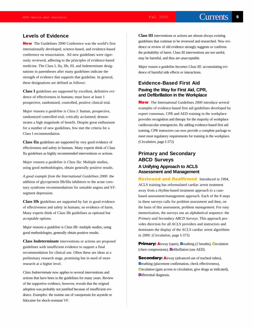
Levels of EvidenceNew The Guidelines 2000 Conference was the world’s first
internationally developed, science-based, and evidence-based
conference on resuscitation. All new guidelines were rigor-
ously reviewed, adhering to the principles of evidence-based
medicine. The Class I, IIa, IIb, III, and Indeterminate desig-
nations in parentheses after many guidelines indicate the
strength of evidence that supports that guideline. In general,
these designations are defined as follows:
Class Iguidelines are supported by excellent, definitive evi-
dence of effectiveness in humans; must have at least 1
prospective, randomized, controlled, positive clinical trial.
Major reasons a guideline is Class I:human, prospective,
randomized controlled trial; critically acclaimed; demon-
strates a high magnitude of benefit. Despite great enthusiasm
for a number of new guidelines, few met the criteria for a
Class I recommendation.
Class IIa guidelines are supported by very good evidence of
effectiveness and safety in humans. Many experts think of Class
IIa guidelines as highly recommended interventions or actions.
Major reasons a guideline is Class IIa:Multiple studies,
using good methodologies, obtain generally positive results.
A good example from the International Guidelines 2000:the
addition of glycoprotein IIb/IIIa inhibitors to the acute coro-
nary syndrome recommendations for unstable angina and ST-
segment depression.
Class IIb guidelines are supported by fair to good evidence
of effectiveness and safety in humans; no evidence of harm.
Many experts think of Class IIb guidelines as optional but
acceptable options.
Major reasons a guideline is Class IIb:multiple studies, using
good methodologies, generally obtain positive results.
Class Indeterminateinterventions or actions are proposed
guidelines with insufficient evidence to support a final
recommendation for clinical use. Often these are ideas at a
preliminary research stage, promising but in need of more
research at a higher level.
Class Indeterminatenow applies to several interventions and
actions that have been in the guidelines for many years. Review
of the supportive evidence, however, reveals that the original
adoption was probably not justified because of insufficient evi-
dence. Examples:the routine use of vasopressin for asystole or
lidocaine for shock-resistant VF.
Class III interventions or actions are almost always existing
guidelines that continue to be reviewed and researched. New evi-
dence or review of old evidence strongly suggests or confirms
the probability of harm. Class III interventions are not useful,
may be harmful, and thus are unacceptable.
Major reason a guideline becomes Class III:accumulating evi-
dence of harmful side effects or interactions.
Evidence-Based First AidPaving the Way for First Aid, CPR,and Defibrillation in the Workplace
New The International Guidelines 2000 introduce several
examples of evidence-based first aid guidelines developed by
expert consensus. CPR and AED training in the workplace
providesrecognition and therapy for the majority of workplace
cardiovascular emergencies. By adding evidence-based first aid
training, CPR instructors can now provide a complete package to
meet most regulatory requirements for training in the workplace.
(Circulation,page I-372)
Primary and SecondaryABCD Surveys A Unifying Approach to ACLSAssessment and Management
Reviewed and Reaffirmed Introduced in 1994,
ACLS training has reformulated cardiac arrest treatment
away from a rhythm-based treatment approach to a case-
based assessment/management approach. Each of the 8 steps
in these surveys calls for problem assessmentand then, on
the basis of this assessment, problem management.For easy
memorization, the surveys use an alphabetical sequence: the
Primary and Secondary ABCD Surveys.This approach pro-
vides direction for all ACLS providers and instructors and
dominates the display of the ACLS cardiac arrest algorithms
in 2000. (Circulation,page I-373)
Primary: Airway (open), Breathing (2 breaths), Circulation
(chest compressions), Defibrillation (use AED).
Secondary: Airway (advanced use of tracheal tubes),
Breathing (placement confirmation, check effectiveness),
Circulation (gain access to circulation; give drugs as indicated),
Differential diagnosis.

6 Fall 2000 ©2000 American Heart AssociationCurrentsCurrents
Education, Training,and Examination
Commitment to Educationand Training Based on CoreLearning ObjectivesOld The core learning objectives of the ECC courses have not
previously been identified and defined.
New The ECC Programs defined the core learning
objectives for each of the resuscitation specialties. The ECC
Subcommittees were asked: “What skills and knowledge should
all participants be able to demonstrate at the end of a successful
course?” (Circulation, pages I-7 to I-9)
Why? Clearly stated learning objectives are necessary to guide
curricula development, teaching techniques, and instructor and
learner evaluation.
Adding specific skills adds specific objectives
AHA courses may add specific skills, depending on course
purpose, the setting, and learners’ scope of practice. For
example, learners must meet specific skill objectives when
any of the following skills are added: child-infant CPR, use
of barrier or mask devices, techniques to correct foreign-
body airway obstruction, or integration of AED use into BLS
for victims older than 8 years.
Pediatric AdvancedLife Support
Core Training Objectives
Immediately after completing PALS trainingand for up to 2 years after, the successfulPALS provider should be able to
1. Reduce the risk of the most commoncauses of injury and death.
2. Recognize and initiate treatment forimpending respiratory failure, shock,and cardiopulmonary arrest.
a. Provide PBLS (open airway,rescue breathing, chestcompressions).
b. Provide advanced support ofoxygenation and ventilation.
c. Obtain vascular access.
d. Initiate appropriate resuscitativefluid and drug therapy.
3. Initiate the first 10 minutes ofresuscitation of the pediatric victimof cardiopulmonary arrest.
4. Provide support for familiesand providers in coping with achild's death
Adult and PediatricBasic Life Support
Core Training Objectives
Immediately after initial basic CPR trainingand for up to 2 years after, a rescuer shouldbe able to
1. Reduce the risk of the most commoncauses of injury and death.
2. Recognize unresponsiveness (or otheremergency situations where resuscitationis appropriate).
3. Phone 911 in a timely fashion (or otheremergency phone number, eg, in-hospital).
4. Provide an open airway (usinghead tilt–chin lift or jaw-thrust techniques).
5. Provide ventilations (breathing) that makethe chest rise, using mouth-to-mouth ormouth-to–barrier device ventilations(bag-mask ventilation by healthcareproviders).
6. Provide chest compressions (usingcompression force that moves sternumdown the appropriate depth for victim’sage [lay rescuer] or that generates apalpable pulse [healthcare provider]).
7. Perform all these skills in a manner thatis safe for the rescuer, the patient, andbystanders.
8. (If healthcare provider) Show proficiencyin bag-mask ventilation for victims ofall ages and use of AEDs for victims8 years of age or older.

©2000 American Heart Association 7Fall 2000 CurrentsCurrents
Open Door for Education andTraining Innovations (Circulation, page I-8)
New Many new adjuncts to education and training have
been developed and accepted for ECC training. Acceptance
can occur if the new approach is evidence-based, validated,
and shown to be effective at increasing skill acquisition and
longer-term skill retention. Some recent examples of
approved techniques follow:
Instructor-directed, videotape-mediated training.Currently accepted: “practice-as-you-watch” videotapes
(Braslow-Kaye-Todd), “watch-then-practice” videotapes
(Stapleton, Aufderheide). Currently not accepted or not vali-
dated: “passive-watching-motivational-informational” videos.
Auditory and/or visual prompts (includes the valuablevoice prompts supplied with most AEDs).These prompts
guide the learner in multiple aspects of performance (ie, com-
pression location, depth, and rate; ventilation rate and volume).
Currently accepted are those prompts that require hands-on prac-
tice such as audible counting devices, sequence directive devices,
and compression-force prompts.
High-tech ACLS and BLS simulatorsguide and improve the
learner’s performance by providing constant device-operator
feedback as the learner attempts the procedure. Currently accept-
ed: Laerdal ALS units, a validated computer-aided learning pro-
gram to maintain healthcare provider skills with AEDs.
Increasing the Value of Examination Part I: Fully Annotated Written ExamsSupporting Pre-test and Post-test Learning
New All ACLS, PALS, and BLS written exams now have
a companion annotated exam. The annotations state why
each of the possible answers in the multiple choice list was
either correct or incorrect and refer the learner to the appropriate
text references and page numbers. After learners complete an
answer sheet, course directors can use student grading,
instructor grading, or colleague grading followed by distribution
of annotated versions. Learners review the annotated version
for discussions of questions that were unclear, misunderstood, or
answered incorrectly. This increases a learner’s independence,
aids self-learning, and frees course time formerly required for
review of written exams. (Circulation, page I-9)
Old From 1992 to 1997 the written exams had only an answer
sheet, with no information on why the right answer was correct
or why the other answers were incorrect. Annotations to the
questions in the written exams were not offered.
Why? Learners and instructors need to know why a particular
answer is either right or wrong. Instructors have complained that
they sometimes did not understand why certain answers were
correct and others were wrong. Exams with annotated answers
have been found to be a valuable self-teaching technique.
Advanced CardiacLife Support
Core Training Objectives
Upon leaving an ACLS course and for 2years after, the successful ACLS providershould be able to
1. Recognize and initiate treatment forprearrest conditions that may leadto a cardiac arrest, including acutecoronary syndromes, respiratoryfailure, and stroke.
2. Manage the first 10 minutes of anarrest due to ventricular fibrillation.
a. Provide BLS care, includingproper operation of an AED.
b. Provide proper operation of aconventional defibrillator.
c. Provide advanced airway supportof oxygenation and ventilationwith secondary confirmation oftracheal tube placement.
d. Obtain vascular access.
3. Correctly treat the 4 arrest rhythms.
a. VT
b. PVT
c. PEA
d. Asystole
8 Fall 2000 ©2000 American Heart AssociationCurrentsCurrents
Part II: Complete Reconstruction of the ECCWritten Exams (for ACLS, PEDS, and BLS)
New New questions for the written exams have been created,
pilot tested, and validated. The questions evaluate mastery of the
major learning objectives. Educational consultants participated in
the writing, evaluation, and validation of these exams.
Old The former written questions did not eliminate ambiguity
and confusion. The subject matter of the questions seldom
aligned with the learning objectives of the course.
Why? This was a long-requested program improvement initia-
tive, undertaken to increase the learning value of written exams
for both the learners and the instructors.
New Focus on Measuring andImproving Instructor QualityNew Emphasis on Consistency inthe Quality of Teaching
New Training centers will create levels of faculty within
the center to facilitate instructor training, mentoring, and moni-
toring. Instructor and instructor-trainer monitoring forms provide
detailed outlines for evaluating instructor and instructor-trainer
performance. Training centers will be able to develop their
instructor corps internally without having to rely on outside
regional faculty. AHA-prepared videos and other standardized
teaching tools allow for core material to be presented consistently
and concisely in every course.
Old Instructor training and monitoring required regional
faculty assistance, which was not always available within
the training center. Instructor monitoring was subjective,
with little or no written guidelines for instructor
performance evaluation.
Why? This initiative enables training centers to meet the
demands for instructor development using internal resources.
Training centers will have the autonomy they need to operate
effectively and efficiently in developing their instructors.
Ethical Concerns inResuscitation
Family Presence DuringResuscitation Attempts Valuedby Families and Loved OnesNew Pediatric critical care nurses pioneered studies
demonstrating that family presence during resuscitation attempts
produces positive psychological effects. This practice is
recommended by the International Guidelines 2000 provided it
is done with planning, staff acceptance, and a designated staff
member who offers this opportunity to the family and remains
with them during their presence at the resuscitation attempt.
(Circulation, page I-19)
Searching for and Honoring“Do Not Attempt Resuscitation”(DNAR) Status In Field, EmergencyDepartment, and HospitalReviewed and Reaffirmed Many individuals, exe-
cuting their right of self-determination, have declared they want
no one to attempt resuscitation if they show indications of dis-
tress (unresponsive, not breathing, no pulse). They wish this even
if they or their family have called the EMS system. This DNAR
decision often takes the form of living wills, advance directives,
or other documents and even bracelets and anklets worn on the
body. Valid expressions of self-determination must be honored;
to do otherwise is unethical and prohibited by law.
(Circulation, pages I-14 to I-16, I-18)
Certification of Death in the Field—No TransportDeath Pronouncement in the Field
Reviewed and Reaffirmed There are very few indi-
cations for transporting a victim of nontraumatic cardiac arrest
who has failed a successfully executed prehospital ACLS resus-
citation effort to an Emergency Department to continue the resus-
citation attempt. The danger to personnel and bystanders is
greater than the probability of resuscitating the victim.
(Circulation, page I-17)
Criteria for Pronouncement of DeathReviewed and Reaffirmed EMS systems should
develop criteria for stopping resuscitation attempts in the out-of-
hospital setting. Advanced life support personnel, in collaboration
with the on-line medical control physician, can gather clinical
©2000 American Heart Association 9Fall 2000 CurrentsCurrents
information which will allow the medical control physician to
declare the patient dead.
This would comply with state law in most states where a proper-
ly licensed physician must certify death.
(Circulation, page I-17)
New With rare exceptions, resuscitation efforts in the field
should cease if the following criteria have been met:
Quality of the resuscitation attempt was satisfactorywith an adequate trial of BLS and ALS as demonstratedby the following:
• Achieved airway control with tracheal intubation or
advanced airway device, confirmed proper tube
placement, and secured tube to prevent dislodgment
• Achieved effective oxygenation and ventilation
• Shocked VF when present
• Gained access to circulatory system and administered
epinephrine (or vasopressin), atropine, and antiarrhythmics
as appropriate
• Considered, searched for, and corrected reversible causes
or special resuscitation circumstances
• Observed continuous and documented pulseless arrest after
all of the above have been accomplished
Reviewed for significant special resuscitationcircumstances:
• Profound hypothermia
• Toxin or drug overdose
Survivor support plans have been established in advanceand they are in compliance with state and local policies
(See new guideline at right: Survivor Support Plans)
• Are medical directors available for real-time consultations
and to authorize cessation of efforts? (Leaving the body at
the scene and supporting the survivors: Class IIa)
For patients meeting above criteria, urgent field-to-hospital
transport with continuing CPR is almost invariably futile.
Because harm to personnel is more likely than help to the
victim, such transports are now classed as Class III (harmful;
no benefit).
Survivor Support Plans
New EMS systems should allow healthcare providers to fol-
low the new international guidelines for stopping
resuscitative efforts in the field. This requires survivor
support plans. EMS systems must develop action steps
to support pronouncement of death in the field:
• EMS personnel must know and understand advance plans
on leaving the body at the scene, how to perform death
certification, and how to transfer to funeral establishments.
• On-scene family advocates (usually designated field
personnel) can help families accept nontransport of the
dead person, reporting the death properly, calling the
funeral home, calling a chaplain or family minister,
completing the death certificate, and providing support
and answering questions. (Circulation, page I-19)
Religious or nondenominational grief counseling helps signifi-
cantly. Determine when and where this counseling is available
and how counselors can be contacted.
Basic Life Support—Adult and Pediatric
This section identifies distinctions in the new BLS
guidelines between those for lay rescuers and those
targeting healthcare providers.
Importance of Early DefibrillationConventional and Automated External
Reviewed and Reaffirmed Since the 1992
Guidelines, early defibrillation has been the preeminent
therapeutic intervention that saves lives of adult victims.
Evidence accumulated in the past decade continues to reaffirm
all guidelines having to do with early defibrillation, including
recommendations for timely defibrillation in public places,
in the homes of high-risk patients, and in commercial aircraft,
airports, hospitals, doctors’ offices, and outpatient clinics.
New Expand authorization for adult victims. The International
Guidelines 2000 highly recommend that authorization to attach
and operate a defibrillator be expanded to nontraditional respon-
ders such as police, firefighters, and security personnel in casi-
nos, on the ground, and in airports. In fact, as reviewed in other
sections, the perceived value of antiarrhythmics, vasopressors,
advanced airway control, oxygenation, and ventilation has
10 Fall 2000 ©2000 American Heart AssociationCurrentsCurrents
declined markedly since 1992. The evidence is disappointingly
weak that any of these interventions convey effective benefit to
cardiac arrest victims when applied universally.
New Remove barriers. Constructive efforts to remove state
and local administrative and regulatory barriers to the use of
AEDs by lay responders are strongly encouraged. In the majority
of states this has required introduction and sponsorship of revi-
sions to state legislative code.
New Insight The relative value of early defibrillation in
reducing the interval between adult sudden cardiac arrest and
first defibrillatory shock by 1 to 2 minutes does more to improve
the probability of survival for an individual patient than all the
medications, airway interventions, and newly designed defibrilla-
tion waveforms combined.
Recognition of Special SituationsThat Modify the “Phone First versusPhone Fast” Guidelines(Circulation, page I-256)
New Providers of adult and pediatric BLS recognize
clinical exceptions to the “phone fast” guideline (applies to chil-
dren up to 8 years) and the “phone first” guideline (applies to
children 8 years and older). The major exception to the “phone
fast” rule is those children (<8 years old) known to be at risk for
VF/VT who experience sudden witnessed collapse. Follow the
“phone first” guideline in such situations. This results in more
rapid arrival of a defibrillator. This situation was noted as a
“special resuscitation situation.”
New (for adults) In parallel to this new PALS guideline, adult
BLS guidelines also recognize 4 special resuscitation situations
where airway compromise, rather than sudden VF/ VT, is the
cause of the arrest. The new BLS guideline for these victims is
“phone fast,” that is, provide 1 minute of CPR before phoning
the EMS system. These situations are
1. Submersion/near-drowning (1 minute CPR; then“phone fast”)
2. Poisoning, drug overdose (1 minute CPR; then“phone fast”)
3. Trauma (1 minute CPR; then “phone fast”)
4. Respiratory arrest (1 minute CPR; then “phone fast”)
Old “Phone fast” was the guideline to follow for infants and
children under 8 years of age. “Phone first” was the guideline to
follow for victims 8 years of age through adult.
Why? To keep the sequence for lay rescuer CPR simple, con-
sistent messages should be given. To maximize survival from
cardiac arrest, however, rescuers should tailor rescue sequences
to best meet the needs of the collapsed victim. As a compromise
between educational simplicity and the needs of the individual
victim, the ECC scientists recommended that the simple message
be continued but that information in the texts address exceptions
to the rule. When appropriate, healthcare providers can suggest
that family members learn a different sequence, if appropriate.
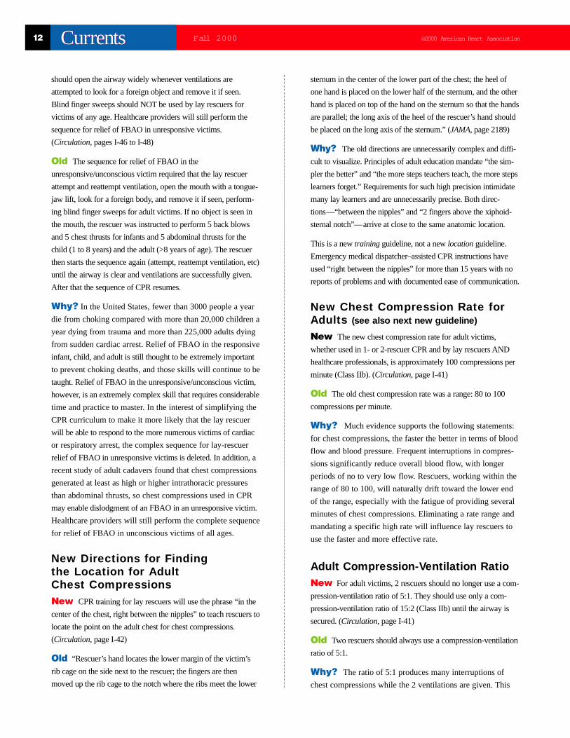
Bag-Mask Ventilation Is aSkill That All BLS HealthcareProviders Must MasterNew Anyone providing prehospital BLS care for adults,
infants, and children should be trained to deliver effective oxy-
genation and ventilation using a bag-mask technique as the pri-
mary method of ventilatory support, particularly if the transport
time is short (Class IIa). (Circulation, pages I-267 to I-268)
Old The need for bag-mask ventilation was not emphasized,
and insertion of a tracheal tube was recommended as soon
as possible.
Why? Bag-mask ventilation provides effective ventilation
when performed by properly trained providers. In a Los Angeles
study, the EMS system had short transport times and the
providers were inexperienced, but trained, in pediatric intubation.
Study data showed that children who received bag-mask ventila-
tion had survival rates equivalent to those who received tracheal
intubation. Bottom line: bag-mask ventilation is a fundamental
skill that should be mastered by all healthcare providers.
Smaller Tidal Volumes DuringAdult Rescue BreathingNew Rescuers should now deliver smaller tidal volumes dur-
ing ventilation with bag-mask ventilation or when supplementary
oxygen is available. Rescue breaths delivered by mouth-to-
mouth or mouth-to–barrier device should average 700 to 1000
mL delivered over 2 seconds. If supplementary oxygen is
©2000 American Heart Association 11Fall 2000 CurrentsCurrents
available, the skilled rescuer should attempt to provide smaller
tidal volumes during mouth-to-mask and bag-mask ventilation,
theoretically 400 to 600 mL over 1 to 2 seconds (Class IIb).
(Circulation,page I-38)
Old Provide tidal volume of 800 to 1200 mL during mouth-
to-mouth or mouth-to-mask or bag-mask ventilation.
Why? Smaller tidal volumes with oxygen supplementation
and/or bag-mask ventilation can support adequate oxygen satura-
tion but reduce the risk of gastric inflation (and its attendant
complications). If the smallest tidal volumes are used, the chest
should rise visibly and the oxygen saturation should be main-
tained. Note that smaller ventilation volumes can be associated
with hypercarbia and acidosis.
Mouth-to-Nose Breathing Is anAcceptable Alternative to Mouth-to-Nose-and-Mouth or Mouth-to-MouthRescue Breathing for an InfantNew Mouth-to-nose breathing is an acceptable alternative
to mouth-to-nose-and-mouth or mouth-to-mouth breathing if
the rescuer is unable to cover the infant’s nose and mouth
(Class IIb). (Circulation, pages I-265 to I-266)
Old Only mouth-to-nose-and-mouth and mouth-to-mouth res-
cue breathing were recommended. Mouth-to-nose breathing in
adults was offered as an alternative in the adult BLS 1992
Guidelines: “This technique is more effective in some patients
than the mouth-to-mouth technique. The mouth-to-nose tech-
nique is recommended when it is impossible to ventilate through
the victim’s mouth, the mouth cannot be opened (trismus), the
mouth is seriously injured, or a tight mouth-to-mouth seal is dif-
ficult to achieve. …” (JAMA, pages 2187-2188)
Why? Studies have shown that some rescuers may have
difficulty covering both the mouth and nose of an infant with
the rescuer’s mouth. In addition, these same studies have
shown that mouth-to-nose breathing may provide effective
ventilation of infants.
Consideration of AlternativeAirway Devices (LMA) forTrained Healthcare ProvidersNew Use of alternative advanced airways should be encour-
aged when rescuers are properly trained in their use (Class
Indeterminate). (Circulation, page I-297)
Old Tracheal intubation was thought to be the gold
standard for airway control.
Why? Several studies have documented the high complication
rate that can occur in some EMS systems when tracheal
intubation is performed by rescuers with limited experience in
performing pediatric intubation. Therefore, alternative techniques
to help isolate the airway and reduce gastric inflation are encour-
aged when rescuers are properly trained.
The “Pulse Check” Should Not BeTaught to Lay Rescuers New In the ABC sequence of CPR, lay rescuers will no longer
be taught to check for a carotid pulse. Instead they will be taught
to look and examine for “signs of circulation,” which include
normal breathing, coughing, or movement (Class IIa). If no signs
of circulation are detected, the rescuer should begin chest com-
pressions and attach an AED, if available. (Circulation, pages I-
39 to I-40 and I-269)
Old Lay rescuers were taught to palpate the carotid artery
located on the same side of the neck as the rescuer and, taking no
more than 10 seconds, decide whether they could feel a pulse. If
they felt no pulse, they would begin chest compressions and
attach the AED.
Why? Lay rescuers perform the pulse check primarily as the
signal to start chest compressions and, if trained as a Heartsaver
AED provider, to call for and attach an AED. Considerable evi-
dence demonstrates that rescuers have trouble locating the cor-
rect place for palpation. They require much more than the recom-
mended upper limit of 10 seconds. Finally, when palpating in the
correct location for as long as needed, the rescuer is unacceptably
inaccurate. A serious type II or false-negative error is committed
10% of the time. This leads to 1 of 10 cardiac arrest patients not
receiving either chest compressions or AED attachment.
Dropping the pulse check will not result in as much potential
harm as keeping the pulse check.
Simplification of Maneuvers for LayRescuer Relief of Foreign-BodyAirway Obstruction in theUnresponsive Victim of Any Age(Class IIb)New Previously recommended maneuvers for relief of
foreign-body airway obstruction (FBAO) in the unconscious
victim will no longer be taught to lay rescuers. Instead the lay
rescuer will begin standard CPR when an unrelieved responsive,
choking victim becomes unresponsive, or an unresponsive
person suspected of an FBAO is encountered, evaluated, and
treated. The only difference from regular CPR is that the rescuer
12 Fall 2000 ©2000 American Heart AssociationCurrentsCurrents
should open the airway widely whenever ventilations are
attempted to look for a foreign object and remove it if seen.
Blind finger sweeps should NOT be used by lay rescuers for
victims of any age. Healthcare providers will still perform the
sequence for relief of FBAO in unresponsive victims.
(Circulation, pages I-46 to I-48)
Old The sequence for relief of FBAO in the
unresponsive/unconscious victim required that the lay rescuer
attempt and reattempt ventilation, open the mouth with a tongue-
jaw lift, look for a foreign body, and remove it if seen, perform-
ing blind finger sweeps for adult victims. If no object is seen in
the mouth, the rescuer was instructed to perform 5 back blows
and 5 chest thrusts for infants and 5 abdominal thrusts for the
child (1 to 8 years) and the adult (>8 years of age). The rescuer
then starts the sequence again (attempt, reattempt ventilation, etc)
until the airway is clear and ventilations are successfully given.
After that the sequence of CPR resumes.
Why? In the United States, fewer than 3000 people a year
die from choking compared with more than 20,000 children a
year dying from trauma and more than 225,000 adults dying
from sudden cardiac arrest. Relief of FBAO in the responsive
infant, child, and adult is still thought to be extremely important
to prevent choking deaths, and those skills will continue to be
taught. Relief of FBAO in the unresponsive/unconscious victim,
however, is an extremely complex skill that requires considerable
time and practice to master. In the interest of simplifying the
CPR curriculum to make it more likely that the lay rescuer
will be able to respond to the more numerous victims of cardiac
or respiratory arrest, the complex sequence for lay-rescuer
relief of FBAO in unresponsive victims is deleted. In addition, a
recent study of adult cadavers found that chest compressions
generated at least as high or higher intrathoracic pressures
than abdominal thrusts, so chest compressions used in CPR
may enable dislodgment of an FBAO in an unresponsive victim.
Healthcare providers will still perform the complete sequence
for relief of FBAO in unconscious victims of all ages.
New Directions for Findingthe Location for AdultChest CompressionsNew CPR training for lay rescuers will use the phrase “in the
center of the chest, right between the nipples” to teach rescuers to
locate the point on the adult chest for chest compressions.
(Circulation, page I-42)
Old “Rescuer’s hand locates the lower margin of the victim’s
rib cage on the side next to the rescuer; the fingers are then
moved up the rib cage to the notch where the ribs meet the lower
sternum in the center of the lower part of the chest; the heel of
one hand is placed on the lower half of the sternum, and the other
hand is placed on top of the hand on the sternum so that the hands
are parallel; the long axis of the heel of the rescuer’s hand should
be placed on the long axis of the sternum.” (JAMA, page 2189)
Why? The old directions are unnecessarily complex and diffi-
cult to visualize. Principles of adult education mandate “the sim-
pler the better” and “the more steps teachers teach, the more steps
learners forget.” Requirements for such high precision intimidate
many lay learners and are unnecessarily precise. Both direc-
tions—“between the nipples” and “2 fingers above the xiphoid-
sternal notch”—arrive at close to the same anatomic location.
This is a new training guideline, not a new locationguideline.
Emergency medical dispatcher–assisted CPR instructions have
used “right between the nipples” for more than 15 years with no
reports of problems and with documented ease of communication.
New Chest Compression Rate forAdults (see also next new guideline)
New The new chest compression rate for adult victims,
whether used in 1- or 2-rescuer CPR and by lay rescuers AND
healthcare professionals, is approximately 100 compressions per
minute (Class IIb). (Circulation, page I-41)
Old The old chest compression rate was a range: 80 to 100
compressions per minute.
Why? Much evidence supports the following statements:
for chest compressions, the faster the better in terms of blood
flow and blood pressure. Frequent interruptions in compres-
sions significantly reduce overall blood flow, with longer
periods of no to very low flow. Rescuers, working within the
range of 80 to 100, will naturally drift toward the lower end
of the range, especially with the fatigue of providing several
minutes of chest compressions. Eliminating a rate range and
mandating a specific high rate will influence lay rescuers to
use the faster and more effective rate.
Adult Compression-Ventilation RatioNew For adult victims, 2 rescuers should no longer use a com-
pression-ventilation ratio of 5:1. They should use only a com-
pression-ventilation ratio of 15:2 (Class IIb) until the airway is
secured. (Circulation, page I-41)
Old Two rescuers should always use a compression-ventilation
ratio of 5:1.
Why? The ratio of 5:1 produces many interruptions of
chest compressions while the 2 ventilations are given. This
©2000 American Heart Association 13Fall 2000 CurrentsCurrents
leads to marked reduction in blood flow and blood pressure.
With a 5:1 ratio the single ventilation, sandwiched between
short periods of rapid chest compressions, leads to faster,
more forceful ventilations from the rescuers. This in turn
leads to greater risks for gastric inflation, regurgitation, aspira-
tion, and severe lung damage.
Reaffirmed The 5:1 ratio should be used in pediatric arrest
by professional responders regardless of whether 1 or 2 rescuers
are involved.
Why? This topic has been reviewed several times since 1992.
There is no evidence to justify a change. Emphasis on oxygena-
tion and ventilation is justified in infants and children based on
the epidemiology of cardiac arrest.
“CPR” Performed Without Mouth-to-Mouth VentilationsReviewed and Reaffirmed CPR with compressions
and ventilations remains the ideal method of maintaining blood
flow until the arrival of a lay responder with an AED or EMS
personnel. However, if unwilling to perform mouth-to-mouth
rescue breathing for an adult victim, the rescuer should access
the EMS system, open the airway, and perform chest
compressions at the rate of approximately 100 compressions
per minute (Class IIa). (Circulation, page I-43)
CPR without ventilation may be taught by emergency
medical dispatchers (Class IIa).
The 2 Thumb–Encircling HandsChest Compression Technique IsRecommended Over the 2-FingerCompression Technique for 2-RescuerInfant CPR by Healthcare Providers
New The 2 thumb–encircling hands technique of chest com-
pression is preferred for chest compressions in infants performed
by healthcare providers when 2 rescuers are available (Class IIb).
(Circulation,page I-351)
Old The 2 thumb–encircling hands technique was an alterna-
tive technique for chest compression in the neonate.
Why? Data show that the 2 thumb–encircling hands tech-
nique can provide better blood flow than the 2-finger technique.
However, a single rescuer providing chest compressions using
this technique may have difficulty alternating between rescue
breathing and chest compressions. For simplicity and retention,
this technique is not taught to the lay rescuer and anyone per-
forming 1-rescuer CPR.
Use of Automated ExternalDefibrillators Is Encouraged forVictims of Cardiac Arrest OlderThan About 8 Years of Age orAbout 25 kg (55 lbs), AlthoughData Is Regarding the Use of AEDsin Pediatric Victims Are Limited
Reviewed An AED can be used in children 8 years of
age and older. The mean weight of an 8-year-old is 25 kg (55 lbs).
At that weight, an adult biphasic defibrillator will provide
nonescalating defibrillation energy doses of 150 J (6 J/kg) for
a 25-kg child. A monophasic defibrillator with escalating
dose will provide approximately 200 J (8 J/kg) for a 25-kg
child initially and then higher doses. Available evidence suggests
that these devices are accurate in differentiating between
shockable and nonshockable rhythms for adolescents. Note
that the guidelines still suggest the use of defibrillators with
adjustable energy dose for in-hospital use in areas that routinely
care for infants and children (Class IIb). (Circulation,page I-271)
Management ofthe Airway and
VentilationNew Airway AdjunctsNew The guideline sections on airway management and
ventilation contain the greatest number of new recommendations.
Most of these recommendations apply to healthcare providers
at both the BLS and ALS level. (Circulation,page I-95)
Why? New evidence. Since the 1992 Guidelines, many
scientists, researchers, corporate engineers, and entrepreneurs
have provided new information, new products, and new insights.
New evidence addresses ventilation volumes, ventilation
rates, maintenance of oxygenation, and prevention of hypercarbia,
acidosis, and aspiration. The international collaboration for
the International Guidelines 2000 increased awareness of
many devices and techniques that would otherwise have
remained unknown to the AHA training network.
The most important airway and ventilation developments come
from quality assessment efforts and outcome evaluation projects
conducted by a number of national and international centers.
These efforts have given us valuable information on the relative
merit of different devices and approaches. More important, this
work has stunned the resuscitation community by unequivocal

14 Fall 2000 ©2000 American Heart AssociationCurrentsCurrents
demonstrations of a sobering fact: healthcare providers in their
best-intentioned efforts to save lives have the capacity to severely
harm their patients.
The Guidelines 2000 Conference experts, leaders, and
participants examined this material carefully, adhering to the
principle of “first, do no harm” (Circulation,page I-380). These
reviews and discussions coalesced into a number of consensus
principles as powerful as any specific guideline. These new
airway guidelines require continuous quality improvement
initiatives that center on outcome assessment. The most
successful efforts are invariably found in programs where the
directors and managers adhere to the following approaches:
1. Examine carefully the ALS and BLS airway support
techniques used in various clinical settings.
2. Move these examinations beyond structural descriptions (how
many medics per population) and process descriptions
(average number of tracheal intubations per medic per year) to
focus on measuring health status outcome (survival to
hospital discharge of patients intubated versus not intubated in
the field).
3. Weigh these outcome evaluations against the training, skill
level, and experience of ALS providers in a given ALS system
or code responders in a given hospital. In addition, all patient
and system characteristics that can affect the evaluation
outcome must be considered (eg, witnessed versus
unwitnessed arrests, bystander CPR versus none, short
versus long transport times to hospitals, field use versus
prohibited use of paralytic agents, and high versus low
annual case frequency).
4. From these evaluations decide if the outcomes are satisfactory.
If not, question whether the sophistication and training
requirements for use of the airway devices match the skill
levels and experience of the emergency care providers.
5. Next, consider alternative action steps that might be
needed for an improvement process.
6. Select and develop action plans that address the problems
identified in this improvement process.
7. Observe the outcomes from the action plans.
8. Adjust training, programs, protocols, and equipment
as necessary.
9. Continue to monitor.
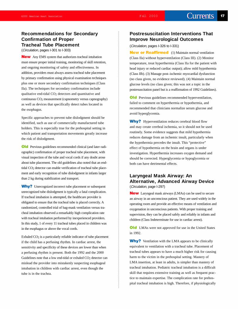
Airway Devices (Bag-Mask, TrachealTube, LMA, Combitube) for TrainedHealthcare Providers New (BLS, ACLS, PALS) Extensive evidence that
became available over the past decade provides these new guide-
lines on airway adjuncts:
New Bag-mask ventilation versus tracheal intubation.
Properly used by skilled BLS providers, the self-inflating, valved
bag with oxygen supplementation and continuous cricoid pres-
sure can be as effective as a tracheal tube in measures of oxy-
genation, ventilation, and protection from aspiration for short
ventilation times. (Circulation,page I-95)
New Dangers of tracheal intubation. Several curriculum
changes in established national training programs allow selected
BLS healthcare providers to be trained in the skills of tracheal
intubation. New evidence, however, reveals that unrecognized,
uncorrected esophageal intubations or tube dislodgments occur
with unacceptable frequency. One project prospectively studied
pediatric, out-of-hospital tracheal intubation attempts by
paramedics. The research leaders discovered that upon arrival at
the Emergency Department, 8% of these pediatric victims were
found to have a tube in the esophagus or hypopharynx. In another
out-of-hospital study of a large, mostly adult group of cardiac
arrest victims, more than 25% were found to have esophageal
or pharyngeal tube placement. (Circulation,page I-100)
New Secondary confirmation techniques. When tracheal intu-
bation is attempted, proper placement of the tube in the trachea
must be confirmed 2 ways: with primary confirmation techniques
and secondary confirmation techniques. Primary techniques
include physical examination: visualization of the tube passing
through the cords, 5-point auscultation, bilateral chest expansion,
tube condensation. In secondary confirmation techniques,
esophageal detector devices are preferred for intubation confir-
mation in adult cardiac arrest victims; end-tidal CO2 detectors are
preferred in non–cardiac arrest victims (Class IIa for victim with
spontaneous perfusion; IIb for victim in cardiac arrest).
(Circulation,page I-101)
New Prevention of tracheal tube dislodgment. After successful
tracheal intubation of adult victims, apply a manufactured tracheal
tube holder to prevent dislodgment, especially for patients who
must be moved and transported while intubated (Class IIa).
Homespun tape and string techniques should be abandoned since
they lack acceptable validation. Note that because the rate of unrec-
ognized tube dislodgments is low in most hospitals and EMS sys-
tems, the sample size required to achieve proper validation of tra-
cheal tube holders is prohibitively large. (Circulation,page I-101)
©2000 American Heart Association 15Fall 2000 CurrentsCurrents
New Detection of tracheal tube dislodgment. After successful
tracheal intubation, use continuous end-tidal CO2 monitoring
(Class IIb) to provide early detection of tube dislodgment. Two
types of devices are acceptable: either the capnometer, which
provides a single numeric CO2 value, or the capnograph, which
provides a continuous visual display of the level of expired CO2.
Capnography is preferred because it provides continuous infor-
mation and is more sensitive but the cost is much higher.
(Circulation,page I-101)
New Four adult alternative advanced airway devices.
(Circulation,page I-98) The Guidelines 2000 Conference evalu-
ated the evidence supporting 4 new alternative airway devices:
the Class IIa esophageal-tracheal Combitube (ETC), the Class IIa
laryngeal mask airway (LMA), the Class Indeterminate pharyn-
gotracheal lumen airway (PTL), and the Class Indeterminate
cuffed oropharyngeal airway (COPA). The LMA and the ETC
alternative airways share the following characteristics:
• Both are advanced airway techniques, are placed orally, and are
inserted past the hypopharyngeal space but not into the trachea.
• Both are inserted blindly, without the need of a laryngoscope
to observe passage through the cords. This single feature is
the reason the LMA and the ETC have attracted so much
attention. Their ease of use, lack of requirements for training
in the difficult skill of laryngoscopy, and considerable
cost-savings are significant variables. These variables
mean that selected, out-of-hospital BLS care providers can
be trained in the use of these devices. This will bring many
of the considerable advantages of the gold standard—
tracheal intubation—to many more victims.
• Both offer protection from aspiration and the severe
consequences of aspirated stomach contents into the lungs.
• In some settings, these devices may be superior to the
bag-mask technique in both ventilation and oxygenation
in adults.
• Of some surprise, in many respects both devices have
proved to be equivalent to the definitive airway
management device, the tracheal tube, in adults.
Old (BLS) Healthcare providers at the BLS level were
taught that their airway device of first choice was a self-inflating
bag with a one-way valve, a side port for supplemental oxygen,
and a properly fitting face mask.
Old (ACLS/PALS) Healthcare providers at the
ACLS/PALS level were taught that the airway device of choice
was the tracheal tube, used with a valved self-inflating bag, with
ports to provide high-flow supplemental oxygen.
Old Policies Versus New GuidelinesBasic-level healthcare providers have been forbidden throughout
most of the United States, often by legislative regulation, from
“invading” the victim’s body during resuscitation efforts. The
S-shaped oropharyngeal airway is the most invasive airway
device ever permitted for BLS personnel, yet even that makes
many EMS medical directors uncomfortable.
With these two new airway devices, the International Guidelines
2000 create a difficult situation for the EMS constituency.
Extensive review of the evidence leads the AHA and the world’s
resuscitation councils to now recommend the use of two new
invasive airway devices. The evidence confirms that under
certain conditions each is clinically superior to the bag-mask
and clinically equivalent to the tracheal tube. Does this mean
paramedics must switch from using the tracheal tube to using
the LMA? Does this mean the basic healthcare provider must
be trained to use the LMA or the ETC and must switch from
the bag-mask to the other devices?
To answer these questions, many factors must be considered. The
AHA and the International Guidelines 2000 Conference cannot
engage in what are now more policy and regulatory factors than
clinical and evidence-based issues. Each EMS system should
review the continuous quality improvement principles, as well as
the evidence supporting the new alternative airway devices,
before making decisions on these new airway guidelines.
16 Fall 2000 ©2000 American Heart AssociationCurrentsCurrents
Pediatric Advanced Life Support
Early identification of critical illness (respiratory failure
and shock) and implementation of advanced life support to
prevent cardiac arrest continue to be emphasized in the new
guidelines. The default sequence of PALS interventions is
based on the most common cause of arrest for a given age
group. New information is provided to help the participant
to identify and treat special resuscitation circumstances that
may alter the ALS intervention approach.
Cardiac Arrest and CardiovascularEmergencies Related to SpecialResuscitation Circumstances: DrugOverdoses, Toxins, ElectrolyteAbnormalities, Asthma, andAnaphylaxis
New Slightly modified approaches to resuscitation and
advanced life support are provided for infants and children with
suspected drug toxicity or poisoning, such as cocaine
or ß-blocker overdose, or suspected electrolyte emergencies.
(Circulation, pages I-322 to I-325)
Old PALS rescuers should “seek and treat” reversible
causes, but no specific information regarding modification of
existing algorithms or treatment approaches was offered.
Why? We now know more about specific arrhythmias and
the cardiovascular effects of drug toxicities and poisonings.
In addition, the old PALS course was not designed to teach
advanced provider therapies and interventions. As more
providers have mastered the previous course content, it was
necessary for the course to evolve to meet the needs of these
trained providers. The advanced life support provider
can now apply PALS guidelines with specific recommendation
for managing common poisonings, toxicologic problems, and
electrolyte abnormalities.
Bag-Mask Ventilation VersusTracheal Intubation by PediatricHealthcare ProvidersReviewed and Reaffirmed All healthcare providers
who provide prehospital care for infants and children must be
trained to provide effective oxygenation and ventilation using
the bag-mask technique. This is an essential core skill for all
healthcare providers.
New For out-of-hospital PALS-level providers, ventilation via
a tracheal tube continues to be recommended under specific con-
ditions. A properly placed and secured tracheal tube is the most
effective and reliable method of assisted ventilation and has long
been considered the gold standard. However, this method
requires initial mastery and continued practice or frequent field
use to maintain safe and effective technical skills.
(Circulation, page I-296)
Tracheal intubation in unconscious patients should be
encouraged only for healthcare providers well trained
in performing this skill, verified by significant and
frequent field experience, and continually monitored
by an ongoing quality-improvement program.
Old In the out-of-hospital setting, for pediatric emergencies,
ventilation via a properly placed tracheal tube is the most
effective and safest ventilatory method. (While this statement
remains true and is still a part of the guidelines, proper
insertion and reliable maintenance of the tracheal tube are
difficult, particularly in EMS systems where paramedics
have infrequent opportunity to attempt intubation.)
Why? Recent research has confirmed new observations
about pediatric airway maintenance and ventilation in the
out-of-hospital setting: (1) properly performed bag-mask
ventilation is safer and more effective than previously realized,
particularly when transport time is short and (2) tracheal tube
intubation is more difficult to master and more dangerous
than previously realized. These observations together suggest
a restatement: bag-mask ventilation may be equivalent to
tracheal tube intubation for respiratory emergencies in some
out-of-hospital pediatric ALS settings.
Bag-mask ventilation, compared with tracheal tube intubation,
can provide equally effective ventilation and oxygenation in
prehospital settings where transport times are short. One urban
EMS system with short transport times and trained providers
with infrequent pediatric intubations in the field observed that
children who required ventilatory support had equivalent sur-
vival rates regardless of whether they received bag-mask venti-
lation or ventilation through a tracheal tube placed in the out-
of-hospital setting. Other studies confirm that in some EMS
systems the success rate for pediatric intubation is mediocre,
with unacceptably high rates of complications.
©2000 American Heart Association 17Fall 2000 CurrentsCurrents
Recommendations for SecondaryConfirmation of ProperTracheal Tube Placement(Circulation, pages I-301 to I-303)
New Any EMS system that authorizes tracheal intubation
must ensure proper initial training, monitoring of skill retention,
and ongoing monitoring of safety and effectiveness. In
addition, providers must always assess tracheal tube placement
by primary confirmation using physical examination techniques
plus one or more secondary confirmation techniques (Class
IIa). The techniques for secondary confirmation include
qualitative end-tidal CO2 detectors and quantitative and
continuous CO2 measurement (capnometry versus capnography)
as well as devices that specifically detect tubes located in
the esophagus.
Specific approaches to prevent tube dislodgment should be
identified, such as use of commercially manufactured tube
holders. This is especially true for the prehospital setting in
which patient and transportation movements greatly increase
the risk of dislodgment.
Old Previous guidelines recommended clinical (and later radi-
ographic) confirmation of proper tracheal tube placement, with
visual inspection of the tube and vocal cords if any doubt arose
about tube placement. The old guidelines also noted that an end-
tidal CO2 detector can enable verification of tracheal tube place-
ment and early recognition of tube dislodgment in infants larger
than 2 kg during stabilization and transport.
Why? Unrecognized incorrect tube placement or subsequent
unrecognized tube dislodgment is typically a fatal complication.
If tracheal intubation is attempted, the healthcare provider is
obligated to ensure that the tracheal tube is placed correctly. A
randomized, controlled trial of bag-mask ventilation versus tra-
cheal intubation observed a remarkably high complication rate
with tracheal intubation performed by inexperienced providers.
In this study, 1 of every 11 tracheal tubes placed in children was
in the esophagus or above the vocal cords.
Exhaled CO2 is a particularly reliable indicator of tube placement
if the child has a perfusing rhythm. In cardiac arrest, the
sensitivityand specificity of these devices are lower than when
a perfusing rhythm is present. Both the 1992 and the 2000
Guidelines note that a low end-tidal or exhaled CO2 detector can
mislead the provider into mistakenly suspecting esophageal
intubation in children with cardiac arrest, even though the
tube is in the trachea.
Postresuscitation Interventions ThatImprove Neurological Outcomes (Circulation, pages I-326 to I-331)
New or Reaffirmed (1) Maintain normal ventilation
(Class IIa) without hyperventilation (Class III). (2) Monitor
temperature, treat hyperthermia (Class IIa for the patient with
head injury or reduced cardiac output); allow mild hypothermia
(Class IIb). (3) Manage post–ischemic myocardial dysfunction
(no class given, no evidence reviewed). (4) Maintain normal
glucose levels (no class given; this was not a topic in the
postresuscitation panel but is a reaffirmation of 1992 Guidelines).
Old Previous guidelines recommended hyperventilation,
failed to comment on hyperthermia or hypothermia, and
recommended that clinicians normalize serum glucose and
avoid hyperglycemia.
Why? Hyperventilation reduces cerebral blood flow
and may create cerebral ischemia, so it should not be used
routinely. Some evidence suggests that mild hypothermia
reduces damage from an ischemic insult, particularly when
the hypothermia precedes the insult. This “protective”
effect of hypothermia on the brain and organs is under
investigation. Hyperthermia increases oxygen demand and
should be corrected. Hyperglycemia or hypoglycemia or
both can have detrimental effects.
Laryngeal Mask Airway: AnAlternative, Advanced Airway Device(Circulation, page I-297)
New Laryngeal mask airways (LMAs) can be used to secure
an airway in an unconscious patient. They are used widely in the
operating room and provide an effective means of ventilation and
oxygenation in unconscious patients. With proper training and
supervision, they can be placed safely and reliably in infants and
children (Class Indeterminate for use in cardiac arrest).
Old LMAs were not approved for use in the United States
in 1992.
Why? Ventilation with the LMA appears to be clinically
equivalent to ventilation with a tracheal tube. Placement of
tracheal tubes appears to have a much higher risk for causing
harm to the victim in the prehospital setting. Mastery of
LMA insertion, at least in adults, is simpler than mastery of
tracheal intubation. Pediatric tracheal intubation is a difficult
skill that requires extensive training as well as frequent prac-
tice to maintain expertise. The complication rate for prehos-
pital tracheal intubation is high. Therefore, if physiologically
18 Fall 2000 ©2000 American Heart AssociationCurrentsCurrents
equivalent alternative airway devices can be placed with a
high success/low complication rate, their use is acceptable.
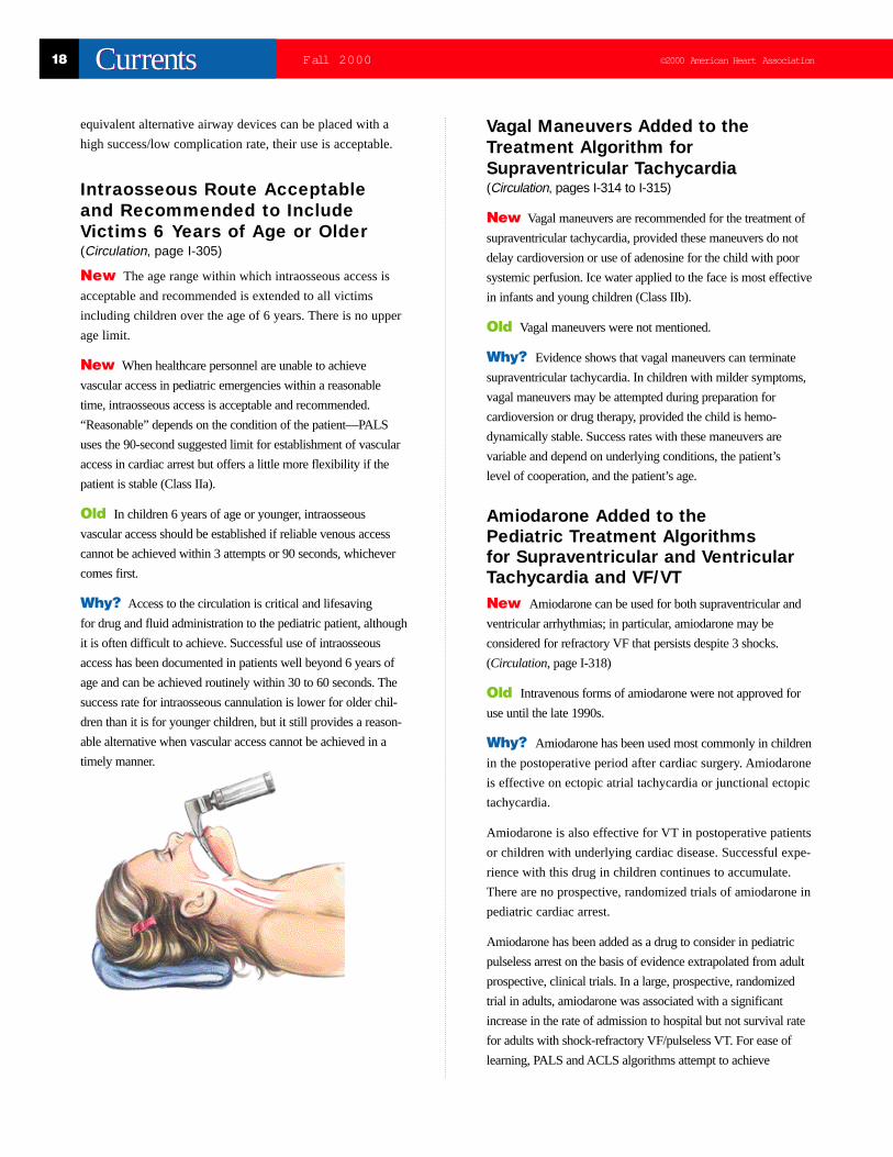
Intraosseous Route Acceptableand Recommended to IncludeVictims 6 Years of Age or Older(Circulation, page I-305)
New The age range within which intraosseous access is
acceptable and recommended is extended to all victims
including children over the age of 6 years. There is no upper
age limit.
New When healthcare personnel are unable to achieve
vascular access in pediatric emergencies within a reasonable
time, intraosseous access is acceptable and recommended.
“Reasonable” depends on the condition of the patient—PALS
uses the 90-second suggested limit for establishment of vascular
access in cardiac arrest but offers a little more flexibility if the
patient is stable (Class IIa).
Old In children 6 years of age or younger, intraosseous
vascular access should be established if reliable venous access
cannot be achieved within 3 attempts or 90 seconds, whichever
comes first.
Why? Access to the circulation is critical and lifesaving
for drug and fluid administration to the pediatric patient, although
it is often difficult to achieve. Successful use of intraosseous
access has been documented in patients well beyond 6 years of
age and can be achieved routinely within 30 to 60 seconds. The
success rate for intraosseous cannulation is lower for older chil-
dren than it is for younger children, but it still provides a reason-
able alternative when vascular access cannot be achieved in a
timely manner.
Vagal Maneuvers Added to theTreatment Algorithm forSupraventricular Tachycardia(Circulation, pages I-314 to I-315)
New Vagal maneuvers are recommended for the treatment of
supraventricular tachycardia, provided these maneuvers do not
delay cardioversion or use of adenosine for the child with poor
systemic perfusion. Ice water applied to the face is most effective
in infants and young children (Class IIb).
Old Vagal maneuvers were not mentioned.
Why? Evidence shows that vagal maneuvers can terminate
supraventricular tachycardia. In children with milder symptoms,
vagal maneuvers may be attempted during preparation for
cardioversion or drug therapy, provided the child is hemo-
dynamically stable. Success rates with these maneuvers are
variable and depend on underlying conditions, the patient’s
level of cooperation, and the patient’s age.
Amiodarone Added to thePediatric Treatment Algorithmsfor Supraventricular and VentricularTachycardia and VF/VT New Amiodarone can be used for both supraventricular and
ventricular arrhythmias; in particular, amiodarone may be
considered for refractory VF that persists despite 3 shocks.
(Circulation, page I-318)
Old Intravenous forms of amiodarone were not approved for
use until the late 1990s.
Why? Amiodarone has been used most commonly in children
in the postoperative period after cardiac surgery. Amiodarone
is effective on ectopic atrial tachycardia or junctional ectopic
tachycardia.
Amiodarone is also effective for VT in postoperative patients
or children with underlying cardiac disease. Successful expe-
rience with this drug in children continues to accumulate.
There are no prospective, randomized trials of amiodarone in
pediatric cardiac arrest.
Amiodarone has been added as a drug to consider in pediatric
pulseless arrest on the basis of evidence extrapolated from adult
prospective, clinical trials. In a large, prospective, randomized
trial in adults, amiodarone was associated with a significant
increase in the rate of admission to hospital but not survival rate
for adults with shock-refractory VF/pulseless VT. For ease of
learning, PALS and ACLS algorithms attempt to achieve

©2000 American Heart Association 19Fall 2000 CurrentsCurrents
consistency. Therefore, despite little evidence about amiodarone
in the specific setting of pediatric pulseless arrest, amiodarone
enters the pediatric algorithms (Class Indeterminate).
Use of High-Dose EpinephrineDe-emphasizedNew High-dose epinephrine for pulseless arrest is
de-emphasized in pediatrics. The recommended initial
resuscitation dose of epinephrine for pediatric cardiac arrest
is 0.01 mg/kg, given by the intravenous or intraosseous route
or 0.1 mg/kg by the tracheal route. Repeated doses are
recommended every 3 to 5 minutes for ongoing arrest. The
same dose of epinephrine is recommended for second and
subsequent doses for unresponsive asystolic and pulseless
arrest, but higher doses of epinephrine (0.1 to 0.2 mg/kg)
by any intravascular route may be considered (Class IIb).
(Circulation, page I-307)
Old The 1992 Guidelines recommended higher doses of epi-
nephrine for second and subsequent doses as Class IIa.
Why? High doses of epinephrine (HDE) improve myocardial
and cerebral blood flow in animals with cardiac arrest. High res-
cue doses of epinephrine were associated with improved survival
rates and neurological outcome compared with historic controls
in a single, nonblinded clinical trial of 20 children with witnessed
cardiac arrest. Large multi-institutional adult studies, well-con-
trolled animal studies, and uncontrolled, retrospective pediatric
data failed to show benefit from HDE. HDE can have adverse
effects including increased myocardial oxygen demand, hyper-
adrenergic state, tachycardia, hypertension, and myocardial
necrosis. There is great interpatient variability in dose response.
It is possible that a dose dangerous for one patient may be
lifesaving for another.
Automated External Defibrillators forTreatment of Children ≥8 Years of AgeNew AEDs may be used in the treatment of children with
out-of-hospital cardiac arrest who are more than 8 years of age.
Attempts to defibrillate VF/pulseless VT detected by an AED
are acceptable and recommended for children ≥8 years of age,
particularly in the out-of-hospital setting (Class IIb).
(Circulation, page I-320)
Old 1992 PBLS and PALS guidelines did not mention use of
AEDs in children. The ACLS textbook recommended their use
in children ≥8 years of age in 1994, and the PALS 1997 text rec-
ommended their use in children ≥8 years of age.
Why? Evidence continues to accumulate that pediatric VF
may be more common than previously suspected and more
common than would be identified by the usual surveillance
methods of emergency medical systems. Early defibrillation
of this VF/pulseless VT is important. Limited data suggests
that AEDs are both sensitive and specific in interpreting
rhythms in older children and adolescents. In children ≥8
years of age, the median weight should be >25 kg, so a 150-J
biphasic shock would deliver no more than 6 J/kg to a child
of average size who is at least 8 years of age. A monophasic
AED defibrillator with escalating shocks would initially
deliver 8 J/kg (200-J initial shock) and could increase to
14 J/kg if a 360-J dose is delivered to a 25-kg child. The
potential for harm from energy levels much greater than the
recommended upper limit of 4 J/kg causes great concern.
This high-energy level is acceptable only until AED
manufacturers develop AEDs with pediatric-appropriate
algorithms and energy levels.
2 Thumb–Encircling HandsTechnique for Chest Compression—See next page.

20 Fall 2000 ©2000 American Heart AssociationCurrentsCurrents
NeonatalResuscitation
Importance of Ventilation inthe Newly Born Infant
Reaffirmed In the newly born infant with heart rate
<100 beats per minute (bpm) and unresponsiveness to
stimulation, the primary concern is to establish adequate
ventilation. (Circulation,page I-350)
Interventions Revised forNeonates With Meconium-StainedAmniotic Fluid
New When meconium is observed in amniotic fluid,
deliver the head and suction meconium from the hypopharynx.
When meconium is observed in amniotic fluid and the newly
born infant demonstrates any of the following: (1) absent or
depressed respirations, (2) heart rate <100 bpm, or (3) poor
muscle tone, direct tracheal suctioning is recommended
(Class I) (Circulation,page I-348). Note that this eliminates
the recommendation for direct tracheal suctioning of the
meconium-stained but vigorous infant, whether the meconi-
um is thick or thin.
Old Direct endotracheal suctioning, using the tracheal tube
as a suction catheter, should be performed if the neonate is
depressed or the meconium is thick or particulate. Tracheal suc-
tioning may not be necessary if the meconium is thin and the
newborn is vigorous.
Why? Some neonates with meconium-stained amniotic
fluid have meconium in the trachea despite suctioning and in
the absence of spontaneous respiration. This suggests meconium
aspiration in utero. A recent randomized clinical trial demonstrated
that tracheal suctioning does not improve outcome and may
cause complications if the infant is active and vigorous.
Indications for ChestCompressions SimplifiedNew Start chest compressions (1) if the heart rate is absent
or (2) if the heart rate remains <60 bpm after 30 seconds of
adequate assisted ventilation. (Circulation,page I-351)
Old Start chest compressions (1) if the heart rate is <60 bpm or
(2) if the heart rate remains 60 to 80 bpm after 30 seconds of
adequate assisted ventilation with 100% oxygen.
New Judging chest compression depth: Use a relative gauge
for chest compressions depth, sufficient to generate a palpable
pulse OR about one third of the anterior-posterior depth of the
chest. Do not use an absolute gauge for chest compression depth.
(Circulation, pages I-351 to I-352)
Reaffirmed Coordinate chest compressions with
ventilation at a ratio of 3:1 with a rate of 120 events/minute
(90 chest compressions and 30 ventilations).
Why? Previous recommendations were complex and difficult
to remember. These simplified guidelines still emphasize the
need for adequate ventilation, which should be provided if the
heart rate is low. Always focus on ventilation and add chest com-
pressions when the heart rate is extremely low (<60 bpm).
Chest Compressions With 2 Thumbsand Encircling FingersThe Preferred Method for 2-RescuerHealthcare Providers
New Compressions should be delivered on the lower third of
the sternum. Acceptable techniques include the 2 thumb–encircling
hands technique and the 2-finger compression technique. The
2 thumb–encircling hands technique is preferred.
(Circulation,page I-351)
Old There are 2 techniques for performing chest compression
in the neonate. Both should be taught. In the preferred technique,
the 2 thumbs are placed on the middle third of the sternum with
the fingers encircling the chest and supporting the back.
Why? Data suggests that the 2 thumb–encircling hands
technique generates higher peak systolic and coronary perfusion
pressures and that providers prefer 2 thumbs with encircling
hands over the 2-finger technique.
Cautions Regarding Need for RescuerSkill in Endotracheal Intubation New Indications for endotracheal intubation can occur at sev-
eral points during neonatal resuscitation. The timing of endotra-
cheal intubation often depends on the skill and experience of the
rescuer. (Circulation,page I-351)
Old Endotracheal intubation was considered the gold
standardwhen advanced airway management was indicated.
The 1992 Guidelines contained no reference to rescuer skill,
rescuer experience, or the need for continued skill maintenance,
continued practice, and in-field experience. The need for
objective assessment of initial competence and objective
assessment of skills over time was not mentioned.
©2000 American Heart Association 21Fall 2000 CurrentsCurrents
Why? Several reports document a high incidence of both
initial esophageal intubations (tube misplacement) as well
as subsequent tube dislodgments, when seriously ill or
injured children are intubated by providers inexperienced
in the technique. Proper bag-mask ventilation can provide
effective ventilation, particularly for short periods.
New Airway Adjunct—the Laryngeal Mask Airway (LMA)Now Available and Acceptable for Use but Only byAppropriately Trained Providers
New Masks that fit over the laryngeal inlet have been shown
to be effective for ventilating newly born full-term infants. The
LMA, when used by appropriately trained providers, shows
promise as an effective alternative for establishing an airway
in the newly born infant. This is especially true in the case of
ineffective bag-mask ventilation or failed endotracheal intubation
(Class Indeterminate). (Circulation,page I-350)
Old The LMA was not available for clinical use until the
late 1990s.
Why? See previous guideline. Note that the LMA is not
recommended routinely and it should not replace the endotracheal
tube for use in neonates with meconium-stained amniotic fluid.
Recommendations forSecondary Confirmation ofTracheal Tube Placement New An exhaled-CO2 monitor is one of several secondary
confirmation devices that should be used to verify tracheal
tube placement. These devices are associated with some
false-negative results (may lead to removal of a properly
placed tube), but few false-positive results (infrequently lead
to thinking an esophageal intubation is in the correct location).
Monitoring of exhaled CO2 can be useful in the secondary
confirmation of tracheal intubation in the newly born, particularly
when clinical assessment is equivocal (Class Indeterminate).
(Circulation, page I-351)
Old Confirmation of tracheal tube placement is accomplished
by clinical assessment, with no mention of use of exhaled
CO2 for secondary confirmation.
Why? Intubation of the small infant can be difficult,
particularly if the rescuer is inexperienced. Several reports
have documented a significant incidence of undetected tube
misplacement or tube dislodgment following intubation of
seriously ill or injured children in the prehospital setting.
Secondary confirmation reduces the risk of undetected tube
misplacement, and continued use of these confirmation
techniques provides early recognition of tube dislodgment.
Caution should be used when applying this technique in
the setting of pulseless arrest or on infants less than 2 kg.
(Circulation, page I-302)
Use of Crystalloid Solutions for InitialVolume Resuscitation Instead ofAlbumin-Containing SolutionsNew The fluid of choice for volume expansion is an isotonic
crystalloid solution such as normal saline or Ringer’s lactate
(Class IIb). Administration of O-negative red blood cells may be
indicated for replacement of large-volume blood loss. Albumin-
containing solutions are less frequently used for initial volume
expansion. (Circulation, page I-352)
Old Blood volume expansion may be accomplished by
using 10 mL/kg of (1) normal saline or Ringer’s lactate,
(2) 5% albumin-saline or other plasma substitute, or
(3) O-negative blood cross-matched with the mother’s blood.
Why? Albumin-containing solutions are less frequently used
because of limited availability, a potential for risk of infectious
disease, and an observed association with increased mortality.
Ethical IssuesNew There are circumstances in which noninitiation or dis-
continuation of resuscitation in the delivery room may be appro-
priate. Specific situations are suggested in the guidelines (eg,
infants with confirmed gestation <23 weeks or birthweight
<400 g, anencephaly, or confirmed trisomy 13 or 18).
(Circulation, pages I-354 to I-355)
Old No mention of noninitiation of resuscitation in the chapter
on neonatal resuscitation; brief discussion in ethics chapter.
Why? Considerable evidence now documents the long-term
outcomes of a large number of premature, small, or impaired
infants following delivery resuscitation. Such information pro-
vides data to support discussions with parents, family members,
and staff about not starting or early discontinuation of resuscita-
tion attempts.
Hypothermia and ResuscitationNew Recent animal and human studies have suggested that
selective (cerebral) hypothermia of the asphyxiated infant may
protect against brain injury. This approach cannot be recom-
mended routinely until appropriate controlled studies in
humans have been performed (Class Indeterminate).
(Circulation,page I-348)
22 Fall 2000 ©2000 American Heart AssociationCurrentsCurrents
Old All newborns have difficulty tolerating a cold
environment. Asphyxiated neonates have a particularly
unstable thermoregulatory system, and recovery from
acidosis is delayed by hypothermia.
Why? This is not a change but a qualification in anticipa-
tion of new information becoming available during the next
several years. It is designed to alert providers about a promis-
ing area of research.
Prevent HyperthermiaNew Avoid hyperthermia, since it can be associated with
perinatal respiratory depression (Class III). (Circulation,page
I-348)
Old No mention of hyperthermia, only of prevention of
heat loss.
Why? New data (1999 publications) linking respiratory
depression with hyperthermia (particularly associated with
maternal fever) has been reported.
Neonatal Guidelines Reviewedand Reaffirmed• Personnel capable of initiating resuscitation should attend
every delivery. (Circulation,page I-345)
• Use 100% oxygen if assisted ventilation is required. There is
insufficient data to justify a change in this recommendation.
(Circulation,page I-350)
Advanced CardiacLife Support
Changes in the ACLS Algorithms For the benefit of experienced ACLS instructors and
providers, explanation of the major guideline changes and
revisions uses the new algorithms as an organizing focal
point. The new algorithms are contained in the special
Circulation supplement containing the full guidelines
(August 22, 2000) and the 2000 ECC Handbook.
Figure 1: ILCOR Universal AlgorithmThe international resuscitation community outside the United
States prefers simple, sparse teaching aids, with the items listed
representing just a hint of the unseen details. For example, the
box labeled “Universal algorithm” is really a representation of all
the actions and details implied by that step. All 4 cardiac arrest
conditions (VF, pulseless VT, asystole, and pulseless electrical
activity) are covered in this one algorithm. Because many learners
may prefer the simple, uncluttered overview to assist learning,
the Universal ILCOR algorithm is retained.
Figure 2: Comprehensive ECC CardiacArrest AlgorithmThis is the AHA ECC version of the ILCOR Universal algo-
rithm, once again providing guidelines for all 4 cardiac arrest
conditions. This algorithm also introduces the Primary and
Secondary ABCD Surveys as a way to organize the rescuers’
thoughts, action sequences, and anticipations. A fair percentage
of the total new ACLS guideline changes are either displayed or
hinted at in this algorithm, especially in Figure 3.
Figure 3: Ventricular Fibrillation/PulselessVentricular TachycardiaThis algorithm conveys most of the important new guideline
changes. The listing of items by “box number” refers to the
VF/pulseless VT algorithm.
Box 1: Primary ABCD Survey This algorithm adheres to the “phone first” action sequence in
which the witness activates the 911-EMS system response. This
is based on an awareness of the high frequency of VF—“get the
defibrillator to the VF patient as fast as possible!”
New The International Guidelines 2000 recommend
changing the treatment sequence if the rescuer possesses
acute knowledge of a non-VF cause for the collapse
(Circulation,page I-147). If the cause of the collapse is NOT
VF/pulseless VT, then the victim is more in need of the
primary ABCs (open airway; check for breathing, provide
rescue breaths; check for signs of life, start chest compressions)
with 1 minute of CPR, in the following situations:
• Submersion/near-drowning (1 minute CPR; then “phone fast”)
• Poisoning, drug overdoses (1 minute CPR; then “phone fast”)
• Trauma (1 minute CPR; then “phone fast”)
• Respiratory arrest (1 minute CPR; then “phone fast”)
Note the simple reference to defibrillators that use biphasic
waveforms. Lack of escalating energy levels by some brands
of AEDs is of little concern as long as the particular wave-
form has been shown to be equivalent in clinical effective-
ness to the monophasic waveform devices.
Box 2: Secondary ABCD SurveyThe major new recommendations regarding management of the
airway, ventilation, and oxygenation are covered in this
Secondary ABCD Survey.
©2000 American Heart Association 23Fall 2000 CurrentsCurrents
1. Place airway device as soon as possible. Two new advanced
airway devices that are recommended as equivalent to the
tracheal tube—the laryngeal mask airway and the Combitube—
do not enter the “endo-tracheal” area, but rather each device is
placed in the hypopharyngeal region, outside of the vocal cords.
(Circulation,page I-147)
2. Confirm airway device placement by primary confirmation
(physical exam) plus secondary confirmation (using a device).
Primary and secondary confirmation of airway device placement
is discussed in other sections.
Box 3: Intravascular Epinephrine 1 mg(Class Indeterminate) OR Vasopressin40 U for Refractory VF/Pulseless VT
New The most effective adult dose of epinephrine remains
1 mg every 3 to 5 minutes After this point, the nomenclature
and dosing of epinephrine quickly become confusing and
contradictory. For example, high-dose epinephrine = 0.1 mg/kg
to 0.2 mg/kg; escalating doses of epinephrine = 1 mg, 3 mg,
5 mg; intermediate dose of epinephrine = 5 mg IV. Some experts
make distinctions between using high-dose epinephrine as the
initial dose or for later doses. In addition, many consider
cumulative doses of epinephrine over the course of the
resuscitation to be more important than single high doses.
New Higher doses of epinephrine are suspected of
causing harm.
Research has not yet demonstrated that routine use of initial 1-mg
doses combined with repeated/escalating doses of epinephrine
can improve survival in cardiac arrest (Class Indeterminate).
Multiple trials of high-dose versus standard-dose epinephrine
as the initial dose given have confirmed that epinephrine
1 mg IV was just as effective as any higher dose.
Since 1992, however, some studies noted that higher-dose or
high-dose epinephrine was associated with worse neurologic
outcomes in those patients who were resuscitated. This trouble-
some evidence suggests that arrest survivors who had received
high-dose epinephrine had more postresuscitation complications
than survivors who had received the standard dose.
High-dose epinephrine remains an acceptable approach, but only
after epinephrine 1 mg has failed (Class IIb). The “barely
acceptable”classification came only because of some studies
in which it was associated with higher resuscitation and
admission rates but not better long-term or neurologically intact
survival. Many experts suggested a Class III recommendation for
all higher doses of epinephrine. The conclusion was to give
higher-dose epinephrine a Class IIb status, meaning its use
was acceptable but no longer recommended. Clinicians who
routinely use high/higher-dose epinephrine should abandon
that practice, saving the higher doses only for cases in which
specific reasons justify the use of an agent with potential for
both benefit and harm.
VasopressinNew Vasopressin is equivalent to epinephrine for
refractory VF/pulseless VT. The initial vasopressor agent for
refractory VF/pulseless VT can be either epinephrine
(Class Indeterminate) or vasopressin (Class IIb). As a
vasoconstrictor, vasopressin appears as effective as epinephrine,
with fewer negative effects on the heart. Despite decades of
use, the effectiveness of epinephrine in human cardiac arrest
has not been shown in prospective, randomized human
clinical trials. (Circulation,page I-146)
Vasopressin lasts much longer (10 to 20 minutes); therefore, only
1 dose is recommended. By consensus, epinephrine 1 mg every 3
to 5 minutes can be resumed after 5 to 10 minutes of no
response to the vasopressin. There is little evidence on this
topic. Deliver the 4th shock 30 to 60 seconds after either epi-
nephrine or vasopressin.
Box 4: Consider Antiarrhythmicsand Buffers
AmiodaroneNew Amiodarone is added to the list of antiarrhythmics
to consider for shock-refractory VF/pulseless VT (Class IIb).
The alternative antiarrhythmic at this point in the algorithm is
lidocaine (Class Indeterminate). (Circulation,page I-149)
Amiodarone requires several time-consuming steps before
administration: a glass ampule must be opened, the drug aspirat-
ed into a syringe with a large-gauge needle and reconstituted
with NS, the needle changed, and then the drug administered
slowly through the IV line. These steps take time. Administration
of amiodarone must NOT delay the 4th or even 5th shock or sub-
sequent epinephrine doses. Instructors should become very familiarwith the guidelines recommendations.
BretyliumNew Bretylium, because of supply problems and a high
incidence of side effects, will no longer be included in the ACLS
list of recommended antiarrhythmics.
24 Fall 2000 ©2000 American Heart AssociationCurrentsCurrents
LidocaineNew Lidocaine remains acceptable for the treatment of
shock-refractory VF, but the levels of evidence supporting a
strong role for lidocaine are at best levels 6, 7, and 8. The
conference conclusions therefore were that lidocaine should
continue to be listed in the algorithm for VF/VT but with an
Indeterminate Class of Recommendation. Lidocaine has not
been recommended for routine prophylaxis of ventricular
arrhythmias in the setting of acute myocardial infarction for
more than 8 years. Conference experts reexamined this ques-
tion and reported that no significant data has accumulated
since 1992 to change the classification of lidocaine to a Class
III (evidence of harm) agent. (Circulation,page I-149)
Magnesium has demonstrated effectiveness for treatment of
VF/pulseless VT in 2 clinical situations: (1) torsades de
pointes, for which it still has a Class IIb recommendation and
(2) VF/VT and other arrhythmias associated with known
hypomagnesemia (Class IIb).
Figure 4: Pulseless Electrical Activity
New The PEA algorithm contains all of the new guide-
lines discussed above in the VF/VT algorithm including tra-
cheal tube insertion, verification of proper tube placement,
epinephrine, and atropine. Note that vasopressin does not
appear in any algorithm other than VF/VT (Class
Indeterminate). (Circulation,page I-150)
Figure 5: Asystole
New The asystole algorithm contains few new clinical rec-
ommendations in terms of the epinephrine, atropine, and pac-
ing that have been the only 3 interventions used for
asystole for more than a decade. Many experts, however,
consider this algorithm to contain the most significant and
important new guidelines changes. (Circulation,page I-152)
New Healthcare providers are advised to specifically
survey the resuscitation scene for a living will, advance
directive, or no-CPR bracelet or anklet and to be particularly
aware and sensitive that asystole often occurs in terminally
ill patients who are expecting to die. The 911 call does not
mean the family or patient has changed the decision to
forego or renounce a no-CPR decision. The call most often
means the patient or family is uncomfortable with death at
home; support, information, and conscientious efforts to
make the patient as comfortable as possible are needed.
(Circulation,page I-152)
New Relatively specific criteria are listed in the algorithm
and in the notes for withholding or stopping resuscitation
efforts. These criteria direct the resuscitation team to consider
the quality of the resuscitation, whether unusual clinical fea-
tures might be present, and whether cease-effort protocols
have been developed and are in place. (Circulation,page I-154)
New Protocols for leaving the body at the scene. The
algorithm notes and the ECC textbook discuss the steps
necessary to adopt these important new protocols for leaving
the body at the scene, including designating an EMS person
designated
to care for the family and survivors and establishing who to
call for completion of death certification documents.
(Circulation,page I-155)
Figure 6: Bradycardias
Reviewed and Reaffirmed Unlike the tachycardia
algorithms, in which massive changes, revisions, and new agents
now appear, clinical research has not identified the need for new
guidelines for bradycardias. The existing intervention sequence
of atropine, transcutaneous pacing, dopamine, and epinephrine
remains intact and unmodified. (Circulation,page I-155)
Figure 7: Tachycardia Overview Plus Wide-Complex TachycardiasTable 7B: Control of Rate and Rhythm inAtrial Fibrillation and FlutterFigure 8: Narrow-ComplexSupraventricular TachycardiasFigure 9: Ventricular Tachycardias Thatare Stable
The most critical new guidelines for the management of
tachycardias are several important management principles:
New Cardiologists who specialize in treatment of arrhythmias
helped develop the new algorithms for tachycardias
(Circulation,page I-158). Proper treatment of tachyarrhythmias
is complicated. ACLS providers can best serve their patients
by understanding and practicing 5 new perspectives on the
ACLS guidelines:
1. Antiarrhythmics are also proarrhythmics. All antiarrhyth-
mic agents are now recognized to possess some degree of
proarrhythmic activity.
2. The use of more than 1 antiarrhythmic to treat tachycardia is
undesirable because it increases the risk of complications.
Administration of more than 1 antiarrhythmic during treatment
of an episode of tachycardia is not routinely recommended.
3. If the patient has impaired myocardial function, most antiar-
rhythmics will make cardiac function worse. Selection of the
©2000 American Heart Association 25Fall 2000 CurrentsCurrents
optimal antiarrhythmic for a given circumstance often requires
knowledge of the patient’s underlying ventricular function.
4. Consider electrical cardioversion as either the intervention
of choice or the second antiarrhythmic. The decision threshold
for performing electrical cardioversion should be much lower
than previously thought. In the “stable versus unstable” deci-
sion, consider this rule of thumb: when an antiarrhythmic is
administered to a patient with persistent tachycardia, that
agent, by definition, renders that patient “unstable.”
5. There is now a much higher priority on arriving at a spe-
cific diagnosis, if time permits, in the acute setting.
New New drugs have been suggested for use for atrial fib-
rillation with class of recommendation dependent on specific
resuscitation circumstance: amiodarone, flecainide,
propafenone, sotalol, ibutilide.
New The hemodynamically stable VT algorithm shows
how rhythm diagnosis is critical and offers a means to an
accurate and specific assessment of whether the patient has
impaired heart function or early congestive heart failure.
Brief Summary of the TachycardiasAlgorithms
New agents and new approaches have been developed to
make treatment of the tachycardias accurate, safe, and
effective (Circulation,page I-159 to I-163)
Atrial Fibrillation and Flutter
New The table summarizes the major treatment considera-
tions for atrial fibrillation and flutter:
• Is the patient clinically unstable?
• Is cardiac function impaired?
• Is Wolff-Parkinson-White syndrome present?
• Has the duration of the atrial fibrillation been >48 hours?
New The treatment is staged depending on several factors:
1. If patient is unstable, cardiovert at once.
2. First control the rate.
3. Then convert the rhythm.
Narrow-Complex SupraventricularTachycardia
New Adenosine still begins the treatment for these
rhythms, but rhythms must then be sorted into junctional
tachycardia versus paroxysmal supraventricular tachycardia
versus multifocal atrial tachycardia.
New Each subset of the supraventricular tachycardia must then
be sorted by preserved cardiac function versus impaired function.
Stable Ventricular Tachycardias
New Treatment depends on whether the VT is polymorphic or
monomorphic and stable or unstable; treatment of polymor-
phic depends on whether the patient has normal or prolonged
QT intervals.
New Amiodarone is the drug of choice for stable ventricu-
lar tachycardia; procainamide is the drug of second choice.
Old Lidocaine was the familiar agent recommended first
for stable wide-complex tachycardias.
Stable Wide-Complex Ventricular Tachycardia
New Amiodarone and sotalol (IV form not yet approved
in the United States) are the drugs of first choice for
wide-complex ventricular tachycardia.
Acute Coronary Syndromes(Circulation, page I-172)
New The prehospital 12-lead ECG should become stan-
dard equipment on all ACLS units that handle acute coronary
syndrome patients (Class IIa).
New Prehospital fibrinolytic therapy is beneficial when
transport time of acutely “infarcting patients” from their
home to the hospital is prolonged (60 minutes or longer).
New Angioplasty is an alternative to fibrinolytic therapy
(Class I) in centers with high volume and experienced operators.
New Patients in cardiogenic shock who are <75 years of
age should be transported to interventional centers for con-
sideration of primary angioplasty and intra-aortic balloon
placement when these centers are experienced and door-to-
balloon times average 90 minutes or less (Class I).
New Patients who are not eligible for fibrinolytic therapy
due to exclusionary criteria should be considered for trans-
port or transfer to interventional centers (Class IIa). Patients
with large anterior infarctions, low blood pressure (SBP <100
mm Hg), and increased heart rate (>100 bpm), or rales >1/3
are also candidates for transfer (Class IIa).
New Antiplatelet therapy with glycoprotein IIb/IIIa
inhibitors for patients with non–Q-wave myocardial infarction
and high-risk unstable angina is recommended (Class IIa).
New Antithrombin therapy with low-molecular-weight
heparins is now an alternative agent for unfractionated
heparin in high-risk unstable angina/non–Q-wave MI, but

26 Fall 2000 ©2000 American Heart AssociationCurrentsCurrents
data for this class of agents is heterogeneous, in part due to
variable anti–factor Xa inhibition (Class Indeterminate).
New The weight-adjusted dose of unfractionated heparin
indicated as adjunct therapy with fibrin-specific lytics
(alteplase, reteplase) is now reduced to 60 U/kg (maximum
4000 U) bolus and 12 IU/kg/h infusion to minimize the inci-
dence of intracerebral hemorrhage with these agents.
New Metabolic manipulation of the infarct with glucose-
potassium-insulin is under continuing investigation. Infusion
of a clinically efficacious dose may be beneficial in diabetic
patients and in those undergoing reperfusion (Class IIb).
New All patients with acute myocardial infarction, including
non–Q-wave MI, should receive aspirin and ß-blockers in the
absence of contraindications (Class I). Patients with large
anterior infarction, left ventricular dysfunction, and ejection
fraction <40% should receive early ACE inhibition in the
absence of hypotension.
Stroke (Circulation, page I-204)
New Intravenous rtPA improves neurologic outcome
in patients with stroke meeting fibrinolytic criteria
when administered within 3 hours of onset (Class I).
New Stroke presenting within 3 hours should be
triaged on an emergent basis with urgency similar to
acute ST-elevation myocardial infarction.
New The use of rtPA in patients within more than 3 hours
but less than 6 hours of symptom onset is under investigation.
A subgroup of such patients may benefit, but use is not
presently recommended (Class Indeterminate).
New Prourokinase has been found to improve neurologic
outcome in patients treated within 3 to 6 hours in 1 complet-
ed but unpublished study. Review of the published data and
additional studies are needed before recommendation of this
fibrinolytic agent is recommended (Class Indeterminate).
New EMS systems should implement a prehospital stroke
protocol to evaluate and rapidly identify patients who may
benefit from fibrinolytic therapy, similar to the protocol for
chest pain patients (Class IIb).
New Patients who may be candidates for fibrinolytic ther-
apy should be transported to hospitals identified as capable
of providing acute stroke care, including 24-hour availability
of CT scan and interpretation (Class IIb).
Postresuscitation Care(Circulation, page I-166)
New Patients who are mildly hypothermic after cardiac
arrest should not be actively rewarmed (Class IIb).
New Active hypothermia after cardiac arrest is under
clinical investigation (Class Indeterminate).
New Febrile patients should be treated with antipyretics
(Class IIa).
New After cardiac arrest, patients who require mechanical
ventilation should have ventilatory parameters maintained in
the normal range (Class IIa).
New Hyperventilation may be harmful and should be avoided
(Class III). An exception is the use of hyperventilation in
patients who have signs of cerebral herniation after resuscitation.
Toxicology
CocaineCocaine use can be associated with serious ventricular
arrhythmias and acute coronary syndromes.
New ß-Blockers in this subset of patients with acute coro-
nary syndromes have been associated with coronary vasocon-
striction and should be avoided (Class III).
New Nitrates should be first-line therapy (Class I) as well
as benzodiazipines (Class IIa).
New α-Adrenergic blocking agents may induce tachycardia
and hypotension and should be reserved for patients who do
not respond to nitrates and benzodiazipines (Class IIb).
Tricyclic antidepressants
Hypotension or ventricular arrhythmias may occur in the set-
ting of tricyclic overdose.
New The induction of a systemic alkalosis with a pH
of 7.50 to 7.55 is the treatment of choice (Class IIa).
New The use of antiarrhythmic agents such as
lidocaine or procainamide has not been studied in
this setting (Class Indeterminate).
©2000 American Heart Association 27Fall 2000 CurrentsCurrents
Opiate overdose
Acute respiratory failure (respiratory acidosis and hypox-
emia) may occur with opiate overdose.
New The expeditious reversal of these abnormalities by
mechanical ventilation before the administration of naloxone.
This reduces incidence of pulmonary edema and serious arrhyth-
mias associated with abrupt catecholamine elevation (Class IIa).
Circulatory and Ventilation/AirwayAdjuncts Approved for Clinical UseNew Laryngeal mask airway and the Combitube are better
alternatives for ventilation than prolonged use of the bag-
mask (Class IIa). (Circulation,page I-98)
New Active compression-decompression CPR (“plunger”
CPR) is an alternative to standard CPR for improving out-
comes when sufficient number of rescue personnel are ade-
quately trained in the use of this device (Class IIb).
(Circulation,page I-107)
New Interposed abdominal compression CPR for in-hos-
pital resuscitation is an alternative to standard CPR when
sufficient personnel trained in the technique are available
(Class IIb). (Circulation,page I-105)
New Vest CPR is an alternative for standard CPR for
hemodynamic support and short-term (6-hour) survival when
there are an adequate number of well-trained, in-hospital per-
sonnel to properly perform this technique (Class IIb).
(Circulation,page I-105)
New Mechanical (piston) CPR may provide hemodynamic
support comparable to standard manual CPR, can reduce rescuer
fatigue, and can be helpful in improving the quality of CPR in
situations where manual chest compressions are difficult (Class
IIb). (Circulation,page I-107)
New Direct cardiac massage CPR is an alternative tech-
nique for resuscitation, considered after initial attempts at
external chest compressions have failed and time of cardiac
arrest is still within the defined window of opportunity (<30
minutes) (Class IIb). (Circulation,page I-107)
New The impedance threshold valve enhances negative
thoracic pressure during the decompression phase of chest
compression with the the active compression-decompression
cardiopump. Early experience suggests some improvement in
hemodynamic and expired CO2 during CPR (Class IIb).
(Circulation,page I-107)
To order ECC textsFor ECC training materials and promotional items contact:
Channing L. Bete Co., Inc.200 State Road
South Deerfield, MA 01373-0200
Phone: 1-800-611-6083
Fax: 1-800-499-6464
www.channing-bete.com
Laerdal Medical Corporation67 Myers Corners Road
PO Box 1840
Wappingers Falls, NY 12590-8840
Phone: 1-888-LMC-4AHA (562-4242)
Fax: 1-800-227-1143 or
1-914-298-4545
www.laerdal.com
Waverly Hispánica, S.A.(Spanish and Portuguese)
Buenos Aires, Argentina
Telephono y Fax:
(5411) 4831-0690
www.waverly.com.ar
WorldPoint ECC151 S. Pfingsten Road, Suite E
Deerfield, IL 60015
Phone: 1-888-322-8350
Fax: 1-888-281-2627
www.worldpoint-ecc.com
These eleven companies are
underwriting the publication of
Currents, enabling the widest
dissemination of information on
emergency cardiovascular care
through free subscriptions.
National Center7272 Greenville AvenueDallas, Texas 75231-4596
70-0068 ISSN 1054-917X
NON-PROFIT ORG.
U.S. POSTAGE PAID
SPRING, TEXAS
PERMIT NO. 50
A G I L E N T T E C H N O L O G I E S
H E A R T S T R E A M O P E R A T I O N
Seattle,WA1-800-934-7372www.agilent.com/healthcare/heart
C H A N N I N G L. B E T E
CO., I N C.South Deerfield, MA1-800-611-6083www.channing-bete.com
C O L E C O M P U T E R
Oklahoma City, OK1-888-479-9093www.okcmasters.com
E M E R G E N C Y F I L T R A T I O N
P R O D U C T S, I N C.Las Vegas, NV1-888-656-3697www.respaide.com
G O L F D I G E S T
Trumbull, CTwww.golfdigest.com
L A E R D A L M E D I C A L
C O R P O R A T I O N
Wappingers Falls, NY1-888-LMC-4AHAwww.laerdal.com
M E D T R O N I C
P H Y S I O - C O N T R O L
Redmond, WA1-800-442-1142 (option 8)www.physiocontrol.com
P R O E D U C A T I O N
Spring, TX1-877-821-2010www.proeducation.com
S U R V I V A L I N K
Minneapolis, MN1-800-991-5465www.survivalink.com
WO R L D P O I N T ECC , I N C .Deerfield, IL1-888-322-8350www.worldpoint-ecc.com
WY E T H - AY E R S T
L A B O R A T O R I E S
St. Davids, PA1-610-902-3565www.ahp.com/wyeth_labs.htm
Fall 2000 ©2000 American Heart AssociationCurrentsCurrents28

We are delighted that we have been able to send you Currents at no charge because of thegeneroussupport of the 11Currents underwriters. Unfortunately,the cost of researching,writing, printing, and mailing Currents to you each quarter keeps going up.
More than 60,000 readers will receive this printed issue of Currents. That’s well overtwice the number of readers just 18 months ago! We’re glad the readership continuesto grow. However, to control costs we may need to charge up to $12 a year to maintainthis level of service.
Did you know you can get Currents on line?
Several hundred subscribers have already switched to reading Currents on theInternet. They get free quarterly email messages when the newest issue of Currentsgoes on line. They simply click on the Internet address of Currents in the emailmessage and go directly to the latest issue.
Go to www.currentsonline.comand select Currents Newsletter. Then DownloadCurrents to read the issue you want. A free Adobe Acrobat download is available ifyou need it. You’ll find all past issues of Currents there, too!
Interested in getting Currents on line?Just let us know your preference …
I’d pay up to $12/year if necessary Send me free email notices soto keep getting Currents by mail. I can read Currents on line.
Name: My email address is:
Street Address:
City, State/Province: Email or phone us your answer.
Or mail back this card. See other side.PostalCode: Country:

i n E m e r g e n c y C a r d i o v a s c u l a r C a r ei n E m e r g e n c y C a r d i o v a s c u l a r C a r e
CurrentsCurrents
Currentsonline.com25700 Interstate 45 NorthSte. 124Spring, TX 77386
PlacePostage
Here
Currents needs your okay andyour email address!
Just tell us you want a freepersonal email notice when eachnew issue goes on line so youcan read Currents on the Internet.
1. Send your email address and request for Currentsemail notices to [email protected]
or
2. Call toll-free in the United States — 877-821-2010
or
3. Fill out and return the card below