currentconcepts …
TRANSCRIPT

NTebtiimi(hwiBel0
Curren
tConcep
ts
3
CURRENTCONCEPTS
Current Concepts in the Management of Brachial
Plexus Birth PalsyHolly B. Hale, MD, Donald S. Bae, MD, Peter M.Waters, MD
Brachial plexus birth palsy, although rare, may result in substantial and chronic impairment.Physiotherapy, microsurgical nerve reconstruction, secondary joint corrections, and muscletranspositions are employed to help the child maximize function in the affected upper extremity.Many present controversies regarding natural history, microsurgical treatment, and secondaryshoulder reconstructive surgery remain unresolved in infants with brachial plexus birth palsies.Recent literature has enhanced our understanding of the pathoanatomy and natural history of theinjury as well as the surgical indications, expected outcomes, and complications; this literaturehas led to improved care of these patients. Based on the present evidence, recommendations forboth microsurgery and shoulder reconstruction with tendon transfer and arthroscopic and openreductions are presented. (J Hand Surg 2010;35A:322–331. © 2010 Published by Elsevier Inc.on behalf of the American Society for Surgery of the Hand.)
Key words Brachial plexus, birth palsy, obstetrical palsy, microsurgery, tendon transfer.
pd9tOwpnstcwnmtcciICtopcd
ATURAL HISTORYhe incidence of brachial plexus birth palsy (BPBP) isstimated to be between 0.4 and 4 per 1000 liveirths.1–3 The range in reported incidence is postulatedo be a result of variance in clinical care and averagenfant birth weights across regions. Perinatal risk factorsnclude large-for-gestational age infants (macrosomia),ultiparous pregnancies, previous deliveries resulting
n BPBP, prolonged labor, breech delivery, and assistedvacuum or forceps) and difficult deliveries.2,3 Onlyalf of patients have 1 or more of these risk factors,hich highlights the concept that the etiology of BPBP
s not yet fully known. Whereas a mechanical basis forPBP is well accepted, delivery by cesarean does notxclude the possibility of birth palsy. However, theikelihood drops from 0.2% without a cesarean to.02% with the procedure.1
From the Department of Orthopaedic Surgery, Children’s Hospital Boston, Boston, MA.
Received for publication November 10, 2009; accepted in revised form November 27, 2009.
No benefits in any form have been received or will be received related directly or indirectly to thesubject of this article.
Corresponding author: Peter M. Waters, MD, Department of Orthopaedic Surgery, Children’sHospital Boston, 300 Longwood Avenue, Boston, MA 02115; e-mail: [email protected].
0363-5023/10/35A02-0030$36.00/0
cdoi:10.1016/j.jhsa.2009.11.026
22 � © Published by Elsevier, Inc. on behalf of the ASSH.
Among affected children, the extent of brachiallexus injury differs greatly, and therefore the prognosisoes, as well. It has previously been reported that up to2% of patients with BPBP have mild injury and spon-aneous recovery within the first 2 months of life.4
ther authors suggest a much lower recovery rate,2,5
ith only 66% of affected children recovering com-letely and 10% to 15% left with considerable perma-ent weakness.2,5 The varying degrees of clinical pre-entation and recovery correlate with different injuryypes, subspecialty referral patterns, and subsequentare. The neuropathoanatomy of peripheral nerve injuryas originally described by Seddon and Sunderland aseurapraxia (type I), axonotmesis (type II–IV), neurot-esis (type V), or avulsion.6 Narakas et al.7 attempted
o categorize brachial plexus injury along a clinicalontinuum. Group I refers to C5–C6 involvement, thelassic Erb’s palsy. This represents 46% of all cases ands associated with the most favorable prognosis. GroupI occurs approximately 30% of the time and refers to5–C7 involvement. Group II carries a worse prognosis
han C5–C6 injury alone. Group III denotes total plex-pathy with flail extremity and occurs in only 20% ofatients. Group IV connotes the most severe form,haracterized by a flail extremity with Horner’s syn-rome, which indicates involvement of the sympathetic
hain and probable avulsion injury.7
ofessio
BRACHIAL PLEXUS BIRTH PALSY 323
CurrentConcepts
For prognosis, it is important to determinewhether the injury is preganglionic or postgangli-onic. Preganglionic avulsion injuries cannot spon-taneously recover motor function. The loss of mo-tor function of other nerves that arise close to thespinal cord can aid in early detection of theseinjuries. Loss of the phrenic, long thoracic, dorsalscapular, suprascapularand thoracodorsal nerves,and sympathetic chainwith resultant Horner’ssyndrome is also suggestiveof preganglionic avulsion in-jury. Preganglionic injuriescarry the worst prognosis formotor recovery.
Most BPBPs are transient;therefore, initial manage-ment consists of supervisedhome therapy to maintainpassive range of motion. In-fants who recover partial an-tigravity upper-trunk muscle strength in the first 2months of life should have a full and complete neuro-logic recovery over the first 1 to 2 years of life.7 Casesin which the return of biceps function occurs after 3months rarely have complete recovery without somenotable limitations in strength or range of motion.Global shoulder function worsens with increasing delayin return of biceps function,8 as suggested by lowerMallet scores found in patients who recovered functionafter 6 months of life, compared with those who recov-ered function between 4 and 6 months.9 A large seriesfrom St. Louis Children’s Hospital suggests that com-plete recovery by natural history occurs only if return ofupper extremity motor function is present at no laterthan 6 months of age.10
Although true natural history studies are rare, a seriesby DiTaranto et al. observing 63 patients without accessto microsurgery reported that no child with full recov-ery of motor function had complete palsy and nerveroot avulsion.11 These observations are similar to thosefound by Eng et al.12 In patients with incomplete returnof function, long-term prognosis is contingent uponneurologic recovery. Nerve root avulsions, the presenceof a total plexopathy in infancy, and Horner’s syndromeare poor prognostic factors.4,7 These patients, as well asthose patients whose nerves are not completely recov-ered by 2 to 3 years of life, will have residual defi-cits.13,14 These deficits often lead to muscle imbalanceabout the upper extremity. Differences in the strength of
EDUCATIONAL OBJEC● List the various types of brachial
findings
● Describe the risk factors for brach
● Compare and contrast children waffected children with regards to
● Explain the role of neurolysis alo
● State the factors that influenceplexus birth palsies
Earn up to 2 hours of CME credit particles and take the online test. Ttest, visit http://www.assh.org/pr
muscles surrounding the shoulder joint can lead to soft
JHS �Vol A, Fe
tissue and joint contractures as well as glenohumeraljoint deformity.11,15
In addition to a better understanding of the naturalhistory of brachial plexus injuries in terms of neurologicrecovery, it is important for research to address theconcerns that families and patients have regarding theappearance and limitations in daily activities resulting
from the injury. Bae et al.noted that upper arm, fore-arm, and hand lengths of theaffected limbs were on aver-age 95%, 94%, and 97%, re-spectively, compared withthe unaffected side. Girth ofthe affected side was alsonoted to be smaller.16 Over37% of patients and familiesthought this difference was“very” or “extremely impor-tant” to them, which high-lights the importance of fac-tors outside of function
alone.16,17 Kirjavainen et al.18 demonstrated asymme-try of the affected limb and further postulated that thisdifference can lead to the postural deformity and ab-normal gait seen in nearly half the patients in theirseries. In addition, participation in physical activitieswas similar to that of the unaffected population; how-ever, two-thirds of the study group experienced symp-toms during the activities primarily attributed to theaffected upper limb. Bae et al. also reported on sportsparticipation by children with BPBPs and showed that,despite lower global function and perception of limita-tion, these children participate in sports at the same rateas their peers. There was no increased injury rate for thechildren in the Bae et al. study group.19 Care providersroutinely use various scoring systems to determinefunction of the affected limb. Outcome measurementscoring systems with actual task performance reportthat the regular use of select systems such as the Pedi-atric Outcomes Data Collection instrument have beenshown to be good proxies for the practical utility of theaffected limb to the patient.20,21
PATIENT EVALUATION AND THERAPYSerial physical examination of children with BPBP isrecommended, because it is essential to predict recov-ery and determine the need for additional therapeutic orsurgical intervention. Passive range of motion and ac-tive muscle strength should be assessed. Assessing in-fants often requires approximation of function by ob-
ES
palsies and their respective clinical
xus palsies
achial plexus birth palsies and un-participation and injury
brachial plexus birth palsies
imate limb function after brachial
issue when you read the relatedthe $20 fee and take this month’snals/jhs.
TIV
plexus
ial ple
ith brsports
ne for
the ult
er JHSo pay
serving spontaneous activity, assessing reflexes (Moro,
bruary

324 BRACHIAL PLEXUS BIRTH PALSY
Curren
tConcep
ts
asymmetric tonic neck, and symmetric tonic neck), andprompting them to reach for objects with and withoutgravity assistance. Researchers examining these chil-dren have used multiple classification methods to stan-dardize examinations. The Medical Research Council,Modified Mallet classification, Toronto Score Test, andHospital for Sick Children Active Movement Scoresgrading systems have all been used; the latter 3 dem-onstrated interobserver and intraobserver reliability.22
Unlike the British Medical Council system, the ActiveMovement Scores scale and Toronto Score Test gradestrength with gravity eliminated, which is useful forinfants who cannot follow commands and therefore canonly be prompted and observed. Passive internal/external rotation of the shoulder is measured both inadduction and at 90° of abduction while stabilizing thescapula against the thorax to assess glenohumeral jointmotion and stability. Palpation of the humeral headwithin the posterior soft spot may indicate posteriorglenohumeral joint subluxation or dislocation. Dynamicinstability of the joint and degree of scapular wingingshould be assessed through the full arc of movementwith special attention when the shoulder is in adductionand internal rotation. Muscle tightness of the pectoralismajor, lattisimus dorsi, and teres major is evaluatedwith direct palpation. The subscapularis can be evalu-ated by measuring the scapular-humeral angle withscapular stabilization. Presence of any contractureshould be recorded and monitored with physical ther-apy interventions.
Therapy should start immediately in infancy. Mostcenters emphasize the prevention of contracture andjoint deformity about the shoulder. Scapular stabiliza-tion and passive glenohumeral mobilization in allplanes is necessary on a frequent basis. This requires asupervised home program with professional monitor-ing. Muscle recovery throughout the affected limb ismonitored and age appropriate functional use empha-sized. Cortical recognition and integration of the af-fected limb is promoted. Failure to improve appropri-ately or worsening of the contracture should promptreferral for further intervention.
MICROSURGICAL INDICATIONSMicrosurgical intervention aims to improve function,often without the expectation that the affected extremitywill completely recover. General consensus is that mi-crosurgical reconstruction should be undertaken for in-fants with global lesions and Horner’s syndrome, by 3months of age.3,4,13,23 As noted previously, withoutmicrosurgical intervention, these patients have lifelong
profound functional deficits. Early timing of surgery isJHS �Vol A, Fe
important in global lesions to minimize motor endplateloss and maximize time for recovery. There has beensome argument that the criteria for early interventionshould be expanded to include patients with initial failextremity with some recovery of biceps, but no signif-icant hand or forearm recovery.24 Conversely, patientswith upper plexus injuries who achieve recovery ofbiceps by 3 months are treated without microsurgicalintervention.
The most controversial element of brachial plexusmanagement remains the timing of surgery for patientswith rupture-type injuries, in which there are varyingdegrees of severity of injury and recovery. Gilbert andTassin,23 as well as other surgeons,13,25 adopted theabsence of return of biceps function by 3 months as anindication for microsurgical intervention. They citedpoorer shoulder outcomes at 5 years and increasedlikelihood for secondary procedures in patients whoregained biceps function after 3 months. Other authorshave adopted more conservative management, citingthat absent elbow flexion alone at 3 months can incor-rectly predict a poor recovery and may lead to unnec-essary microsurgical intervention.8,26 These authors re-port that patients who regained biceps function between4 and 6 months of age were able to achieve globalshoulder function with secondary tendon transfers8,26
comparable to the function of those who underwentmicrosurgical procedures at 3 months of age. In 1 study,the 5- to 6-month biceps recovery transition seemed tobest demarcate outcome,8 but ultimately, the exact tim-ing is still unknown.
Microsurgery for extraforaminal ruptures may beindicated beyond 3 to 6 months of life. Clarke andCurtis routinely used return of biceps function at 9months of age to determine microsurgical interven-tion.27 The child’s ability to bring a cookie (the “cookietest”) to his or her mouth is a defining factor guidingtreatment. Chuang et al. retrospectively reviewed78 infants with 4 years of follow up and determined thatimprovement in shoulder and elbow function was sim-ilar for patients treated with microsurgery during theinfant period or later. Hand function, however, waspoorer when surgery was performed after infancy.28
Grossman et al. reported on a small series with childrenundergoing surgery after return of biceps function at 10months of age or later. The goal of late intervention inthis small group was to augment shoulder abductionand external rotation even after partial neural recovery.Surgical technique in this series included nerve graftingand nerve transfers to the suprascapular nerve, with
good results reported in all children.29bruary
BRACHIAL PLEXUS BIRTH PALSY 325
CurrentConcepts
MICROSURGICAL PROCEDURES
Many of the recent advances in the microsurgical treat-ment of BPBP have been in techniques for reinnerva-tion of the musculature of the upper extremity. Thespectrum of nerve surgery historically used includesneurolysis, neuroma resection, and nerve grafting.Nerve transfers30 and nerve conduits have led to anexpansion of procedures available for nerve reconstruc-tion. Neurolysis alone is no longer indicated inBPBPs.31
The most common anatomic finding in BPBP is aneuroma-in-continuity of the upper trunk located at thejunction of the C5 and C6 nerve roots. Clarke et al.described superior long-term results with neuroma re-section and nerve grafting compared with neurolysisalone.31 After excision of the neuroma, direct repair israrely performed owing to the difficulty of coaptinghealthy nerve ends without tension, although some sur-geons continue to use this technique and describe fa-vorable results in younger patients.32 Nerve graftsbridge the viable nerve tissue proximal and distal to theinjury.3 Autologous sural nerve grafts are typicallyused, although other donor grafts from the affected limbhave also been employed.33 Neuroma resection andnerve grafting are the standard of microsurgical care forrupture injuries34 to which other techniques need to becompared.
Nerve transfer procedures are becoming progres-sively more common in the surgical treatment of bra-chial plexus injuries. Transfers are now used duringprimary surgeries in lieu of or in conjunction withresection and grafting, and in late cases to augmentfunction in the setting of partial neurologic recovery.Unlike nerve grafting, nerve transfer has the benefit ofonly 1 microsurgical interface and is a direct motor-to-motor neural connection.
Nerve transfers were initially described for improv-ing biceps and rotator cuff function. The function of theinfraspinatus and supraspinatus can be restored directlytransferring the terminal motor branches of the spinalaccessory nerve to the suprascapular nerve. In the caseof upper-trunk lesions with a shortage of viable axons,transfer to the suprascapular nerve is often per-formed.5,25,33 Reinnervation of both the deltoid androtator cuff muscles can lead to better shoulder outcomethan supraspinatus alone. Employing the spinal acces-sory transfer technique in conjunction with the use ofthe long head of the triceps motor branch of the radialnerve transfer to the anterior portion of the axillarynerve has improved abduction over reinnervation of the
suprascapular nerve alone.35JHS �Vol A, Fe
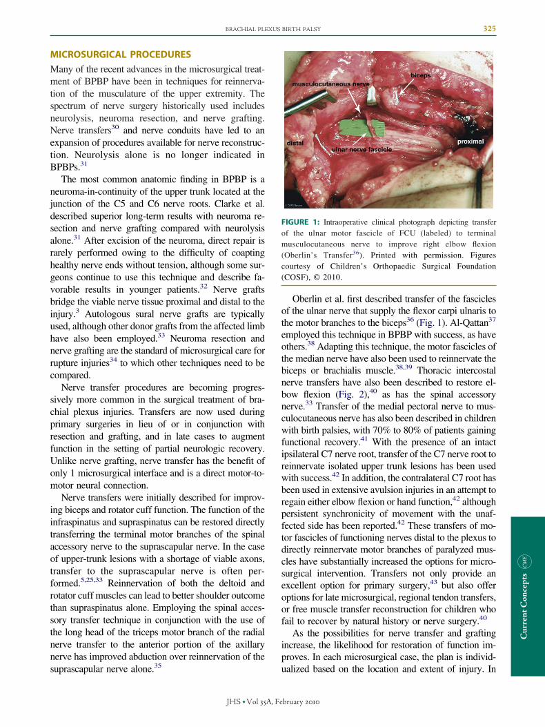
Oberlin et al. first described transfer of the fasciclesof the ulnar nerve that supply the flexor carpi ulnaris tothe motor branches to the biceps36 (Fig. 1). Al-Qattan37
employed this technique in BPBP with success, as haveothers.38 Adapting this technique, the motor fascicles ofthe median nerve have also been used to reinnervate thebiceps or brachialis muscle.38,39 Thoracic intercostalnerve transfers have also been described to restore el-bow flexion (Fig. 2),40 as has the spinal accessorynerve.33 Transfer of the medial pectoral nerve to mus-culocutaneous nerve has also been described in childrenwith birth palsies, with 70% to 80% of patients gainingfunctional recovery.41 With the presence of an intactipsilateral C7 nerve root, transfer of the C7 nerve root toreinnervate isolated upper trunk lesions has been usedwith success.42 In addition, the contralateral C7 root hasbeen used in extensive avulsion injuries in an attempt toregain either elbow flexion or hand function,42 althoughpersistent synchronicity of movement with the unaf-fected side has been reported.42 These transfers of mo-tor fascicles of functioning nerves distal to the plexus todirectly reinnervate motor branches of paralyzed mus-cles have substantially increased the options for micro-surgical intervention. Transfers not only provide anexcellent option for primary surgery,43 but also offeroptions for late microsurgical, regional tendon transfers,or free muscle transfer reconstruction for children whofail to recover by natural history or nerve surgery.40
As the possibilities for nerve transfer and graftingincrease, the likelihood for restoration of function im-proves. In each microsurgical case, the plan is individ-
FIGURE 1: Intraoperative clinical photograph depicting transferof the ulnar motor fascicle of FCU (labeled) to terminalmusculocutaneous nerve to improve right elbow flexion(Oberlin’s Transfer36). Printed with permission. Figurescourtesy of Children’s Orthopaedic Surgical Foundation(COSF), © 2010.
ualized based on the location and extent of injury. In
bruary

326 BRACHIAL PLEXUS BIRTH PALSY
Curren
tConcep
ts
total avulsions, Haerle and Gilbert24 argued that priorityshould be given to microsurgical reconstruction of thehand, specifically with attention to the median nerve,because infants can recover useful hand function afternerve grafting and nerve transfer.5,24
Carlstedt44 and Amr et al.45 have done a limitednumber of procedures attempting to directly graft intothe spinal cord for root avulsions. They have reportedgood preliminary results in their small series; yet, thisprocedure continues to carry substantial risk and uni-versal adoption remains unlikely.
Recent approval by the Food and Drug Administra-tion of synthetic collagen nerve conduits has led toincreased interest in the use of these nerve guidancechannels in microsurgery of BPBP. Previously, syn-thetic tubes constructed from materials such as polyg-lycolic acid, polylactide-co-caprolactone, and siliconeyielded poor results. There are no trials at this time tocompare collagen synthetic nerve grafts with autolo-gous nerve grafts. The advantages of synthetic graftsover conventional autologous grafts include eliminatingdonor site morbidity, increasing the amount of graftmaterial available, and providing direct conduits forneural growth factors produced by the proximal seg-ment to reach the distal segment. A preliminary seriesby Ashley et al. described 5 children treated with col-
FIGURE 2: Intraoperative photograph of intercostal nervestransferred to recipient nerve in right upper limb. Printed withpermission. Figures courtesy of Children’s Orthopaedic SurgicalFoundation (COSF), © 2010.
lagen tubes, with the result of a good recovery for 4 of
JHS �Vol A, Fe
the 5 patients (Motor Scale Composite � 0.6), and 3 of5 attaining an excellent recovery (Motor Scale Com-posite � 0.75) at the 2-year follow-up visit.46 Althoughlarger studies will be needed, the data from this smallstudy suggest that collagen matrix tubes could be a safealternative to autologous nerves for select short-segment brachial plexus repairs.
PROGRESSION OF DISEASE AT THE SHOULDERUp to 35% of infants and children with BPBPs experi-ence some degree of shoulder weakness, contracture, orjoint deformity.2,5 Only infants who recover completelywithin the first 2 months of life are spared from somedegree of long-term sequelae.8,26 Variable nerve recov-ery of upper-plexus injuries leads to soft tissue contrac-tures resulting from relatively unaffected internal rota-tors and adductors versus weak external rotators andabductors.47–49 Multiple authors have postulated thatthis muscular imbalance results in deformity of theglenohumeral joint, leading to further limitation inrange of motion, strength, and function for the pa-tient.3,50–52 Recent analyses of muscle volume andcross-sectional area of the shoulder region in childrenwith chronic brachial plexus injuries support this hy-pothesis.49,53
Previous studies describe development of internalrotation contractures of the shoulder in approximately50% to 70% of patients,2,8,32 which can be present inpatients as young as 5 months of age with incompleterecovery. The contracture develops primarily owing toweakness in infraspinatus and teres minor muscles andunopposed action of the internal rotators, which aredually innervated and therefore relatively unaffected.The etiology of muscular atrophy and hypoplasia ispartly due to denervation, but also occurs because thenormal passive stretch of the muscles is lost.54 Thisfurther highlights the importance of maintaining fullpassive range of motion in young children. The signif-icant difference in bulk and strength of the pectoralismajor and subscapularis compared with the externalrotators has been shown qualitatively by magnetic res-onance imaging studies.49 Compared with the unaf-fected side, the mean difference in the ratios of cross-sectional area was 30% for the pectoralis major toexternal rotator ratio and 10% for the subscapularis toexternal rotator ratio.49,53,54 In addition, having lowmuscular volumes of the infraspinatus and subscapu-laris predicted worsening subluxation with less than30% of the anterior humeral head covered.54
In addition to the physical impairments resultingfrom muscle weakness and soft tissue contracture of the
shoulder, progressive loss of passive external rotationbruary

BRACHIAL PLEXUS BIRTH PALSY 327
CurrentConcepts
and internal contracture can lead to glenohumeral jointdysplasia and joint instability.32,47,55,56 Weakness ofthe external rotators and subsequent contracture leads topositioning of the joint predominately in adduction andinternal rotation, which in turn leads to increased stressalong the posterior aspect of the glenoid and progres-sive deformity.47 These eccentric forces across the jointcause the glenoid to become increasingly retrovertedand the humeral head to become flat and to subluxate ina posterior direction.49,53,54
Sixty to eighty percent of children who do not re-cover full motor function have some degree of gleno-humeral deformity. Moderate glenohumeral deformitydevelops within the first 2 years of life in 60% to 70%of children with internal contractures. Deformityprogresses with age, correlating to the magnitude ofshoulder internal rotation contracture.3,55 Long-term ef-fects of glenohumeral dysplasia remain unknown, butsome authors have postulated that longstanding gleno-humeral deformity may predispose the shoulder to painor arthrosis. Children with established internal rotationcontracture and glenohumeral joint incongruity are un-likely to regain optimal shoulder function without in-tervention. Soft tissue lengthening procedures and re-duction of the joint have been shown to arrestprogressive dysplasia and promote remodeling of thejoint.
IMAGINGDifferent imaging modalities, including radiographs,arthrograms, ultrasound, computed tomography scans,and magnetic resonance imaging have been used toassess joint and bony development in BPBP. In thefirst few years of life, the humeral head and gle-noid are mostly nonossified cartilage, precludingvisualization with plain radiographs or computedtomography. For that reason most centers use mag-netic resonance imaging scans because they offerthe ability to visualize the cartilaginous surfaces ofthe glenohumeral joint in young children.52,53 Inolder children, computed tomography scans havebeen used to evaluate the bony structures afterossification. In young children, both modalitiesrequire sedation. Ultrasonography has been used tovisualize the joint in real time, is minimally inva-sive, and does not require sedation. Arthrogramshave been shown to correlate well with magneticresonance imaging findings, especially at the endsof the spectrum of deformity; however, they haveto be performed with sedation or general anesthe-sia. Arthrography has usually been performed at
the time of surgery, thus decreasing their utility inJHS �Vol A, Fe
preoperative planning.55 Increasingly, as it be-comes apparent that the severity of the glenohu-meral deformity influences the success of second-ary reconstruction, most surgeons advocatepreoperative magnetic resonance imaging to assessfor underlying osseous and joint deformities inpatients with internal rotation contractures.34,57
Various classifications have been proposed for grad-ing severity of glenohumeral deformity in BPBPs. Themost commonly used are those of Birch and Achan,58
Waters et al.,59 and Pearl and Edgerton.55 All 3 classi-fication schemes describe increasing severity as increas-ing subluxation of the humeral head within an increas-ingly retroverted glenoid and eventual flattening of boththe glenoid and humeral head.47,52,53,57,59 With retro-version of the glenoid, a “pseudoglenoid” is formedwith false articulation of the humeral head with theposterior aspect of the glenoid. The classification in thestudy by Waters et al. describes the progression fromnormal (type I) to posterior glenoid deformity (type II),to humeral head subluxation and further glenoid dys-plasia (type III), to the development of a false glenoid(type IV), and to flattening of both the glenoid and thehumeral head (type V) (Fig. 3). The degree of jointdeformity can help guide the surgeon in selectingsurgical procedures, especially as our understand-ing of glenoid remodeling after soft tissue andjoint procedures increases.59 Recently, Clarke etal. suggested that the biceps angle, measured as theangle between the biceps tendon and a line per-pendicular to the axis of the scapula, may better
FIGURE 3: Axial magnetic resonance image of left shoulderdemonstrating posterior humeral head subluxation andincreased glenoid retroversion in the setting of longstandingbrachial plexus birth palsy. Printed with permission. Figurescourtesy of Children’s Orthopaedic Surgical Foundation(COSF), © 2010.
characterize rotational deformity than classic clas-
bruary

328 BRACHIAL PLEXUS BIRTH PALSY
Curren
tConcep
ts
sification schemes and should be used as anothermeasurement of osseous and joint deformity.60
SURGICAL MANAGEMENT OF THE SHOULDERIndications for surgical intervention involving theshoulder include infantile dislocation, persistent internalrotation contracture refractory to physiotherapy, limita-tion of active abduction and external rotation functionwith plateauing of neural recovery, and progressiveglenohumeral deformity. The principles of treatment foreach individual include contracture release, muscle re-balancing, and joint reduction. The age at interventionand type of procedure depend on the problem and itsseverity. Ultimately, the limitation in function as well asseverity of glenohumeral deformity influence the typeand timing of secondary reconstructive options. Thoseoptions include extra-articular musculotendinouslengthenings and tendon transfers with or without intra-articular release performed as either an open or arthro-scopic procedure. In the setting of more severe osseousdeformity, alternative options such as derotational hu-meral osteotomies or glenoid osteotomies may be con-sidered.
Fairbank and Sever described initial musculotendi-nous procedures in the early 20th century, which in-volved z-lengthening of the subscapularis and and pec-toralis major.61 Later, L’Espisocopo described transferof the teres major and lattisimus dorsi to the rotator cuffin addition to lengthening of the internal rotators (Fig.4). Many authors, including Sever and Fairbank, advo-cate not opening the joint to prevent anterior-inferiordislocation or stiffness. Chen et al. suggested that im-
FIGURE 4: Intraoperative appearance of latissimus dorsi andteres major tendons (marked by traction sutures) harvested fortransfer to rotator cuff of left upper extremity. Printed withpermission. Figures courtesy of Children’s Orthopaedic SurgicalFoundation (COSF), © 2010.
provement of both abduction and external rotation of
JHS �Vol A, Fe
the shoulder could be achieved by a combined transferof trapezius to the humerus and traditional transfer ofthe lattisimus dorsi and teres major to the rotator cuff.62
A series by Hoffer and Phipps involving patients whounderwent release of the pectoralis major with transferof lattisimus dorsi and teres major to the posteriorrotator cuff reported that an extra-articular techniquemay be sufficient to restore function and maintainproper shoulder joint position.63 However, later seriesemploying a similar technique in more dysplastic jointsproduced inconsistent results, which suggests that in thepresence of considerable glenohumeral dysplasia, ex-traarticular procedures alone are insufficient for jointreduction or remodeling.34,64,65
One of the most important concepts to surface fromthe literature in recent years is the effect of soft tissueprocedures on osseous formation of the shoulder joint.The data suggest that without concurrent intra-articularprocedures, improvement in shoulder motion can begained, but little correction of underlying osseous de-formity occurs.34,47,48,66 An initial series by Hui andTorode67 reported that open reduction of the glenohu-meral joint accompanied by tendon lengthening andtransfers could reduce abnormal glenoid retroversion inyoung patients with BPBP. Importantly, interventionalso led to joint remodeling with improvement in gle-noid retroversion in 30% of their patients.
Recent studies have increasingly looked at bothfunctional outcomes and this potential for remodelingof the glenohumeral joint after soft tissue and jointreduction procedures.34,65,67,68 Waters et al.49 showedan improvement in the grade of glenohumeral dysplasiain 83% of patients with combined intra-articular andextra-articular procedures. Glenoid version angles im-proved from –39 to –18 and the percentage of thehumeral head anterior to the middle of the glenoidimproved from 13% to 38%. Using an arthroscopicapproach, Pearl et al.,65 Kozin et al.,69 and others68
have also demonstrated that the release of the subscap-ularis tendon or anterior release of the glenohumeraljoint capsule, in isolation or combined with latissimusdorsi and teres major tendon transfers, leads to im-provement in passive external rotation and centering ofthe humeral head within the glenoid. In the arthroscopicseries by Pearl et al., external rotation was achieved in40 of the 41 children, and postoperative imaging dem-onstrated improvement in glenohumeral dysplasia in 12of the 15 patients for whom imaging was available.With either open or arthroscopic releases, care must betaken not to lose internal rotation power resulting fromextensive release of the anterior capsule and subscapu-
laris.bruary

BRACHIAL PLEXUS BIRTH PALSY 329
CurrentConcepts
Recently Elhassan and Shin described a novel trans-fer of middle and lower segments of trapezius withAchilles tendon allograft augmentation to the infraspi-natus for external rotation in older patients experiencingtraumatic brachial plexopathies. This technique offersan alternative to a latissimus or teres major transfer toachieve external rotation.70
The role of botulinum toxin A is expanding and isunder investigation in the treatment of brachial plexusinjuries. It has been used to prevent contractures in theimmediate postoperative period after microsur-gery.29,71,72 It has been used with serial casting as anadjunct to physical therapy.73,74 The effect of the toxinis to reduce muscle tone and force to restore passiverange of motion and joint alignment.29,54,72 Price et al.postulated that there may also be a central nervoussystem effect in children with BPBP similar to theeffect in cerebral palsy.75
Older patients with advanced shoulder joint defor-mity may best be served by a derotational humeralosteotomy to improve function. Multiple stud-ies25,26,76,77 have shown that upper extremity functionis significantly improved by orienting the arc of shoul-der rotation into a more functional range. This wasquantitatively illustrated by increases in Mallet scoresfor global shoulder function, including hand to mouthand hand to neck,76 as well as passive abduction andexternal rotation.77
Whether glenoid osteotomy has a role in the treat-ment of moderate glenoid deformity is still unclear.Hopyan and Clarke (Hopyan S, presented at the Pedi-atric Orthopedic Society of North America annualmeeting, 2009) reported this technique for more severecases of glenohumeral dysplasia. A triangle tilt surgeryhas also been described to improve the limitation ofexternal rotation of the shoulder by eliminating possibleimpingement of the distal acromioclavicular triangleagainst the humeral head, which Nath et al. identified asthe etiology of the internal rotation contracture. Theauthors described this procedure involving osteotomiesof the medial clavicle and medial spine of the scapula aspreliminarily effective in restoring external rotation.78
Given the options for surgical intervention, radio-graphic and physical examination of the patient mustguide treatment. In patients with no or minimal gleno-humeral dysplasia, musculotendinous lengthenings andtendon transfers may be sufficient to improve globalshoulder function. Conversely, patients with the mostsevere humeral head flattening, glenoid deformity, andposterior dislocation may benefit from a derotationalhumeral osteotomy to place the extremity in a more
functional position.37,76 Patients who fall along the con-JHS �Vol A, Fe
tinuum between these extremes will likely benefit fromextra-articular rebalancing with joint reduction proce-dures.76
Infants who experience BPBP have a wide range ofclinical presentations, and their care and treatmentshould be tailored. Recent developments in nerve, softtissue, and osseous procedures have led to better func-tional outcomes. Despite these advances, the propertiming of the intervention and type of surgical proce-dure for children who do not recover motor functionspontaneously continue to be controversial topics. Aswe continue to learn about the sequlae after injury andthe effects of various treatment options, the optimalmanagement of infants with BPBP can only be clarifiedthrough prospective multicenter studies such as the oneunder way through the Pediatric Orthopedic Society ofNorth America.
REFERENCES1. Foad SL, Mehlman CT, Ying J. The epidemiology of neonatal
brachial plexus palsy in the United States. J Bone Joint Surg 2008;90A:1258–1264.
2. Hoeksma AF, ter Steeg AM, Nelissen RG, van Ouwerkerk WJ,Lankhorst GJ, de Jong BA. Neurological recovery in obstetric bra-chial plexus injuries: an historical cohort study. Dev Med ChildNeurol 2004;46:76–83.
3. Water PM. Obstetric brachial plexus injuries: evaluation and man-agement. J Am Acad Orthop Surg 1997;5:205–214.
4. Laurent JP, Lee R, Shenaq S, Parke JT, Solis IS, Kowalik L.Neurosurgical correction of upper brachial plexus birth injuries.J Neurosurg 1993;79:197–203.
5. Pondaag W, Lee R, Shenaq S, Parke JT, Solis IS, Kowalik L. Naturalhistory of obstetric brachial plexus palsy: a systematic review. DevMed Child Neurol 2004;46:138–144.
6. Sunderland S. Nerves and nerve injuries. London: Churchill Living-stone, 1978.
7. Narakas AO. [Injuries of the brachial plexus and neighboring pe-ripheral nerves in vertebral fractures and other trauma of the cervicalspine]. Orthopade 1987;16:81–86.
8. Waters PM. Comparison of the natural history, the outcome ofmicrosurgical repair, and the outcome of operative reconstruction inbrachial plexus birth palsy. J Bone Joint Surg 1999;81A:649–659.
9. Mallet J. [Obstetrical paralysis of the brachial plexus. II. Therapeu-tics. Treatment of sequelae. Priority for the treatment of the shoulder.Method for the expression of results]. Rev Chir Orthop ReparatriceAppar Motil 1972;58(Suppl 1):166–168.
10. O’Brien DF, Park TS, Noetzel MJ, Weatherly T. Management ofbirth brachial plexus palsy. Childs Nerv Syst 2006;22:103–112.
11. DiTaranto P, Campagna L, Price AE, Grossman JA. Outcome fol-lowing nonoperative treatment of brachial plexus birth injuries.J Child Neurol 2004;19:87–90.
12. Eng GD, Binder H, Getson P, O’Donnell R. Obstetrical brachialplexus palsy (OBPP) outcome with conservative management. Mus-cle Nerve 1996;19:884–891.
13. Bain JR, Dematteo C, Gjertsen D, Hollenberg RD. Navigating thegray zone: a guideline for surgical decision making in obstetricalbrachial plexus injuries. J Neurosurg Pediatr 2009;3:173–180.
14. Strombeck C, Krumlinde-Sundholm L, Forssberg H. Functional out-come at 5 years in children with obstetrical brachial plexus palsywith and without microsurgical reconstruction. Dev Med Child Neu-
rol 2000;42:148–157.15. Nehme A, Kany J, Sales-De-Gauzy J, Charlet JP, Dautel G, Cahuzac
bruary

330 BRACHIAL PLEXUS BIRTH PALSY
Curren
tConcep
ts
JP. Obstetrical brachial plexus palsy: prediction of outcome in upperroot injuries. J Hand Surg 2002;7B:9–12.
16. Bae DS, Ferretti M, Waters PM. Upper extremity size differences inbrachial plexus birth palsy. Hand (N Y) 2008;3:297–303.
17. McDaid PJ, Kozin SH, Thoder JJ, Porter ST. Upper extremitylimb-length discrepancy in brachial plexus palsy. J Pediatr Orthop2002;22:364–366.
18. Kirjavainen MO, Remes VM, Peltonen J, Helenius IJ, RautakorpiSM, Vähäsarja VJ, et al. Permanent brachial plexus birth palsy doesnot impair the development and function of the spine and lowerlimbs. J Pediatr Orthop B 2009 Jul 9 [Epub ahead of print].
19. Bae DS, Zurakowski D, Avallone N, Yu R, Waters PM. Sportsparticipation in selected children with brachial plexus birth palsy.J Pediatr Orthop 2009;29:496–503.
20. Bae DS, Waters PM, Zurakowski D. Correlation of pediatric out-comes data collection instrument with measures of active movementin children with brachial plexus birth palsy. J Pediatr Orthop 2008;28:584–592.
21. Yang LJ, Anand P, Birch R. Limb preference in children withobstetric brachial plexus palsy. Pediatr Neurol 2005;33:46–49.
22. Bae DS, Waters PM, Zurakowski D. Reliability of three classifica-tion systems measuring active motion in brachial plexus birth palsy.J Bone Joint Surg 2003;85A:1733–1738.
23. Gilbert A, Tassin JL. [Surgical repair of the brachial plexus inobstetric paralysis]. Chirurgie 1984;110:70–75.
24. Haerle M, Gilbert A. Management of complete obstetric brachialplexus lesions. J Pediatr Orthop 2004;24:194–200.
25. Terzis JK, Kokkalis ZT. Shoulder function following primary axil-lary nerve reconstruction in obstetrical brachial plexus patients. PlastReconstr Surg 2008;122:1457–1469.
26. Al-Qattan MM. The outcome of Erb’s palsy when the decision tooperate is made at 4 months of age. Plast Reconstr Surg 2000;106:1461–1465.
27. Clarke HM, Curtis CG. An approach to obstetrical brachial plexusinjuries. Hand Clin 1995;11:563–580; discussion 580–581.
28. Chuang DC, Mardini S, Ma HS. Surgical strategy for infant obstet-rical brachial plexus palsy: experiences at Chang Gung MemorialHospital. Plast Reconstr Surg 2005;116:132–142; discussion 143–144.
29. Grossman JA, DiTaranto P, Yaylali I, Alfonso I, Ramos LE, PriceAE. Shoulder function following late neurolysis and bypass graftingfor upper brachial plexus birth injuries. J Hand Surg 2004;29B:356–358.
30. El-Gammal TA, Fathi NA. Outcomes of surgical treatment of bra-chial plexus injuries using nerve grafting and nerve transfers. JReconstr Microsurg 2002;18:7–15.
31. Clarke HM, DiTaranto P, Yaylali I, Alfonso I, Ramos LE, Price AE.Obstetrical brachial plexus palsy: results following neurolysis ofconducting neuromas-in-continuity. Plast Reconstr Surg 1996;97:974–982; discussion 983–984.
32. Kirjavainen M, Remes V, Peltonen J, Kinnunen P, Pöyhiä T, Te-laranta T, et al. Long-term results of surgery for brachial plexus birthpalsy. J Bone Joint Surg 2007;89A:18–26.
33. Midha R. Nerve transfers for severe brachial plexus injuries: areview. Neurosurg Focus 2004;16:E5.
34. Waters PM, Bae DS. Effect of tendon transfers and extra-articularsoft-tissue balancing on glenohumeral development in brachialplexus birth palsy. J Bone Joint Surg 2005;87A:320–325.
35. Bertelli JA, Ghizoni MF. Reconstruction of C5 and C6 brachialplexus avulsion injury by multiple nerve transfers: spinal accessoryto suprascapular, ulnar fascicles to biceps branch, and triceps long orlateral head branch to axillary nerve. J Hand Surg 2004;29A:131–139.
36. Oberlin C, Béal D, Leechavengvongs S, Salon A, Dauge MC, SarcyJJ. Nerve transfer to biceps muscle using a part of ulnar nerve forC5-C6 avulsion of the brachial plexus: anatomical study and report
of four cases. J Hand Surg 1994;19A:232–237.JHS �Vol A, Fe
37. Al-Qattan MM. Oberlin’s ulnar nerve transfer to the biceps nerve inErb’s birth palsy. Plast Reconstr Surg 2002;109:405–407.
38. Noaman HH, Shiha AE, Bahm J, Oberlin’s ulnar nerve transfer to thebiceps motor nerve in obstetric brachial plexus palsy: indications,and good and bad results. Microsurgery 2004;24:182–187.
39. Zhao X, Lao J, Hung LK, Zhang GM, Zhang LY, Gu YD. Selectiveneurotization of the median nerve in the arm to treat brachial plexuspalsy: surgical technique. J Bone Joint Surg 2005;87A(Suppl 1):122–135.
40. Kawabata H, Shibata T, Matsui Y, Yasui N. Use of intercostal nervesfor neurotization of the musculocutaneous nerve in infants withbirth-related brachial plexus palsy. J Neurosurg 2001;94:386–391.
41. Wellons JC, Tubbs RS, Pugh JA, Bradley NJ, Law CR, Grabb PA.Medial pectoral nerve to musculocutaneous nerve neurotization forthe treatment of persistent birth-related brachial plexus palsy: an11-year institutional experience. J Neurosurg Pediatr 2009;3:348–353.
42. Gu YD. Contralateral C7 root transfer over the last 20 years inChina. Chin Med J (Engl) 2007;120:1123–1126.
43. Shigematsu K, Yajima H, Kobata Y, Kawamura K, Maegawa N,Takakura Y. Oberlin partial ulnar nerve transfer for restoration inobstetric brachial plexus palsy of a newborn: case report. J BrachialPlex Peripher Nerve Inj 2006;1:3.
44. Carlstedt T, Grane P, Hallin RG, Norén G. Return of function afterspinal cord implantation of avulsed spinal nerve roots. Lancet 1995;346:1323–1325.
45. Amr SM, Essam AM, Abdel-Meguid AM, Kholeif AM, MoharramAN, El-Sadek RE. Direct cord implantation in brachial plexus avul-sions: revised technique using a single stage combined anterior (first)posterior (second) approach and end-to-side side-to-side graftingneurorrhaphy. J Brachial Plex Peripher Nerve Inj 2009;4:8.
46. Ashley WW Jr, Weatherly T, Park TS. Collagen nerve guides forsurgical repair of brachial plexus birth injury. J Neurosurg 2006;105(6 Suppl):452–456.
47. Kozin SH, Chafetz RS, Barus D, Filipone L. Magnetic resonanceimaging and clinical findings before and after tendon transfers aboutthe shoulder in children with residual brachial plexus birth palsy. JShoulder Elbow Surg 2006;15:554–561.
48. Newman CJ, Morrison L, Lynch B, Hynes D. Outcome of subscap-ularis muscle release for shoulder contracture secondary to brachialplexus palsy at birth. J Pediatr Orthop 2006;26:647–651.
49. Waters PM, Monica JT, Earp BE, Zurakowski D, Bae DS. Correla-tion of radiographic muscle cross-sectional area with glenohumeraldeformity in children with brachial plexus birth palsy. J Bone JointSurg 2009;91A:2367–2375.
50. Moukoko D, Ezaki M, Wilkes D, Carter P. Posterior shoulderdislocation in infants with neonatal brachial plexus palsy. J BoneJoint Surg 2004;86A:787–793.
51. Pagnotta A, Haerle M, Gilbert A. Long-term results on abductionand external rotation of the shoulder after latissimus dorsi transferfor sequelae of obstetric palsy. Clin Orthop Relat Res 2004;426:199–205.
52. van der Sluijs JA, van der Meij M, Verbeke J, Manoliu RA, Wuis-man PI. Measuring secondary deformities of the shoulder in childrenwith obstetric brachial plexus lesion: reliability of three methods.J Pediatr Orthop 2003;12B:211–214.
53. Pöyhiä TH, Nietosvaara YA, Remes VM, Kirjavainen MO, PeltonenJI, Lamminen AE. MRI of rotator cuff muscle atrophy in relation toglenohumeral joint incongruence in brachial plexus birth injury.Pediatr Radiol 2005;35:402–409.
54. van Gelein Vitringa VM, van Kooten EO, Jaspers RT, MullenderMG, van Doorn-Loogman MH, van der Sluijs JA. An MRI study onthe relations between muscle atrophy, shoulder function and gleno-humeral deformity in shoulders of children with obstetric brachialplexus injury. J Brachial Plex Peripher Nerve Inj 2009;4:5.
55. Pearl ML, Edgerton BW. Glenoid deformity secondary to brachialplexus birth palsy. J Bone Joint Surg 1998;80A:659–667.
56. Kozin SH. Correlation between external rotation of the glenohumeral
bruary

BRACHIAL PLEXUS BIRTH PALSY 331
joint and deformity after brachial plexus birth palsy. J Pediatr Orthop2004;24:189–193.
57. Pearl ML. Arthroscopic release of shoulder contracture secondary tobirth palsy: an early report on findings and surgical technique.Arthroscopy 2003;19:577–582.
58. Birch R, Achan P. Peripheral nerve repairs and their results inchildren. Hand Clin 2000;16:579–595.
59. Waters PM, Smith GR, Jaramillo D. Glenohumeral deformity sec-ondary to brachial plexus birth palsy. J Bone Joint Surg 1998;80A:668–677.
60. Clarke SE, Kozin SH, Chafetz RS. The biceps tendon as a measureof rotational deformity in residual brachial plexus birth palsy. J Pe-diatr Orthop 2009;29:490–495.
61. Sever JW. Obstetric paralysis: report of eleven hundred cases.JAMA 1925;85:1862–1865.
62. Chen L, Gu YD, Hu SN. Applying transfer of trapezius and/orlatissimus dorsi with teres major for reconstruction of abduction andexternal rotation of the shoulder in obstetrical brachial plexus palsy.J Reconstr Microsurg 2002;18:275–280.
63. Hoffer MM, Phipps GJ. Closed reduction and tendon transfer fortreatment of dislocation of the glenohumeral joint secondary tobrachial plexus birth palsy. J Bone Joint Surg 1998;80A:997–1001.
64. van der Sluijs JA, van Ouwerkerk WJ, de Gast A, Wuisman PI,Nollet F, Manoliu RA. Deformities of the shoulder in infantsyounger than 12 months with an obstetric lesion of the brachialplexus. J Bone Joint Surg 2001;83B:551–555.
65. Pearl ML, Edgerton BW, Kazimiroff PA, Burchette RJ, Wong K.Arthroscopic release and latissimus dorsi transfer for shoulder inter-nal rotation contractures and glenohumeral deformity secondary tobrachial plexus birth palsy. J Bone Joint Surg 2006;88A:564–574.
66. El-Gammal TA, Saleh WR, El-Sayed A, Kotb MM, Imam HM, FathiNA. Tendon transfer around the shoulder in obstetric brachial plexusparalysis: clinical and computed tomographic study. J Pediatr Orthop2006;26:641–646.
67. Hui JH, Torode IP. Changing glenoid version after open reduction of
shoulders in children with obstetric brachial plexus palsy. J PediatrJOURNAL CME QUESTIONS
JHS �Vol A, Fe
68. Pedowitz DI, Gibson B, Williams GR, Kozin SH. Arthroscopictreatment of posterior glenohumeral joint subluxation resulting frombrachial plexus birth palsy. J Shoulder Elbow Surg 2007;16:6–13.
69. Kozin SH, Boardman MJ, Chafetz RS, Williams GR, Hanlon A.Arthroscopic treatment of internal rotation contracture and glenohu-meral dysplasia in children with brachial plexus birth palsy. J Shoul-der Elbow Surg 2010;19:102–110.
70. Elhassan B, Bishop A, Shin A. Trapezius transfer to restore externalrotation in a patient with a brachial plexus injury: a case report.J Bone Joint Surg 2009;91A:939–944.
71. Price AE, Ditaranto P, Yaylali I, Tidwell MA, Grossman JA. Bot-ulinum toxin type A as an adjunct to the surgical treatment of themedial rotation deformity of the shoulder in birth injuries of thebrachial plexus. J Bone Joint Surg 2007;89B:327–329.
72. Vekris MD, Lykissas MG, Beris AE, Manoudis G, Vekris AD,Soucacos PN. Management of obstetrical brachial plexus palsy withearly plexus microreconstruction and late muscle transfers. Micro-surgery 2008;28:252–261.
73. Desiato MT, Risina B. The role of botulinum toxin in the neuro-rehabilitation of young patients with brachial plexus birth palsy.Pediatr Rehabil 2001;4:29–36.
74. Rollnik JD, Hierner R, Schubert M, Shen ZL, Johannes S, Tröger M,et al. Botulinum toxin treatment of cocontractions after birth-relatedbrachial plexus lesions. Neurology 2000;55:112–114.
75. Price AE, Grossman JA, Tidwell M. Potential for remodeling of theglenoid in children with brachial plexus palsy and shoulder sublux-ation/dislocation. J Pediatr Orthop 2004;24:346.
76. Waters PM, Bae DS. The effect of derotational humeral osteotomyon global shoulder function in brachial plexus birth palsy. J BoneJoint Surg 2006;88A:1035–1042.
77. Kirkos JM, Kyrkos MJ, Kapetanos GA, Haritidis JH. Brachial plexuspalsy secondary to birth injuries. J Bone Joint Surg 2005;87B:231–235.
78. Nath RK, Amrani A, Melcher SE, Eichhorn MG. Triangle tilt sur-gery in an older pediatric patient with obstetric brachial plexus
Orthop 2003;23:109–113. injury. Eplasty 2009;9:e26.
Current Concepts in the Management of Brachial Plexus Birth Palsy
What is the most common roots injury in brachial plexus birth palsy?
a. C5
b. C5 and C6
c. C5, C6, and C7
d. C5, C6, C7, C8, and T1
What is the surgical treatment for extra-foraminal ruptures in childrenwith brachial plexus birth palsies?
a. Neurolysis
b. Resection and direct repair
c. Resection and nerve grafting
d. Resection and nerve conduit
CurrentConcepts
e. C8 and T1 e. Nerve transfers
To take the online test and receive CME credit, go to http://www.assh.org/professionals/jhs.
bruary