current knowledge and perspectives of potential impacts of
TRANSCRIPT

REVIEW Open Access
Current knowledge and perspectives ofpotential impacts of Salmonella enterica onthe profile of the gut microbiotaNesreen H. Aljahdali1,2, Yasser M. Sanad1,3,4, Jing Han1 and Steven L. Foley1*
Abstract
In the past decade, the initial studies of the gut microbiota started focusing on the correlation of the compositionof the gut microbiota and the health or diseases of the host, and there are extensive literature reviews pertainingto this theme. However, little is known about the association between the microbiota, the host, and pathogenicbacteria, such as Salmonella enterica, which is among the most important foodborne pathogens and identified asthe source of multiple outbreaks linked to contaminated foods causing salmonellosis. Secretion systems, flagella,fimbriae, endotoxins, and exotoxins are factors that play the most important roles in the successful infection of thehost cell by Salmonella. Infections with S. enterica, which is a threat to human health, can alter the genomic,taxonomic, and functional traits of the gut microbiota. The purpose of this review is to outline the state ofknowledge on the impacts of S. enterica on the intestinal microbiota and highlight the need to identify the gutbacteria that could contribute to salmonellosis.
Keywords: Gut microbiota, Salmonella enterica, Host cell-micbobe interaction
BackgroundAn enteric pathogen is a microbe that impacts thegastrointestinal tract (GIT) and causes gastrointestinaldiseases. These infectious pathogens, including bacteriasuch as Escherichia, Campylobacter, Shigella, Yersinia,Salmonella, and other genera, protozoa such as amoeba,rotavirus, and other pathogenic microorganisms, are re-sponsible for causing gastroenteritis [1]. Among entericpathogens there is often an age-associated bias with thedevelopment of gastroenteritis upon exposure. For ex-ample, Escherichia coli (E. coli) causes enteric disease inpeople most commonly during early and late ages,whereas rotaviruses are the most common among in-fants and young children. Similarly, Campylobacter in-fections occur most often in early childhood into young
adulthood, while Salmonella infections have higher ratesin infants and people over 65 [2]. Salmonella infectionsare a significant global public health threat and contrib-ute to morbidity and mortality worldwide [3]. The Sal-monella genus is generally considered to be divided intotwo species: S. enterica and S. bongori. Although, S. bon-gori appears adapted to cold-blooded animals, it can in-fect humans, but accounts for less than 1% of humaninfections [4, 5]. On the other hand, several of the sub-species of S. enterica are more commonly isolated fromwarm-blooded animals. S. enterica includes six subspe-cies: S. enterica subsp. enterica, S. enterica subsp. sala-mae, S. enterica subsp. arizonae, S. enterica subsp.diarizonae, S. enterica subsp. houtenae, and S. entericasubsp. indica. Among these subspecies, S. enterica subsp.salamae, and S. enterica subsp. arizonae, are more com-monly isolated from cold-blooded animals [6, 7]. S.enterica includes more than 2600 serotypes that differfrom each other based on the polysaccharide portion of
© The Author(s). 2020 Open Access This article is licensed under a Creative Commons Attribution 4.0 International License,which permits use, sharing, adaptation, distribution and reproduction in any medium or format, as long as you giveappropriate credit to the original author(s) and the source, provide a link to the Creative Commons licence, and indicate ifchanges were made. The images or other third party material in this article are included in the article's Creative Commonslicence, unless indicated otherwise in a credit line to the material. If material is not included in the article's Creative Commonslicence and your intended use is not permitted by statutory regulation or exceeds the permitted use, you will need to obtainpermission directly from the copyright holder. To view a copy of this licence, visit http://creativecommons.org/licenses/by/4.0/.The Creative Commons Public Domain Dedication waiver (http://creativecommons.org/publicdomain/zero/1.0/) applies to thedata made available in this article, unless otherwise stated in a credit line to the data.
* Correspondence: [email protected] of Microbiology, National Center for Toxicological Research, U.S.Food and Drug Administration, 3900 NCTR Rd, Jefferson, AR 72079, USAFull list of author information is available at the end of the article
Aljahdali et al. BMC Microbiology (2020) 20:353 https://doi.org/10.1186/s12866-020-02008-x

lipopolysaccharide layer (O antigen) and/or the filament-ous portion of the flagella (H antigen) [8]. Scallan et al(2011) estimated that nontyphoidal Salmonella accountfor approximately 28% of foodborne illness-associateddeaths [9]. The predominant subspecies associated withsevere disease is S. enterica subsp. enterica and amongits serotypes, there is also variability in the outcomes ofdisease with some serovars causing relatively severe out-comes. For example, S. enterica serovar Heidelberg con-tributes to about 7% of the Salmonella-related deaths inthe U.S. [10] and 11% of reported invasive infections,which are relatively high percentages considering thatthey typically cause under 5% of infections [11].S. enterica is a highly diverse Gram-negative bacterial
species that can be divided into typhoidal and nonty-phoidal Salmonella serovars. Typhoidal Salmonella sero-vars share virulence properties that were obtainedthrough convergent evolution and therefore these viru-lence genes are absent from most non-typhoidal Sal-monella serovars [12]. For instance, S. Typhi has specificvirulence factors, including typhoid toxin and Vi antigen[7, 12, 13]. Nontyphoidal S. including Typhimurium,Enteritidis, Heidelberg, Newport, Weltevreden, Choler-aesuis, Saintpaul, Infantis and Javiana cause gastroenter-itis, while typhoidal S. including Typhi and Paratyphiserovars commonly cause typhoid fever [13]. Nontyphoi-dal serotypes can be transferred between humans andanimals, whereas typhoidal serotypes are only transmis-sible among humans [14]. Notably, nontyphoidal Sal-monella disseminates rapidly in people with an impairedimmune system and in neonates [15]. Ninety-five per-cent of S. enterica infections are associated with con-sumption of contaminated food products [7]. More than2600 serotypes of Salmonella have been identified [16,17]. To clarify links between Salmonella serotypes andfood products, Jackson and colleagues (2013) indicatedthat more than 80% of outbreaks caused by serotypesEnteritidis, Heidelberg, and Hadar were associated witheggs or poultry, while greater than 50% of outbreakscaused by serotypes Javiana, Litchfield, Mbandaka,Muenchen, Poona, and Senftenberg were attributed toplant commodities. Serotypes Typhimurium and New-port were linked to a wide variety of food commodities[18]. These organisms invade the GIT causing salmonel-losis, which is typically characterized by a self-limitinggastroenteritis symptom, such as diarrhea, fever, abdom-inal cramps, and vomiting [19].The GIT is host to diverse taxa from across the tree of
life, such as bacteria, archaea, fungi, protozoa, and virusesthat make up the gut microbiota [20]. The gut harbors ahighly diverse microbial community, which impacts thehost’s nutrition, physiology, and immune system [21, 22].The composition of the gut microbiota remains relativelystable within healthy people throughout their lifetime
[23]. However, specific shifts in the composition and di-versity have been linked to diet, diseases, and susceptibilityto infection. For instance, alteration of the intestinalmicrobiota has been associated with acute inflammationthat can be triggered by enteric pathogens [24]. Salmon-ella and other pathogens have been widely studied; how-ever, the interactions between enteric pathogens andintestinal microbes are not well understood. In this reviewwe will summarize the knowledge of the interaction be-tween Salmonella and intestinal microbiota that is cur-rently available and clarify the research that needs to beundertaken to understand the consequences of thesesinteractions.
Gut microbiotaHuman gut microbiota/microbiomeThe human body hosts up to 100 trillion (1014) mi-crobes, with the majority residing in the GIT, which hasbecome the most investigated microbial community inrecent years [20, 25]. Most of the microbiota in the GITare primarily anaerobic bacteria. Typically, 97% of thebacteria in the GIT are strict anaerobes, and only 3%constitute the aerobic bacteria (facultative anaerobes)[26]. The collective pan-genome of bacterial cells is lar-ger than the human genome [25]. There are large differ-ences in microbial load in different regions of the GIT.To illustrate this, Helicobacter pylori resides in the stom-ach at a concentration of 102–103 cells/ml. The mucosaof the small intestine is dominated by the phyla Bacter-oidetes and members of the Clostridiales cluster XIVand IV, and the lumen contains members of the Entero-bacteriaceae with a biomass of 104–105 cell/ml [22, 25].The large intestine contains species from the phyla Bac-teroidetes and Firmicutes with amounts in the range of1011–1012, with other phyla including Proteobacteria,Verrucomicrobia, and Actinobacteria being less repre-sented (Fig. 1) [22, 25].Generally, the composition of the gut microbiota shifts
throughout life as people transition from newborns toinfants to young adults to elders. The GIT of newbornsis expected to be sterile at birth. However, major shiftstake place during and immediately after birth due to thecolonization with aerobic bacteria (Enterococcaceae andStreptococcus) [27]. The gut microbiota composition ofinfants is highly dynamic with low levels of total bacteria[28]. The microbiota of infants is dominated by somemembers of Clostridium, Bifidobacteria, and facultativeanaerobes like E. coli, while elderly people generally havehigher levels of Bacteroidetes and facultative anaerobeslike E. coli [29]. In young adults the composition of thegut microbiota is dominated by Bacteroidetes and Firmi-cutes with smaller portions of Actinobacteria, Verruco-microbia, and Proteobacteria [25].
Aljahdali et al. BMC Microbiology (2020) 20:353 Page 2 of 15

A generally symbiotic relationship between the host andthe gut microbiota has been known to be strongly associ-ated with health [22]. The host provides a nutrient-richand hospitable environment for the gut microbiota. Inparallel, the gut microbiota is extremely important as itsupports the host by enhancing metabolism, maturationof the immune system, developing the GIT, and protect-ing against pathogens [26, 30]. Also, intestinal bacteria de-grade undigested foods by two main metabolic pathways:saccharolytic and proteolytic pathways. Non-digestiblecarbohydrates are degraded into monomeric sugars thatcan be converted to beneficial products, such as short-chain fatty acids (SCFAs), principally acetate, propionate,and butyrate. These products have been shown to de-crease the risk of developing gastrointestinal disorders,cancer, and other metabolic syndromes [31–33]. Peptideand amino acids, on the other hand, are hydrolyzed intoshort or branched-chain fatty acids and other metabolicelements, some of which are possibly toxic to the host,such as uremic toxins [34, 35]. The gut microbiota usuallylives within the host in a commensal manner; however,many external factors can alter the balance of this micro-biota composition, potentially leading to gastrointestinaldiseases, such as salmonellosis.
SalmonellosisSalmonella gastroenteritisSalmonella infections are significant economic and publichealth concerns, costing an estimated 3.7 billion dollarsper year [36, 37]. According to the Centers for Diseases
Control and Prevention (CDC), it is estimated that mem-bers of the Salmonella genus cause 1.35 million infectionsleading to 26,500 hospitalizations and 420 deaths per yearin the United States [38]. Salmonellosis can manifest inseveral disease syndromes including Salmonella gastro-enteritis, inflammation, enteric fever, bacterium, and othersyndromes [39, 40]. Salmonella gastroenteritis is the pre-dominant form of salmonellosis and is characterized bystomach cramps, diarrhea, fever, and sometimes vomiting[3]. Human salmonellosis is most commonly associatedwith consumption of contaminated foods, resulting in theability of Salmonella to colonize and persist in the GIT [7,41–43]. It has been reported that the highesthospitalization rates are among the elderly and young chil-dren [38, 44]. As previously mentioned, the gut microbiotacomposition of infants and old people are highly dynamicwith higher percentages of facultative anaerobes like E.coli [29]. Thus, the ability of Salmonella to invade the GITis relatively high when the bacterial population of the GITis less stable due to higher levels of Proteobacteria [2].Furthermore, young children have immune systems thatare still developing (immunocompromised) that also con-tributed to their higher prevalence of salmonellosis com-pared to adults [45]. Details of the interactions ofSalmonella and the GIT will be explored in greater detailthroughout the review.The plasticity of bacterial genomes is known in Sal-
monella species to influence the acquisition of genesthrough horizontal and vertical gene transfer [46]. Thisplasticity can be achieved with the presence of mobile
Fig. 1 Normal gastrointestinal tract of humans harbors the high relative abundance of commensal bacteria, such as Bacteroidetes and Firmicuteswith smaller portions of Actinobacteria, Verrucomicrobia, and Proteobacteria
Aljahdali et al. BMC Microbiology (2020) 20:353 Page 3 of 15

genetic elements (MGEs), such as plasmids [11]. Plas-mids play vital roles in the ability of Salmonella to sur-vive in different food animal sources and causeinfections in humans [9]. Plasmids are self-replicatinggenetic elements that can allow for gene transfer be-tween different bacteria. The presence of plasmids canimpact the ability of S. enterica to cause disease andavoid treatment strategies due to the presence of anti-microbial resistance and virulence genes that they carry.These factors have allowed for the dissemination of epi-demic clones over large geographical distances that havecontributed to significant morbidity and mortality [47,48]. Several plasmid types have been identified carryingantimicrobial resistance and virulence genes [7]. Hori-zontal gene transfer, with plasmids or other MGEs, canimpact the host range of the bacterium [7]. The acquisi-tion of genes can be important for colonization of patho-gens in the host cell. S. enterica and other pathogens canenter the host’s GIT through the fecal-oral route, andthe effector proteins they harbor can manipulate andovercome the intestinal epithelial barrier [49]. The abil-ity of Salmonella and other enteric pathogens to invadethe GIT is relatively high when the colonic microbiota isless stable due to higher numbers of Proteobacteriaduring infections [2]. Despite the role of the intestinalepithelium as a protective barrier against bacterial infec-tions, the genetics of Salmonella itself play a significantrole in survival and growth in diversified host environ-ments [7, 50]. Several strategies allow S. enterica to ef-fectively compete with the gut microbiota and overcomecolonization, such as the expression of an assortment ofvirulence factors and the exploitation of intestinal in-flammatory processes.S. enterica harbor the Salmonella pathogenicity island-
1 (SPI-1) encoded type III secretion system (T3SS) andSalmonella pathogenicity island-2 (SPI-2) encoded T3SS,which facilitate the attachment, invasion, and internal-ization of Salmonella during infection in the host cell.To illustrate, S. Typhimurium contains genes, such asthose for the Salmonella invasive proteins (Sips) andSalmonella outer proteins (Sops) encoded in the SPI-1T3SS. These proteins alter the actin cytoskeleton of in-testinal epithelial cells, resulting in membrane rufflingand bacterial internalization [51]. Furthermore, SopE in-duces nitrate production by the host, which boosts Sal-monella growth in the host cell [52]. Once Salmonella isengulfed within intestinal epithelial cells, the host cellmembrane is rearranged leading to the formation of amembrane-bound organelle termed a Salmonella con-taining vacuole (SCV), where Salmonella can replicate tohigh numbers before exiting the cell and infecting newhost cells [53]. The SPI-2 T3SS genes are expressed in-side the SCV, resulting in the rapid induction of intes-tinal inflammation [54]. In addition to SPIs, plasmids,
carrying virulence genes, are essential for the infectionprocess to host cells in order to ensure nutrient supply[55], compete against commensal bacteria [56], avoidkilling by innate immune system, and manipulate thehost to establish infection [57].
Inflammatory responseThe innate immune system plays a crucial role incontrolling infections when Salmonella has been de-tected. To illustrate, the O-antigen and lipid A of Sal-monella are detected by the innate immune systemelements including complement component 3 andmacrophages, which result in the production of pro-inflammatory cytokines, such as IL-22, IL-18, TNF-α,and other cytokines [58]. Thereby, the induction ofcytokines culminates the host defense pathway, in-cluding neutrophil recruitment, macrophage activa-tion, and the release of an antimicrobial protein [24].Cattle infected with S. Typhimurium displayed amassive infiltration of neutrophils following infection[59]. Neutrophils limit pathogen loads in the mucosaand in the intestinal lumen at later stages of infection[60]. Macrophages also contribute to pathogen clear-ance; for instance, proteins called toll-like receptors(TLRs) on the surface of macrophages can recognizepathogen-associated molecular patterns (PAMPs) andeliminate the pathogens [61]. Moreover, macrophagesproduce nitric oxide (NO), which diffuses across cel-lular membranes to combat pathogens [62]. Addition-ally, during S. Typhimurium infection, IL-18 plays avital for induction of inflammation within the first 12h of infection and recruits neutrophil and mature nat-ural killer (NK) cells to the site of infection. The NKcells express perforin, which plays a major role in theinduction of mucosal inflammation [63]. This inflam-mation plays important roles in the pathogenesis ofSalmonella in the GIT.Microbial communities play a fundamental role in
regulating immunity in the GIT [22]. The intestinalmicrobiota mediates colonization resistance againstenteric pathogens through activation of antimicrobialhost immune mechanisms. For instance, Lactobacillusreuteri plays an important role in the induction of IL-22, a cytokine that enhances the mucosal barrieragainst pathogens [58, 64]. Another important supportof the immune response modulated by the microbiotainvolves the stimulation of IL-1B, which results in therecruitment of neutrophils to the site of the infection[65]. However, infections with Salmonella result fromcompetition with the gut microbiota during an intes-tinal inflammatory response [66]. To illustrate thisphenomenon, during a S. Typhimurium infection,neutrophils that migrate into the lumen of the colonrelease reactive oxygen species (ROS), which oxidizes
Aljahdali et al. BMC Microbiology (2020) 20:353 Page 4 of 15

thiosulfate to form tetrathionate that can be used byS. Typhimurium as an anaerobic respiratory electronacceptor allowing for competition with the microbiota[24, 67]. Moreover, NO, which is produced by macro-phage, can be exploited by Salmonella and used togenerate nitrate, which can be used as a terminalelectron acceptor [52].The more rapid growth of S. Typhimurium in the
intestine is due in part to its ability to utilize ethanol-amine, which is released from the epithelial tissue[68]. After inflammation is induced, lipocalin-2, a hostantimicrobial protein is released into the intestinallumen in response to IL17- and IL-22 [69]. Lipocalin-2 binds to enterobactin that is produced by membersof the Enterobacteriaceae in the microbiome, but notsalmochelin that is produced by Salmonella [70]. Thesequestration of enterobactin, but not salmochelin, al-lows for the S. Typhimurium to bloom in the lumenof the inflamed intestine and result in a bacteriostaticactivity for some commensal bacteria, such as E. coli[70, 71]. Additionally, S. Typhimurium induces ex-pression of colicin Ib and Ia genes, which increasethe fitness of S. Typhimurium in competition againstcommensal E. coli [21]. Thus, Salmonella elicits anacute intestinal inflammatory response from the host,which enhances its transmission and growth in theGIT. Once the Salmonella has colonized the GIT, thealteration of the gut microbiota composition and thehorizontal gene transfer (HGT) between Salmonellaand commensal bacteria can occur (Fig. 2).
Interaction between gut microbes and SalmonellaAlteration microbial composition in the gut caused by S.entericaThe number of Enterobacteriaceae is relatively low whenthe gut microbiota has developmentally stabilized in theGIT [25]. The microbiol communities produce a diver-sity of products, such as SCFAs, secondary bile acids,and bacteriocins that provide resistance againstcolonization by pathogens in the GIT. The commensalmicrobiota protects the host from enteric pathogens[72]. For example, in an in vivo study, microcin, pro-duced by E. coli Nissle (EcN), can limit the growth ofcompeting Enterobacteriaceae, including commensal E.coli, and pathogenic Salmonella in the inflamed gut [73].Conversely, infections with Salmonella can impact thehost intestinal microbial composition (Table 1). A recentstudy found that infections with S. Typhimurium re-sulted in the alteration of the gut microbiota compos-ition in the ceca of pigs. There were significant increasesin the population of Anaerobacter, Barnesiella, Pediococ-cus, Sporacetigenium, Turicibacter, Catenibacterium,Prevotella, Pseudobutyrivibrio, and Xylanibacter in theinfected pigs compared to the control groups [74].Furthermore, in an in vivo setting, S. Typhimurium in-fections in pigs impacted the microbial diversity at theileum mucous. This change was reflected in a rise innumbers of the potentially pathogenic bacteria Citrobac-ter, with a corresponding decrease in Bifidobacterium,Lactobacillus, and Ruminococcus, which are often con-sidered beneficial to gut health [75]. Moreover, it was
Fig. 2 During infection with Salmonella, the gut shifts to the low relative abundance of commensal bacteria such as, Lachnospiraceae, Clostridialeswith a higher portion of members of Enterobacteriaceae, E. coli. Neutrophils migrate and release ROS, which oxidizes thiosulfate to tetrathionateused by Salmonella. Lipocalin-2 release from the intestinal lumen and bind to enterobactin, but not salmochelin
Aljahdali et al. BMC Microbiology (2020) 20:353 Page 5 of 15

reported that infections with S. Typhimurium resulted ina reduction of specific microbiota species, such asSCFA-producing bacteria [76]. More recently it wasfound that S. Typhimurium-infected mice disturbed thegut microbiota composition with an increase in the rela-tive abundance of Enterobacteriaceae, including Entero-bacter cancerogenus, Proteus penneri, and Escherichiafergusonii, but an overall decrease in bacterial diversity[77]. Barman et al (2008) found that infections with S.Typhimurium resulted in the reduction of the total bac-terial number in the cecum and large intestine of mice[78]. They found the relative abundances of Lactobacil-lus, Enterococcus, Eubacterium rectale, and Clostridiumcoccoides were significantly lower in S. Typhimurium in-fected mice compared to uninfected controls [78].Similar findings demonstrated that S. Enteritidis can
affect the composition of the gut microbiota by changingthe relative abundance of certain microbes. It was foundthat chickens inoculated with S. Enteritidis over an ex-tended period had an altered relative abundance of gen-era at different time points [79]. Blautia, Shuttleworthia,and Anaerostipes were less abundant, but Anaerotruncus,Bacillus, Enterococcus, Anaerostipes, Flavonifractor andIntestinimonas were more abundant in the infectedchicken than the control group [79]. Another studyfound that the relative abundance and the overall diver-sity of the microbiota populations significantly changedat the family level after infections with S. Enteritidis [80].The study demonstrated that Salmonella colonization inthe GIT of the chicken had a significant inverse correl-ation between the Enterobacteriaceae and Lachnospira-ceae families, with an increase of Enterobacteriaceaemembers [80]. Also, a previous report studying hatchedchicks found that infection with S. Enteritidis caused a
minor numerical increase in the members of Enterobac-teriaceae, but Ruminococcaceae decreased, althoughthese results were not significant [81]. Likewise, Juricovaet al (2013) demonstrated that infections with S. Enteri-tidis can alter the number of bacteria at the ordertaxonomic level [82]. The relative abundance of Entero-bacteriales was higher in the infected chickens than inthe control group. This increase corresponded to a de-cline in the relative abundance of Clostridiales, Lactoba-cillales, and Bifidobacteriales [82] (Fig. 3). Interestingly,it is important to note that there are other pathogensthat can impact the diversity and abundance of the gutmicrobiota. Thus, there is interest to know how otherpathogenic bacteria can alter the composition of the gutbacteria. Previous studies have indicated that the intes-tinal communities in patients with enteric bacterial in-fections had lower species richness and diversity,compared to apparently healthy people [83]. For in-stance, patients infected with different pathogens, suchas Campylobacter, Salmonella, Shiga toxin-producingE.coli (STEC), and Shigella had high abundance of Pro-teobacteria members, while higher abundances of Bac-teroidetes and Firmicutes were observed in healthypeople [83]. The study found that the relative percentageof Proteobacteria was different between the populationscolonized with different pathogenic bacteria. To illus-trate, the relative abundances of Proteobacteria was 37%in patients infected with Campylobacter, followed by29% with Salmonella, 18% with STEC, and 38% withShigella [83]. Furthermore, the authors noted that genusEscherichia predominated in the fecal microbiome of pa-tients infected with pathogens such as Campylobacter,Salmonella, Shigella and STEC, where the mean per-centage of Escherichia were 0.21, 0.14, 0.24, and 0.21,
Table 1 Summary of the effect of S. enterica on the gut microbiota composition
S. enterica Impact of infection on gut microbiota Method for Analyses of GutMicrobiota
Reference
S. Typhimurium infectedpig
Increase in Anaerobacter, Barnesiella, Pediococcus, Sporacetigenium,Turicibacter, Catenibacterium, Prevotella, Pseudobutyrivibrio, and Xylanibacter
Roche 454 GS-FLX sequencer [74]
S. Typhimurium infectedpig
Increase Citrobacter but decrease Bifidobacterium, Lactobacillus, Clostridiumspp., and Ruminococcus
Illumina MiSeq sequencer [75]
S. Typhimurium-infectedmice
Increase Enterobacteriaceae members, such as Enterobacter cancerogenus,Proteus penneri, and Escherichia fergusonii
Illumina MiSeq sequencer [77]
S. Typhimurium-infectedmice
Decrease Lactobacillus spp., Enterococcus spp., Eubacterium rectale, andClostridium coccoides
Quantitative real-time PCRamplification
[78]
S. Enteritidis infectedchicken
Increase Anaerotruncus, Bacillus, Enterococcus, Anaerostipes, Flavonifractorand Intestinimonas but decrease Blautia, Shuttleworthia, and Anaerostipes
Illumina MiSeq sequencer [79]
S. Enteritidis infectedyoung chicken
Increase Enterobacteriaceae members but decrease Lachnospiraceae family Illumina MiSeq sequencer [80]
S. Enteritidis infectedchicken
Increase Enterobacteriaceae family but decrease Ruminococcaceaemembers
Pyrosequencing 454 sequencer [81]
S. Enteritidis infectedchicken
Increase Enterobacteriales bacteria but decrease Clostridiales, Lactobacillales,and Bifidobacteriales
Quantitative real-time PCRamplification
[82]
Aljahdali et al. BMC Microbiology (2020) 20:353 Page 6 of 15

respectively, compared to uninfected people (0.01) [83].Thus, once the alteration of the microbial profile in theGIT happens, the effective conjugative transfer canoccur among bacteria [21].
Horizontal gene transfer between S. enterica andcommensal bacteriaHGT or lateral gene transfer (LGT) is the exchangeof genetic material between unicellular and/or multi-cellular organisms by means other than by the verti-cal transmission of genetics between generations [84].A few recent studies have started to focus on theprevalence of antimicrobial resistance (AMR) genes inthe commensal microbiota. The gut microbiota showsgreater rate of HGT than that of bacteria in other en-vironments [85]. HGT can occur via three mainmechanisms: transformation, transduction or conjuga-tion [86]. Persistent temperature, nutrient influx, andthe high relative abundance of microbes in the gutform an appropriate environment for HGT amongbacteria. The plasticity of microbial metagenome isbelieved to be attributable to HGT between microbes[87, 88]. It has been reported that different bacteriacan carry identical genes [89]. For example, a studyreported that a bile salt hydrolase (bsh) gene, encod-ing resistance to bile found in Bacteroides, Bifidobac-terium, Clostridium, Lactobacillus, and Enterococcus,could be obtained by HGT [90]. The members of En-terobacteriaceae are prime examples by whichconjugation-mediated HGT has occurred at a rela-tively high rate in the inflamed gut [21]. In normal
gut, the proportion of Enterobacteriaceae is very lowcompared to other taxa. Thus, effective conjugativeplasmid transfer is low among the Enterobacteriaceaedue to the low density of donor and recipient bacteriacausing a decreased rate of conjugation-mediatedHGT [21, 91].Although, contact-dependent conjugation between En-
terobacteriaceae is inhibited by commensal microbiota,the inflammatory response to pathogens can boost thefrequency of conjugative HGT [21]. Infections with en-teric Salmonella can cause Enterobacteriaceae to thrive,which can lead to increased HGT between S. entericaand commensal microbes (Table 2). Consequently, theintestinal microbiota can act as reservoir for virulenceand antimicrobial resistance genes [87, 92]. Stecher andcolleagues (2012) found that the colicin-plasmid p2 wasable to transfer from S. Typhimurium to commensal E.coli at a high rate in an in vivo mouse colitis model [21].Another study found that the transfer of a p3464b plas-mid, which carried blaCTX-M-9 resistance gene, from S.Virchow isolated from a chicken farm to E. coli hap-pened at a higher rate in vivo than in in vitro studies[93]. Further, Faure et al (2010) confirmed that this re-sistance plasmid was transferred from S. Virchow to acommensal E. coli isolated from the human GIT using agnotobiotic mouse model [94]. A recent study demon-strated that pIFM3844 plasmid, harboring multidrug re-sistance genes and blaCTX-M1 gene, was transferred fromS. Typhimurium to commensal E. coli in an in vitrochicken gut model at a relatively high rate [95]. In earlystudy, Aviv et al (2016) found that pESI megaplasmid,
Fig. 3 During infection with Salmonella, the horizontal gene transfer (HGT) can occur between Salmonella and commensal bacteria, such ascommensal E. coli
Aljahdali et al. BMC Microbiology (2020) 20:353 Page 7 of 15
carrying multidrug resistance and virulence genes, canbe horizontally transferred to commensal E. coli of themice gut microbiota from S. Infantis [96].On the other hand, plasmid-mediated antibiotic resist-
ance transfer may also occur in the opposite direction,from the commensal bacteria to S. enterica. For example,a study suggested that pSA831R plasmid carrying theblaCTX-M-3 gene, encoding resistance to ceftriaxonefound in S. Anatum, could be acquired from other mem-bers of the family Enterobacteriaceae through the ex-change of genetic materials in the GIT of patients [97].Archambaud et al (1991) found that S. Kedougou iso-lated from the stools and a blood culture of a patientlikely acquired a plPl849 plasmid carrying blaTEM-3 genefrom Klebsiella pneumoniae in the GIT of individualpatients [98]. Also, the 72-MDa plasmid containingblaCMY-2 gene was likely transferred from E. coli to S.Newport present in the GIT of turkeys [99]. Anotherstudy found that S. Heidelberg acquired an IncK2 plas-mid carrying blaCMY-2 gene from commensal E. coli afterinoculation of S. Heidelberg into chicken ceca in anin vitro study [100]. Smith (1977) found that R plasmidencoded resistance to streptomycin could be transferredfrom E. coli to S. Lomita in the GIT of sheep [101]. Plas-mids and other mobile genetics elements not only canbe transmitted between S. enterica and commensal
bacteria, but also can be transferred among diverse bac-teria to disseminate genes into a variety of interactingbacterial communities. It would be very interesting toknow another horizontal gene transfer can occur amongmicroorganisms.
HGT among other microorganisms associated with the GITGenes can be disseminated among microorganism inboth in vitro and in vivo studies (Table 1 supplement).It was shown that resistance plasmids that contain genesencoding resistance to at least 14 antibiotics were trans-ferred from Serratia liquefaciens isolated from the urineof a patient to E. coli originating from humans [102].Likewise, the transfer of plasmids carrying multiple anti-microbial resistance genes from K. pneumoniae isolatedfrom patient to the E. coli K12 strain occurred at a rela-tively high rate in the GIT of mice, compared to anin vitro assay [103]. Another study found that IncI1 plas-mid carrying an extended-spectrum β-lactamase genewas able to be transferred from E. coli originating frompoultry to E. coli isolated from a human [104]. Interest-ingly, plasmids can be conjugatively transferred fromGram-negative to Gram-positive bacteria in some cases[105]. Trieucuot et al (1987) demonstrated that thepAT187 plasmid encoded resistance to kanamycin(aphA-3) could be transferred from E. coli to Entero-coccus faecalis, Streptococcus lactis, Streptococcus agalac-tiae, Bacillus thuringiensis, Listeria monocytogenes andStaphylococcus aureus [105]. On the other hand, theconjugal transfer of the plasmid could also occur fromGram-positive to Gram-negative bacteria. To illustrate,in an in vitro assay it was found that the pBR322-pAMII1 chimeric plasmid designated pATl91, encodingresistance to kanamycin (aphA-3), erythromycin (erm),and β-lactamase, could be transferred from E. faecalis toE. coli [106]. Likewise, in germ-free mice, the pBR322-pAMβ1 chimeric vector designated pAt191 plasmid, en-coding resistance to kanamycin (aphA-3), was trans-ferred from E. faecalis to E. coli, indicating that theconjugation could account for the resistance gene flux inbacteria observed in the GIT [107]. Shoemaker and col-leagues (2000) confirmed that the Gram-negative Bacter-oides species were able to acquire erm(B) and tet(Q)genes, encoding resistance to erythromycin and tetracyc-line from E. faecalis and other Gram-positive bacteria inthe GIT of patients [108]. Because the GIT containsdensely populated bacteria, there is opportunity for thetransfer of genetic elements among bacteria in the GIT.The cumulative set of antimicrobial resistance genesthat is harbored by the gastrointestinal microbiota iscalled the gastrointestinal resistome [109, 110]. There-fore, there is considerable interest to understand as towhat extent bacteria can disseminate these genes inthe GIT [111].
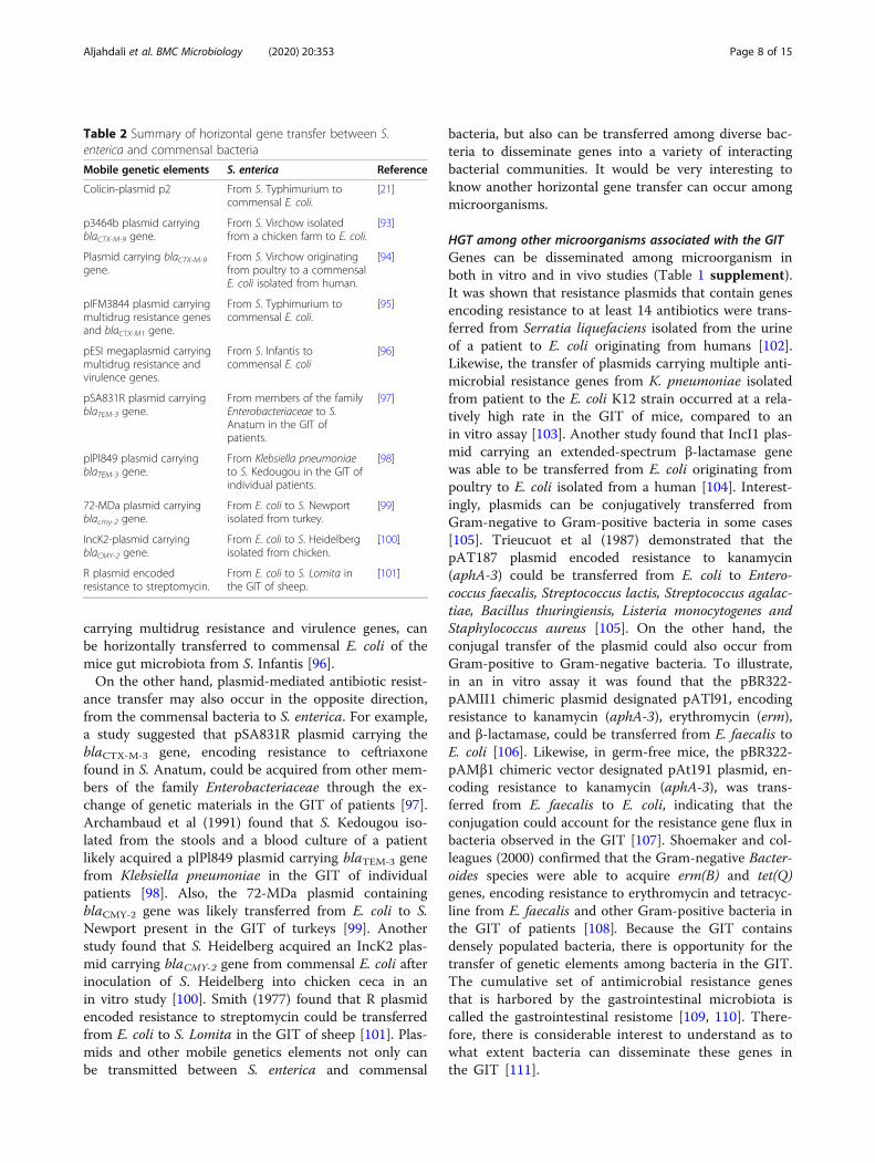
Table 2 Summary of horizontal gene transfer between S.enterica and commensal bacteria
Mobile genetic elements S. enterica Reference
Colicin-plasmid p2 From S. Typhimurium tocommensal E. coli.
[21]
p3464b plasmid carryingblaCTX-M-9 gene.
From S. Virchow isolatedfrom a chicken farm to E. coli.
[93]
Plasmid carrying blaCTX-M-9gene.
From S. Virchow originatingfrom poultry to a commensalE. coli isolated from human.
[94]
pIFM3844 plasmid carryingmultidrug resistance genesand blaCTX-M1 gene.
From S. Typhimurium tocommensal E. coli.
[95]
pESI megaplasmid carryingmultidrug resistance andvirulence genes.
From S. Infantis tocommensal E. coli
[96]
pSA831R plasmid carryingblaTEM-3 gene.
From members of the familyEnterobacteriaceae to S.Anatum in the GIT ofpatients.
[97]
plPl849 plasmid carryingblaTEM-3 gene.
From Klebsiella pneumoniaeto S. Kedougou in the GIT ofindividual patients.
[98]
72-MDa plasmid carryingblacmy-2 gene.
From E. coli to S. Newportisolated from turkey.
[99]
IncK2-plasmid carryingblaCMY-2 gene.
From E. coli to S. Heidelbergisolated from chicken.
[100]
R plasmid encodedresistance to streptomycin.
From E. coli to S. Lomita inthe GIT of sheep.
[101]
Aljahdali et al. BMC Microbiology (2020) 20:353 Page 8 of 15

Further evidence for conjugative transfer of resistancegenes carried by transposons is illustrated by the mem-bers of Firmicutes in the GIT. It was shown that trans-poson Tn1545, which carries multiple drug resistancedeterminants such as those for kanamycin (aphA-3),erythromycin (ermAM), and tetracycline (tetM), can betransferred from E. faecalis to L. monocytogenes in theGIT of gnotobiotic mice at a high rate, compared toin vitro experiments [112]. Moubareck and colleagues(2003) found that transposon Tn1546, which carriesvanA and multiple other antibiotic resistance genes,such as ermB, tet(L), ant (6), and tetM, can be horizon-tally transferred from E. faecium originating from pigs toE. faecium isolated from humans at a high frequency inthe GIT of gnotobiotic mice [113]. This study suggestedthat different resistance genes can be conjugativelytransferred from an E. faecium strain of animal origin toa human-origin bacterium of the same species [113].Earlier studies found that the transposon Tn1546 carry-ing vanA gene was transferred from an E. faecium isolateof chicken origin to an E. faecium isolate of human ori-gin in the intestines of human volunteers [114]. Like-wise, another study confirmed that the vanA gene,encoding resistance to vancomycin, can be transferredfrom E. faecium originating from pigs and poultry to E.faecalis originating from human in the GIT of gnoto-biotic mice [115]. Launay and colleagues (2006) demon-strated that transposon Tn1549, which carries the vanB2gene, can be transferred from Clostridium symbiosum toE. faecium and E. faecalis in the GIT of gnotobiotic miceat a high rate, compared to in vitro experiments [116].Also, another study confirmed that the vanB gene, en-coding resistance to vancomycin, was transferred amongE. faecium in the GIT of patients [117]. It is of centralimportance to know that conjugative transfer of genescan occur among Bacteroidetes members in the GIT.For instance, it was found conjugative plasmid (pRRI4),encoding to tetracycline resistance gene, was transferredfrom Prevotella ruminicola to Bacteroides spp [118]. Astudy indicated that the transfer of Tn5030 carrying clin-damycin resistance (ermFU) gene can occur among Bac-teroides species [119]. Conjugal transfer of plasmids andconjugative transposons among bacteria appears to beimportant to the HGT in the GIT. Consequently, theefficiency of HGT among bacteria can be affected byseveral factors such as SOS response, stresshormones, antibiotic treatment, inflammation, andbacteria-derived factors such as quorum sensing mol-ecules. It is of considerable interest to know the fac-tors that influence HGT.
Factors influencing HGT with S. entericaThe findings from recent studies indicated that theantibiotic-induced SOS response, which is a global stress
response to DNA damage, could promote HGT in bac-teria. For example, Bearson and Brunelle (2015) foundthat the induction of SOS response by antibiotics, suchas fluoroquinolones (ciprofloxacin, enrofloxacin anddanofloxacin), could facilitate the transfer of plasmidfrom S. Typhimurium DT120 and DT104 to a recipientkanamycin-susceptible Salmonella [120]. Furthermore,there is also increasing evidence that the impact of anti-biotic intake on increased HGT is 3-fold stronger in theresistome of people treated with antimicrobials com-pared to untreated people (19 and 5%, respectively)[121]. In addition to antibiotics, norepinephrine (NE), astress hormone, can contribute significantly to HGT be-tween bacteria. A recent study found that NE enhancedHGT of a conjugative plasmid carrying AMR genes fromS. Typhimurium to commensal E. coli due to upregu-lated expression of tra genes in the presence of NE[122]. A major factor that can influence HGT is inflam-mation in the GIT. Stecher et al (2012) found that infec-tions with S. Typhimurium resulted in an inflammatoryresponse, which prompted HGT of the colicin-plasmidp2 from S. Typhimurium to commensal E. coli [21].Moreover, other studies reported that some gut
bacteria-derived factors associated with quorum sensingmay promote HGT [123]. Quorum sensing signalingmolecules are synthesized by gut microbiota and func-tion to control population density and synchronize bac-terial behaviors [124]. One important class of signalingmolecules are referred as autoinducers, which are themajor signaling molecules involved in quorum sensing.The concentration of autoinducers increase as the bac-teria replicate and increase in number allowing for sens-ing of population densities [125]. The most commonclass of autoinducers are acyl homoserine lactones(AHLs) [124]. S. enterica and other Gram-negative bac-teria encode SdiA, which is a homolog of the well char-acterized AHL sensor LuxR, but they do not synthesizetheir own AHLs [126]. However, S. Typhimurium useSdiA as a sensor to detect and respond a variety ofAHLs in GIT [127], and potentially influence HGT inthe GIT [123]. Interestingly, MuCuddin et al (2006)found that the rumen protozoa are a influencing factorin bacterial gene transfer, enhancing transfer a plasmidcarrying the blaCMY-2 gene from Klebsiella to Salmonellain both in vitro and in vivo studies of bovine, caprine,and ovine species [128].
Promotion or inhibition of S. enterica growth by gutbacteriaSalmonella infections lead to changes in the gut micro-biota composition, certain gut bacteria harvest moleculesthat serve as nutrients or signals to aid in promotion orlimitation of the growth of Salmonella [129, 130]. Theexploitation of microbiota-derived molecules is a critical
Aljahdali et al. BMC Microbiology (2020) 20:353 Page 9 of 15

issue for both the colonization or decolonization of thehost cells by enteric pathogens (Table 3). For example,Bacteroides thetaiotaomicron harvests the fucose, galact-ose, sialic acid from the gut epithelium [129, 131, 132].These sugars can be used as a source of carbon by S.Typhimurium to promote its expansion in the GIT(Fig. 4) [129]. Also, hydrogen, which is a central inter-mediate of microbiota metabolism, can be used as an en-ergy source to enhance the growth of S. Typhimuriumduring the early stages of infection [133]. This growthwas enhanced by S. Typhimurium hyb hydrogenase,which facilitates consumption of hydrogen [133]. More-over, SCFAs that are produced by members of the GITmicrobiota play an important role in colonization ofpathogenic bacteria in the GIT. To illustrate, it wasshown that the high concentration of acetate in the dis-tal ileum enhanced the expression of the invasion genesof SPI-1 encoded T3SS through sensor kinase (BarA)and response regulator (SirA) pathways (Fig. 4) [134]. Inbrief, acetate can be converted to acetylphosphate byacetate kinase (AckA), which could phosphorylate BarAand SirA. SirA is essential for the expression of SPI-1 in-vasion genes [134].Conversely, propionate and butyrate suppressed the
expression of the invasion genes of SPI-1 encoded T3SS[134]. Jacobson and colleagues (2018) demonstrated thatthe production of propionate by Bacteroides spp. limitedthe growth of S. Typhimurium by disrupting intracellu-lar pH homeostasis in an in vivo study [130]. Anotherstudy found that pre-incubation of S. Enteritidis withpropionate and butyrate could decrease the invasion ofthe intestinal epithelial cells in an in vitro avian model[135]. Although the intestinal microbiota is complex andthe role of most of the bacteria in providing benefit tothe host is not clear, bacterial species of the generaLactobacillus have been shown to supply protection
against enteric infections. Peng and colleagues (2015)suggested that Lactobacillus casei could inhibit thegrowth of pathogens by 99% [136]. More specifically,they found that linoleic acids that were produced by L.casei, limited the growth of S. Typhimurium [137]. Fur-thermore, Makras et al (2006) found that the inhibitoryactivity of four of six examined Lactobacillus strainsagainst S. Typhimurium was solely due to lactic acidproduction, while that of the remaining two was due tolactic acid plus another unknown substance [138]. Inaddition, indole is produced by commensal E. coli andcould be important in the intestinal epithelial cell re-sponse to pathogens [139]. Evidence provided by the au-thors observed that indole downregulated the expressionof the SPI-1 T3SS genes of S. Typhimurium [140]. Re-markably, there are other pathogens that can also exploitnutrients or molecules for the successful infection ofhost cells, and it would be very interesting to know otherpathogenic bacteria compete and use the molecules har-vested by the gut bacteria (Table 2 supplement).It has been found that fucose harvested by B. thetaio-
taomicron repressed the expression of virulence genes inenterohaemorrhagic E. coli (EHEC) serovar O157:H7encoded T3SS through the FusK and FusR signaling cas-cade [132]. On the other hand, B. thetaiotaomicronmodified the metabolites by increasing succinate, whichcan lead to enhance EHEC virulence gene expressionthrough the transcription factor, Cra, which is function-ally sensitive to succinate [141]. Takao et al (2014) foundthat butyrate produced by the gut microbiota enhancedthe expression of leuO gene that activated the locus forenterocyte effacement (LEE) genes and flagella biosyn-thesis genes in EHEC-encoded T3SS [142]. Likewise, bu-tyrate enhanced the expression of the Shiga toxin (Stx)receptor globotriaosylceramide (Gb3) on the colonic epi-thelium and increased susceptibility to EHEC infection
Table 3 Summary of certain members of the gut microbiota promotion or inhibition of S. enterica growth in the GIT
Gut microbiota Type of molecules produced by gutmicrobiota
The result of study Reference
Bacteroidesthetaiotaomicron
Fucose, galactose, sialic acid Enhance the growth of S. Typhimurium [129, 131,132]
Microbiota-derivedH2
Hydrogen (H2) Enhance the growth of S. Typhimurium during the early stageinfection
[133]
Microbiota- derivedSCFAs
Acetate Enhance the expression of the invasion genes of SPI-1 encoded T3SSof S. Typhimurium
[134]
Microbiota- derivedSCFAs
Propionate and butyrate Suppress the expression of the invasion genes of SPI-1 encoded T3SSof S. Typhimurium
[134]
Microbiota- derivedSCFAs
Propionate Limit S. Typhimurium growth [130]
Microbiota- derivedSCFAs
Propionate and butyrate Decrease the invasion of the intestinal epithelial cells in an in vitroavian model of S. Enteritidis
[135]
Lactobacillus casei Linoleic acids Limit S. Typhimurium growth [137]
Commensal E. coli Indole Downregulated genes of SPI-1 encoded T3SS of S. Typhimurium [139, 140]
Aljahdali et al. BMC Microbiology (2020) 20:353 Page 10 of 15

[143]. Acyl-homoserine lactones (AHLs) produced bysome members of Bacteroidetes can be used by EHECthrough sensor protein SdiA to successfully colonize inthe intestinal epithelium of cattle [144]. However, mole-cules that are modified by the gut microbiota can be de-tected by pathogens and control their virulence genes.For instance, the metabolic conversion of bile acids intodeoxycholic acid by some members of the gut bacteria,such as Bifidobacterium bifidum, can decrease the ex-pression of virulence genes in Vibrio cholerae encodedtype VI secretion system (T6SS), which is used to killother bacteria [145].
ConclusionThe human gastrointestinal microbiota is a complex ofmicroorganisms that has received much attention be-cause of its impact on human health and disease. Recentinsights into the interaction between Salmonella, thehost and its microbiota, found that Salmonella hasevolved molecular machineries that allows them to adaptto the inflamed intestine and compete with the gutmicrobiota. Thereby genes can be transferred horizon-tally between pathogens and microbial communities thatlead to changes in the GIT bacterial structure and theirbehavior. Together, this interplay could result in risks tohuman health, for example, the human colon can serveas an environment that acts as a reservoir for antimicro-bial resistance and mobile genetics elements. The trans-fer of MGEs harboring multiple resistance genes andvirulence factors from pathogens to human intestinal
bacteria has centered around the questions such as: whathappens to the transferred MGEs once entering the gutmicrobiota, and which mechanisms that certain gut bac-teria use for HGT can contribute to increasing the viru-lence factors associated with salmonellosis? Althoughseveral recent studies started to focus on understandingthe shifts in the taxonomic composition of the develop-ing microbiota from infancy to adulthood; the review ofthe literature showed that much remains to be learneddue to the limited knowledge of the effect of Salmonellainfection on the microbial composition, as well as on theMGEs in the gut microbiota, including the transmissionand persistence of antimicrobial resistance genes.S. Typhimurium has been widely studied as a pathogen
and is known to create its own niche in the intestine bycausing inflammation, alteration the composition of gutmicrobiota, and using nutrients produced by gut micro-biota. We reviewed many of the latest insights describingthe interactions between the microbiota, the host, andpathogenic bacteria in animal models, and it is evidentthat further studies are needed to better understand theinteraction of the gastrointestinal microbiota of differenthosts and Salmonella serotypes most associated with in-fections. S. Typhimurium has developed mechanisms torapidly transfer the genes into the gut bacteria at ahigher rate in vivo than found in in vitro studies. Al-though the gut microbiota likely influences S. Typhimur-ium infection kinetics, the effects of molecules producedby gut bacteria on the expression of virulence genes in S.Typhimurium is not yet well defined. SCFA produced by
Fig. 4 Bacteroides thetaiotaomicron, harvests the fucose, galactose, sialic acid from the gut epithelium and used as a source of carbon bySalmonella to promote its expansion in the GIT. Acetate produced by commensal microbiota enhanced expression of the invasion genes of SPI-1encoded T3SS of Salmonella
Aljahdali et al. BMC Microbiology (2020) 20:353 Page 11 of 15

bacteria may have utility as therapeutics targets or thefor successful prevention against Salmonella infection.Likewise, approaches to impact quorum sensing path-ways in Salmonella and other enteric pathogens couldpotentially minimize the role of HGT on AMR and viru-lence factor transmission conserving potential thera-peutic options for control of infections.
Supplementary informationSupplementary information accompanies this paper at https://doi.org/10.1186/s12866-020-02008-x.
Additional file 1.
AbbreviationsGIT: Gastrointestinal tract; SCFAs: Short-chain fatty acids; MGEs: Mobilegenetic elements; SPI-1: Salmonella pathogenicity island-1; SPI-2: Salmonellapathogenicity island − 2; T3SS: Type III secretion system; Sips: Salmonellainvasive proteins; Sops: Salmonella outer proteins; SCV: Salmonella containingvacuole; NK: Natural Killer; TLRs: Toll-like receptors; PAMPs: Pathogen-associated molecular patterns; NO: Nitric oxide; ROS: Reactive oxygenspecies; HGT: Horizontal gene transfer; EcN: E. coli Nissle; STEC: Shiga toxin-producing E. coli; LGT: Lateral gene transfer; AMR: Antimicrobial resistance;MLS: Macrolides, lincosamide and streptogramin; NE: Norepinephrine;BarA: Sensor kinase; SirA: Response regulator; AckA: Acetate kinase;EHEC: Enterohaemorrhagic E. coli; AHLs: Acyl-homoserine lactones;T6SS: Type VI secretion system
AcknowledgementsThe authors would like to recognize Drs. Ashraf Khan, Saeed Khan and CarlCerniglia for their critical review of the manuscript. Dr. Nesreen Aljahdali wassupported in part by an appointment to the Research Participation Programat the National Center for Toxicological Research administered by the OakRidge Institute for Science and Education (ORISE) through an interagencyagreement between the U.S. Department of Energy and the U.S. Food andDrug Administration. The opinions expressed in this manuscript are solelythe responsibility of the authors and do not necessarily represent the officialviews and policy of the Food and Drug Administration or Department ofHealth and Human Services.
Authors’ contributionsNA wrote the first draft of the manuscript. NA and SF coordinated thewriting of the manuscript. YS and JH contributed to manuscript writing. Allcontributed to the editing and refinement of the final version of themanuscript. The author(s) read and approved the final manuscript.
FundingNot applicable.
Availability of data and materialsNot applicable.
Ethics approval and consent to participateNot applicable.
Consent for publicationNot applicable.
Competing interestsThe authors declare that they do not have competing interests.
Author details1Division of Microbiology, National Center for Toxicological Research, U.S.Food and Drug Administration, 3900 NCTR Rd, Jefferson, AR 72079, USA.2Biological Science Department, College of Science, King Abdul-AzizUniversity, Jeddah, Saudi Arabia. 3Department of Agriculture, University of
Arkansas, Pine Bluff, AR, USA. 4Department of Parasitology and AnimalDiseases, Veterinary Research Division, National Research Centre, Giza, Egypt.
Received: 6 July 2020 Accepted: 12 October 2020
References1. Tsolis RM, Young GM, Solnick JV, Bäumler AJ. From bench to bedside:
stealth of enteroinvasive pathogens. Nat Rev Microbiol. 2008;6(12):883–92.2. Kolling G, Wu M, Guerrant RL. Enteric pathogens through life stages. Front
Cell Infect Microbiol. 2012;2:8.3. National Salmonella Surveillance [https://www.cdc.gov/nationalsurveillance/
salmonella-surveillance.html]. Accessed 15 Oct 2020.4. Nastasi A, Mammina C, Villafrate MR, Massenti MF, Scarlata G, Diquattro M.
Multiple typing of strains of Salmonella enterica subsp. Bongori ser. 48:Z35:-isolated in southern Italy. Ann Inst Pasteur Microbiol. 1988;139(5):605–12.
5. Pui CF, Wong WC, Chai LC, Tunung R, Jeyaletchumi P, Noor Hidayah MS,Ubong A, Farinazleen MG, Cheah YK, Son R. Salmonella: a foodbornepathogen. Int Food Res J. 2011;18(2):4–473.
6. Su LH, Chiu CH. Salmonella: Clinical importance and evolution ofnomenclature. Chang Gung Med J. 2007;30(3):210–9.
7. Foley SL, Johnson TJ, Ricke SC, Nayak R, Danzeisen J. Salmonellapathogenicity and host adaptation in chicken-associated serovars. MicrobiolMol Biol Rev. 2013;77(4):582–607.
8. Voogt N, Wannet WJ, Nagelkerke NJ, Henken AM. Differences betweennational reference laboratories of the European community in their abilityto serotype Salmonella species. Eur J Clin Microbiol Infect Dis. 2002;21(3):204–8.
9. Scallan E, Hoekstra RM, Angulo FJ, Tauxe RV, Widdowson MA, Roy SL, JonesJL, Griffin PM. Foodborne illness acquired in the United States-majorpathogens. Emerg Infect Dis. 2011;17(1):7–15.
10. Kennedy M, Villar R, Vugia DJ, Rabatsky-Ehr T, Farley MM, Pass M, Smith K,Smith P, Cieslak PR, Imhoff B, et al. Hospitalizations and deaths due toSalmonella infections, FoodNet, 1996-1999. Clin Infect Dis. 2004;38:S142–8.
11. Han J, Lynne AM, David DE, Tang HL, Xu JS, Nayak R, Kaldhone P, LogueCM, Foley SL. DNA sequence analysis of plasmids from multidrug resistantSalmonella enterica serotype Heidelberg isolates. PLoS One. 2012;7(12):8.
12. Hiyoshi H, Tiffany CR, Bronner DN, Bäumler AJ. Typhoidal Salmonellaserovars: ecological opportunity and the evolution of a new pathovar. FEMSMicrobiol Rev. 2018;42(4):527–41.
13. Johnson R, Mylona E, Frankel G. Typhoidal Salmonella: distinctive virulencefactors and pathogenesis. Cell Microbiol. 2018;20(9):e12939.
14. Information for Healthcare Professionals and Laboratories [https://www.cdc.gov/salmonella/general/technical.html]. Accessed 15 Oct 2020.
15. Haselbeck AH, Panzner U, Im J, Baker S, Meyer CG, Marks F. Currentperspectives on invasive nontyphoidal Salmonella disease. Curr Opin InfectDis. 2017;30(5):498–503.
16. Jones TF, Ingram LA, Cieslak PR, Vugia DJ, Tobin-D'Angelo M, Hurd S, MedusC, Cronquist A, Angulo FJ. Salmonellosis outcomes differ substantially byserotype. J Infect Dis. 2008;198(1):109–14.
17. Brenner FW, Villar RG, Angulo FJ, Tauxe R, Swaminathan B. Salmonellanomenclature. J Clin Microbiol. 2000;38(7):2465–7.
18. Jackson BR, Griffin PM, Cole D, Walsh KA, Chai SJ. Outbreak-associatedSalmonella enterica serotypes and food commodities, United States, 1998-2008. Emerg Infect Dis. 2013;19(8):1239–44.
19. Parry CM, Threlfall EJ. Antimicrobial resistance in typhoidal andnontyphoidal Salmonellae. Curr Opin Infect Dis. 2008;21(5):531–8.
20. Walter J, Ley R. The human gut microbiome: Ecology and recentevolutionary changes. Annu Rev Microbiol. 2011;65:411–29 Palo Alto:Annual Reviews; Edited by Gottesman S, Harwood CS, vol. 65.
21. Stecher B, Denzler R, Maier L, Bernet F, Sanders MJ, Pickard DJ, Barthel M,Westendorf AM, Krogfelt KA, Walker AW, et al. Gut inflammation can boosthorizontal gene transfer between pathogenic and commensalEnterobacteriaceae. Proc Natl Acad Sci U S A. 2012;109(4):1269–74.
22. Martin R, Miquel S, Ulmer J, Langella P, Bermudez-Humaran LG. Gutecosystem: how microbes help us. Benefic Microbes. 2014;5(3):219–33.
23. Caporaso JG, Lauber CL, Costello EK, Berg-Lyons D, Gonzalez A, StombaughJ, Knights D, Gajer P, Ravel J, Fierer N, et al. Moving pictures of the humanmicrobiome. Genome Biol. 2011;12(5):8.
24. Thiennimitr P, Winter SE, Baumler AJ. Salmonella, the host and itsmicrobiota. Curr Opin Microbiol. 2012;15(1):108–14.
Aljahdali et al. BMC Microbiology (2020) 20:353 Page 12 of 15

25. Ley RE, Peterson DA, Gordon JI. Ecological and evolutionary forces shapingmicrobial diversity in the human intestine. Cell. 2006;124(4):837–48.
26. Noverr MC, Huffnagel GB. Does the microbiota regulate immune responsesoutside the gut? Trends Microbiol. 2004;12(12):562–8.
27. Ohoka M, Ito T, Kitsunezaki M, Nomoto K, Bando Y, Ishii M. Changes inneonatal microbiota distribution influenced by the environment of theneonatal intensive care unit in the first month of life. J Neonatal Biol. 2016;5(2):1–7.
28. Palmer C, Bik EM, DiGiulio DB, Relman DA, Brown PO. Development of thehuman infant intestinal microbiota. PLoS Biol. 2007;5(7):1556–73.
29. Mariat D, Firmesse O, Levenez F, Guimaraes VD, Sokol H, Dore J, Corthier G,Furet JP. The Firmicutes/Bacteroidetes ratio of the human microbiotachanges with age. BMC Microbiol. 2009;9:6.
30. Gill SR, Pop M, DeBoy RT, Eckburg PB, Turnbaugh PJ, Samuel BS, Gordon JI,Relman DA, Fraser-Liggett CM, Nelson KE. Metagenomic analysis of thehuman distal gut microbiome. Science. 2006;312(5778):1355–9.
31. Gibson GR, Roberfroid MB. Dietary modulation of the human colonicmicrobiota - introducing the concept of prebiotics. J Nutrition. 1995;125(6):1401–12.
32. Duncan SH, Hold GL, Barcenilla A, Stewart CS, Flint HJ. Roseburia intestinalissp nov., a novel saccharolytic, butyrate-producing bacterium from humanfaeces. Int J Syst Evol Microbiol. 2002;52:1615–20.
33. Wong JMW, de Souza R, Kendall CWC, Emam A, DJA J. Colonic health:Fermentation and short chain fatty acids. J Clin Gastroenterol. 2006;40(3):235–43.
34. Evenepoel P, Meijers BKI, Bammens BRM, Verbeke K. Uremic toxinsoriginating from colonic microbial metabolism. Kidney Int. 2009;76:S12–9.
35. Macfarlane GT, Cummings JH, Allison C. Protein-degradation by humanintestinal bacteria. J Gen Microbiol. 1986;132:1647–56.
36. Hoffman S, Maculloch B, Batz M. Economic Burden of Major FoodborneIllnesses Acquired in the United States. EIB-140, U.S. Deptartment ofAgriculture, Economic Research Service. 2015. https://www.ers.usda.gov/publications/pub-details/?pubid=43987. Accessed 15 Oct 2020.
37. Cost Estimates of Foodborne Illnesses, Cost of Foodborne Illness Estimatesfor Salmonella (non-typhoidal) [https://www.ers.usda.gov/data-products/costestimates-of-foodborne-illnesses.aspx#48498]. Accessed 15 Oct 2020.
38. National Enteric Disease Surveillance Salmonella Annual Report [ https://www.cdc.gov/salmonella/index.html]. Accessed 15 Oct 2020.
39. Darwin KH, Miller VL. Molecular basis of the interaction of Salmonella withthe intestinal mucosa. Clin Microbiol Rev. 1999;12(3):405.
40. Foley SL, Lynne AM. Food animal-associated Salmonella challenges:pathogenicity and antimicrobial resistance. J Anim Sci. 2008;86(14 Suppl):E173–87.
41. Hanning IB, Nutt JD, Ricke SC. Salmonellosis outbreaks in the United Statesdue to fresh produce: sources and potential intervention measures.Foodborne Pathog Dis. 2009;6(6):635–48.
42. Howard ZR, O'Bryan CA, Crandall PG, Ricke SC. Salmonella Enteritidis in shelleggs: current issues and prospects for control. Food Res Int. 2012;45(2):755–64.
43. Ricke SC. Insights and challenges of Salmonella infection of laying hens.Curr Opin Curr Opin Food Sci. 2017;18:43–9.
44. Trevejo R, Starr M. Re: "epidemiology of salmonellosis in California, 1990-1999: morbidity, mortality, and hospitalization costs" - reply. Am J Epidemiol.2004;159(1):104–5.
45. Bula-Rudas FJ, Rathore MH, Maraqa NF. Salmonella infections in childhood.Adv Pediatr Infect Dis. 2015;62(1):29–58.
46. Jakociune D, Bisgaard M, Pedersen K, Olsen JE. Demonstration of persistentcontamination of a cooked egg product production facility with Salmonellaenterica serovar Tennessee and characterization of the persistent strain. JAppl Microbiol. 2014;117(2):547–53.
47. Hancock D, Besser T, Gay J, Rice D, Davis M, Gay C. The globalepidemiology of multiresistant Salmonella enterica serovar TyphimuriumDT104. Washington: Amer Soc Microbiology; 2000.
48. Rabsch W, Tschape H, Baumler AJ. Non-typhoidal salmonellosis: emergingproblems. Microb Infect. 2001;3(3):237–47.
49. Foster JW. Salmonella acid shock proteins are required for the adaptive acidtolerance response. J Bacteriol. 1991;173(21):6896–902.
50. Foley SL, Nayak R, Hanning IB, Johnson TJ, Han J, Ricke SC. Populationdynamics of Salmonella enterica serotypes in commercial egg and poultryproduction. Appl Environ Microbiol. 2011;77(13):4273–9.
51. Lostroh CP, Lee CA. The Salmonella pathogenicity island-1 type III secretionsystem. Microb Infect. 2001;3(14–15):1281–91.
52. Lopez CA, Winter SE, Rivera-Chavez F, Xavier MN, Poon V, Nuccio SP, Tsolis RM,Baumler AJ. Phage-Mediated Acquisition of a Type III Secreted Effector ProteinBoosts Growth of Salmonella by Nitrate Respiration. mBio. 2012;3(3):10.
53. Haraga A, Ohlson MB, Miller SI. Salmonellae interplay with host cells. Nat RevMicrobiol. 2008;6(1):53–66.
54. Knodler LA, Steele-Mortimer O. Taking possession: biogenesis of theSalmonella-containing vacuole. Traffic. 2003;4(9):587–99.
55. Khajanchi BK, Hasan NA, Choi SY, Han J, Zhao SH, Colwell RR, Cerniglia CE,Foley SL. Comparative genomic analysis and characterization ofincompatibility group FIB plasmid encoded virulence factors of Salmonellaenterica isolated from food sources. BMC Genomics. 2017;18:14.
56. Kaldhone PR, Carlton A, Aljahdali N, Khajanchi BK, Sanad YM, Han J, Deck J,Ricke SC, Foley SL. Evaluation of incompatibility group I1 (IncI1) plasmid-containing Salmonella enterica and assessment of the plasmids inbacteriocin production and biofilm development. Front Vet Sci. 2019;6:298.
57. Gokulan K, Khare S, Rooney AW, Han J, Lynne AM, Foley SL. Impact ofplasmids, including those encoding VirB4/D4 type iv secretion systems, onSalmonella enterica serovar Heidelberg virulence in macrophages andepithelial cells. PLoS One. 2013;8(10):13.
58. Pham TA, Clare S, Goulding D, Arasteh JM, Stares MD, Browne HP, Keane JA,Page AJ, Kumasaka N, Kane L, et al. Epithelial IL-22RA1-mediatedfucosylation promotes intestinal colonization resistance to an opportunisticpathogen. Cell Host Microbe. 2014;16(4):504–16.
59. Santos RL, Zhang S, Tsolis RM, Baumler AJ, Adams LG. Morphologic andmolecular characterization of Salmonella Typhimurium infection in neonatalcalves. Vet Pathol. 2002;39(2):200–15.
60. Loetscher Y, Wieser A, Lengefeld J, Kaiser P, Schubert S, Heikenwalder M,Hardt WD, Stecher B. Salmonella transiently reside in luminal neutrophils inthe inflamed gut. PLoS One. 2012;7(4):11.
61. Broz P, Ohlson MB, Monack DM. Innate immune response to SalmonellaTyphimurium, a model enteric pathogen. Gut Microbes. 2012;3(2):62–70.
62. Schairer DO, Chouake JS, Nosanchuk JD, Friedman AJ. The potential of nitricoxide releasing therapies as antimicrobial agents. Virulence. 2012;3(3):271–9.
63. Muller AA, Dolowschiak T, Sellin ME, Felmy B, Verbree C, Gadient S,Westermann AJ, Vogel J, LeibundGut-Landmann S, Hardt WD. An NK cellperforin response elicited via il-18 controls mucosal inflammation kineticsduring Salmonella gut infection. PLoS Pathog. 2016;12(6):30.
64. Zheng Y, Valdez PA, Danilenko DM, Hu Y, Sa SM, Gong Q, Abbas AR,Modrusan Z, Ghilardi N, de Sauvage FJ, et al. Interleukin-22 mediates earlyhost defense against attaching and effacing bacterial pathogens. Nat Med.2008;14(3):282–9.
65. Sassone-Corsi M, Raffatellu M. No vacancy: how beneficial microbescooperate with immunity to provide colonization resistance to pathogens. JImmunol. 2015;194(9):4081–7.
66. Santos RL, Raffatellu M, Bevins CL, Adams LG, Tukel C, Tsolis RM, Baumler AJ.Life in the inflamed intestine, Salmonella style. Trends Microbiol. 2009;17(11):498–506.
67. Winter SE, Thiennimitr P, Winter MG, Butler BP, Huseby DL, Crawford RW,Russell JM, Bevins CL, Adams LG, Tsolis RM, et al. Gut inflammation providesa respiratory electron acceptor for Salmonella. Nature. 2010;467(7314):426–9.
68. Thiennimitr P, Winter SE, Winter MG, Xavier MN, Tolstikov V, Huseby DL,Sterzenbach T, Tsolis RM, Roth JR, Baumler AJ. Intestinal inflammation allowsSalmonella to use ethanolamine to compete with the microbiota. Proc NatlAcad Sci U S A. 2011;108(42):17480–5.
69. Hausmann A, Hardt WD. The interplay between Salmonella enterica serovarTyphimurium and the intestinal mucosa during oral infection. MicrobiolSpectr. 2019;7(2):16.
70. Flo TH, Smith KD, Sato S, Rodriguez DJ, Holmes MA, Strong RK, Akira S,Aderem A. Lipocalin 2 mediates an innate immune response to bacterialinfection by sequestrating iron. Nature. 2004;432(7019):917–21.
71. Raffatellu M, George MD, Akiyama Y, Hornsby MJ, Nuccio SP, Paixao TA,Butler BP, Chu HT, Santos RL, Berger T, et al. Lipocalin-2 resistanceconfers an advantage to Salmonella enterica serotype Typhimurium forgrowth and survival in the inflamed intestine. Cell Host Microbe. 2009;5(5):476–86.
72. Ducarmon QR, Zwittink RD, Hornung BVH, van Schaik W, Young VB, KuijperEJ. Gut microbiota and colonization resistance against bacterial entericinfection. Microbiol Mol Biol Rev. 2019;83(3):29.
73. Sassone-Corsi M, Nuccio SP, Liu H, Hernandez D, Vu CT, Takahashi AA,Edwards RA, Raffatellu M. Microcins mediate competition amongEnterobacteriaceae in the inflamed gut. Nature. 2016;540(7632):280.
Aljahdali et al. BMC Microbiology (2020) 20:353 Page 13 of 15

74. Borewicz KA, Kim HB, Singer RS, Gebhart CJ, Sreevatsan S, Johnson T,Isaacson RE. Changes in the porcine intestinal microbiome in response toinfection with Salmonella enterica and Lawsonia intracellularis. PLoS One.2015;10(10):16.
75. Arguello H, Estelle J, Zaldivar-Lopez S, Jimenez-Marin A, Carvajal A,Lopez-Bascon MA, Crispie F, O'Sullivan O, Cotter PD, Priego-Capote F,et al. Early Salmonella Typhimurium infection in pigs disruptsmicrobiome composition and functionality principally at the ileummucosa. Sci Rep. 2018;8:12.
76. Drumo R, Pesciaroli M, Ruggeri J, Tarantino M, Chirullo B, Pistoia C, PetrucciP, Martinelli N, Moscati L, Manuali E, et al. Salmonella enterica SerovarTyphimurium exploits inflammation to modify swine intestinal microbiota.Front Cell Infect Microbiol. 2016;5:13.
77. Bratburd JR, Keller C, Vivas E, Gemperline E, Li LJ, Rey FE, Currie CR. GutMicrobial and Metabolic Responses to Salmonella enterica SerovarTyphimurium and Candida albicans. mBio. 2018;9(6):14.
78. Barman M, Unold D, Shifley K, Amir E, Hung K, Bos N, Salzman N. Entericsalmonellosis disrupts the microbial ecology of the murine gastrointestinaltract. Infect Immun. 2008;76(3):907–15.
79. Liu LY, Lin LL, Zheng LN, Tang H, Fan XZ, Xue NG, Li M, Liu M, Li XY. Cecalmicrobiome profile altered by Salmonella enterica, serovar Enteritidisinoculation in chicken. Gut Pathog. 2018;10:14.
80. Mon KK, Saelao P, Halstead MM, Chanthavixay G, Chang HC, Garas L, Maga EA,Zhou H. Salmonella enterica serovar Enteritidis infection alters the indigenousmicrobiota diversity in young layer chicks. Front Vet Sci. 2015;2:61.
81. Videnska P, Sisak F, Havlickova H, Faldynova M, Rychlik I. Influence ofSalmonella enterica serovar Enteritidis infection on the composition ofchicken cecal microbiota. BMC Vet Res. 2013;9:8.
82. Juricova H, Videnska P, Lukac M, Faldynova M, Babak V, Havlickova H, SisakF, Rychlik I. Influence of Salmonella enterica Serovar Enteritidis infection onthe development of the cecum microbiota in newly hatched chicks. ApplEnviron Microbiol. 2013;79(2):745–7.
83. Singh P, Teal TK, Marsh TL, Tiedje JM, Mosci R, Jernigan K, Zell A, NewtonDW, Salimnia H, Lephart P, et al. Intestinal microbial communities associatedwith acute enteric infections and disease recovery. Microbiome. 2015;3:12.
84. Soucy SM, Huang J, Gogarten JP. Horizontal gene transfer: building the webof life. Nat Rev Genet. 2015;16(8):472–82.
85. Jeong H, Arif B, Caetano-Anolles G, Kim KM, Nasir A. Horizontal genetransfer in human-associated microorganisms inferred by phylogeneticreconstruction and reconciliation. Sci Rep. 2019;9:18.
86. Lerner A, Matthias T, Aminov R. Potential effects of horizontal geneexchange in the human gut. Front Immunol. 2017;8:14.
87. Stecher B, Maier L, Hardt WD. 'Blooming' in the gut: how dysbiosismight contribute to pathogen evolution. Nat Rev Microbiol. 2013;11(4):277–84.
88. Hehemann JH, Correc G, Barbeyron T, Helbert W, Czjzek M, Michel G.Transfer of carbohydrate-active enzymes from marine bacteria to Japanesegut microbiota. Nature. 2010;464(7290):908–U123.
89. Salyers AA, Gupta A, Wang YP. Human intestinal bacteria as reservoirs forantibiotic resistance genes. Trends Microbiol. 2004;12(9):412–6.
90. Begley M, Hill C, Gahan CG. Bile salt hydrolase activity in probiotics. ApplEnviron Microbiol. 2006;72(3):1729–38.
91. Scott KP. The role of conjugative transposons in spreading antibioticresistance between bacteria that inhabit the gastrointestinal tract. Cell MoleLife Sci. 2002;59(12):2071–82.
92. Stecher B, Robbiani R, Walker AW, Westendorf AM, Barthel M, Kremer M,Chaffron S, Macpherson AJ, Buer J, Parkhill J, et al. Salmonella entericaserovar typhimurium exploits inflammation to compete with the intestinalmicrobiota. PLoS Biol. 2007;5(10):2177–89.
93. Faure S, Perrin-Guyomard A, Delmas JM, Laurentie M. Impact of therapeutictreatment with beta-lactam on transfer of the bla (CTX-M-9) resistance genefrom Salmonella enterica Serovar Virchow to Escherichia coli in Gnotobioticrats. Appl Environ Microbiol. 2009;75(17):5523–8.
94. Faure S, Perrin-Guyomard A, Delmas JM, Chatre P, Laurentie M. Transfer ofplasmid-mediated CTX-M-9 from Salmonella enterica serotype Virchow toEnterobacteriaceae in human Flora-associated rats treated with Cefixime.Antimicrob Agents Chemother. 2010;54(1):164–9.
95. Card RM, Cawthraw SA, Nunez-Garcia J, Ellis RJ, Kay G, Pallen MJ, WoodwardMJ, Anjum MF. An In Vitro Chicken Gut Model Demonstrates Transfer of aMultidrug Resistance Plasmid from Salmonella to Commensal Escherichiacoli. mBio. 2017;8(4):15.
96. Aviv G, Rahav G, Gal-Mor O. Horizontal transfer of the Salmonella entericaserovar Infantis resistance and virulence plasmid pESI to the Gut Microbiotaof Warm-Blooded Hosts. mBio. 2016;7:5.
97. Su LH, Chiu CH, Chu C, Wang MH, Chia JH, Wu TL. In vivo acquisition ofceftriaxone resistance in Salmonella enterica serotype anatum. AntimicrobAgents Chemother. 2003;47(2):563–7.
98. Archambaud M, Gerbaud G, Labau E, Marty N, Courvalin P. Possible in vivotransfer of beta-lactamase tem-3 from Klebsiella pneumoniae to Salmonella-Kedougou. J Antimicrob Chemother. 1991;27(4):427–36.
99. Poppe C, Martin LC, Gyles CL, Reid-Smith R, Boerlin P, McEwen SA, PrescottJF, Forward KR. Acquisition of resistance to extended-spectrumCephalosporins by Salmonella enterica subsp enterica serovar Newport andEscherichia coli in the Turkey poult intestinal tract. Appl Environ Microbiol.2005;71(3):1184–92.
100. Oladeinde A, Cook K, Lakin SM, Woyda R, Abdo Z, Looft T, Herrington K,Zock G, Lawrence JP, Thomas JC, et al. Horizontal gene transfer andacquired antibiotic resistance in Salmonella enterica Serovar Heidelbergfollowing in vitro incubation in broiler ceca. Appl Environ Microbiol. 2019;85(22):16.
101. Smith MG. Transfer of R factors from Escherichia coli to Salmonella in therumen of sheep. J Med Microbiol. 1977;10(1):29–35.
102. Duval-Iflah Y, Raibaud P, Tancrede C, Rousseau M. R-plasmic transfer fromSerratia liquefaciens to Escherichia coli in vitro and in vivo in the digestivetract of gnotobiotic mice associated with human fecal flora. Infect Immun.1980;28(3):981–90.
103. Schjorring S, Struve C, Krogfelt KA. Transfer of antimicrobial resistanceplasmids from Klebsiella pneumoniae to Escherichia coli in the mouseintestine. J Antimicrob Chemother. 2008;62(5):1086–93.
104. Smet A, Rasschaert G, Martel A, Persoons D, Dewulf J, Butaye P, Catry B,Haesebrouck F, Herman L, Heyndrickx M. In situ ESBL conjugation fromavian to human Escherichia coli during cefotaxime administration. J ApplMicrobiol. 2011;110(2):541–9.
105. Trieucuot P, Carlier C, Martin P, Courvalin P. Plasmid transfer by conjugationfrom Escherichia coli to gram-positive bacteria. FEMS Microbiol Lett. 1987;48(1–2):289–94.
106. Trieucuot P, Carlier C, Courvalin P. Conjugative plasmid transfer fromEnterococcus faecalis to Escherichia coli. J Bacteriol. 1988;170(9):4388–91.
107. Doucetpopulaire F, Trieucuot P, Andremont A, Courvalin P. Conjugaltransfer of plasmid dna from Enterococcus faecalis to Escherichia coli indigestive tracts of gnotobiotic mice. Antimicrob Agents Chemother.1992;36(2):502–4.
108. Shoemaker NB, Vlamakis H, Hayes K, Salyers AA. Evidence for extensiveresistance gene transfer among Bacteroides spp. and among Bacteroides andother genera in the human colon. Appl Environ Microbiol. 2001;67(2):561–8.
109. Bag S, Shankar Ghosh T, Banerjee S, Mehta O, Verma J, Dayal M, DesigamaniA, Kumar P, Saha B, Kedia S, et al. Molecular insights into antimicrobialresistance traits of commensal human gut microbiota. Microb Ecol. 2019;77(2):546–57.
110. van Schaik W. The human gut resistome. Philos Trans R Soc B-Biol Sci. 2015;370(1670):9.
111. Salyers AA. Gene transfer in the mammalian intestinal tract. Curr OpinBiotechnol. 1993;4(3):294–8.
112. Doucetpopulaire F, Trieucuot P, Dosbaa I, Andremont A, Courvalin P.Inducible transfer of conjugative transposon Tn1545 from Enterococcusfaecalis to Listeria monocytogenes in the digestive tracts of gnotobiotic mice.Antimicrob Agents Chemother. 1991;35(1):185–7.
113. Moubareck C, Bourgeois N, Courvalin P, Doucet-Populaire F. Multipleantibiotic resistance gene transfer from animal to human enterococci in thedigestive tract of gnotobiotic mice. Antimicrob Agents Chemother. 2003;47(9):2993–6.
114. Lester CH, Frimodt-Moller N, Sorensen TL, Monnet DL, Hammerum AA. Invivo transfer of the vanA resistance gene from an Enterococcus faeciumisolate of animal origin to an E-faecium isolate of human origin in theintestines of human volunteers. Antimicrob Agents Chemother. 2006;50(2):596–9.
115. Bourgeois-Nicolaos N, Moubareck C, Mangeney N, Butel MJ, Doucet-Populaire F. Comparative study of vanA gene transfer from Enterococcusfaecium to Enterococcus faecalis and to Enterococcus faecium in the intestineof mice. FEMS Microbiol Lett. 2006;254(1):27–33.
116. Launay A, Ballard SA, Johnson PDR, Grayson ML, Lambert T. Transfer ofvancomycin resistance transposon Tn1549 from Clostridium symbiosum to
Aljahdali et al. BMC Microbiology (2020) 20:353 Page 14 of 15

Enterococcus spp. in the gut of gnotobiotic mice. Antimicrob AgentsChemother. 2006;50(3):1054–62.
117. Howden BP, Holt KE, Lam MMC, Seemann T, Ballard S, Coombs GW, TongSYC, Grayson ML, Johnson PDR, Stinear TP. Genomic insights to control theemergence of vancomycin-resistant Enterococci. mBio. 2013;4(4):9.
118. Shoemaker NB, Wang GR, Salyers AA. Evidence for natural transfer of atetracycline resistance gene between bacteria from the human colon andbacteria from the bovine rumen. Appl Environ Microbiol. 1992;58(4):1313–20.
119. Halula M, Macrina FL. Tn5030: a conjugative transposon conferringclindamycin resistance in Bacteroides species. Rev Infect Dis. 1990;12(Suppl2):S235–42.
120. Bearson BL, Brunelle BW. Fluoroquinolone induction of phage-mediatedgene transfer in multidrug-resistant Salmonella. Int J Antimicrob Agents.2015;46(2):201–4.
121. Li J, Rettedal EA, van der Helm E, Ellabaan M, Panagiotou G, Sommer MOA.Antibiotic treatment drives the diversification of the human gut Resistome.Genom Proteom Bioinf. 2019;17(1):39–51.
122. Peterson G, Kumar A, Gart E, Narayanan S. Catecholamines increaseconjugative gene transfer between enteric bacteria. Microb Pathog. 2011;51(1–2):1–8.
123. Zeng XM, Lin J. Factors influencing horizontal gene transfer in the intestine.Anim Health Res Rev. 2017;18(2):153–9.
124. Jimenez AG, Sperandio V. Quorum sensing and the gut microbiome.London: Academic Press Ltd-Elsevier Science Ltd; 2019.
125. Galloway WR, Hodgkinson JT, Bowden SD, Welch M, Spring DR. Quorumsensing in gram-negative bacteria: small-molecule modulation of AHL andAI-2 quorum sensing pathways. Chem Rev. 2011;111(1):28–67.
126. Hudaiberdiev S, Choudhary KS, Vera Alvarez R, Gelencsér Z, Ligeti B, LambaD, Pongor S. Census of solo LuxR genes in prokaryotic genomes. Front CellInfect Microbiol. 2015;5:20.
127. Dyszel JL, Smith JN, Lucas DE, Soares JA, Swearingen MC, Vross MA, YoungGM, Ahmer BM. Salmonella enterica serovar Typhimurium can detect acylhomoserine lactone production by Yersinia enterocolitica in mice. J Bacteriol.2010;192(1):29–37.
128. McCuddin Z, Carlson SA, Rasmussen MA, Franklin SK. Klebsiella to Salmonellagene transfer within rumen protozoa: implications for antibiotic resistanceand rumen defaunation. Vet Microbiol. 2006;114(3–4):275–84.
129. Ng KM, Ferreyra JA, Higginbottom SK, Lynch JB, Kashyap PC, Gopinath S,Naidu N, Choudhury B, Weimer BC, Monack DM, et al. Microbiota-liberatedhost sugars facilitate post-antibiotic expansion of enteric pathogens. Nature.2013;502(7469):96.
130. Jacobson A, Lam L, Rajendram M, Tamburini F, Honeycutt J, Pham T, VanTreuren W, Pruss K, Stabler SR, Lugo K, et al. A Gut Commensal-ProducedMetabolite Mediates Colonization Resistance to Salmonella Infection. CellHost Microbe. 2018;24(2):296.
131. Chow WL, Lee YK. Free fucose is a danger signal to human intestinalepithelial cells. Br J Nutr. 2008;99(3):449–54.
132. Pacheco AR, Curtis MM, Ritchie JM, Munera D, Waldor MK, Moreira CG,Sperandio V. Fucose sensing regulates bacterial intestinal colonization.Nature. 2012;492(7427):113.
133. Maier L, Vyas R, Cordova CD, Lindsay H, Schmidt TSB, Brugiroux S,Periaswamy B, Bauer R, Sturm A, Schreiber F, et al. Microbiota-derivedhydrogen fuels Salmonella Typhimurium invasion of the gut ecosystem. CellHost Microbe. 2013;14(6):641–51.
134. Lawhon SD, Maurer R, Suyemoto M, Altier C. Intestinal short-chain fattyacids alter Salmonella typhimurium invasion gene expression and virulencethrough BarA/SirA. Mol Microbiol. 2002;46(5):1451–64.
135. Van Immerseel F, De Buck J, Pasmans F, Velge P, Bottreau E, Fievez V,Haesebrouck F, Ducatelle R. Invasion of Salmonella enteritidis in avianintestinal epithelial cells in vitro is influenced by short-chain fatty acids. Int JFood Microbiol. 2003;85(3):237–48.
136. Peng MF, Reichmann G, Biswas D. Lactobacillus casei and its byproductsalter the virulence factors of foodborne bacterial pathogens. J Funct Food.2015;15:418–28.
137. Peng MF, Tabashsum Z, Patel P, Bernhardt C, Biswas D. Linoleic acidsoverproducing Lactobacillus casei limits growth, survival, and virulence ofSalmonella Typhimurium and Enterohaemorrhagic Escherichia coli. FrontMicrobiol. 2018;9:14.
138. Makras L, Triantafyllou V, Fayol-Messaoudi D, Adriany T, Zoumpopoulou G,Tsakalidou E, Servin A, De Vuyst L. Kinetic analysis of the antibacterialactivity of probiotic lactobacilli towards Salmonella enterica serovar
Typhimurium reveals a role for lactic acid and other inhibitory compounds.Res Microbiol. 2006;157(3):241–7.
139. Bansal T, Alaniz RC, Wood TK, Jayaraman A. The bacterial signal indoleincreases epithelial-cell tight-junction resistance and attenuates indicators ofinflammation. Proc Natl Acad Sci U S A. 2010;107(1):228–33.
140. Nikaido E, Giraud E, Baucheron S, Yamasaki S, Wiedemann A, Okamoto K,Takagi T, Yamaguchi A, Cloeckaert A, Nishino K. Effects of indole on drugresistance and virulence of Salmonella enterica serovar Typhimuriumrevealed by genome-wide analyses. Gut Pathog. 2012;4(1):5.
141. Curtis MM, Hu ZP, Klimko C, Narayanan S, Deberardinis R, Sperandio V. Thegut commensal Bacteroides thetaiotaomicron exacerbates enteric infectionthrough modification of the metabolic landscape. Cell Host Microbe. 2014;16(6):759–69.
142. Takao M, Yen HL, Tobe T. LeuO enhances butyrate-induced virulenceexpression through a positive regulatory loop in enterohaemorrhagicEscherichia coli. Mol Microbiol. 2014;93(6):1302–13.
143. Zumbrun SD, Melton-Celsa AR, Smith MA, Gilbreath JJ, Merrell DS, O'BrienAD. Dietary choice affects Shiga toxin-producing Escherichia coli (STEC)O157:H7 colonization and disease. Proc Natl Acad Sci U S A. 2013;110(23):E2126–33.
144. Hughes DT, Terekhova DA, Liou L, Hovde CJ, Sahl JW, Patankar AV, GonzalezJE, Edrington TS, Rasko DA, Sperandio V. Chemical sensing in mammalianhost-bacterial commensal associations. Proc Natl Acad Sci U S A. 2010;107(21):9831–6.
145. Bachmann V, Kostiuk B, Unterweger D, Diaz-Satizabal L, Ogg S, Pukatzki S.Bile salts modulate the Mucin-activated type VI secretion system ofpandemic Vibrio cholerae. Plos Neglect Trop Dis. 2015;9(8):22.
Publisher’s NoteSpringer Nature remains neutral with regard to jurisdictional claims inpublished maps and institutional affiliations.
Aljahdali et al. BMC Microbiology (2020) 20:353 Page 15 of 15