current drug targets - infectious disorders 201-213 … drug targets - infectious disorders, 2001,...
TRANSCRIPT

Current Drug Targets - Infectious Disorders, 2001, 1, 201-213 201
E. Coli MurG: A Paradigm for a Superfamily of Glycosyltransferases
Ha, S., Gross, B. and Walker, S.*
Chemistry Department, Princeton University, Princeton, NJ 08544, USA
Abstract: MurG is an essential bacterial glycosyltransferase that is involved in thebiosynthesis of peptidoglycan. The enzyme is found in all organisms that synthesizepeptidoglycan and is a target for the design of new antibiotics. A direct assay to studyMurG was reported recently, followed shortly by the crystal structure of E. coli MurG.This first MurG structure, combined with sequence data on other glycosyltransferases,has revealed that MurG is a paradigm for a large family of metal ion-independentglycosyltransferases found in both eukaryotes and prokaryotes. A better understandingof MurG could lead to the development of new drugs to combat antibiotic resistantinfections, and may also shed light on a broad class of glycosyltransferases.
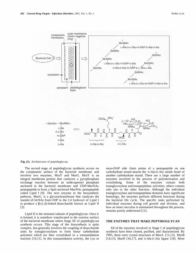
THE PROBLEM OF ANTIBIOTIC RESISTANCE eukaryotes. Peptidoglycan is comprised of linear chains of arepeating β-linked disaccharide unit held together by peptidecrosslinks (Fig. (1)) [2]. One of the sugars in the repeatingdisaccharide, N-acetyl muramic acid, is not made ineukaryotic cells. Furthermore, the peptides in the crosslinkscontain both D-amino acids and unusual backbone linkages,two additional features not found in eukaryotes. Thereforeinhibitors of enzymes involved in many steps ofpeptidoglycan synthesis are likely to be specific for bacterialcells. Because the basic elements of the peptidoglycan layerare remarkably similar across bacterial strains, suchinhibitors may well have broad-spectrum activity. Structuralinformation on enzymes involved in peptidoglycan synthesisis an important first step toward the development of newinhibitors of this pathway.
Overuse of antibiotics in medicine and agriculture has ledto extensive resistance against most classes of clinically usedantibiotics [1]. Genes that confer resistance can be transferredreadily from one bacterial strain to another, producing multi-drug resistant organisms. Multi-drug resistant bacteria are aparticular threat in hospitals, and thousands of patients nowdie each year from hospital-acquired bacterial infections thatare resistant to antibiotic treatment. We need to identify newantibacterial agents before particularly virulent strains ofmulti drug resistant bacteria - vancomycin-resistantstaphylococcal stains, for example - spread from hospitals tothe community at large. Structural and mechanisticinformation on essential bacterial enzymes could lead to thedesign of new antibiotics. Hence, scientists in both academiaand industry have redoubled their efforts to identify andcharacterize key bacterial enzymes. THE THREE STAGES OF PEPTIDOGLYCAN
BIOSYNTHESIS
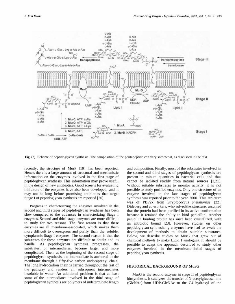
THE BACTERIAL CELL WALL AS A TARGET The biosynthetic pathway to peptidoglycan occurs inthree distinct stages. The first stage involves the conversionof UDP-N-acetyl glucosamine to UDP-N-acetyl muramylpentapeptide [5] (Fig. (2)). This stage occurs in thecytoplasm of the bacterial cell and requires at least sevenenzymes [5]. The first two enzymes, MurA and MurB,convert UDP-GlcNAc to UDP-MurNAc [6,7]. The rest of theenzymes are ligases that form the amide bonds of the peptidechain. The composition of the peptide chain variessomewhat between organisms [8]. For example, in E. colithe peptide chain contains meso-diaminopimelic acid in thethird position, whereas in most gram positive organismslysine is found in the third position. In some gram positiveorganisms, a string of glycines is attached to the lysine atposition 3. Nevertheless, certain features of the peptide chainare broadly conserved. For example, the peptide chaintypically contains five amino acids in the linear sequence;the terminal dipeptide is usually D-Ala-D-Ala; and thesecond amino acid is coupled through its side chain. Thefunctional differences between peptides with differentcompositions are poorly understood.
Bacterial cell membranes are subjected to high osmoticpressures because bacterial cells generate high internalconcentrations of ions and metabolites under normal growthconditions. Bacterial membranes are able to withstand thesehigh pressures because concentric layers of a crosslinkedpolymer called peptidoglycan (Fig. (1)) surround them [2-4].Compounds that damage the peptidoglycan layer causebacterial cells to lyse. Therefore all of the enzymes involvedin the biosynthesis of peptidoglycan are potential targets forthe design of new antibiotics.
Although there are other essential metabolic pathways inbacteria - DNA, RNA, and protein synthesis, for example -peptidoglycan synthesis may have advantages as a target forinhibition because there are no parallels to this pathway in
*Address correspondence to this author at the Chemistry Department,Princeton University, Princeton, NJ 08544, USA; phone: (609) 258-1149;fax: (609) 258-2617; email: [email protected]
1568-0053/01 $28.00+.00 © 2001 Bentham Science Publishers Ltd.
202 Current Drug Targets - Infectious Disorders, 2001, Vol. 1, No. 2 Walker et al.
Fig. (1). Architecture of peptidoglycan.
The second stage of peptidoglycan synthesis occurs onthe cytoplasmic surface of the bacterial membrane andinvolves two enzymes, MraY and MurG. MraY is anintegral membrane protein that catalyzes a pyrophosphateexchange reaction between an undecaprenyl phosphateanchored in the bacterial membrane and UDP-MurNAcpentapeptide to form a lipid anchored MurNAc pentapeptidecalled Lipid I [9]. The next enzyme in the biosyntheticpathway, MurG, is a glycosyltransferase that catalyzes thetransfer of GlcNAc from UDP to the C4 hydroxyl of Lipid Ito produce a β-(1,4)-linked disaccharide known as Lipid II[3].
meso-DAP side chain amine of a pentapeptide on onecarbohydrate strand attacks the D-Ala-D-Ala amide bond ofanother carbohydrate strand. There are a large number ofenzymes involved in the process of polymerization andcrosslinking. Some of the enzymes contain bothtransglycosylase and transpeptidase activities; others containonly one or the other function. Although the individualtransglycosylase and transpeptidase domains have significanthomology, the enzymes perform different functions duringthe bacterial life cycle. The specific tasks performed byindividual enzymes during cell growth and division, andhow an intact sacculus is maintained throughout the process,remains poorly understood [11].
Lipid II is the minimal subunit of peptidoglycan. Once itis formed, it is somehow translocated to the exterior surfaceof the bacterial membrane where Stage III of peptidoglycansynthesis occurs. This stage of the biosynthesis is quitecomplex, but generally involves the coupling of disaccharideunits by transglycosylases to form linear carbohydratepolymers which are then crosslinked in a transamidationreaction [10,11]. In this transamidation activity, the Lys or
THE ENZYMES THAT MAKE PEPTIDOGLYCAN
All of the enzymes involved in Stage I of peptidoglycansynthesis have been cloned, purified, and characterized. By1995, there were crystal structures of MurA [12,13], MurB[14,15], MurD [16,17], and D-Ala-D-Ala ligase [18]. More
E. Coli MurG Current Drug Targets - Infectious Disorders, 2001, Vol. 1, No. 2 203
Fig. (2). Scheme of peptidoglycan synthesis. The composition of the pentapeptide can vary somewhat, as discussed in the text.
recently, the structure of MurF [19] has been reported.Hence, there is a large amount of structural and mechanisticinformation on the enzymes involved in the first stage ofpeptidoglycan synthesis. This information may prove usefulin the design of new antibiotics. Good screens for evaluatinginhibitors of the enzymes have also been developed, and itmay not be long before promising antibiotics that targetStage I of peptidoglycan synthesis are reported [20].
and composition. Finally, most of the substrates involved inthe second and third stages of peptidoglycan synthesis arepresent in minute quantities in bacterial cells and thuscannot be isolated readily from natural sources [3,21].Without suitable substrates to monitor activity, it is notpossible to study purified enzymes. Only one structure of anenzyme involved in the late stages of peptidoglycansynthesis was reported prior to the year 2000. This structurewas of PBP2x from Streptococcus pneumoniae [22].Dideberg and co-workers, who solved the structure, assumedthat the protein had been purified in its active conformationbecause it retained the ability to bind penicillin. Anotherpenicillin binding protein has since been crystallized, withan antibiotic bound [23]. However, studies on otherpeptidoglycan synthesizing enzymes have had to await thedevelopment of methods to obtain suitable substrates.Below, we describe studies on MurG that grew out ofchemical methods to make Lipid I analogues. It should bepossible to adapt the approach described to study otherenzymes involved in the membrane-linked stages ofpeptidoglycan synthesis.
Progress in characterizing the enzymes involved in thesecond and third stages of peptidoglycan synthesis has beenslow compared to the advances in characterizing Stage Ienzymes. Second and third stage enzymes are more difficultto study for two reasons. The first reason is that theseenzymes are all membrane-associated, which makes themmore difficult to overexpress and purify than the soluble,cytoplasmic Stage I enzymes. The second reason is that thesubstrates for these enzymes are difficult to obtain and tohandle. As peptidoglycan synthesis progresses, thesubstrates, or intermediates, become larger and morecomplicated. Then, at the beginning of the second stage ofpeptidoglycan synthesis, the intermediate is anchored to themembrane through a fifty-five carbon undecaprenyl chain.The long hydrocarbon chain is carried throughout the rest ofthe pathway and renders all subsequent intermediatesinsoluble in water. An additional problem is that at leastsome of the intermediates involved in the third stage ofpeptidoglycan synthesis are polymers of indeterminate length
HISTORICAL BACKGROUND OF MurG
MurG is the second enzyme in stage II of peptidoglycanbiosynthesis. It catalyzes the transfer of N-acetylglucosamine(GlcNAc) from UDP-GlcNAc to the C4 hydroxyl of the
204 Current Drug Targets - Infectious Disorders, 2001, Vol. 1, No. 2 Walker et al.
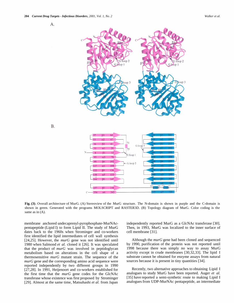
Fig. (3). Overall architecture of MurG. (A) Stereoview of the MurG structure. The N-domain is shown in purple and the C-domain isshown in green. Generated with the programs MOLSCRIPT and RASTER3D. (B) Topology diagram of MurG. Color coding is thesame as in (A).
membrane anchored undecaprenyl-pyrophosphate-MurNAc-pentapeptide (Lipid I) to form Lipid II. The study of MurGdates back to the 1960s when Strominger and co-workersfirst identified the lipid intermediates of cell wall synthesis[24,25]. However, the murG gene was not identified until1980 when Salmond et al. cloned it [26]. It was speculatedthat the product of murG was involved in peptidoglycanmetabolism based on alterations in the cell shape of athermosensitive murG mutant strain. The sequence of themurG gene and the corresponding amino acid sequence werereported independently by two different groups in 1990[27,28]. In 1991, Heijenoort and co-workers established forthe first time that the murG gene codes for the GlcNActransferase whose existence was first proposed by Strominger[29]. Almost at the same time, Matsuhashi et al. from Japan
independently reported MurG as a GlcNAc transferase [30].Then, in 1993, MurG was localized to the inner surface ofcell membrane [31].
Although the murG gene had been cloned and sequencedby 1990, purification of the protein was not reported until1998 because there was simply no way to assay MurGactivity except in crude membranes [30,32,33]. The lipid Isubstrate cannot be obtained for enzyme assays from naturalsources because it is present in tiny quantities [34].
Recently, two alternative approaches to obtaining Lipid Ianalogues to study MurG have been reported. Auger et al.[35] have reported a semi-synthetic route to making Lipid Ianalogues from UDP-MurNAc pentapeptide, an intermediate
E. Coli MurG Current Drug Targets - Infectious Disorders, 2001, Vol. 1, No. 2 205
which can be isolated in large quantities from bacterial cells.In this approach, UDP-MurNAc pentapeptide was treatedwith snake venom phosphodiesterase to cleave thepyrophosphate linkage and the anomeric sugar phosphatewas then coupled chemically to a thirty five carbon activatedlipid phosphate. This 35 carbon Lipid I analogue was foundto be a substrate for MurG, but has not been utilized infurther studies.
similar functions with regard to binding diphosphates. Forexample, both substrates of MurG contain diphosphates thatplay a critical role in binding. Furthermore, sequenceanalysis (see below) shows that MurG contains threeconserved glycine-rich loops near the cleft, two located in theN-domain and one in the C-domain. Finally, MurG is notdependent on metal ions for activity. There must be somemechanism by which the anionic substrates are stabilized forbinding which does not involve metal ion coordination. Theglycine-rich loops in the two Rossmann domains wouldprovide such a mechanism.
We developed a convergent total synthesis of Lipid I andanalogs [36-38]. Although time consuming, a fully syntheticapproach has advantages over isolation or semi-synthesis.The chief advantage is that is possible to vary independentlythe three major components of Lipid I - the sugar, thepeptide, and the lipid chain - in order to evaluate their roles[36-38]. Using synthetic substrate analogs, we have beenable to monitor the activity of E. coli MurG duringpurification. Although it is normally associated with thecytoplasmic membrane [31], MurG can be readily removedfrom the membrane with Triton X-100. Furthermore, thesolubilized enzyme is active and accepts Lipid I substratescontaining hydrocarbon chains at least as short as tencarbons [38]. Hence, a membrane interface is not required tomaintain the active conformation of MurG. Moreover, MurGcan be concentrated to high levels (>10mg/mL) withoutprecipitating. Although this behavior was unexpected, itafforded us the opportunity to try to grow crystals.
The preceding analysis combined with the two-domainstructure of E. coli MurG suggests a binding model in whicheach substrate binds in a separate domain with itsdiphosphate in contact with at least one glycine-rich loop.The substrates are brought together for reaction across thecleft. Although we do not yet have a co-complex of E. coliMurG with either substrate, a comparison of the MurGstructure to that of another glycosyltransferase, T4 phage β-glucosyltransferase has provided considerable insight into thedonor binding domain [43,44].
TOPOLOGICAL SIMILARITY TO BGT
T4 phage β-glucosyltransferase (BGT) was the first NDP-glycosyltransferase ever crystallized [43,44]. The BGTstructure was reported five years before any otherglycosyltransferase structures appeared, but its relevance to abroader understanding of glycosyltransferase structure andmechanism was not clear. BGT has no significant homologyto any other glycosyltransferase. Although it is a TDP/UDPglycosyltransferase, it cannot be classified in any of theexisting glycosyltransferases families. Moreover, certainfunctional differences between BGT and otherglycosyltransferases made BGT appear to be a special case.For example, BGT is not membrane-associated like mostother glycosyltransferases. In addition, the acceptor in theBGT reaction is a segment of duplex DNA, which is largerthan a typical glycosyltransferase acceptor. BGT is proposedto clasp the double helix with loops extending from bothdomains. A hydroxymethylcytosine residue is proposed toflip out of the helix and into the cleft between the twodomains where it is glycosylated [44].
STRUCTURE OF E. COLI MurG
Crystals of E. coli MurG containing a C-terminalLEHHHHHH sequence were grown at room temperatureusing the hanging-drop vapor diffusion method in a NaMESbuffer (pH 6.5) containing 0.5 M (NH4)2SO4 and 0.2%Triton X-100. Triclinic crystals belonging to the P1 spacegroup and having two molecules per asymmetric unit grewto a typical size of 0.2 mm × 0.1 mm × 0.1 mm within aweek. The crystal structure was solved by a combination ofanomalous scattering and multiple isomorphous replacementand refined to 1.9 Å. The structure is shown in Fig. (3) [39].MurG contains two domains separated by a cleft that isapproximately 20Å deep × 18Å wide at the widest point.Both domains adopt an α/β open-sheet motif, also known asa Rossmann fold. A common Rossmann motif contains sixβ strands connected by α helices. The β strands form atwisted parallel sheet with the strand order reading 654123[40]. As shown in the topology diagram (Fig. (3)) for MurG,both domains approximate a classic Rossmann fold and havehigh structural homology despite a lack of sequence identity.
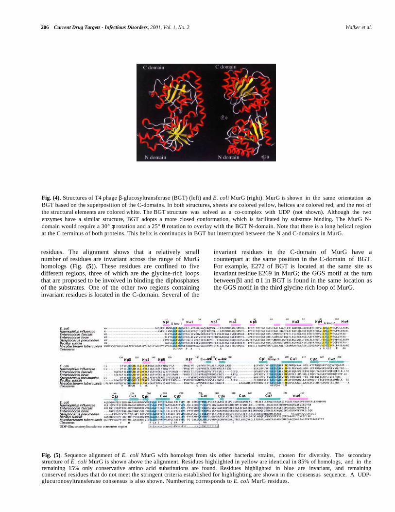
In spite of the functional differences between E. coliMurG and BGT, a structural comparison of MurG and BGThas revealed striking similarities. Like MurG, BGT is atwo-domain protein in which both domains have an α/βopen sheet fold. The N-terminal Rossmann domain containsa helix that originates in the C-terminal domain just as inMurG (Fig. (4)). Thus, the two proteins are topologicallyalmost identical even though they share little sequencehomology (11%).
The Rossmann fold was first identified in proteins thatcontain domains that bind to diphosphate-containingcofactors such as NADH [41]. In a typical Rossman domain,the diphosphates are bound near a P-loop, a glycine-richstretch of amino acids located at the carboxyl terminus of oneβ strand and its adjacent α helix. It is believed that thepositively charged helix dipole of the helix connecting the P-loop to the preceding element of β sheet interacts favorablywith the anionic substrate [42]. Thus, no metal ions orpositively charged residues are necessary to stabilize thenegative charges. Several facts suggest that the two domainsin MurG not only look like Rossman domains, but perform
The structural homology between BGT and E. coliMurG is particularly good in the C-domains (RMSD =2.218Å over 89 aligned Cα atoms), and the similarity iseven more pronounced when the invariant residues in MurGare considered. It is possible to identify the residues mostcritical for binding and catalysis in MurG by aligning theavailable MurG sequences and identifying the invariant
206 Current Drug Targets - Infectious Disorders, 2001, Vol. 1, No. 2 Walker et al.
Fig. (4). Structures of T4 phage β-glucosyltransferase (BGT) (left) and E. coli MurG (right). MurG is shown in the same orientation asBGT based on the superposition of the C-domains. In both structures, sheets are colored yellow, helices are colored red, and the rest ofthe structural elements are colored white. The BGT structure was solved as a co-complex with UDP (not shown). Although the twoenzymes have a similar structure, BGT adopts a more closed conformation, which is facilitated by substrate binding. The MurG N-domain would require a 30° φ rotation and a 25° θ rotation to overlay with the BGT N-domain. Note that there is a long helical regionat the C terminus of both proteins. This helix is continuous in BGT but interrupted between the N and C-domains in MurG.
residues. The alignment shows that a relatively smallnumber of residues are invariant across the range of MurGhomologs (Fig. (5)). These residues are confined to fivedifferent regions, three of which are the glycine-rich loopsthat are proposed to be involved in binding the diphosphatesof the substrates. One of the other two regions containinginvariant residues is located in the C-domain. Several of the
invariant residues in the C-domain of MurG have acounterpart at the same position in the C-domain of BGT.For example, E272 of BGT is located at the same site asinvariant residue E269 in MurG; the GGS motif at the turnbetween β1 and α1 in BGT is found in the same location asthe GGS motif in the third glycine rich loop of MurG.
Fig. (5). Sequence alignment of E. coli MurG with homologs from six other bacterial strains, chosen for diversity. The secondarystructure of E. coli MurG is shown above the alignment. Residues highlighted in yellow are identical in 85% of homologs, and in theremaining 15% only conservative amino acid substitutions are found. Residues highlighted in blue are invariant, and remainingconserved residues that do not meet the stringent criteria established for highlighting are shown in the consensus sequence. A UDP-glucuronosyltransferase consensus is also shown. Numbering corresponds to E. coli MurG residues.
E. Coli MurG Current Drug Targets - Infectious Disorders, 2001, Vol. 1, No. 2 207
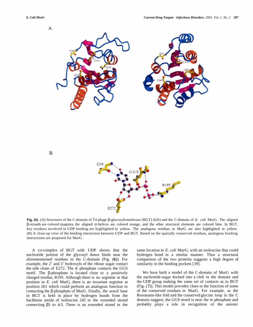
Fig. (6). (A) Structures of the C-domain of T4 phage β-glucosyltransferase (BGT) (left) and the C-domain of E. coli MurG. The alignedβ-strands are colored magenta, the aligned α-helices are colored orange, and the other structural elements are colored blue. In BGT,key residues involved in UDP binding are highlighted in yellow. The analogous residues in MurG are also highlighted in yellow.(B) A close-up view of the binding interaction between UDP and BGT. Based on the spatially conserved residues, analogous bindinginteractions are proposed for MurG.
A co-complex of BGT with UDP shows that thenucleotide portion of the glycosyl donor binds near theaforementioned residues in the C-domain (Fig. (6)). Forexample, the 2’ and 3’ hydroxyls of the ribose sugar contactthe side chain of E272. The α phosphate contacts the GGSmotif. The β-phosphate is located close to a positivelycharged residue, R195. Although there is no arginine at thatposition in E. coli MurG, there is an invariant arginine atposition 261 which could perform an analogous function incontacting the β phosphate of MurG. Finally, the uracil basein BGT is held in place by hydrogen bonds from thebackbone amide of isoleucine 245 in the extended strandconnecting β3 to α3. There is an extended strand in the
same location in E. coli MurG, with an isoleucine that couldhydrogen bond in a similar manner. Thus a structuralcomparison of the two proteins suggests a high degree ofsimilarity in the binding pockets [39].
We have built a model of the C-domain of MurG withthe nucleotide-sugar docked into a cleft in the domain andthe UDP group making the same set of contacts as in BGT(Fig. (7)). This model provides clues to the function of someof the conserved residues in MurG. For example, as theRossmann-like fold and the conserved glycine loop in the Cdomain suggest, the GGS motif is near the α-phosphate andprobably plays a role in recognition of the anionic

208 Current Drug Targets - Infectious Disorders, 2001, Vol. 1, No. 2 Walker et al.
Fig. (7). A close-up view of the proposed donor binding pocket in the MurG C-domain with UDP-GlcNAc manually docked in place.The invariant residues are colored magenta. The carbonyl oxygen of I245 is shown in red and the backbone nitrogen is shown in blue.
diphosphate. R261 is close to the β-phosphate and mayfunction to stabilize the developing negative charge on theleaving group. E269 contacts the 2’ and 3’ hydroxyls of theribose sugar and helps position the glycosyl donor.
324-354). The helix is interrupted at the region betweendomains. A modest rearrangement of this linker couldfacilitate a large change in relative domain orientation.
THE MEMBRANE ASSOCIATION SITEOur model for how UDP binds to MurG is supported byexperimental data. For example, MurG binds significantlybetter to UDP than to CDP, ADP or GDP. The purinenucleotides are presumably excluded from the nucleotidebinding pocket on the basis of size, whereas CDP binding isdisfavored because CDP presents an inappropriate pattern ofhydrogen bond donors and acceptors to the binding pocket.Only pyrimidines containing a hydrogen bond acceptor atthe 4 position and a donor at the 3 position arecomplementary to the backbone amide. Finally, mutationalanalysis indicates that E269 is an important residue inbinding the nucleotide-sugar donor (unpublished results fromthis laboratory).
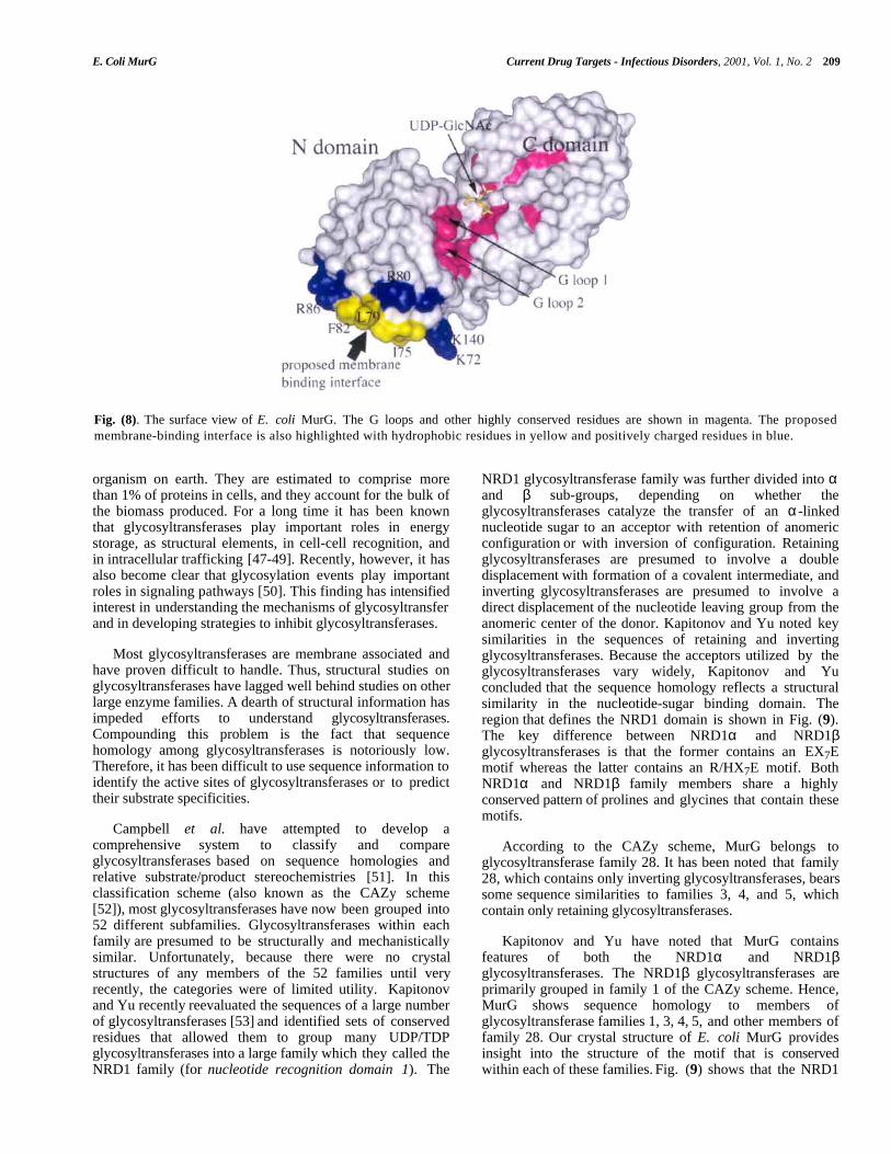
Many glycosyltransferases are anchored to membranes viaa transmembrane spanning helix. MurG has notransmembrane helix but nevertheless associates withbacterial membranes. We have found that MurG can beremoved from membranes with Triton X-100. Earlier reportssuggest that membrane association is also decreased in thepresence of high salt concentrations [45,46]. The crystalstructure of MurG shows that the N-domain contains aconcave hydrophobic patch surrounded by basic residues(Fig. (8)). We speculate that this hydrophobic and basicpatch is the membrane association site. Consistent with thishypothesis, the crystal structure of E. coli MurG shows thatthe isooctylphenyl rings of three Triton X-100 moleculesoccupy the hydrophobic patch. An examination of thestructure suggests that this patch could contact themembrane surface without impeding access of either theLipid I acceptor substrate or the UDP-GlcNAc donorsubstrate to their respective binding sites [39]. In addition,the orientation of the Lipid I substrate with respect to entryinto the binding site would be appropriate for reaction.
THE ACCEPTOR BINDING SITE
Our structural analysis suggests that the pyrophosphateportion of the acceptor contacts at least one of the twoglycine-rich loops located in the N-domain near the cleft.The MurNAc sugar probably contacts residues in the loopspanning residues 125-133, which contains a number ofinvariant residues, including a conserved HEQN motif. Withthe pyrophosphate anchored near G-loop 2 and the sugarbound in the aforementioned loop, the C4 hydroxyl on theacceptor would then protrude into the cleft between domains.Examination of the substrate-free structure suggests,however, that the domains are too far apart for reaction tooccur. Binding of one or both substrates may, therefore,facilitate a conformational change that brings the twodomains closer together. Such a change is thought to occurin BGT [43]. The two domains in MurG are connected by alinker region (residues 158-165) and a long α helix (residues
MurG AND OTHER GLYCOSYLTRANSFERASES
Glycosyltransferases are an enormous family of enzymesthat catalyze the transfer of sugar moieties from activateddonor molecules to specific acceptor molecules, formingglycosidic bonds. The most common activated donors arenucleotide sugars. The acceptors can be other sugars, lipids,proteins, DNA, or virtually any small molecule containing ahydroxyl. Glycosyltransferases are present in virtually every
E. Coli MurG Current Drug Targets - Infectious Disorders, 2001, Vol. 1, No. 2 209
Fig. (8). The surface view of E. coli MurG. The G loops and other highly conserved residues are shown in magenta. The proposedmembrane-binding interface is also highlighted with hydrophobic residues in yellow and positively charged residues in blue.
organism on earth. They are estimated to comprise morethan 1% of proteins in cells, and they account for the bulk ofthe biomass produced. For a long time it has been knownthat glycosyltransferases play important roles in energystorage, as structural elements, in cell-cell recognition, andin intracellular trafficking [47-49]. Recently, however, it hasalso become clear that glycosylation events play importantroles in signaling pathways [50]. This finding has intensifiedinterest in understanding the mechanisms of glycosyltransferand in developing strategies to inhibit glycosyltransferases.
NRD1 glycosyltransferase family was further divided into αand β sub-groups, depending on whether theglycosyltransferases catalyze the transfer of an α-linkednucleotide sugar to an acceptor with retention of anomericconfiguration or with inversion of configuration. Retainingglycosyltransferases are presumed to involve a doubledisplacement with formation of a covalent intermediate, andinverting glycosyltransferases are presumed to involve adirect displacement of the nucleotide leaving group from theanomeric center of the donor. Kapitonov and Yu noted keysimilarities in the sequences of retaining and invertingglycosyltransferases. Because the acceptors utilized by theglycosyltransferases vary widely, Kapitonov and Yuconcluded that the sequence homology reflects a structuralsimilarity in the nucleotide-sugar binding domain. Theregion that defines the NRD1 domain is shown in Fig. (9).The key difference between NRD1α and NRD1βglycosyltransferases is that the former contains an EX7Emotif whereas the latter contains an R/HX7E motif. BothNRD1α and NRD1β family members share a highlyconserved pattern of prolines and glycines that contain thesemotifs.
Most glycosyltransferases are membrane associated andhave proven difficult to handle. Thus, structural studies onglycosyltransferases have lagged well behind studies on otherlarge enzyme families. A dearth of structural information hasimpeded efforts to understand glycosyltransferases.Compounding this problem is the fact that sequencehomology among glycosyltransferases is notoriously low.Therefore, it has been difficult to use sequence information toidentify the active sites of glycosyltransferases or to predicttheir substrate specificities.
Campbell et al. have attempted to develop acomprehensive system to classify and compareglycosyltransferases based on sequence homologies andrelative substrate/product stereochemistries [51]. In thisclassification scheme (also known as the CAZy scheme[52]), most glycosyltransferases have now been grouped into52 different subfamilies. Glycosyltransferases within eachfamily are presumed to be structurally and mechanisticallysimilar. Unfortunately, because there were no crystalstructures of any members of the 52 families until veryrecently, the categories were of limited utility. Kapitonovand Yu recently reevaluated the sequences of a large numberof glycosyltransferases [53] and identified sets of conservedresidues that allowed them to group many UDP/TDPglycosyltransferases into a large family which they called theNRD1 family (for nucleotide recognition domain 1). The
According to the CAZy scheme, MurG belongs toglycosyltransferase family 28. It has been noted that family28, which contains only inverting glycosyltransferases, bearssome sequence similarities to families 3, 4, and 5, whichcontain only retaining glycosyltransferases.
Kapitonov and Yu have noted that MurG containsfeatures of both the NRD1α and NRD1βglycosyltransferases. The NRD1β glycosyltransferases areprimarily grouped in family 1 of the CAZy scheme. Hence,MurG shows sequence homology to members ofglycosyltransferase families 1, 3, 4, 5, and other members offamily 28. Our crystal structure of E. coli MurG providesinsight into the structure of the motif that is conservedwithin each of these families. Fig. (9) shows that the NRD1

210 Current Drug Targets - Infectious Disorders, 2001, Vol. 1, No. 2 Walker et al.
Fig. (9). (A) Sequence alignment of E. coli MurG with selected NDR1β family proteins. Conserved residues are in bold. The secondarystructure of MurG is shown above the sequence. The Swiss-Prot number is given in parenthesis, except when noted. 1. MurG fromEscherichia coli (P17443), 2. glucuronosyltransferase 1A from Homo sapiens (P22309), 3. ceramide 1-β-galactosyltransferase fromHomo sapiens (O00196), 4. macrolide glycosyltransferase from Streptomyces lividans (Q54387), 5. daunosamine transferase fromStreptomyces peucetius (Q54824), 6. zeaxanthin glucosyltransferase from Erwinia ananus (P21686), 7. oleandomycinglycosyltransferase from Streptomyces antibioticus (Q53685), 8. flavonol O3-glucosyltransferase from Perilla frutescens (O04114),9. UDP rhamnose:anthocyanidin-3-glucoside rhamnosyltransferase from Petunia hybrida (Q43716), 10. baumycin glycosyltransferasfrom Streptomyces sp. C5 (Q53881), 11. monogalactosyldiacylglycerol synthase (MGD) from Arabidopsis thaliana (O82730). Allsequences, except for MurG and MGD (family 28), are from CAZy family 1. (B) Sequence alignment of E. coli MurG with selectedNDR1α family proteins. Conserved residues are in bold. The secondary structure of MurG is shown above the sequence. The Swiss-Prot number is given in parenthesis except when noted. 1. MurG from Escherichia coli (P17443), 2. AceA from Acetobacter xylinum(Q44571), 3. monoglucosyldiacylglycerol synthase (MGlcDAG) from Acholeplasma laidlawii (AF349769 - GeneBank), 4.mannosyltransferase MtfB from Synechocystis sp. (P74013), 5. glycogen synthase from Homo Sapiens (P13807) (Two possibleNRD1α domains appear in this sequence.), 6. Sucrose synthase from Spinacia oleracea (P31928). With the exception of glycogensynthase (family 3), all members are from CAZy family 4.
motif, which is specified by a conserved pattern of prolinesand glycines on which are grafted certain key residues,adopts an α−β−α fold. The model we have presented forhow the donor substrate binds suggests a function for someof the conserved residues of the NRD1 motif. In the invertingglycosyltransferases, for example, the R/H residue thatprecedes the first α-helix of the α−β−α fold plays a role instabilizing the leaving group (through e.g., electrostaticinteractions or protonation). This interpretation is similar tothat proposed by Kapitonov and Yu, who also suggestedthat the corresponding glutamate in retainingglycosyltransferases is the nucleophile that forms thecovalent intermediate. Our model also suggests that theglutamate that follows eight residues later helps bind the
ribose sugar. In contrast, Kapitonov and Yu had proposedthat this residue plays a catalytic role and deprotonates theincoming acceptor hydroxyl.
Analysis of MurG mutants supports the proposal that theglutamate in the R/HX7E motif is involved in donor bindingbut not catalysis. Mutation of E269 to A or to D increasesthe Km of the UDP-GlcNc substrate significantly but hasonly a modest effect on kcat (unpublished results). Othershave reported that mutation of the corresponding glutamatein AceA, a retaining UDP-mannosyltransferase whichcontains an EX7E sequence characteristic of the NRD1αfamily, decreases but does not abolish activity [54].Therefore, it was suggested that this second glutamate
E. Coli MurG Current Drug Targets - Infectious Disorders, 2001, Vol. 1, No. 2 211
probably plays a role in donor binding but not catalysis. Incontrast, mutation of the first glutamate led to a completeloss of activity, consistent with its critical catalytic role.
in the donor binding domain [39], and MurG is a twodomain enzyme, it has been suggested that the MurG is amodular enzyme [63]. As defined by Khosla and Harbury, amodular device is a multicomponent system in whichindividual components can be interchanged with functionallydistinct analogues from related systems. That MurG ismodular is supported by the fact that eukaryoticglucuronosyltransferases genes can be spliced at differentsites, resulting in a change of acceptor specificity [64,65].The prospect of domain swapping these enzymes to matedifferent donor and acceptor specificity is tantalizing. Ifdomain swapping is possible, the directed evolution ofglycosyltransferases to accomplish different or uniqueglycosylation reactions could proceed rapidly. The ability toengineer glycosyltransferase selectivity could be useful for thesynthesis of glycoconjugates and for understanding the rolesof oligosaccharides in biological systems.
One of the most surprising findings to come out of ourcrystallographic studies is that MurG has a very similardonor-binding site to BGT. BGT could not be grouped intoany of the 52 families in the CAZy scheme. Furthermore, itdoes not contain a characteristic NRD1 motif. We have nowshown, however, that BGT has both an overall fold and anα-β-α subdomain similar to the conserved subdomain ofMurG. We think that BGT evolved from an NRD1glycosyltransferase, but lost the sequence identity as theloops separating the elements of secondary structure becamelonger. Several long loops in the N and C-domains of BGTare proposed to clasp the duplex DNA acceptor, drawing ittowards the cleft between domains where the active site islocated. The combination of the MurG and the BGTstructures, and the sequence homologies between MurG andother NRD1 family members, is beginning to shed quite abit of light on a large number of glycosyltransferases. (Itshould be noted that others have already used the MurGstructure in predicting the three-dimensional fold of themannosyltransferase AceA [54] and the bacterial lipidglycosyltransferase MGlcDAG synthase [55]).
The structure of MurG also makes it possible to thinkabout the rational design of glycosyltransferase inhibitors.There are very few examples of designed glycosyltransferaseinhibitors, in part because the structural or mechanisticinformation needed to design selective glycosyltransferaseinhibitors has not been available [66]. It is generally agreedthat glycosyltransfer proceeds through an oxocarbeniumintermediate like a glycosidase reaction [67-69], but therehas not been sufficient detailed information to designtransition state analogs. Glycosidase inhibitors that mimicoxocarbenium ion intermediates do not tend to inhibitglycosyltransferases very well [70]. One possible reasoncould be that the carboxylate present in the active site ofglycosidases is not present in NDP-glycosyltransferases.Instead, the negatively charged NDP leaving group itselfmay stabilize the developing positive charge on the anomericcarbon. Consistent with this hypothesis, Wong and co-workers have demonstrated synergistic inhibition with aglycosidase inhibitor and a nucleotide diphosphate [71].Inhibitors containing features that mimic the NDP group inthe transition state as well as the oxocarbenium ion may bemore effective.
THE DXD FAMILY OF GLYCOSYLTRANS-FERASES
Six other glycosyltransferase crystal structures haveappeared in the last two years. These structures include asingle member from CAZy families 2 [56], 6 [57], 7 [58], 8[59], 13 [60], and 43 [61]. All bear remarkable structuralsimilarity to each other, but not to MurG or BGT. Theseglycosyltransferases contain a characteristic DXD motif,which is believed to be part of a metal binding site thathelps position the nucleotide diphosphate and which isessential for reaction. A DXD motif is also found in families10, 12, 34, 44, and 49 [52]. All these families probably haveimportant similarities in their mechanisms [62].
Some efforts to design glycosyltransferase inhibitors thatinclude pyrophosphate mimics have been reported. It hasgenerally been assumed, however, that the diphosphate bindsa divalent metal ion. For example, Wong and co-workershave investigated hexoses as pyrophosphate mimics on theassumption that the pyrophosphate-metal complex forms achair-like structure in which the negative charges areneutralized by the metal [72]. Although one superfamily ofglycosyltransferases may be metal-ion dependent, evidence isaccumulating that the large group of glycosyltransferasesrepresented by the MurG/BGT superfamily is not. Theconformation of pyrophosphate in these glycosyltransferasesis uncertain and may vary widely due to the inherentflexibility of the diphosphate linkage. Furthermore, in theseglycosyltransferases, the charges are not neutralized by ametal ion. To design inhibitors of these glycosyltransferases,we need more structural information, including structures ofco-complexes. Work on solving co-complexes of MurG withboth substrates is currently going on in our laboratory andmay provide information to use in inhibitor design soon.
Despite the large numbers of glycosyltransferases and thelimited sequence homology, the structural comparisonsshow that glycosyltransferases probably fall into a smallnumber of superfamilies. So far, only two differentsuperfamilies of glycosyltransferases have been identified.These two superfamilies evidently represent two differentways of binding nucleotide sugars and catalyzingglycosyltransfer. The superfamily to which MurG belongs isapparently not dependent on metal ions for catalysis whereasthe DXD superfamily is. More structural and mechanisticinformation will be necessary to understand how thesedifferent families of enzymes catalyze glycosyltransfer.
FUTURE DIRECTIONS
The crystal structure of E. coli MurG has revealed thatNature has adapted a common nucleotide-sugar bindingdomain for a wide variety of different glycosylation reactions.Since a conserved supersecondary structure apparently exists
212 Current Drug Targets - Infectious Disorders, 2001, Vol. 1, No. 2 Walker et al.
REFERENCES [23] Lee, W.; McDonough, M. A.; Kotra, L.; Li, Z. H.;Silvaggi, N. R.; Takeda, Y.; Kelly, J. A.; Mobashery, S.Proc. Natl. Acad. Sci. USA, 2001, 98 , 1427-1431.[1] Walsh, C. Nature, 2000, 406, 775-781.
[24] Anderson, J. S.; Matsuhashi, M.; Haskin, M. A.;Strominger, J. L. J. Biol. Chem., 1967, 242, 3180-3190.
[2] Park, J. T. The Murein Sacculus, in Escherichia coli andSalmonella: Cellular and Molecular Biology, ASMPress: Washington, D.C., 1996.
[25] Anderson, J. S.; Matsuhashi, M.; Haskin, M. A.;Strominger, J. L. Proc. Natl. Acad. Sci. USA, 1965, 53 ,881-889.
[3] van Heijenoort, J. Murein Synthesis, in Escherichia coliand Salmonella: Cellular and Molecular Biology, ASMPress: Washington, D.C., 1996.
[26] Salmond, G. P. C.; Lutkenhaus, J. F.; Donachie, W. D. J.Bacteriol., 1980, 144, 438-440.[4] Ghuysen, J.-M. Bacterial Cell Wall. New Comprehensive
Biochemistry, Elsevier: Amsterdam, 1994; Vol. 27.[27] Mengin-Lecreulx, D.; Texier, L.; van Heijenoort, J. Nucl.
Acid Res., 1990, 18 , 2810.[5] Bugg, T. D. H.; Walsh, C. T. Natural Product Reports,1992, 199-215.
[28] Ikeda, M.; Wachi, M.; Jung, H. K.; Ishino, F.;Matsuhashi, M. Nucl. Acid Res., 1990, 18 , 4014.[6] Marquardt, J. L.; Siegele, D. A.; R., K.; Walsh, C. T. J.
Bacteriol., 1992, 174, 5748-5752.[29] Mengin-Lecreulx, D.; Texier, L.; Rousseau, M.; van
Heijenoort, J. J. Bacteriol., 1991, 173, 4625-4636.[7] Benson, T. E.; Marquardt, J. L.; Marquardt, A. C.;Etzkorn, F. A.; Walsh, C. T. Biochemistry, 1993, 32 ,2024-2030. [30] Ikeda, M.; Wachi, M.; Matsuhashi, M. J. Gen. Appl.
Microbiol., 1992, 38 , 53-62.[8] Betina, V. The Chemisty and Biology of Antibiotics,
Elsevier Scientific Publishing Company, 1983. [31] Bupp, K.; van Heijenoort, J. J. Bacteriol., 1993, 175,1841-1843.
[9] Ikeda, M.; Wachi, M.; Jung, H. K.; Ishino, F.;Matsuhashi, M. J. Bacteriol., 1991, 173, 1021-1026. [32] Tamura, G.; Sasaki, T.; Matsuhashi, M.; Takatsuki, A.;
Yamasaki, M. Agr. Biol. Chem., 1976, 40 , 447-449.[10] Holtje, J.-V. Microbiol. Mol. Biol. Rev., 1998, 62 , 181-
203. [33] Axelrod et al. have developed an improved assay:Branstrom, A. A.; Midha, S.; Longley, C. B.; Han, K.;Baizman, E. R.; Axelrod, H. R. Anal. Biochem., 2000,280, 315-319.
[11] van Heijenoort, J. Glycobiology, 2001, 11 , 25R-35R.
[12] Schonbrunn, E.; Sack, S.; Eschenburg, S.; Perrakis, A.;Krekel, F.; Amrhein, N.; Mandelkow, E. Structure, 1996,4, 1065-1075.
[34] van Heijenoort, Y.; Gomez, M.; Derrien, M.; Ayala, J.;van Heijenoort, J. J. Bacteriol., 1992, 174, 3549-3557.
[35] Auger, G.; Crouvoisier, M.; Caroff, M.; van Heijenoort,J.; Blanot, D. Lett. Peptide Sci., 1997, 4, 371-376.
[13] Skarzynski, T.; Mistry, A.; Wonacott, A.; Hutchinson, S.E.; Kelly, V. A.; Duncan, K. Structure, 1996, 4, 1465-1474.
[36] Men, H.; Park, P.; Ge, M.; Walker, S. J. Am. Chem. Soc.,1998, 120, 2484-2485.[14] Benson, T. E.; Filman, D. J.; Walsh, C. T.; Hogle, J. M.
Nat. Struct. Biol., 1995, 2, 644-653.[37] Ha, S.; Chang, E.; Lo, M.-C.; Men, H.; Park, P.; Ge, M.;
Walker, S. J. Am. Chem. Soc., 1999, 121, 8415-8426.[15] Benson, T. E.; Walsh, C. T.; Hogle, J. M. Structure, 1996,4, 47-54.
[38] Ye, X.-Y.; Lo, M.-C.; Brunner, L.; Walker, D.; Kahne, D.;Walker, S. J. Am. Chem. Soc ., 2001, 123, 3155-3156.[16] Bertrand, J. A.; Auger, G.; Fanchon, E.; Martin, L.;
Blanot, D.; van Heijenoort, J.; Dideberg, O. EMBO J.,1997, 16 , 3416-3425. [39] Ha, S.; Walker, D.; Shi, Y.; Walker, S. Protein Sci., 2000,
9, 1045-1052.[17] Bertrand, J. A.; Auger, G.; Martin, L.; Fanchon, E.;
Blanot, D.; Beller, D. L.; van Heijenoort, J.; Dideberg, O.J. Mol. Biol., 1999, 289, 579-590.
[40] Branden, C.; Tooze, J. Introduction to protein structure,Gerland Publishing Inc.: New York, 1998.
[41] Rossmann, M. G.; Moras, D.; Olsen, K. W. Nature, 1974,250, 194-199.
[18] Fan, C.; Moews, P. C.; Walsh, C. T.; Knox, J. R. Science,1994, 266, 439-443.
[42] Hol, W. G.; van Duijnen, P. T.; Berendsen, H. J. Nature,1978, 273, 443-446.
[19] Yan, Y.; Munshi, S.; Leiting, B.; Anderson, M. S.;Chrzas, J.; Chen, Z. J. Mol. Biol., 2000, 304, 435-445.
[43] Vrielink, A.; Ruger, W.; Driessen, H. P.; Freemont, P. S.The EMBO Journal, 1994, 13 , 3413-3422.
[20] Wong, K. K.; Pompliano, D. L. Adv. Exp. Med. Biol.,1998, 456, 197-217.
[44] Morera, S.; Imberty, A.; Aschke-Sonnenborn, U.; Ruger,W.; Freemont, P. S. J. Mol. Biol., 1999, 292, 717-730.
[21] Kohlrausch, U.; Wientjes, F. B.; Holtje, J. V. J. Gen.Microbiol., 1989, 135, 1499-1506.
[45] Taku, A.; Fan, D. B. J. Biol. Chem., 1976, 251, 1889-1895.
[22] Gordon, E.; Mouz, N.; Duee, E.; Dideberg, O. J. Mol.Biol., 2000, 299, 477-485.
E. Coli MurG Current Drug Targets - Infectious Disorders, 2001, Vol. 1, No. 2 213
[46] Taku, A.; Fan, D. B. J. Biol. Chem., 1976, 251, 6154-6156.
[60] Unligil, U. M.; Zhou, S.; Yuwaraj, S.; Sarkar, M.;Schachter, H.; Rini, J. M. EMBO J., 2000, 19 , 5269-5280.
[47] Gagneux, P.; Varki, A. Glycobiology, 1999, 9, 747-755. [61] Pedersen, L. C.; Tsuchida, K.; Kitagawa, H.; Sugahara,K.; Darden, T. A.; Negishi, M. J. Biol. Chem., 2000, 275,34580-34585.[48] Monsigny, M.; Midoux, P.; Mayer, R.; Roche, A. C.
Biosci. Rep., 1999, 19 , 125-132.[62] For two recent reviews on glycosyltransferase structure
and mechanism, see: a) Breton, C.; Imberty, A. Curr.Opin. Struct. Biol., 1999, 9, 563-571; b) Unligil, U. M.;Rini, J. M. Curr. Opin. Struct. Biol., 2000, 10 , 510-517.
[49] Reuter, G.; Gabius, H. J. Cell Mol. Life. Sci., 1999, 55 ,368-422.
[50] Wells, L.; Vosseller, K.; Hart, G. W. Science, 2001, 291,2376-2378. [63] Khosla, C.; Harbury, P. B. Nature, 2001, 409, 247-52.
[51] Campbell, J. A.; Davies, G. J.; Bulone, V.; Henrissat, B.Biochem. J., 1997, 326, 929-939.
[64] Strassburg, C. P.; Oldhafer, K.; Manns, M. P.; Tukey, R.H. Mol. Pharmacol., 1997, 52 , 212-220.
[52] An excellent source for current glycosyltransferaseclassification. http://afmb.cnrs-mrs.fr/~pedro/CAZY/gtf.html
[65] Koiwai, O.; Hasada, K.; Yasui, Y.; Sakai, Y.; Sato, H.;Watanabe, T. Biochem. Genet., 1995, 33 , 111-122.
[66] See Kim et al. and references therein: Kim, Y. J.;Ichikawa, M.; Ichikawa, Y. J. Am. Chem. Soc., 1999,121, 5829-5830.
[53] Kapitonov, D.; Yu, R. K. Glycobiology, 1999, 9, 961-978.
[54] Abdian, P. L.; Lellouch, A. C.; Gautier, C.; Ielpi, L.;Geremia, R. A. J. Biol. Chem., 2000, 275, 40568-40575.
[67] Bruner, M.; Horenstein, B. A. Biochemistry, 2000, 39 ,2261-2268.
[55] Berg, S.; Edmanm, M.; Li, L.; Wikstroem, M.;Wieslander, A. 2001, in press.
[68] Murray, B. W.; Takayama, S.; Schultz, J.; Wong, C.-H.Biochemistry, 1996, 35 , 11183-11195.
[56] Charnock, S. J.; Davies, G. J. Biochemistry, 1999, 38 ,6380-6385.
[69] Tvaroska, I.; Andre, I.; Carver, J. P. J. Am. Chem. Soc.,2000, 122, 8762-8776.
[57] Gastinel, L. N.; Bignon, C.; Misra, A. K.; Hindsgaul, O.;Shaper, J. H.; Joziasse, D. H. EMBO J., 2001, 20 , 638-49.
[70] Platt, F. M.; Neises, G. R.; Reinkensmeier, G.; Townsend,M. L. ; Perry, V. H.; Proia, R. L.; Winchester, B.; Dwek,R. A.; Buttlers, T. D. Science, 1996, 276, 428-431.
[58] Gastinel, L. N.; Cambillau, C.; Bourne, Y. EMBO J.,1999, 18 , 3546-3557. [71] Qiao, L.; Murray, B. W.; Shimazaki, M.; Schultz, J.;
Wong, C.-H. J. Am. Chem. Soc ., 1996, 118, 7653-7662.[59] Persson, K.; Ly, H. D.; Dieckelmann, M.; Wakarchuk, W.
W.; Withers, S. G.; Strynadka, N. C. Nat. Struct. Biol.,2001, 8, 166-175.
[72] Wang, R.; Steensma, D. H.; Takaoka, Y.; Yun, J. W.;Kajimoto, T.; Wong, C.-H. Bioorg. Med. Chem., 1997, 5,661-672.