curative effect assessed with computed tomography ...€¦ · curative effect assessed with...
TRANSCRIPT

Curative effect assessed with computed tomography perfusion imagingfollowing intracranial and extracranial bypass for ischemic cerebrovasculardisease.
Man Gao1#, Yi Zhang1#, Jiaxin Chi2*
1Department of Neuroradiology, Tianjin Huanhu Hospital, Tianjin, PR China2Department of Neurosurgery, Tianjin Huanhu Hospital, Tianjin, PR China#These authors have equally contributed to this study.
Abstract
By utilizing Computed Tomography (CT) perfusion technology, we analyzed hemodynamiccharacteristics before and after intracranial and extracranial vascular anastomosis for ischemiccerebrovascular disease and evaluated therapeutic efficacy. Fifty-seven patients with ischemiccerebrovascular disease underwent intracranial and extracranial vascular anastomoses. CT perfusionwas evaluated before and after anastomosis. The preoperative diagnoses included Transient IschemicAttack (TIA), Reversible Ischemic Neurological Deficit (RIND), and atypical symptoms of cerebralischemia. Evaluation of middle cerebral artery distribution and cerebral watershed area indicatedobvious delay in CT perfusion. Regional cerebral blood flow was slightly reduced or normal, andperfusion in the cerebral watershed area after anastomosis recovered to normal. The area of perfusiondelay was obviously reduced after anastomosis. CT perfusion indicated improvement of 84.2%. CTperfusion technology can accurately evaluate the hemodynamic status of unilateral anterior circulationarterial stenosis, and surgical treatment can improve this hemodynamic barrier. The curative effect wasobjectively and accurately evaluated using CT perfusion technology.
Keywords: Ischemic cerebral vascular disease, CT perfusion, Intracranial and extracranial vascular anastomosis,Middle cerebral artery stenosis.
Accepted on August 04, 2017
IntroductionCarotid artery stenosis or occlusion is common, involves theinternal carotid and middle cerebral arteries, and is frequentlycaused by atherosclerosis or arteritis [1-3]. Embolic cerebralinfarction can occur because of detached plaque or thrombus,and can even result from decreased blood velocity, which actsas a hemodynamic barrier. Timely blood reperfusion canreduce the risk of infarction in patients with carotid arterystenosis [4-6]. Therefore, before surgical intervention,evaluation should be performed to determine whether cerebralischemia will respond to blood reperfusion after anastomosis.Evaluation of cerebral blood flow perfusion can accuratelyreflect the blood supply of brain tissue, thereby providing animportant reference for design of a clinical treatment plan[7-10]. In this research, we retrospectively analyzed theefficacy of anastomosis in 57 patients with unilateral middlecerebral artery stenosis who underwent surgery; the patientswere evaluated before and after anastomosis, using 64-sliceComputed Tomography (CT) and Perfusion CT (PCT).
Methods
Clinical dataWe retrospectively analyzed perfusion images in 57 patientswith cerebral ischemia before and after middle cerebral arteryanastomosis, between July 2008 and December 2013. Thepatients included 39 men and 18 women, with mean age 54.6 ±5.1 y (range, 46-70 y). All patients had a history of cerebralischemia, 40 had a Transient Ischemic Attack (TIA), 8 had aReversible Ischemic Neurological Deficit (RIND), and 9 had acerebral infarction. Cerebral angiography showed that allpatients had some degree of internal carotid artery stenosis andunilateral middle cerebral artery stenosis. Agitated oruncooperative patients with impaired consciousness wereexcluded. All patients underwent 64-slice spiral CT perfusionimaging 1 d before and 5 d after anastomosis. All patientsunderwent digital subtraction angiography or 64-slice spiralCT perfusion imaging (CT angiography CTA)) beforeanastomosis, and were found to have cerebrovascular diseasedue to unilateral middle cerebral artery stenosis. Informedconsent was obtained from the patients or their families. After
ISSN 0970-938Xwww.biomedres.info
Biomed Res 2017 Volume 28 Issue 17 7498
Biomedical Research 2017; 28 (17): 7498-7502

examination, the patients underwent superficial temporalartery-middle cerebral artery anastomosis.
A GE Light Speed CT unit was used for all imaging. Thisstudy was conducted in accordance with the declaration ofHelsinki. This study was conducted with approval from theEthics Committee of Tianjin Huanhu Hospital. Our study willissue an informed consent letter to each patient and familymember for their approval and signature confirmation.
Image and data processingThe frontal, central, and posterior white matter in the half-ovalcentral layer was assessed for Cerebral Blood Flow (CBF),Cerebral Blood Volume (CBV), and Mean Transit Time (MTT)in the bilateral internal carotid artery supply area. Results forthe paretic side were compared with the computer-generatedrelative values of rCBF, rCBV and rMTT at correspondinglocations. Each Region of Interest (ROI) measured about 100mm2. When obtaining the ROI, one should confirm the infarctarea and large blood vessel supply region. Bilateral absolutevalues before anastomosis and relative values before and afteranastomosis and recovery of perfusion should be compared inthe ischemic areas.
Statistical analysisThe measurement data are shown as x̄ ± s. Statistical analysiswas performed using SPSS 13.0 software. The measurementdata were evaluated with the paired t-test and analysis ofvariance. A P value<0.05 was considered statisticallysignificant.
Results
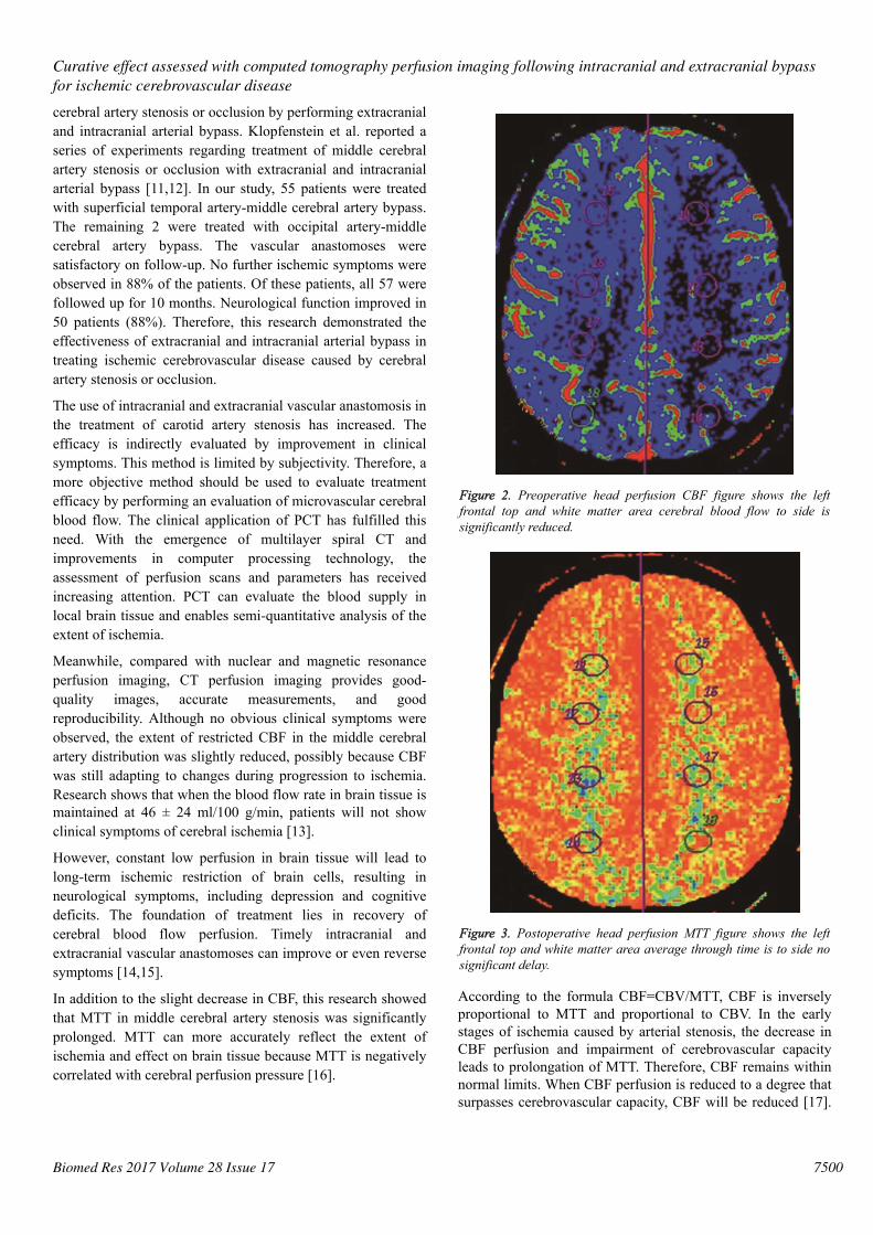
PCT evaluation of intracranial status beforeanastomosisBefore surgery, rCBF and MTT were determined for thearterial distribution and watershed areas on the paretic andunaffected sides. Analysis of variance showed the following:(1) There was no significant difference in regional cerebralblood flow in all patients (P>0.05), indicating no obviousabnormality in flow on the paretic side; (2) MTT in the middlecerebral artery distribution on the paretic side was obviouslydelayed (P<0.05), indicating perfusion delay in the two areas(Figures 1 and 2).
PCT evaluation of intracranial status afteranastomosisThe hemodynamic index was obtained before anastomosis inall patients. The paretic side was compared with the unaffectedside. Analysis of variance showed the following: (1) regionalcerebral blood flow remained normal (P>0.05); (2) MTT in thewatershed area after anastomosis was similar to MTT in theunaffected side, without statistical significance (P>0.05), butMTT in the middle cerebral artery distribution was relatively
prolonged when compared with MTT in the unaffected side(P=0.01), indicating lack of recovery to the normal state.
Figure 1. Preoperative head perfusion MTT figure shows the leftfrontal top and white matter area average through time is to sidesignificantly prolongs.
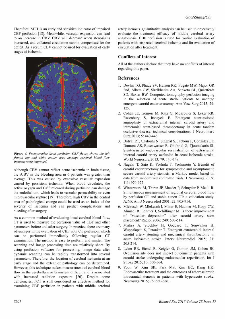
Comparison of changes in PCT hemodynamicparametersIn all patients, PCT parameters were compared before and afteranastomosis and paired examination and analysis wereperformed (Figures 3 and 4). The results were as follows: (1)There was no obvious difference in regional cerebral bloodflow before and after anastomosis (P>0.05); (2) MTT in themiddle cerebral artery distribution was shorter (P=0.021) afteranastomosis. Therefore, surgery improved the delay in MTT.After anastomosis, blood flow improved in 48 of the 57patients, but no MTT improvement was observed in 9 patients.MTT was significantly shorter after anastomosis (P<0.05).Color-coded images showed pathological changes more clearlyfor MTT than for CBF. The extent of cerebral ischemia wasmore defined. CT follow-up after anastomosis showed a smallamount of bleeding in the ischemic area in 6 cases.Retrospective analysis of CTA before anastomosis showed thatthe mean rCBV was greater in the bleeding area in 6 patientswith hemorrhage.
DiscussionMiddle cerebral artery stenosis or occlusion is an importantcause of ischemic cerebrovascular disease. Patients withsignificant occlusion, who do not respond to conservativetreatment, should be considered for extracranial andintracranial arterial bypass or intracranial vascular stenting.Research has long been conducted on treatment of middle
Gao/Zhang/Chi
7499 Biomed Res 2017 Volume 28 Issue 17
cerebral artery stenosis or occlusion by performing extracranialand intracranial arterial bypass. Klopfenstein et al. reported aseries of experiments regarding treatment of middle cerebralartery stenosis or occlusion with extracranial and intracranialarterial bypass [11,12]. In our study, 55 patients were treatedwith superficial temporal artery-middle cerebral artery bypass.The remaining 2 were treated with occipital artery-middlecerebral artery bypass. The vascular anastomoses weresatisfactory on follow-up. No further ischemic symptoms wereobserved in 88% of the patients. Of these patients, all 57 werefollowed up for 10 months. Neurological function improved in50 patients (88%). Therefore, this research demonstrated theeffectiveness of extracranial and intracranial arterial bypass intreating ischemic cerebrovascular disease caused by cerebralartery stenosis or occlusion.
The use of intracranial and extracranial vascular anastomosis inthe treatment of carotid artery stenosis has increased. Theefficacy is indirectly evaluated by improvement in clinicalsymptoms. This method is limited by subjectivity. Therefore, amore objective method should be used to evaluate treatmentefficacy by performing an evaluation of microvascular cerebralblood flow. The clinical application of PCT has fulfilled thisneed. With the emergence of multilayer spiral CT andimprovements in computer processing technology, theassessment of perfusion scans and parameters has receivedincreasing attention. PCT can evaluate the blood supply inlocal brain tissue and enables semi-quantitative analysis of theextent of ischemia.
Meanwhile, compared with nuclear and magnetic resonanceperfusion imaging, CT perfusion imaging provides good-quality images, accurate measurements, and goodreproducibility. Although no obvious clinical symptoms wereobserved, the extent of restricted CBF in the middle cerebralartery distribution was slightly reduced, possibly because CBFwas still adapting to changes during progression to ischemia.Research shows that when the blood flow rate in brain tissue ismaintained at 46 ± 24 ml/100 g/min, patients will not showclinical symptoms of cerebral ischemia [13].
However, constant low perfusion in brain tissue will lead tolong-term ischemic restriction of brain cells, resulting inneurological symptoms, including depression and cognitivedeficits. The foundation of treatment lies in recovery ofcerebral blood flow perfusion. Timely intracranial andextracranial vascular anastomoses can improve or even reversesymptoms [14,15].
In addition to the slight decrease in CBF, this research showedthat MTT in middle cerebral artery stenosis was significantlyprolonged. MTT can more accurately reflect the extent ofischemia and effect on brain tissue because MTT is negativelycorrelated with cerebral perfusion pressure [16].
Figure 2. Preoperative head perfusion CBF figure shows the leftfrontal top and white matter area cerebral blood flow to side issignificantly reduced.
Figure 3. Postoperative head perfusion MTT figure shows the leftfrontal top and white matter area average through time is to side nosignificant delay.
According to the formula CBF=CBV/MTT, CBF is inverselyproportional to MTT and proportional to CBV. In the earlystages of ischemia caused by arterial stenosis, the decrease inCBF perfusion and impairment of cerebrovascular capacityleads to prolongation of MTT. Therefore, CBF remains withinnormal limits. When CBF perfusion is reduced to a degree thatsurpasses cerebrovascular capacity, CBF will be reduced [17].
Curative effect assessed with computed tomography perfusion imaging following intracranial and extracranial bypassfor ischemic cerebrovascular disease
7500Biomed Res 2017 Volume 28 Issue 17
Therefore, MTT is an early and sensitive indicator of impairedCBF perfusion [18]. Meanwhile, vascular expansion can leadto an increase in CBV. CBV will decrease when stenosis isincreased, and collateral circulation cannot compensate for thedeficit. As a result, CBV cannot be used for evaluation of earlystages of ischemia.
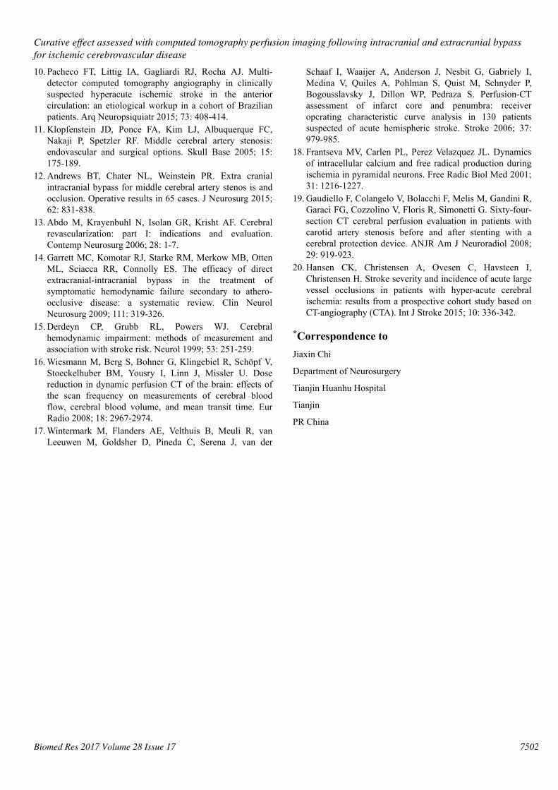
Figure 4. Postoperative head perfusion CBF figure shows the leftfrontal top and white matter area average cerebral blood flowincrease were improved.
Although CBV cannot reflect acute ischemia in brain tissue,the rCBV in the bleeding area in 6 patients was greater thanaverage. This was caused by excessive vascular expansioncaused by persistent ischemia. When blood circulates, theactive oxygen and Ca2+ released during perfusion can damagethe endothelium, which leads to vascular permeability or evenmicrovascular rupture [19]. Therefore, high CBV in the centralarea of pathological change could be used as an index of theseverity of ischemia and can predict complications andbleeding after surgery.
As a common method of evaluating local cerebral blood flow,CT is used to measure the perfusion value of CBF and otherparameters before and after surgery. In practice, there are manyadvantages in the evaluation of CBF with CT perfusion, whichcan be performed immediately following regular CTexamination. The method is easy to perform and master. Thescanning and image processing time are relatively short. Byusing perfusion software for processing, image data afterdynamic scanning can be rapidly transformed into severalparameters. Therefore, the location of cerebral ischemia at anearly stage and the extent of pathology can be determined.However, this technique makes measurement of cerebral bloodflow in the cerebellum or brainstem difficult and is associatedwith increased radiation exposure [20]. Despite somedeficiencies, PCT is still considered an effective method forexamining CBF perfusion in patients with middle cerebral
artery stenosis. Quantitative analysis can be used to objectivelyevaluate the treatment efficacy of middle cerebral arteryanastomosis. CBF perfusion is used for routine evaluation ofpatients with suspected cerebral ischemia and for evaluation ofcirculation after treatment.
Conflicts of InterestAll of the authors declare that they have no conflicts of interestregarding this paper.
References1. Devlin TG, Phade SV, Hutson RK, Fugate MW, Major GR
2nd, Albers GW, Sirelkhatim AA, Sapkota BL, QuartfordtSD, Baxter BW. Computed tomography perfusion imagingin the selection of acute stroke patients to undergoemergent carotid endarterectomy. Ann Vasc Surg 2015; 29:125.
2. Cohen JE, Gomori M, Rajz G, Moscovici S, Leker RR,Rosenberg S, Itshayek E. Emergent stent-assistedangioplasty of extracranial internal carotid artery andintracranial stent-based thrombectomy in acute tandemocclusive disease: technical considerations. J NeurointervSurg 2013; 5: 440-446.
3. Dalyai RT, Chalouhi N, Singhal S, Jabbour P, Gonzalez LF,Dumont AS, Rosenwasser R, Ghobrial G, Tjoumakaris SI.Stent-assisted endovascular recanalization of extracranialinternal carotid artery occlusion in acute ischemic stroke.World Neurosurg 2013; 79: 143-148.
4. Nagaki T, Sato K, Yoshida T, Yoshimoto Y. Benefit ofcarotid endarterectomy for symptomatic and asymptomaticsevere carotid artery stenosis: a Markov model based ondata from randomized controlled trials. J Neurosurg 2009;111: 970-977.
5. Wintermark M, Thiran JP, Maeder P, Schnyder P, Meuli R.Simultaneous measurement of regional cerebral blood flowby perfusion CT and stable xenon CT: a validation study.AJNR Am J Neuroradiol 2001; 22: 905-914.
6. Mlekusch W, Mlekusch I, Minar E, Haumer M, Kopp CW,Ahmadi R, Lehrner J, Schillinger M. Is there improvementof “vascular depression” after carotid artery stentplacement? Radiol 2006; 240: 508-514.
7. Mishra A, Stockley H, Goddard T, Sonwalker H,Wuppalapati S, Patankar T. Emergent extracranial internalcarotid artery stenting and mechanical thrombectomy inacute ischaemic stroke. Interv Neuroradiol 2015; 21:205-214.
8. Leker RR, Eichel R, Keigler G, Gomori JM, Cohen JE.Occlusion site does not impact outcome in patients withcarotid stroke undergoing endovascular reperfusion. Int JStroke 2015; 10: 560-564.
9. Yoon W, Kim SK, Park MS, Kim BC, Kang HK.Endovascular treatment and the outcomes of atheroscleroticintracranial stenosis in patients with hyperacute stroke.Neurosurg 2015; 76: 680-686.
Gao/Zhang/Chi
7501 Biomed Res 2017 Volume 28 Issue 17
10. Pacheco FT, Littig IA, Gagliardi RJ, Rocha AJ. Multi-detector computed tomography angiography in clinicallysuspected hyperacute ischemic stroke in the anteriorcirculation: an etiological workup in a cohort of Brazilianpatients. Arq Neuropsiquiatr 2015; 73: 408-414.
11. Klopfenstein JD, Ponce FA, Kim LJ, Albuquerque FC,Nakaji P, Spetzler RF. Middle cerebral artery stenosis:endovascular and surgical options. Skull Base 2005; 15:175-189.
12. Andrews BT, Chater NL, Weinstein PR. Extra cranialintracranial bypass for middle cerebral artery stenos is andocclusion. Operative results in 65 cases. J Neurosurg 2015;62: 831-838.
13. Abdo M, Krayenbuhl N, Isolan GR, Krisht AF. Cerebralrevascularization: part I: indications and evaluation.Contemp Neurosurg 2006; 28: 1-7.
14. Garrett MC, Komotar RJ, Starke RM, Merkow MB, OttenML, Sciacca RR, Connolly ES. The efficacy of directextracranial-intracranial bypass in the treatment ofsymptomatic hemodynamic failure secondary to athero-occlusive disease: a systematic review. Clin NeurolNeurosurg 2009; 111: 319-326.
15. Derdeyn CP, Grubb RL, Powers WJ. Cerebralhemodynamic impairment: methods of measurement andassociation with stroke risk. Neurol 1999; 53: 251-259.
16. Wiesmann M, Berg S, Bohner G, Klingebiel R, Schöpf V,Stoeckelhuber BM, Yousry I, Linn J, Missler U. Dosereduction in dynamic perfusion CT of the brain: effects ofthe scan frequency on measurements of cerebral bloodflow, cerebral blood volume, and mean transit time. EurRadio 2008; 18: 2967-2974.
17. Wintermark M, Flanders AE, Velthuis B, Meuli R, vanLeeuwen M, Goldsher D, Pineda C, Serena J, van der
Schaaf I, Waaijer A, Anderson J, Nesbit G, Gabriely I,Medina V, Quiles A, Pohlman S, Quist M, Schnyder P,Bogousslavsky J, Dillon WP, Pedraza S. Perfusion-CTassessment of infarct core and penumbra: receiveropcrating characteristic curve analysis in 130 patientssuspected of acute hemispheric stroke. Stroke 2006; 37:979-985.
18. Frantseva MV, Carlen PL, Perez Velazquez JL. Dynamicsof intracellular calcium and free radical production duringischemia in pyramidal neurons. Free Radic Biol Med 2001;31: 1216-1227.
19. Gaudiello F, Colangelo V, Bolacchi F, Melis M, Gandini R,Garaci FG, Cozzolino V, Floris R, Simonetti G. Sixty-four-section CT cerebral perfusion evaluation in patients withcarotid artery stenosis before and after stenting with acerebral protection device. ANJR Am J Neuroradiol 2008;29: 919-923.
20. Hansen CK, Christensen A, Ovesen C, Havsteen I,Christensen H. Stroke severity and incidence of acute largevessel occlusions in patients with hyper-acute cerebralischemia: results from a prospective cohort study based onCT-angiography (CTA). Int J Stroke 2015; 10: 336-342.
*Correspondence toJiaxin Chi
Department of Neurosurgery
Tianjin Huanhu Hospital
Tianjin
PR China
Curative effect assessed with computed tomography perfusion imaging following intracranial and extracranial bypassfor ischemic cerebrovascular disease
7502Biomed Res 2017 Volume 28 Issue 17