culturing of epidermal cells by manipulating the …
TRANSCRIPT

www.wjpps.com Vol 6, Issue 8, 2017.
2358
Balakrishnan. World Journal of Pharmacy and Pharmaceutical Sciences
CULTURING OF EPIDERMAL CELLS BY MANIPULATING THE
MECHANISM OF REGENERATION IN SPONGES AND
EXTRACTION OF ANTIBIOTICS FROM MARINE SPONGES OF BAY
OF BENGAL
Srinivasan Balakrishnan*
Dept of Advanced Zoology and Biotechnology, Loyola College, Chennai,
Tamilnadu, India.
INTRODUCTION
Among all metazoan phyla, sponges are known to produce the largest
number of bioactive compounds, some of them are rich in human
therapeutic value. Therefore, an increasing interest in basic cell
biology research up to biochemical engineering can be observed,
aiming at the production of bioactive compounds from sponges, under
completely controlled conditions. Sponges are Sessile, soft- bodied
marine invertebrates which lack obvious physical defenses and
biosynthesize bioactive, non-primary or secondary metabolites to
protect themselves and maintain homeostasis (Atta way and Zaborsky,
1993). Sponges have antibacterial, antifungal, antiviral, anti-
helminthic, anticoagulant, antitumor, cytotoxic, antidiabetic, anti inflammatory, antimalarial,
and antiplatelet, antiprotozoal, antileukemic, anti tuberculosis, and immunomodulatory
activities (Butler, 2004).Sponges are an important source of secondary metabolites with
pharmaceutical interest. This is the main reason for the increasing interest of sponge culture
in recent years. The use of sponges dates back many centuries. As early as 700 BC, Homer
and Aristotle described the use of sponges in medicine (Voultsiadou, 2007). Until the end of
the nineteenth century, sponges had been widely used in surgery (Müller et al., 2004b). For
example, sponges soaked with extracts of opium were used to anaesthetize patients prior to
surgery, and extracts of Spongia tosta were found to be effective in the treatment of scrofula
(Müller et al., 2004b). Besides their biomedical application, sponges also had commercial
value as bath sponges, because of their absorptive properties. Until 1960, commercial bath
WORLD JOURNAL OF PHARMACY AND PHARMACEUTICAL SCIENCES
SJIF Impact Factor 6.647
Volume 6, Issue 8, 2358-2388 Review Article ISSN 2278 – 4357
*Corresponding Author
Srinivasan Balakrishnan
Dept of Advanced Zoology
and Biotechnology, Loyola
College, Chennai,
Tamilnadu, India.
Article Received on
21 June 2017,
Revised on 12 July 2017,
Accepted on 02 Aug 2017
DOI: 10.20959/wjpps20178-9480

www.wjpps.com Vol 6, Issue 8, 2017.
2359
Balakrishnan. World Journal of Pharmacy and Pharmaceutical Sciences
sponge farming was a lucrative business spreading from the Mediterranean to the Atlantic
and Pacific Ocean (Bernard, 1968; Duckworth, 2009). From 1960, due to the invention of the
less expensive synthetic bath sponge, farming of the natural bath sponge decreased and now
only occupies a small niche in the market for high-quality applications (Hogg et al., 2010).
Current biotechnological applications of sponges are more directed towards the production of
marine natural products and biomaterials. Besides pharmaceutical products many other
applications of sponge derived biomaterials have been identified. For example, the silicon
skeleton of glass sponges may serve as a blueprint for the production of very efficient fibre
optics (Sundar et al., 2003) and biosilica- producing enzymes from sponges have been
applied in nanotechnology (Schröder et.al.,2010).
Fig: 1 Phylogenetic position of the Porifera between the Urmetazoa and the Urbilateria.
The major evolutionary novelties which have to be attributed to the Urmetazoa are those
molecules which mediate apoptosis and control morphogenesis, the immune molecules and
primarily the cell adhesion molecules. The siliceous sponges with the two classes
Hexactinellida and Demospongiae emerged first and finally the Calcarea, which possess a
calcareous skeleton, appeared. These three classes of Porifera are living fossils that
provide a reservoir for molecular biological studies. The Archaeocyatha, sponge related
www.wjpps.com Vol 6, Issue 8, 2017.
2360
Balakrishnan. World Journal of Pharmacy and Pharmaceutical Sciences
animals with a calcareous skeleton, became extinct. The Calcarea are very likely a sister
group of the Cnidaria. From the latter phylum the Ctenophora evolved which comprise not
only an oral/aboral polarity but also a biradial symmetry. Finally the Urbilateria emerged
from which the Protostomia and the Deuterostomia originated. Very likely the Urmetazoa
emerged between the two major ‘snowball earth events’, the Sturtian glaciation (710–680
MYA) and the Varanger-Marinoan ice ages (605–585 MYA). In the two poriferan classes
Hexactinellida and Demospongiae the skeleton is composed of amorphous and hydrated
silica, while the spicules of Calcarea are composed of Cacarbonate. The latter biomineral
is also prevalent in Protostomia and also in Deuterostomia. In vertebrates the bones are
composed of Ca-phosphate [apatite].
In fact, sponges are the only animal able to polymerize silica to generate massive skeletal
elements in a single reaction at ambient temperature and pressure (biosintering) (Müller et al.,
2009). According to a survey, sources have stated that there has been no recent research on
sponge culture techniques in India. The optimal culture system depends on the species to be
cultured, while some species easily produce sponge aggregates after dissociation
(primmorphs), others show a great capacity to regenerate after fragmentation (explants).One
major obstacle is the limited availability of larger quantities of defined sponge material, “the
so- called supply problem”. In this Research Project, different approaches used so far for
producing sponge biomass by culturing methods as well as some significant modifications are
introduced for the maintenance of sponges are studied. In recent years, great efforts have
been made to set up in vitro culture systems for the cultivation of sponge cells. One of the
major advantages of cell cultures is the possibility to control and manipulate the cultivation
conditions depending on the sponge species and the target metabolite. Successful attempts to
produce sponge metabolites using culturing of epidermal cells of sponges by manipulating
the mechanism of regeneration in sponges are discussed.
Literature Review
Shear effects on suspended marine sponge cells was done by F. Garc´ıa Camachoa, E.H.
Belarbi a, M.C. Cer´on Garc´ıa a, A. S´anchez Mir´on a, T. Chile b, Y. Chisti b, E. Molina
Grima states that, a four-step mechanistic model was shown to describe the kinetics of cell
death and fragmentation. The damage to cells was not depending on cell–cell interactions.
The forces in the agitated fluid killed the viable cells by impact, which was not accompanied
by cell rupture (i.e. the cell was left dead, but intact). Biochemistry and Cell Biology of Silica

www.wjpps.com Vol 6, Issue 8, 2017.
2361
Balakrishnan. World Journal of Pharmacy and Pharmaceutical Sciences
Formation in Sponges done by: werner e.g. mu¨ ller, anatoli krasko, gae¨ l le pennec, and
Heinz c. schro¨ der states that The spicules occur in the cytoplasm and the extracellular space
and also in the nucleus (as silicate crystals) of some sponge cells; the spicules are formed by
the enzyme called Silicatein. The enzyme is dependent on ferric ion. Silicatein also has
proteolytic (cathepsin-like) activity. The morphogenetic activity of silicate is underscored by
the finding that this ferric ion increases gene expression of silicatein and collagen. Based on
these findings, it is concluded that both ferric ion and silicate stimulate the activity of
silicatein. Furthermore, it is proposed that the growing spicules are surrounded by the
scavenger receptor which might be considered as a docking molecule for the collagen matrix
into which the spicules are embedded. Cell culture from sponges: pluripotency and
immortality so` nia de Caralt1, Marı´a J. Uriz and Rene´ H.
Wijffels states that Sponges have an enormous potential for the Development of new medical
drugs. Thus, although several Bottlenecks remain, efforts to develop a technology for
continuous cell cultures, such as the use of embryonic stem. Cells to form a cell line and
research into the control of apoptosis are worthwhile and might become successful in the
interim. Establishment of a primary cell culture from a sponge: primmorphs from Suberites
domuncula Werner e. g. miller.
Fig: 2.1 A large barrel sponge. Courtesy of Jonathan Bird, Oceanic Research Group.
Matthias wiensl, renato et.al steffen, heinz. schroder, adovan to conclude, the primmorph
system described by Custodio et al. (1998) can be used in the future for a variety of
applications in the following main directions:(1) As a bioreactor to produce bioactive
compounds from sponges and (2) For the detection of potential cytostatic compounds causing

www.wjpps.com Vol 6, Issue 8, 2017.
2362
Balakrishnan. World Journal of Pharmacy and Pharmaceutical Sciences
a transition from telomerase-positive to telomerase- negative cells. Advances in the
production of sponge biomass Aplysina aerophoba A model sponge for ex situ sponge
biomass production Rudolf Haussmann, Marco Vitello b, Frank Leitermann a, Christoph
Syldatk a Obtaining that is at present extremely difficult because most sponges are relatively
rare in nature and their mass cultivation in the laboratory has not yet been accomplished. In
this study the possibility of culturing Aplysina aerophoba fragments in laboratory was
examined. While a substantial biomass increase was not yet observed, we achieved
fragmented sponge tissue to develop into a functional sponge as a first success. Antibacterial
Activity of Marine Sponge Extracts against Fish Pathogenic Bacteria G. Annie Selva Sonlal,
A.P. Lipton and R. Paul Raj. Suggest that fractionation and purification of the crude methanol
extract of A. elongata has potential in the development of novel Antibiotic substances for
managing common bacterial diseases in aquaculture. The most active species was A. elongata
which inhibited 100% and 87.5% of the tested bacterial isolates at 20°C and 30°C
respectively. For most sponge-derived bioactive compounds, it is not clear whether they are
produced by the sponge or the symbiont (which can contribute up to 40% of the sponge
volume). Metabolites known to be produced by the sponge are, for example, avarol, found
in Dysidea avara (Uriz et al., 1996) and stevensine, found in Axinella corrugata (Andrade et
al., 1999). On the other hand, it has been demonstrated that Oscillatoria spongelia, a
cyanobacterial symbiont, produces antimicrobial polybrominated biphenyl ethers and might
keep the sponge free of other bacteria (Unson et al., 1994).
Fig: 2.2 A dye squirted around the base of a purple tube sponge colors the jet emerging
from the osculum at the top of the sponge. Courtesy of Jonathan Bird, Oceanic Research
Group.

www.wjpps.com Vol 6, Issue 8, 2017.
2363
Balakrishnan. World Journal of Pharmacy and Pharmaceutical Sciences
MATERIALS AND METHODOLOGY
Collection of sponges
Sponged were collected by, hand-picking at the coastal regions of Pamban Bridge (9°19’ N
latitude and 79°23’ E longitude). Sponges were obtained as entangled specimens from fishing
nets set at depths of 10-15 m off Pamban coast of Rameswaram. The churning seawater in
this area during January-February and May-August lead to dislodging and entanglement of
marine organisms in nets. The collected specimens were washed in filtered seawater. Eight
species of sponges were collected, and, were identified by Dr.G.Sivaleela of Zoological
Survey of India, Chennai, as Clathria Gargonoides, Haliclona fibulata, Sigmadocia
petrosionides, Dysidea herbacea, Hyattella intestinalis, Gelliodes pumila, Stellitethya repens
and Suberitus carnosus all of which belong to the class Demospongiae Sollas.
Formation of sponge cell lines
In general, animal cell cultures are obtained by isolating cells from an axenic piece of tissue.
These cells are cultured in medium that will support proliferation. This stage is called
primary cell culture and most cell types will undergo a limited number of divisions and then
enter senescence. Continuous cell lines are generally obtained from spontaneously
immortalized primary cell cultures (e.g., random mutagenesis) or are derived from cancerous
tissue (Freshney, 2005). Another approach towards developing a continuous cell line is the
insertion and expression of immortalizing genes (e.g., SV40LT, hTERT) in primary cells.
Immortalizing genes interfere with the regulatory pathways for cell division, and in this way
cause unlimited cell division, resulting in a continuous cell line (Freshney, 2005).
In principle, sponges have great potential for cell culture because of the presence of totipotent
stem cells (i.e. archaeocytes) and because cells can be easily dissociated from its tissue, due
to their very loosely packed cellular organization. However, despite efforts by several
research groups, a continuous sponge cell line has not yet been developed, and the number of
primary sponge cell cultures developed is very limited (Rinkevich, 2005; Pomponi, 2006;
Schippers et al., 2012). This is related to the following aspects.
Due to the presence of symbionts inside the sponge, it is practically impossible to obtain
an axenic piece of sponge tissue. Researchers have used antibiotics and antimycotics to
reduce growth of contaminants (Pomponi and Willoughby, 1994), but still contamination
occurs (Ilan et al., 1996).
Also obtaining proliferating starting material remains problematic, since most dissociated

www.wjpps.com Vol 6, Issue 8, 2017.
2364
Balakrishnan. World Journal of Pharmacy and Pharmaceutical Sciences
sponge cells go into apoptosis and die (Schippers et al., 2011 However, Pomponi and
Willoughby (2000) were able to show sponge cell division when exposed to the mitogen,
phytohaemagglutinin. Other options for obtaining proliferating starting material are the
use of primmorphs, gemmules or larvae.
Primmorphs are reaggregated sponge cells which have the ability to proliferate in vitro,
although no net growth has been observed (Sipkema et al., 2003). Gemmules and larvae
both contain a package of stem cells, and can develop into a juvenile sponge in vitro.
Nutritional requirements of sponge cells are still poorly understood. Most of the sponge
cell culture media are based on mammalian cell culture media, but certain components
such as fatty acids or growth factors, are expected to be different for sponge cells.
The establishment of sponge cell lines is still hampered by three issues: the presence of
contaminants in the starting material, the lack of knowledge on nutritional requirements and
the lack of proliferating starting material. This thesis focusses on the last item; sponge cell
proliferation).
Fig: 3.1 Schematic representation of different sponge material for stem cell cultures. Adult
sponges have differentiated cells (e.g. choanocytes, pinacocytes and sclerocyetes) and
pluripotent cells (archeocytes). Ripe sponges harbour high numbers of embryos, which are
composed of blastomeres (stem cells) that increase in number per sponge unit during the
later stages of the reproduction cycle. The embryos are released as larvae, which can be
cultured as a source of stem cells and (ii) sponge juveniles for obtaining sponge biomass.

www.wjpps.com Vol 6, Issue 8, 2017.
2365
Balakrishnan. World Journal of Pharmacy and Pharmaceutical Sciences
Abbreviations – archeocytes; c – choanocytes; ca – canal; ch – choanocyte chamber; e –
embryo; – endopinacocyte; g – gemmule; s – spicules; sc – sclerocyte.
Explants were cultured under two Methods as follows.
1. In vitro Culturing Technique.
2. Closed aquarium system.
In this study only the invitro culturing of marine sponges Clathiria gorgonoides and Dysidea
herbaceae is performed
Invitro cell culture technique
Cell or tissue culture (both subsequently referred to as in vitro culture) is a preferred method
because the system can be defined and controlled. Sponges have great potential for cell
culture because of the presence of totipotent stem cells and because cells can be easily
dissociated from its tissue, due to their loosely cellular organization. Despite efforts by
several research groups, a continuous sponge cell line has not yet been developed, and the
number of primary sponge cell cultures developed is very limited (Rinkevich, 2005). The
only report of a continuous sponge cell culture was Klautau et al. (1993, 1994); however, this
cell line was subsequently identified as a protozoan (Custodio et al., 1995).
DISSOCIATION OF SPONGE CELLS
The first step in obtaining a primary sponge cell culture involves dissociation of the cells into
a monodisperse suspension. Several dissociation techniques, Such as Mechanical, chemical,
enzymatic and spontaneous, have been studied.
The most successful dissociation method is a combination of mechanical and chemical
dissociation (Pomponi and Willoughby, 1994; Rinkevich et al., 1998). If a monodisperse cell
suspension is needed, the method described by Pomponi and Willoughby (1994) is generally
used, whereas the method described by Custodioet al. (1998) is applied when re-aggregation
of sponge cells (i.e. to formprimmorphs) is needed. The dissociation method, will result in a
mixture of cell types, for example, archaeocytes, choanocytes, pinacocytes, collencytes.
Archaeocytes and choanocytes are considered to be the pluripotent stem cells in sponges.
These stem cell-like cells have the capacity to proliferate and to differentiate into other cell
types. Most cell types in a sponge are expected to be terminally differentiated and not able to
divide. Consequently, selecting and enriching for archaeocytes and choanocytes may result in

www.wjpps.com Vol 6, Issue 8, 2017.
2366
Balakrishnan. World Journal of Pharmacy and Pharmaceutical Sciences
a proliferating sponge cell culture. By using a process which involving differential
centrifugation, selective aggregation in low Ca2/Mg2 seawater, differential adherence in
artificial seawater and by centrifugation, eventually obtained an archaeocyte-enriched
fraction greater than 80%, and proliferation was demonstrated by BrdU incorporation
Steps which are involved in cell dissociation of marine sponges
Cut sponge in very small parts of 2–3 cm from the mother sponge in water with the help
of sterile razor sharp scalpel.
Wash in fresh artificial sea water to Remove debris and mince into small pieces of 2-5mm
in length.
Cut parts of sponge where transferred to Petri dishes containing CMFSW- EDTA in the
ratio of 10:1.Soak for 20 minutes in sterile CMFSW-EDTA.
Filter through 70 mm nylon mesh /sterile gauze now the cells are easily released to
remove spicules.
To form crude cell suspension centrifuge at 300rpm for 5minutes to enrich the sponge cell
content which where in pellet and remove most of the bacteria in supernatant.
The supernatant was discarded and cells in the pellet were resuspended in CMFSW-
EDTA.
Table1: Publications about Primary sponge cell culture.
Sponge species Reference Proliferation Comments
Ircinia Muscarum &
Sarcotragus foetidus
De Rosa et al. (2001,
2003) Yes, but.
No data of sponge cell growth over
time are shown
Suberites domuncula De Rosa et al. (2003)
and Zhang et al. (2004) Yes
Sterilized sponge cell proliferates
approximately after 3 days
Hymeniacidon
heliophila
Pomponi and
Willoughby (1994) Yes
PHA-stimulated cells doubled after
72 h
Ephydatia muelleri Imsiecke et al. (1995) Yes
Cells were taken from hatched
gemmules Eventually, cultures were
dominated by bacteria
Acanthella acuta Nickel et al. (2000) No -
Dysidea avara Nickel et al. (2000) and
De Rosa et al.(2003) No -
Hymeniacidon
heliophila
Pomponi and
Willoughby (1994) Yes
PHA-stimulated
cells doubled after 72 h

www.wjpps.com Vol 6, Issue 8, 2017.
2367
Balakrishnan. World Journal of Pharmacy and Pharmaceutical Sciences
Fig: 3.2 Dissociation method described by Pomponi and Willoughby (1994).
Finally resuspend pellet into culture medium for further regeneration of cells. Phyto-
haemogluttinin a mitosis inducing lectin was also added.
EDTA10mM was added which helps in dissociating cells by chelating any multivalent
metal ions in the sponge surface.
Medium for sponge cell culture
Preparation of a culture medium is very essential to support the growth of Sponge cells.
Generally, an axenic inoculum is used to initiate a primary cell Culture from animal tissue
(Freshney, 2005). It is not very easy to isolate such.
An axenic part from sponges, since both eukaryotic and prokaryotic symbionts are
intermingled with the sponge in the tissue. This made it very difficult to develop a specific
nutrient media for the growth of sponge cells, since adding nutrients for sponge cell growth
will also stimulate growth of other contaminants present in the sponges. Therefore, measures
have been taken to limit the growth of contaminants, such as bacteria, fungi, protozoa etc. To
prevent growth of these contaminants, antibiotics and antimycotics can be added to the
medium, but most of these only temporarily inhibit growth of bacteria and fungi. This may be
due to the fact that some bacteria in sponges may have antibiotic resistance. In addition, high
degree of antibiotics and antimycotics can also have a negative effect on the viability of the
sponge cells. Despite these difficulties, researchers have tried to develop culture media for
sponge cells. The most basic culture medium consists of filtered seawater which provides the
appropriate salinity and pH, and contains some dissolved nutrients and trace elements which
are generally present in the sea. Sponge cells could be maintained in filtered seawater for
several weeks without contaminants overgrowing the culture. However, since no growth of e
sponges a.

www.wjpps.com Vol 6, Issue 8, 2017.
2368
Balakrishnan. World Journal of Pharmacy and Pharmaceutical Sciences
Table 2: Publications about sponge cell culture media.
Media Antibiotics Supplements Contaminations Sponge species
SW Penicillin,
streptomycin
Glutamine, pyruvate, iron
citrate, silicon, RPMI 1640,
Marine Broth 2216
No Suberites domuncula
SW - - Yes Xestospongia muta
M199 in
CMF Rifampicin 5% FBS, 1.5% PHA No Hymeniacidonheliophila
Modified
ASW
Inorganic salts (e.g. ferric
iron NaCl,) amino acids,
sugars, vitamin C, DMEM,
RPMI 1640, 1.5% PHA
No Hymeniacidonperlevis
DMEM in
ASW
Nystatin,
rifampicin,
gentamycin,
Penicillin,
Ciprofloxacin
- Yes
Acanthella acuta
Dysidea avara
Cell was observed (Schippers et al., 2011), it is concluded that the dissolved nutrients in
seawater are not sufficient to support long-term growth of sponge cells. Other sponge cell
culture media (reviewed by Pomponi, 2006) have mostly been derived from existing animal
cell culture media, which contain amino acids, vitamins and glucose. Since these media are
rich in nutrients, the use of antibiotics and antimycotics is also essential. Despite the use of
antibiotics and antimycotics, contamination can still occur (Ilan et al., 1996), and the addition
of cell culture media alone does not support sponge growth.Growth factors is essential. For
example, the addition of PHA (phytohemagglutinin), a mitogenic lectin, stimulated cell
division (Pomponi et al.,1997; Sun et al. 2007) and the addition of cholesterol resulted in
increased cell density compared to cell cultures without cholesterol (De Rosa et al.,
2001,2003). Evidently, cells in vitro are unable to biosynthesize sterols, and the addition of
sterols is needed to satisfy membrane requirements. Ferric iron was also found to
significantly improve cell viability. So to prepare a new culture medium to culture sponge
cells the methods are as follows, the medium consists of the parent sea water from where the
sponges subjected for culture process is taken and it is sterilized by filter sterilization
technique. The antibiotics like penicillin and streptomycin was also sterilized and added to
the filtered fresh parent sea water. Now the substratum like mussels, rocks, and other
calcareous fossil remains which found in-depth of the sea to which the sponge holds was
placed inside the medium which is also sterilized by acid wash and finally with double
distilled water to remove the acidified nature. Next is to provide the exact temperature, light
intensity, Pressure and pH and make the water in the medium not to stagnant, provide a little

www.wjpps.com Vol 6, Issue 8, 2017.
2369
Balakrishnan. World Journal of Pharmacy and Pharmaceutical Sciences
motion to the water by the blower. By enabling this suitable environment for sponge culture
process we can expect the proliferation of sponges will takes place.
Primmorph
Fig: 3.3 Method to generate primmorphs, described by Custodio et al. (1998).
Abbreviations
FSW- filtered seawater
CMFSW calcium- and magnesium-free seawater. EDTA-Ethylene Diamine Tetra Acetic
Acid.
SW- seawater AB- antibiotics
The sponge taken from the sea is cut into pieces of 1 mm.
Wash with sterile Calcium Magnesium free fresh sea water.
Transfer the sponges into 50ml test tube containing 40ml CMF-EDTA solution.
Gentle shaking of the test tube for 30minutes.
Discard the solution and fill the test tube with new CMF-EDTA solution.
Gentle shaking for 30 minutes and the solution is collected.
Filter the collected water through 40micrometer nylon mesh and isolate the sponge cells.
Isolated sponge cells are subjected to centrifugation at 500rpm for 5minutes.
Repeat the centrifugation process for two times and resuspend the solution to sea water
and antibiotic solution.
www.wjpps.com Vol 6, Issue 8, 2017.
2370
Balakrishnan. World Journal of Pharmacy and Pharmaceutical Sciences
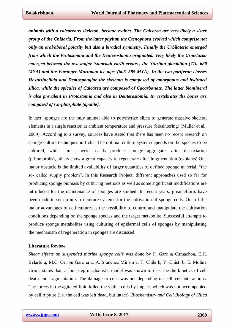
Collect the primmorphs and wash with sea water and place them in well plates for further
experiments.
Fig: 3.4 Fig: 3.5
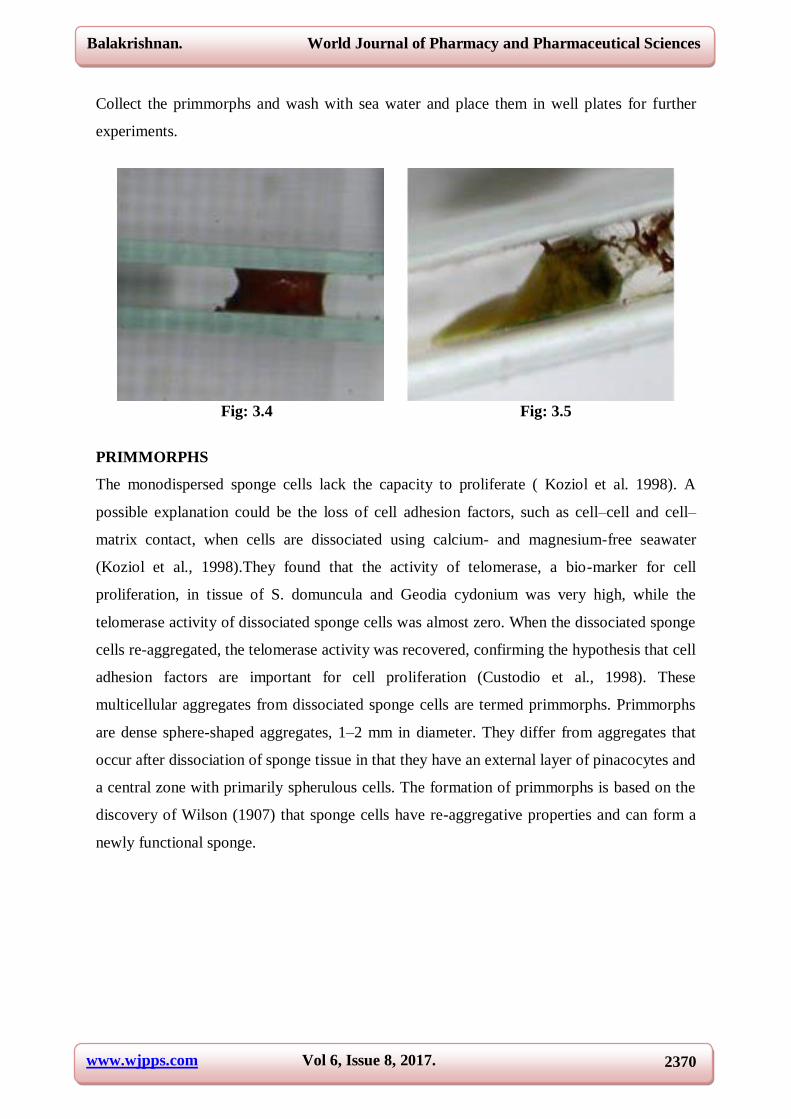
PRIMMORPHS
The monodispersed sponge cells lack the capacity to proliferate ( Koziol et al. 1998). A
possible explanation could be the loss of cell adhesion factors, such as cell–cell and cell–
matrix contact, when cells are dissociated using calcium- and magnesium-free seawater
(Koziol et al., 1998).They found that the activity of telomerase, a bio-marker for cell
proliferation, in tissue of S. domuncula and Geodia cydonium was very high, while the
telomerase activity of dissociated sponge cells was almost zero. When the dissociated sponge
cells re-aggregated, the telomerase activity was recovered, confirming the hypothesis that cell
adhesion factors are important for cell proliferation (Custodio et al., 1998). These
multicellular aggregates from dissociated sponge cells are termed primmorphs. Primmorphs
are dense sphere-shaped aggregates, 1–2 mm in diameter. They differ from aggregates that
occur after dissociation of sponge tissue in that they have an external layer of pinacocytes and
a central zone with primarily spherulous cells. The formation of primmorphs is based on the
discovery of Wilson (1907) that sponge cells have re-aggregative properties and can form a
newly functional sponge.

www.wjpps.com Vol 6, Issue 8, 2017.
2371
Balakrishnan. World Journal of Pharmacy and Pharmaceutical Sciences
Fig: 3.6
Marine sponges produce secondary metabolites that can be used as a natural source for the
design of new drugs and cosmetics. There is, however, a supply problem with these natural
substances for research and eventual commercialization of the products. In Vitro sponge
culture is nowadays one of the most reliable methods to supply pharmaceutical companies
with sufficient quantities of the target compound.
Cell Proliferation of Sponges
Cell proliferation has been observed in primmorphs by means of BrdU incorporation
(Custodio et al., 1998; Zhang et al., 2003b); however, an increase in total biomass over time
has not been observed. Possibly there is equilibrium in the primmorphs between proliferating
and apoptotic cells (Koziol et al., 1998). A possible explanation for the lack of increase in
biomass is the deficiency of nutrients in the media. Primmorphs were generally cultivated in
nutrient-poor medium, such as natural seawater or artificial seawater without the addition of
any nutrients (Custodio et al., 1998; Nickel et al., 2001; Sipkema et al., 2003c; Zhang et al.,
2003a, Valisano et al., 2006b). Only Muller et al. (2000) and Krasko et al. (2002) added 0.1%
(v/v) marine broth 2216 or 0.2% RPMI 1640, respectively, to their media, but they do not

www.wjpps.com Vol 6, Issue 8, 2017.
2372
Balakrishnan. World Journal of Pharmacy and Pharmaceutical Sciences
show data for nutrient uptake by the primmorphs. To be able to obtain continuous biomass
growth, it is important to supply appropriate nutrients and growth factors.The cell culture of
sponge Clathria gorgonoides was not successful because from which the cells are
dissociated, during dissociation it was damaged and lost its proliferating nature eventhough
some callus like structure was arised but they are found to be the culture of symbiotic micro-
organisms associated with sponges, but the culture of Dysidea herbacea is successful because
the cell are not dissociated but as whole mass of 1-2mm of length as a tissue fragment or
otherwise called as primmorph is cultured in this process the mass of sponge ex-plant placed
in the medium was doubled in four days and further growth is not observed due to addition of
antibiotics such as penicillin and streptomycin to control the contamination which eventually
affects on the primmorphs of sponges which subjected to cultivation. So only the primary cell
culture of sponges was obtained. For future investigation of sponge culture, extra care must
be given to the methods of cultivation and addition of antibiotics to develop Immortal sponge
cell lines.
Discussion on Genomic Based Approach for sponge cell culture
Sponges are a rich source of bioactive compounds which have the potential to provide future
medicines, such as new antibiotics and anticancer drugs (Blunt et al., 2009; Sipkema et al.,
2005a). However, the limited availability of sponge biomass hampers the development of
these potential drugs into commercial products. The use of in vitro sponge cell cultures is a
potential alternative for biological supply of sponge-derived products, but until now a
continuous sponge cell line has not been developed (Pomponi, 2006; De Caralt et al., 2007b;
Schippers et al., 2012).This is primarily due to significant gaps in our understanding of
sponge cell proliferation and death, nutritional requirements and cultivation conditions.
Recent developments in the field of genomics and transcriptomics can help us to get more
insight in sponge growth and death as well as nutritional requirements. Recently, the first
genome of a sponge has been sequenced, and information about genes specific for
multicellularity, such as control of cell proliferation and death, were revealed (Srivastava et
al., 2010). These genes play a significant role in regulating cell growth, which is crucial for
multicellular animals, since unrestricted growth of cells (= cancer) is detrimental for the other
cells and the organism as a whole. For the development of a continuous cell line, the
possibility to attain unrestricted growth is needed, per definition. Accordingly, most
mammalian cell lines are derived from cancerous tissue or are actively immortalized, e.g., by
inserting viral genes. The occurrence of cancer in an organism shows that cells of that

www.wjpps.com Vol 6, Issue 8, 2017.
2373
Balakrishnan. World Journal of Pharmacy and Pharmaceutical Sciences
organism can, in principle, be immortalized and the tumors themselves can serve as a source
of immortal cells. Although many genes involved in the development of cancer are also
present in sponges, cancerous growth in sponges has not been observed yet, neither in situ
nor in vitro. The genes of interest discussed in this study focus on genes involved in cell
proliferation, cell death and cell adhesion, since these genes play a significant role in
maintaining homeostasis in multicellular animals. Deregulation of these genes can cause
unlimited growth of cells (i.e. stimulation of cell proliferation in combination with prevention
of apoptosis), which is essential for the development of a continuous cell line. Most sponge
cultivation studies observe the change of biomass (e.g., volume, weight, cell numbers, protein
content) over time and do not measure expression of certain genes and their related products.
We suggest to study gene expression (tools to study gene expression are mentioned in box 1)
in proliferating and non-proliferating conditions to obtain insight in the role of known genes
involved in cell proliferation and cell death and find other genes of interest. Following
conditions could be compared.
Larvae and gemmules vs. adult sponge.
Juveniles of larvae and gemmules have the potential to proliferate in vitro, whereas an adult
sponge in general lacks this capacity. A plausible explanation for this could be the large
number of stem cells present in larvae and gemmules. Using sponge stem cells for cell culture
has already been proposed by De Caralt et al. (2007b) and Rinkevich (2011). Since juveniles
of larvae and gemmules are growing and developing into a sponge, genes related to cell
proliferation are expected to be more actively expressed than in an adult sponge which is in
homeostasis. By comparing gene expression profiles of juveniles with adult sponges, we can
confirm if specific genes involved in cell proliferation are more actively expressed.
Primary sponge cell cultures vs. adult sponge.
In general, primary sponge cell cultures lack proliferative capacity. Previous studies already
showed that dissociation of sponge cells resulted in increased caspase activity (Schippers et
al., 2011) which is an indicator for apoptosis, and decreased telomerase activity (Koziol et al.,
1998) which is a bio-marker for cell proliferation. Nevertheless, Pomponi et al. (1997) were
able to demonstrate sponge cell growth when PHA was added to the growth medium.
Subsequently Willoughby (2002) compared gene expression levels of sponge cells cultivated
with and without PHA. She found that PHA effects expression levels of proliferative and
anti-apoptotic genes in marine sponge cells. This research has not been followed up, but it

www.wjpps.com Vol 6, Issue 8, 2017.
2374
Balakrishnan. World Journal of Pharmacy and Pharmaceutical Sciences
would be of value to study the expression of the genes mentioned in primary sponge cell
cultures stimulated with mitogens like PHA and to compare with gene expression in the adult
sponge.
Another alternative is exposure of sponge cells to carcinogens or UV-radiation. Carcinogens
are substances which can induce cancer, which is in most cases related to mutagenesis.
Exposure to carcinogens can for example lead to increased oncogene expression and can
therefore also be applied to transform cells.
Examples of carcinogens which lead to immortal transformation in mammalian cells are UV-
radiation (Wazer et al., 1994), heavy metals (Hamilton et al., 1998) and (Stampfer and
Bartley, 1985). By exposing sponge cells to these substances, we can alter the activity of e.g.
tumor suppressor genes and proto-oncogenes and possibly immortalize sponge cells.
Fig: 3.7 Proposed strategy of a genomic-based approach towards a sponge cell line.
In addition, for future study researchers can search for additional genes in sponges important
for regulating cell proliferation and death by studying the transcriptome of proliferating
sponge cells (e.g. larvae, gemmules, PHA stimulated sponge cells) in comparison with non-
www.wjpps.com Vol 6, Issue 8, 2017.
2375
Balakrishnan. World Journal of Pharmacy and Pharmaceutical Sciences
proliferating sponge cells (e.g. adult sponge, non-PHA stimulated sponge cells).
Finally, a promising method to develop a continuous sponge cell line is to insert
immortalizing genes, such as SV40 LT together with TERT. To develop a continuous sponge
cell line using immortalizing genes, the following prerequisites need to be met: metabolically
active primary cells, a promoter that is recognized by the RNA polymerase of the sponge,
effective DNA delivery method, functional immortalizing genes, and a method to obtain
stable insertion of the genes (e.g. appropriate selectable marker for sponges.
Preparation of Methanol Extract of Sponge
The sponges collected are washed in deionised water and they are subjected to shade dry till
they reach crispy condition then they are powdered in the blender. Now fifty (50 gm) gram of
shade dried sponge powder is mixed with Methanol and homogenized. Keep the mixture at
shaker at 100 rpm for 24 hours and then they are centrifuged at 12000 rpm for 10 minutes.
Then the supernatant was filtered thorough Whatman No: 1 filter paper or syringe filter.
The filtrate was dried to evaporate the solvents at room temperature. Te sediment extract i.e.
pellet was weighed and dissolved in five percentage (5% DMSO), and refrigerated for future
study.
Qualitative Analysis of Active Metabolites from Sponge Extract
Terpenoids, steroids, Alkaloids, Saponins, Glycosides were screened from marine sponges by
adopting following protocol.
Terpenoid and steroid
Four milligram of the extract was treated with the0.5 ml of Acetic Anhydride and 0.5 ml
of Chloroform.
Then concentrated solution was added slowly and red violet color was observed for
terpenoid and green bluish color for steroids.
Alkaloid
The extract as evaporated to dryness and residue was heated with the boiling water bath
with 2% hydrochloric acid.
After cooling the mixture was cooled and treated with few drops of Mayer’s reagent.
The samples were then observed for the presence of turbidity or Yellow Precipitation.

www.wjpps.com Vol 6, Issue 8, 2017.
2376
Balakrishnan. World Journal of Pharmacy and Pharmaceutical Sciences
Saponins
Frothing test was identified the presence of saponins.
100ml of extract was added 5 ml distilled water.
Frothing Persistence indicated the presence of positive result ie.the presence of Saponin.
Glycoside
To the solution of the extract glacial acetic acid is added few drops of ferric chloride and
concentrated sulphuric acid.
Observed for red brown colorations at the junctions of the tube and bluish green color in
the upper layer.
Preparation for antibacterial sensitivity test
A cross- streak method was used for antimicrobial activity. Single streak of was made on
surface of the modified Muller- Hinton Agar Plates Supplemented with 2% of NaCl and
incubated at 28 °C. The pathogenic bacterial strains, such as E. coli, H. influenzae, Klebsiella
pneumoniae, Enterococcus faecalis which were streaked and incubated at 28 °C and the
incubation distance was measured after 24-48 hrs. A control plate was also maintained
without inoculating the microbes to assess the normal growth of bacteria.
Antibacterial Assay
Antibacterial activity was determined against E. coli, K. pneumoniae, H. influenzaes, E.
faecalis, using the paper disk assay method (12).Whatman No. 1 filter paper disk of 6-mm
diameter was sterilized by autoclaving for 15 min at 120 0C. The sterile disks were
impregnated with different extracts (500 Ag/ ml).Agar plates were surface inoculated
uniformly from the broth culture of the tested microorganisms. In all cases, the concentration
was approximately 1.2X10⁸ CFU/ml. The impregnated disks were placed on the medium
suitably spaced apart and the plates were incubated at 37 0C for 24 h. Disk of Streptomycin
(400 Ag/ml) was used as a positive control. The diameter (mm) of the Zone of inhibition
(halos) caused by the methanolic extracts of sponges was examined.

www.wjpps.com Vol 6, Issue 8, 2017.
2377
Balakrishnan. World Journal of Pharmacy and Pharmaceutical Sciences
Table 3: shows the measurement of zone of inhibition i.e., Antibacterial activity of
methanolic extract of different marine sponges against the different disease causing
pathogenic bacteria.
NAME OF THE
SPONGE
Name of the tested micro-organisms
(Zone of Inhibition in Diameters mm)
E.Coli H.influenzae K.Pneumoniae E.faecalis
Clathria gorgonoides 5±2.58 _ 10±1.91* 9±4.11
Sigmodocia petrosioides 9±0.36 8±1.49 12±0.09* 6.3±2.6
Haliclona fibulata - 11±1.49* 3.41±0.21 13±3.68*
Suberitus carnosus 3.65±0.6 5±0.02 11±0.50* -
(-) no inhibition of growth during testing Asterisks (*) indicates the values which are
significantly different.
Fig: 4.1 Fig: 4.2
Fig 4.1 and 4.2 shows the Secondary screening of antibacterial activity of the Marine
sponges against the various Bacterial Pathogens.
The Present study clearly shows that the maximum peak of activity was noted on two sponge
species such as Sigmodocia petrosioides and Haliclona fibulata crude extract showed the
peak activity 12±0.09 and 13±3.68 bioactivity against the pathogen of K.Pneumoniae and
E.faecalis respectively.Subsequently another maximum zone of inhibition (11±0.50) formed
with the sponge Suberitus carnosus extract on K. pneumoniae. The present result denoted on
both sponge extract antimicrobial activity at 5% level of statistically significant.
Research has indicated that the secondary metabolites of sponges play an important role in
their defense against infectious pathogens. This systematic investigation of marine
environments is reflected in the large number of novel compounds especially reported in the
literature over the past decade. Marine natural products especially sponges could yield new
drugs to cure the severe diseases. The quest for drugs from the sea has yielded an impressive
www.wjpps.com Vol 6, Issue 8, 2017.
2378
Balakrishnan. World Journal of Pharmacy and Pharmaceutical Sciences
list of natural products mostly from invertebrates such as sponges that are either in the late
stages of clinical trials, or have already entered the market. Some of the Sponge derived
bioactive compounds presently available in the market are Ara-A (antiviral), Ara-C
(anticancer) and Manoalide (phospholipase A2 inhibitor), while IPL512602 (anti-
inflammatory), KRN 7000 (anticancer), LAF389 (anticancer), Discodermolide (anticancer)
and HTI286 (anticancer) are under clinical trial. Besides their pharmaceutical potential,
sponges are an important to explain classification patterns and phylogenetic relationships.
During the year 1998 Hattori demonstrated new ceramide compound from marine sponge.
Possible Biogenetic Relevance with Manzamine portrayed novel Lipid contents of the sponge
Haliclona sp. Sponges produce a wide array of secondary metabolites ranging from
derivatives of amino acids and nucleosides to macrolides, porphyrins, terpenoids, aliphatic
cyclic peroxides and sterols.
ANTIBACTERIAL ANALYSIS
Marine sponges are potential sources of unique bioactive metabolites and many of these
compounds are valuable for medicinal uses. The present study was also the part of the
project, undertaken to assess the potential antibacterial spectrum of various marine sponges
against pathogenic organisms. The evaluation of wide array of marine sponges for potential
use in chemotherapy poses problems of procurement of materials and of techniques of
screening for significant drug activity. From the present result clearly expressed the
maximum peak antibacterial.
Activity was noted on two sponges such as Sigmodocia petrosioides and Haliclona fibulata
crude extract showed that the peak activity bioactivity against12±0.09 and 13±3.68 the
pathogens of K.Pneumoniae and E.faecalis respectively. Subsequently another maximum
zone of inhibition (11±0.50) formed with the sponge Suberitus carnosus extract on K.
pneumoniae. In this study also concluded the dominant bioactivity denotes on the marine
sponge Haliclona fibulata possessed an important Antimicrobial compound such as 2-
Methoxy-1, 4-Benzenediol identified through a GC-MS analysis. Hence, the present results
profounded the promising antibacterial effect on Four active marine sponges against
pathogenic strains so this study concluded all the four marine sponges possessed excellent
source of antibacterial bioactive compound thus they act as a excellent peculiar antibacterial
agent.

www.wjpps.com Vol 6, Issue 8, 2017.
2379
Balakrishnan. World Journal of Pharmacy and Pharmaceutical Sciences
Fig: 4.3 GCMS spectrum of Methanol extract of Haliclona fibulata.
ANNEXURE 1
Fig:5.1 Dysidea herbacea. Fig:5.2 Haliclona fibulata
Fig:5.3 Sigmodocia petrosioides. Fig:5.4 Stellitetheya repens.

www.wjpps.com Vol 6, Issue 8, 2017.
2380
Balakrishnan. World Journal of Pharmacy and Pharmaceutical Sciences
Fig: 5.5 Hyattella intestinalis. Fig: 5.6 Gellioides pumila.
Fig: 5.7 Suberitus carnosus. Fig: 5.8 Clathria gorgonoides.
ANNEXURE 2
Fig: 6.1 Fig:6.2
www.wjpps.com Vol 6, Issue 8, 2017.
2381
Balakrishnan. World Journal of Pharmacy and Pharmaceutical Sciences
Fig: 6.3. Fig:6.4
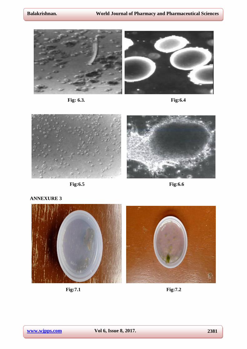
Fig:6.5 Fig:6.6
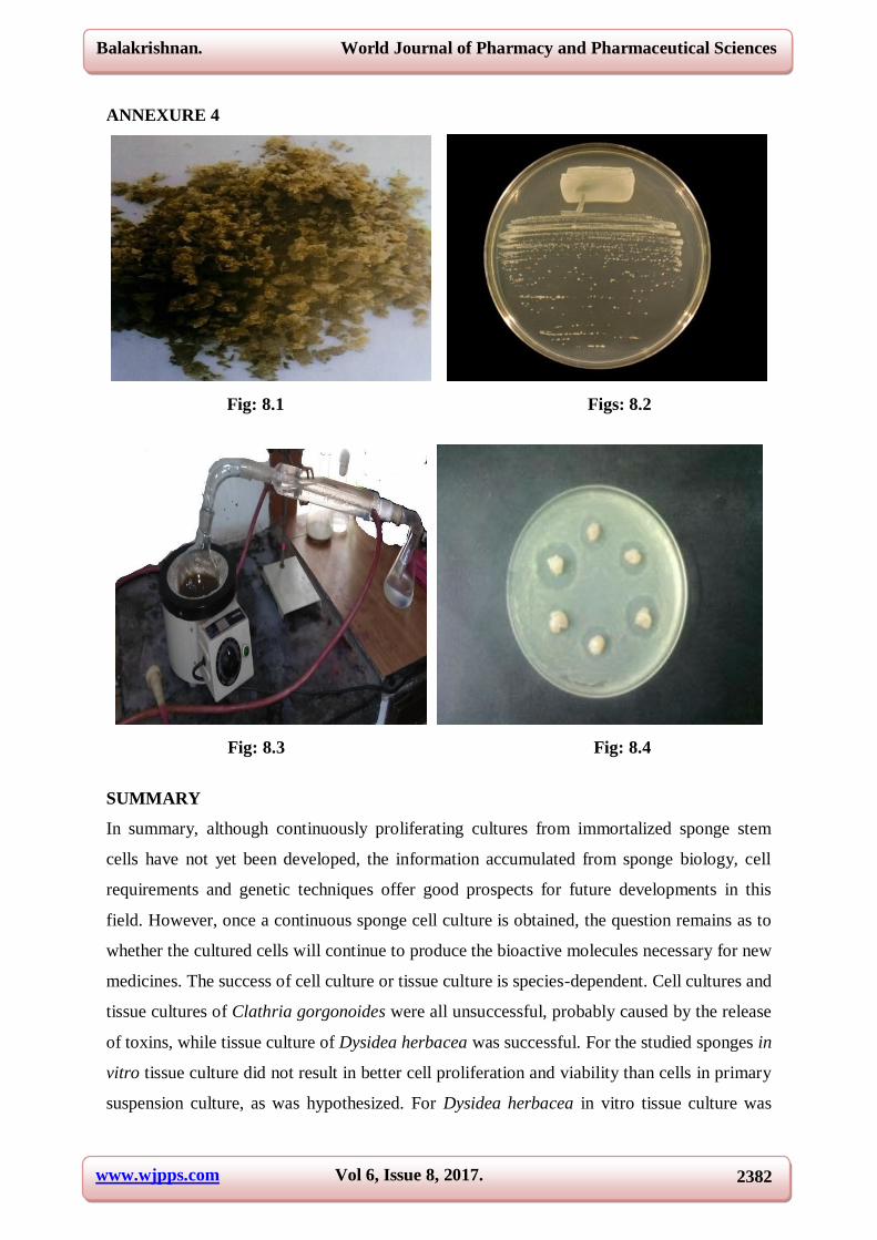
ANNEXURE 3
Fig:7.1 Fig:7.2
www.wjpps.com Vol 6, Issue 8, 2017.
2382
Balakrishnan. World Journal of Pharmacy and Pharmaceutical Sciences
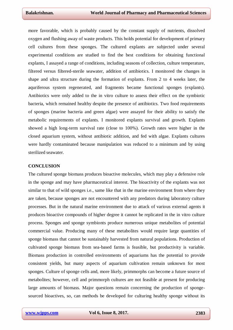
ANNEXURE 4
Fig: 8.1 Figs: 8.2
Fig: 8.3 Fig: 8.4
SUMMARY
In summary, although continuously proliferating cultures from immortalized sponge stem
cells have not yet been developed, the information accumulated from sponge biology, cell
requirements and genetic techniques offer good prospects for future developments in this
field. However, once a continuous sponge cell culture is obtained, the question remains as to
whether the cultured cells will continue to produce the bioactive molecules necessary for new
medicines. The success of cell culture or tissue culture is species-dependent. Cell cultures and
tissue cultures of Clathria gorgonoides were all unsuccessful, probably caused by the release
of toxins, while tissue culture of Dysidea herbacea was successful. For the studied sponges in
vitro tissue culture did not result in better cell proliferation and viability than cells in primary
suspension culture, as was hypothesized. For Dysidea herbacea in vitro tissue culture was
www.wjpps.com Vol 6, Issue 8, 2017.
2383
Balakrishnan. World Journal of Pharmacy and Pharmaceutical Sciences
more favorable, which is probably caused by the constant supply of nutrients, dissolved
oxygen and flushing away of waste products. This holds potential for development of primary
cell cultures from these sponges. The cultured explants are subjected under several
experimental conditions are studied to find the best conditions for obtaining functional
explants, I assayed a range of conditions, including seasons of collection, culture temperature,
filtered versus filtered-sterile seawater, addition of antibiotics. I monitored the changes in
shape and ultra structure during the formation of explants. From 2 to 4 weeks later, the
aquriferous system regenerated, and fragments became functional sponges (explants).
Antibiotics were only added to the in vitro culture to assess their effect on the symbiotic
bacteria, which remained healthy despite the presence of antibiotics. Two food requirements
of sponges (marine bacteria and green algae) were assayed for their ability to satisfy the
metabolic requirements of explants. I monitored explants survival and growth. Explants
showed a high long-term survival rate (close to 100%). Growth rates were higher in the
closed aquarium system, without antibiotic addition, and fed with algae. Explants cultures
were hardly contaminated because manipulation was reduced to a minimum and by using
sterilized seawater.
CONCLUSION
The cultured sponge biomass produces bioactive molecules, which may play a defensive role
in the sponge and may have pharmaceutical interest. The bioactivity of the explants was not
similar to that of wild sponges i.e., same like that in the marine environment from where they
are taken, because sponges are not encountered with any predators during laboratory culture
processes. But in the natural marine environment due to attack of various external agents it
produces bioactive compounds of higher degree it cannot be replicated in the in vitro culture
process. Sponges and sponge symbionts produce numerous unique metabolites of potential
commercial value. Producing many of these metabolites would require large quantities of
sponge biomass that cannot be sustainably harvested from natural populations. Production of
cultivated sponge biomass from sea-based farms is feasible, but productivity is variable.
Biomass production in controlled environments of aquariums has the potential to provide
consistent yields, but many aspects of aquarium cultivation remain unknown for most
sponges. Culture of sponge cells and, more likely, primmorphs can become a future source of
metabolites; however, cell and primmorph cultures are not feasible at present for producing
large amounts of biomass. Major questions remain concerning the production of sponge-
sourced bioactives, so, can methods be developed for culturing healthy sponge without its

www.wjpps.com Vol 6, Issue 8, 2017.
2384
Balakrishnan. World Journal of Pharmacy and Pharmaceutical Sciences
endosymbionts? Can endosymbiotic bacteria be cultured in the absence of live sponge tissue
and cells, to produce metabolites of interest? Studies are needed of sponge nutrition and how
nutrition can influence growth and metabolite production. What might be the influence of
precursor feeding? All these and many other questions remain to be answered.
ACKNOWLEDGEMENT
I thank the Lord Almighty for being gracious and merciful unto me. He has been mindful of
me and blessed me with abundance. I stand to testify His unchanging love and compassion
towards, which has filled me with strength to endure. Glory to God, He Lifted me up, I thank
“THE LORD ALMIGHTY” who is the owner of strength and confidence in my life.
Firstly, I would like to express my sincere gratitude to my Guide Prof. Dr.D.Sudarsanam for
the continuous support of my minor Research Project and related studies, for his patience,
motivation, and immense knowledge. His guidance helped me in all the time of research and
writing of this thesis. I could not have imagined having a better advisor and mentor for my
Minor Research Project.
Besides my advisor, I would like to thank the rest of Professors, scholars and friends who
helped me in completion of the project: Prof.Dr.S.John William, Prof.Dr.JMV.Kalaiarasi,
Head of the department, Dr.Albin T. Flemming, Prof.Dr.John Milton, Prof.Dr.M.Raja, and
Prof.Dr.P.Agustian for their constant support and encouragement to finish this project and
Ms.Vasantha supriya, Ms.Maria, Mr. Joel, Mr.Gowrisankar for their insightful comments
and encouragement, but also for the hard question which incented me to widen my research
from various perspectives and my friends V.Anthony Ponnaya and S.Frederick for studious
help to complete my project in the best way.
My sincere thanks to Rev.Dr.Fr.M.Arockiasamy Xavier, S.J, Principal- Loyola College, Prof.
Dr. S.Vincent- Dean of Research and Rev.Fr. Dr.S.Ignacimuthu, S.J. Director, Entomology
Research Institute, Loyola College who provided me an opportunity to perform a Minor
Research Project, and who gave access to the laboratory and research facilities respectively.
Dr.G.Sivaleela, Scientist, Zoological Survey of India, for her help in Identifying the Sponge
Specimen collected from Pamban coast of Rameswaram, Prof. T. Yuvarani, Department of
Biotechnology, D.G.Vaishnav College for her idea about the sponges and culturing
techniques. Without their precious support it would not be possible to conduct this research.
www.wjpps.com Vol 6, Issue 8, 2017.
2385
Balakrishnan. World Journal of Pharmacy and Pharmaceutical Sciences
Last but not the least; I would like to thank my Parents for their continuous support and love.
REFERENCES
1. Abdelmohsen, U. R., Pimentel-Elardo, S. M., Hanora, A., Radwan, M., Abou- El-Ela, S.
H., Ahmed, S., and Hentschel, U. Isolation, phylogenetic analysis and anti-infective
activity screening of marine sponge-associated actinomycetes. Marine Drugs, 2010; 8:
399–412.
2. Achtman, M., and Wagner, M. Microbial diversity and the genetic nature of microbial
species. Nature Reviews Microbiology, 2008; 6: 431–440.
3. Andrade, P., Willoughby, R., Pomponi, S. A., and Kerr, R. G. Biosynthetic studies of the
alkaloid, stevensine, in a cell culture of the marine sponge Teichaxinella morchella.
Tetrahedron Letters, 1999; 40: 4775–4778.
4. Battershill, C. N., and Page, M. J. Sponge aquaculture for drug production. Aquaculture
Update, 1996; 16: 5–6.
5. Bayer, K., Schmitt, S., and Hentschel, U. Physiology, phylogeny and in situ evidence for
bacterial and archaeal nitrifiers in the marine sponge Aplysina aerophoba. Environmental
Microbiology, 2008; 10: 2942–2955.
6. Becerro, M. A., Turon, X., and Uriz, M. J. Multiple functions for secondary metabolites
in encrusting marine invertebrates. Journal of Chemical Ecology, 1997; 23: 1527–1547.
7. Belarbi, E. H., Contreras Gomez, A., Chisti, Y., Garcia Camacho, F., and Molina Grima,
E. Producing drugs from marine sponges. Biotechnology Advances, 2003; 21: 585–598.
8. Bergman, O., Haber, M., Mayzel, B., Anderson, M., Shpigel, M., Hill, R., and Ilan, M.
Marine-based cultivation of Diacarnus sponges and the bacterial community composition
of wild and maricultured sponges and their larvae. Marine Biotechnology, 2011.
9. Carballo, J. L., Yanez, B., Zubia, E., Ortega, M. J., and Vega, C. Culture of explants from
the sponge Mycale cecilia to obtain bioactive mycalazal-type metabolites. Marine
Biotechnology, 2010; 12: 516–525.
10. Castresana, J. Selection of conserved blocks from multiple alignments for their use in
phylogenetic analysis. Molecular Biology and Evolution, 2000; 17: 540– 552.
11. Chernogor, L., Denikina, N., Belikov, S., and Ereskovsky, A. Long- term cultivation of
primmorphs from freshwater Baikal sponges; Lubomirskia baikalensis. Marine
Biotechnology, 2011; 1–11.
12. De Rosa, S., De Caro, S., Iodice, C., Tommonaro, G., Stefanov, K., and Popov, S.
Development in primary cell culture of demosponges. Journal of Biotechnology, 2003;

www.wjpps.com Vol 6, Issue 8, 2017.
2386
Balakrishnan. World Journal of Pharmacy and Pharmaceutical Sciences
100: 119–125.
13. Duckworth, A. R., and Pomponi, S. A. (2005). Relative importance of bacteria,
microalgae and yeast for growth of the sponge Halichondria melanadocia (De
Laubenfels, A laboratory study. Journal of Experimental Marine Biology and Ecology,
1936; 323: 151–159.
14. Duckworth, A. R., Samples, G. A., Wright, A. E., and Pomponi, S. A. In vitro culture of
the tropical sponge Axinella corrugata (Demospongia): Effect of food cell concentration
on growth, clearance rate and biosynthesis of stevensine. Marine Biotechnology, 2003; 5:
519–527.
15. Funayama, N. The stem cell system in demosponges: Insights into the origin of somatic
stem cells. Development, Growth and Differentiation, 2010; 521–14.
16. Funayama, N., Nakatsukasa, M., Hayashi, T., and Agata, K. Isolation of the choanocyte
in the fresh water sponge, Ephydatia fluviatilis and its lineage marker, Ef annexin.
Development, Growth and Differentiation, 2005; 47: 243–253.
17. Funayama, N., Nakatsukasa, M., Mohri, K., Masuda, Y., and Agata, K. Piwi expression
in archeocytes and choanocytes in demosponges: Insights into the stem cell system in
demosponges. Evolution and Development, 2010; 12: 275–287.
18. Garcia Camacho, F., Chileh, T., Ceron Garcia, M. C., Sanchez Miron, A., Belarbi, E. H.,
Chisti, Y., and Molina Grima, E. A bioreaction-diffusion model for growth of marine
sponge explants in bioreactors. Applied Microbiology and Biotechnology, 2006a; 73:
525–532.
19. Garcia Camacho, F., Chileh, T., Ceron Garcı´a, M. C., Sanchez Miro´n, A., Belarbi, E.
H., Contreras Go´mez, A., and Molina Grima, E. Sustained growth of explants from
Mediterranean sponge Crambe crambe cultured in vitro with enriched RPMI 1640.
Biotechnology Progress, 2006b; 22: 781–790.
20. Gerce, B., Schwartz, T., Voigt, M., Ruhle, S., Kirchen, S., Putz, A., Proksch, P., Obst, U.,
Syldatk, C., and Hausmann, R. Morphological, bacterial, and secondary etabolite changes
of Aplysina aerophoba upon long-term maintenance under artificial conditions. Microbial
Ecology, 2009; 58: 865–878.
21. Holmes, B., and Blanch, H. Genus-specific associations of marine sponges with group I
crenarchaeotes. Marine Biology, 2007; 150: 759–772.
22. Holmes, B., and Blanch, H. Possible taxonomic trends in the success of primary
aggregate formation in marine sponge cell cultures. Marine Biotechnology, 2008; 10:
99–109.

www.wjpps.com Vol 6, Issue 8, 2017.
2387
Balakrishnan. World Journal of Pharmacy and Pharmaceutical Sciences
23. Holmes, B. M. Marine Sponges: Systematics, Symbiosis and Cell Culture. UC Berkeley,
Berkeley, USA, 2006.
24. Klautau, M., Custodio, M. R., and Borojevic, R. Cell cultures of sponges Clathrina and
Polymastia. In Vitro Cellular and Developmental Biology, 1993; 29A: 97–99.
25. Klautau, M., Custodio, M. R., and Borojevic, R. In vitro culture of primary cell lines from
marine sponges. In “Sponges in Time and Space” (R. Van Soest, T. M. G. Van Kempen
and J. C. Braekman, eds), pp. 401–406. Rotterdam, Balkema, 1994.
26. Klo¨ppel, A., Pfannkuchen, M., Putz, A., Proksch, P., and Bru¨mmer, F. Ex situ
cultivation of Aplysina aerophoba close to in situ conditions: Ecological, biochemical and
histological aspects. Marine Ecology, 2008; 29: 259–272.
27. Molinski, T. F., Dalisay, D. S., Lievens, S. L., and Saludes, J. P. Drug development from
marine natural products. Nature Reviews, 2009; 8: 69–85.
28. Morrison-Gardiner, S. Dominant fungi from Australian coral reefs. Fungal Diversity,
2002; 9: 105–121.
29. Muller, W. E. G., Bohm, M., Batel, R., De Rosa, S., Tommonaro, G., Muller, I. M., and
Schroder, H. C. Application of cell culture for the production of bioactive compounds
from sponges: Synthesis of avarol by primmorphs from Dysidea avara. Journal of
Natural Products, 2000; 63: 1077–1081.
30. Pomponi, S. A. The oceans and human health: The discovery and development of marine-
derived drugs. Oceanography, 2001; 14: 28–42.
31. Pomponi, S. A. Biology of the Porifera: Cell culture. Canadian Journal of Zoology, 2006;
84: 167–174.
32. Pomponi, S. A., and Willoughby, R. Sponge cell culture for the production of bioactive
metabolites. In “Sponges in Time and Space” (R. Van Soest, T. M. G. Van Kempen and J.
C. Braekman, eds). Balkema, Rotterdam, 1994.
33. Pomponi, S. A., and Willoughby, R. Development of sponge cell cultures for biomedical
applications. In “Aquatic Invertebrate Cell Culture” (B. Austin and C. Mothersill, eds).
Springer, London, 2000.
34. Rinkevich, B. Cell cultures from marine invertebrates: New insights for capturing endless
stemness. Marine Biotechnology, 2011; 13: 345–354.
35. Rinkevich, B., Blisko, R., and Ilan, M. Further steps in the initiation of cell cultures from
embryos and adult sponge colonies. In Vitro Cellular and Developmental Biology Animal,
1998; 34: 753–756.
36. Schippers, K. J., Martens, D. E., Pomponi, S. A., and Wijffels, R. H. Cell cycle analysis

www.wjpps.com Vol 6, Issue 8, 2017.
2388
Balakrishnan. World Journal of Pharmacy and Pharmaceutical Sciences
of primary sponge cell cultures. In Vitro Cellular and Developmental Biology Animal,
2011; 47: 302–31.
37. Wilson, H. V. On some phenomena of coalescence and regeneration in sponges. Journal
of Experimental Zoology, 1907; 5: 245–258.
38. Zhang, X. Y., Le Pennec, G., Steffen, R., Muller, W. E. G., and Zhang, W. Application of
a MTT assay for screening nutritional factors in growth media of primary sponge cell
culture. Biotechnology Progress, 2004; 20: 151–155.
39. Zhao, Q. Y., Zhang, W., Jin, M. F., Yu, X. J., and Deng, M. C. Formulation of a basal
medium for primary cell culture of the marine sponge Hymeniacidon perleve.
Biotechnology Progress, 2005; 21: 1008–1012.
40. Zientz, E., Dandekar, T., and Gross, R. Metabolic interdependence of obligate
intracellular bacteria and their insect hosts. Microbiology and Molecular Biology
Reviews, 2004; 68: 745–770.