cultured limbal stem therapy for corneal chemical injury
TRANSCRIPT

2/12/2020
1
Cultured limbal stem therapy for Corneal chemical injury
Limbal cell deficiency causes
May occur as a result of depletion of stem cells ordestruction of their stromal niche such as in:
Congenital: AniridiaIdiopathic conditionsChemical/thermal burnIatrogenic: surgery or contact lens useAutoimmune: Stevens Johnson syndrome and OCP

2/12/2020
2
Introduction
Conditions with limbal cell deficiency represent a MAJOR CLINICAL CHALLANGE.
Conventional corneal transplants alone are destined to fail , due to absence of epithelial cells progenes
Introduction
Limbal stem cells gained clinical interest since their location was exactly dicovered and described
Limbal Stem cell therapy (LSCT), weather Auto or Allograft , is an old technique for treatmeant of limbal cell deficency diseases (LSCD)
Pellegrini et al in 1997 first described transplantation of ex vivo expanded limbal epithelial stem cells to restore corneal epithelial surface in patients with LSCD
Now its considered as an advanced therapy medical product (ATMP)

2/12/2020
3
Design of study In this pilot study we have chosen to deal with one etiology
of LSCD, which is chemical burns.
Five patients were selected for this study , all of them have previous occular chemical burns. Other new patients were added consecutively.
Study started 2014
In all affected eyes the limbus and cornea was totally damaged with conjunctivalization.
Vision was hand movement in these eyes.
The surgical procedure was started after receiving the usual medical treatment for chemical burns for 6 months .

2/12/2020
4
LSC culture procedure:
• Fresh or frozen amniotic is prepared from Eye bank.
• Denuded by trypsin/ EDTA
Amniotic membrane
• Limbal biopsy obtained from healthy contra lateral eye or cadaveric limbal rim
Limbal Explants
• HCE medium
• DMEM/F12
• FCS 10%
• EGF
• Cholera toxin
• Insulin
Culture for 14 days
Feeder-free Limbal Culture Procedure
Fresh frozen amniotic membrane (AM) 5x5 cm ispurchased from Eye Bank. AM was screened forinfectious diseases e.g. HCV, HBV and HIV.
AM is denuded by thermolysin enzme (125 µg/mL) inphosphate buffered saline (D-PBS).
AM was stretched and limbal explants were culturedover epithelial side.
Culture was submerged in growth medium for 10-14days in CO2 incubator at 37 ˚C, 98% humidity and 5%CO2.

2/12/2020
5
Preparing the implant(Cell factory)
Auto explants 2x1 mm were excised from other eye limbus , transferred to the cell culture lab in a sterile tube carrying collection medium.
Allo grafts are transferred in the same way from fresh excised limbal corneal button rings.
These grafts would be implanted on denuded amniotic membranes in culture plates with selective growth media for corneal limbal cells (cell factory)
SerumMedium
Growth factors
• Human Corneal Epithelial (HCE medium)
• DMEM/F12
• Fetal calf serum (FCS) 10% or autologous
• Epidermal growth factor (EGF) 10 ng / ml
• Cholera toxin 100 nm/ml
• Insulin 5µg / ml
2/12/2020
6
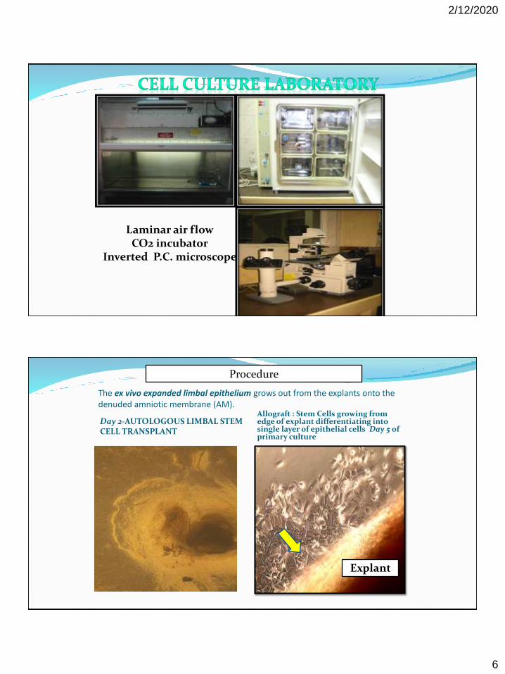
Laminar air flowCO2 incubator
Inverted P.C. microscope
The ex vivo expanded limbal epithelium grows out from the explants onto the denuded amniotic membrane (AM).
Allograft : Stem Cells growing from edge of explant differentiating into single layer of epithelial cells Day 5 of primary culture
Explant
Day 2-AUTOLOGOUS LIMBAL STEM CELL TRANSPLANT
Procedure

2/12/2020
7
Higher magnification STEM CELLS
New epithelium
AMNIOTIC memb
Day 2 growth Day 10
DAY 12 AFTER PRIMARY EXPLANT CULTURE

2/12/2020
8
Explant
Epithelium
Epithelium
40x 100x
When the AM is sufficiently covered by expanded limbal epithelial cells,within 14 days, it is transplanted to the eye affected by LSCD.
Auto & Allograft (cadaveric rim) explant culture showing confluent multilayered epithelium at Day 14 of culture ,corneal phenotype was proven by RT-PCR using P 63 & CK3/12(moclecular test)
Surgical steps Conjuntiva is dissected through a 360 degree periotomy,
peeled from underlying cornea
The cultured amniotic membrane is transferred with cell facing up and sutured to corneal limbus with 8/0 nylon
Cultered amniotic membrane was left to stablize and enrich the corneal surface with cultered limbal cells
Pkp with a secondry cultured amniotic membrane was done to restore vision ,after 6 months
Patients who had Allografts ,received cyclosporin 1% eye drops 3 times daily
2/12/2020
9
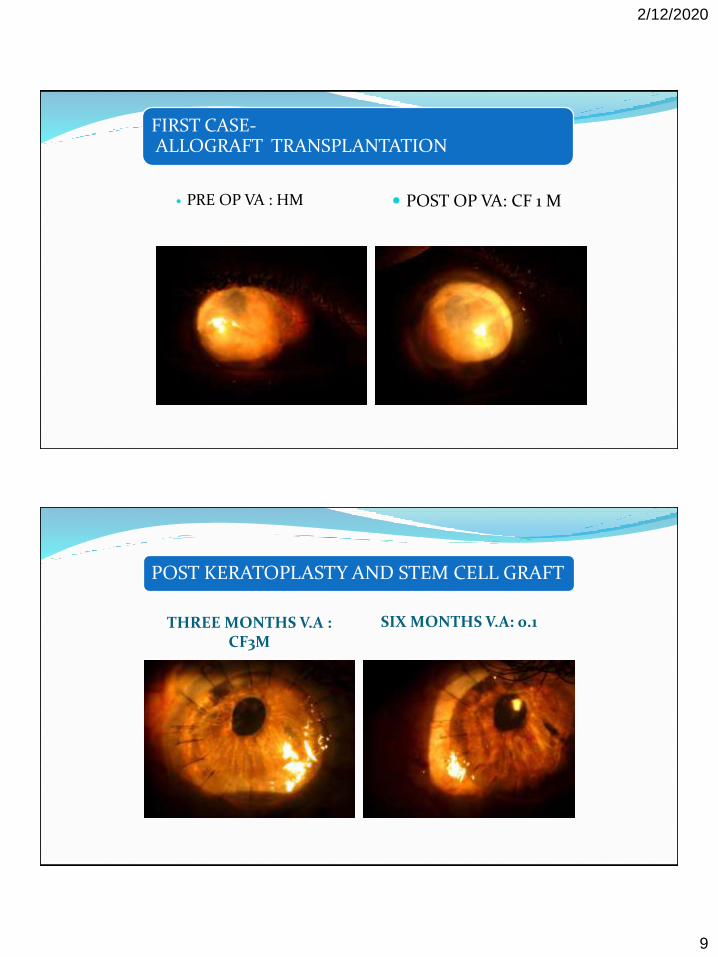
FIRST CASE-ALLOGRAFT TRANSPLANTATION
PRE OP VA : HM POST OP VA: CF 1 M
POST KERATOPLASTY AND STEM CELL GRAFT
THREE MONTHS V.A : CF3M
SIX MONTHS V.A: 0.1

2/12/2020
10
H&E200X
H&E100X
CK 19-VE
PAS-VE
2/12/2020
11
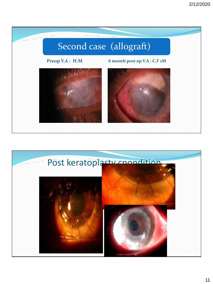
Second case (allograft)
Preop V.A : H.M 6 month post op V.A : C.F 2M
Post keratoplasty cnondition
2/12/2020
12
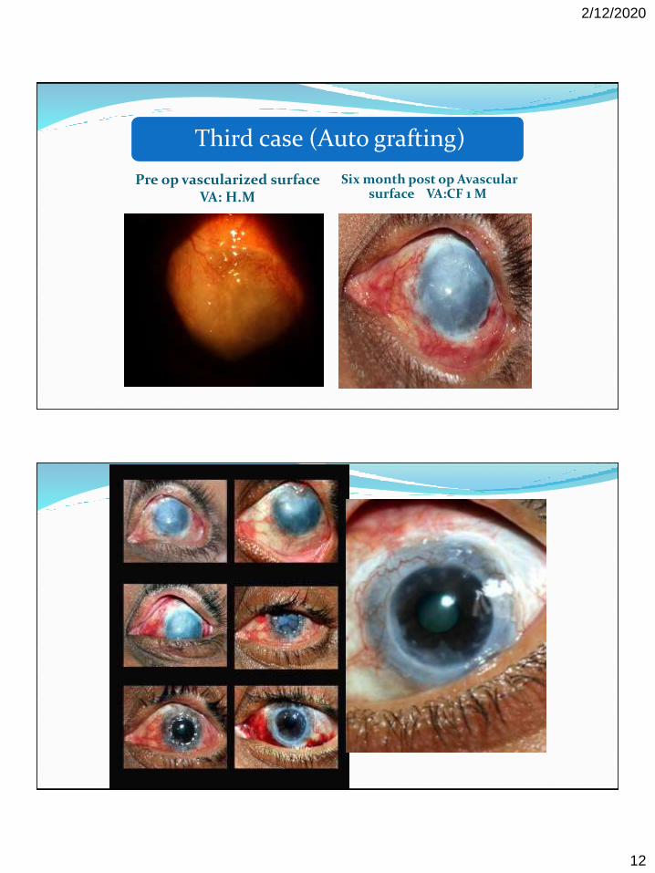
Third case (Auto grafting)
Pre op vascularized surface VA: H.M
Six month post op Avascular surface VA:CF 1 M

2/12/2020
13
Fourth case(Allograft)
Fifth patientAuto graft failure
2/12/2020
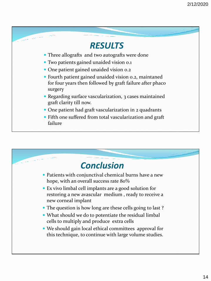
14
RESULTS Three allografts and two autografts were done
Two patients gained unaided vision 0.1
One patient gained unaided vision 0.2
Fourth patient gained unaided vision 0.2, maintaned for four years then followed by graft failure after phaco surgery
Regarding surface vascularization, 3 cases maintained graft clarity till now.
One patient had graft vascularization in 2 quadrants
Fifth one suffered from total vascularization and graft failure
Conclusion Patients with conjunctival chemical burns have a new
hope, with an overall success rate 80%
Ex vivo limbal cell implants are a good solution for restoring a new avascular medium , ready to receive a new corneal implant
The question is how long are these cells going to last ?
What should we do to potentiate the residual limbal cells to multiply and produce extra cells
We should gain local ethical committees approval for this technique, to continue with large volume studies.

2/12/2020
15
THANK YOU
1- Holoclones : Diameter of 6-10 µm. These cells have a high proliferating capability with ≤5% aborted colonies and ≥100 cell doublings;2- Meroclones : Young TA cells with intermediate proliferating capacity having a diameter of 10-18 µm. These cells usually have 5-95% aborted colonies; 3- Paraclones TD cells with 15-20 cell doublings and very low proliferative capability. These cells are 18-36 µm long in diameter.
1 2 3

2/12/2020
16
Allograft : Stem Cells growing from edge of explant differentiating into single layer of epithelial cells Day 5 of primary culture