ct of osteomyelitis of the spine - american journal of ... of the spine may be a difficult...
TRANSCRIPT

Cornelia Golimbu' Hossein Firooznia
Mahvash Rafii
This article appears in the November / December 1983 issue of AJNR and the January 1984 issue of AJR .
Received December 15, 1983; accepted after revision June 15, 1983.
"AII authors: Department of Radiology, New York University Medical Center, 560 First Ave., New York, NY 10016. Address reprint requests to C. Golimbu.
AJNR 4:1207-1211 , Nov/ Dec 1983 0 195- 6108 / 83 / 0406-1207 $00.00 © American Roentgen Ray Society
CT of Osteomyelitis of the Spine
1207
Computed tomography (CT) were performed in 17 adults with osteomyelitis of the spine. The dominant features were paravertebral soft-tissue swelling , abscess formation , and bone erosion. In two patients there were no findings indicative of osteomyelitis on conventional radiographs, but CT revealed paravertebral abscesses and bone lysis, helping to establish the diagnosis of osteomyelitis. CT was found helpful in the evaluation of the patients suspected of spinal osteomyelitis, chiefly because of its ability to detect early erosion of spongy vertebral bone, disk involvement, paravertebral soft-tissue swelling or abscess, and extension of the pathology into the spinal canal. Furthermore, CT facilitated closed-needle biopsy, helping to establish the pathologic diagnosis.
Osteomyelitis of the sp ine may be a difficult diagnostic problem. The c linical manifestations are often nonspecific, simply fever of unknown origin or septicem ia, and there may be no localizing signs. The definitive diagnosis may be delayed for a number of weeks until characterist ic erosive bone changes become evident [1-6]. Conventional radiography may fail to reveal the initial vertebral involvement. In our experience, early detection of erosive changes of the spongy vertebral bone is usually not possible by conventional rad iography. Computed tomography (CT), on the other hand, displ ays the anatomy of the sp ine in an ax ial plane , thus overcoming the problem of overlapping of bone surfaces, and in some instances may reveal otherwise undetectable bone erosion . CT is particularly su ited for study of the paravertebral abscesses [7] and sp inal canal abnormalities [8, 9]. We report our experience with 17 patients with osteomyeliti s evaluated by CT during a 4 year period.
Materials and Methods
Seventeen adults (12 men and five women) were studied ; th ey were 29-79 years old (mean , 54 years). Eight had tuberculosis and nine had pyogenic infec tion s of th e spine. Seven were habitu al intravenous heroin users. One patient had urinary trac t infec tion preceding the clinica l onset of osteomyeliti s. Another was on long-term cort icosteroid treatment for rheumatoid arthriti s. One patient was in a state of immunosuppression. Three patients were referred for CT examination because of fever of unknown origin . The 14 patients with c linica lly diagnosed osteomyelitis were investigated by CT to determine th e extent of involvement of the soft tissues and spinal canal. The site of involvement was found to be in th e lumbar spine in nine patients, lumbosacral in one, th oracic in six, and cervica l
in one. The patients were studied on GE 8800 and EMI 6000 scanners. The area of interest was
scanned with sequenti al 5-mm-thick slices. Four pat ients had foll ow-up CT examinations. Frontal and lateral conventional rad iographs were available in all patients. Conventional tomography was performed in fi ve patients.

1208 GOLIMBU ET AL. AJNR :4 , Nov./Dec. 1983
A 8
Fig. 1.-Case 1. Staphytococca l osteomyelitis L3 -L4 . S I P aorti c graft surgery. Anteroposterior (AP) (A) and lateral (8) views of lumbar spine. Loss of L3-L4 disk space and destruction of adjacent vertebral end-plates. Clips in abdomen from previous graft surgery. C , CT of inferior aspect of L3 after
Results
In all patients, CT demonstrated paravertebral soft-t issue swelling . This was marked and was assoc iated with psoas abscesses containing low-density necrotic locu lations in four patients with tuberculosis and in one with pyogenic infection. None of these abscesses were detected by conventional radiographic methods. In one patient with pyogen ic infection, CT demonstrated gas bubbles in a presacral abscess .
The extent of the trabecular bone destruction seen on CT was invariably greater than that estimated by conventional radiography . In two of the 17 patients there were no signs of osteomyeliti s on conventional films , but CT revealed erosion of the cancellous bone of the vertebral bodies and loss of the cortical margin of the end-plates. In one patient with thoracic spine osteomyeliti s CT revealed distinct destruction of the anterior cortex of the rib near the costovertebral joint, a finding not detected with other methods.
The narrowing of the disk space was at times difficult to notice on primary axial CT images. Reference to the lateral scout images and, in se lected cases, sagittal and coronal reconstruction made this information easily noticeable.
CT demonstrated spinal canal involvement in two patients with posterior extension of the inflammatory mass and detritus of bone originating in an infected disk space. In another patient extension of the paraspinal abscess into the neural foramen was observed .
Follow-up CT in four patients demonstrated emergence
c oral and intravenous contrast. Destru cti on of ver tebra l body L3 with extension of bone detritus in an terior aspect of spi nal canal. Soft-tissue mass containing ca lcificat ions in paravertebral space. Clear fat plane separates paravertebral soft-tissue swelling from aorti c graft.
of reactive osteosc lerosis, calcification in the paraspinal abscesses , and a decrease in the soft-tissue swel ling. The following three cases illustrate the importance of CT in the diagnosis and management of these patients .
Representative Case Reports
Case 1
A 65-year-old man with previous graft replacement of an abdominal aortic aneurysm was admitted for intermittent fever of several weeks ' duration and diffuse back pain. Repeated blood cu ltu res were positive for Staphylococcus aureus. The initial diagnosis was septicemia, for which he received a variety of ant ibiotics. The continuous seeding of the bloodstream and lack of response to antibiotics were attributed to localization of the infection in the abdominal aortic graft. Physical examinat ion was unremarkable, except for lumbar spine tenderness.
Radiographs of the lumbar spine demonstrated disk space loss at L3-L4 with destruction of the adjacent vertebral end-plates (figs. 1 A and 18). Abdominal CT demonstrated a normal aspect of the aortic graft. There was a paravertebral abscess containing calcific as well as low-density areas extending into the psoas muscle. A c lear fat plane was seen separat ing the psoas abscess from the posterior wall of the aortic graft. Marked destruction of the vertebral body L3 was seen on CT, with extension of the bone detritus posteriorly into the spinal canal (fig. 1 C).
Comment: CT was the optimal method to disclose the separation between the paravertebral abscess and the aortic graft. It added the information of posterior extension of the vertebral body destruction , with involvement of the spinal canal.
AJNR:4, Nov./ Dec. 1983 CT OF SPINAL OSTEOMYELITIS 1209
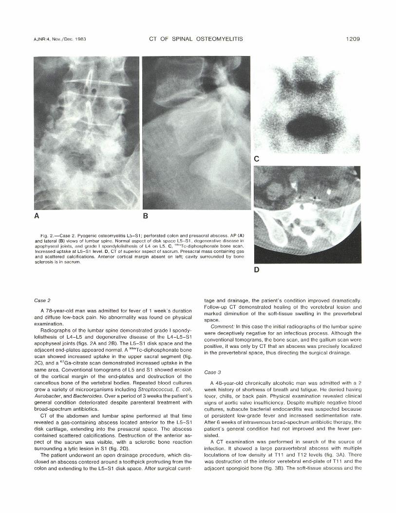
Fig . 2. -Case 2. Pyogenic osteomyelitis L5-S1 ; perforated colon and presacral abscess. AP (A) and lateral (8) views of lumbar spine. Normal aspect of disk space L5-S1, degenerat ive disease in apophyseal jo ints, and grade I spondylolisthesis of L4 on L5. C , 99mTc-d iphosphonate bone scan. Increased uptake at L5-S1 level. D , CT of superior aspect of sacrum. Presacral mass containing gas and scattered calc ifications. Anterior cortical margin absent on left ; cavity surrounded by bone sclerosis is in sacrum.
Case 2
A 78-year-old man was admitted for fever of 1 week 's duration and diffuse low-back pain. No abnormality was found on physical examination.
Radiographs of the lumbar spine demonstrated grade I spondylolisthesis of L4-L5 and degenerative disease of the L4-L5-S1 apophyseal joints (figs . 2A and 28). The L5-S1 disk space and the adjacent end-plates appeared normal. A 99mTc-d iphosphonate bone scan showed increased uptake in the upper sacral segment (fig . 2C), and a 67Ga-citrate scan demonstrated increased uptake in the
same area . Convent ional tomograms of L5 and S1 showed erosion of the cortical margin of the end-plates and destruction of the cancellous bone of the vertebral bodies. Repeated blood cultures grew a variety of microorganisms including Streptococcus, E. coli , Aerobacter, and Bacteroides. Over a period of 3 weeks the patient 's general condition deteriorated despite parenteral treatment with broad-spectrum antibiotics.
CT of the abdomen and lumbar spine performed at that time revealed a gas-containing abscess located anterior to the L5-S1 disk carti lage, extending into the presacral space . The abscess contained scattered calc ifications. Destruction of the anterior aspect of the sacrum was visib le, with a sc lerot ic bone reaction surrounding a lytic lesion in S1 (fig. 20).
The patient underwent an open drainage procedure, which disclosed an abscess centered around a toothpick protrud ing from the colon and extend ing to the L5-S1 disk space. After surgical curet-
D
tage and drainage, the patient 's condition improved dramatically. Follow-up CT demonstrated healing of the vere tebral lesion and marked diminution of the soft-tissue swelling in the prevertebral space.
Comment: In this case the initial rad iograph s of th e lumbar spine were deceptively negative for an infec tious process. Although the conventional tomograms, the bon e scan, and the gallium scan were positive, it was on ly by CT that an abscess was precisely localized in the prevertebral space, thus directing th e surgical drainage.
Case 3
A 48-year-old chronica lly alcoholi c man was ad mitted with a 2 week history of shortness of breath and fatigue. He denied having fever, chills, or back pain. Physical examination revealed c linica l signs of aorti c valve insuffic iency. Despite multiple negative blood cu ltures , subacute bacterial endocard itis was suspected because of persi stent low-grade fever and increased sed imen tation rate. After 6 weeks of intravenous broad-spectrum antibiotic therapy , the patient 's general cond ition had not improved and the fever per
sisted. A CT examination was performed in search of the source of
infection. It showed a large paravertebral abscess with multiple loculations of low density at T11 and T1 2 level s (fig. 3 A) . There was destruction of the inferior veretebral end-plate of T11 and the
adjacent spongioid bone (fig . 38). The soft-tissue abscess and the

1210 GOLIMBU ET AL. AJNR:4, Nov, l Dec , 1983
A B
o
bone destruction extended into the right neural foramen (fig , 3C) , Marked reacti ve sclerosis of bone was seen in the T1 2 vertebral body (fig, 3A) , It was only at this point that conventional radiog raphic studies of the thoracolumbar spine were made, Th ey revealed a paraveretebral soft-ti ssue mass, narrowing of the disk space T11 -T1 2, and reacti ve bone production of T1 2 (figs, 3 D and 3E) , Needle aspirate of the paravertebral abscess grew Mycobac terium tuberculosis,
Discussion
Osteomyelitis has increased in frequency in the last 2 decades, This has been attributed to a number of factors , includ ing the rise in drug addiction [1 , 10, 11], use of intravenous indwell ing catheters [1] , and more aggressive urologic instrumentations in pati ents with urinary sepsis [3 , 4]. In such instances , inte rmittent bacteremia presumably leads to hematogenous spread of the infection to the spine, with the initial focus of infection involving the spongy verte-
c
Fig , 3 ,-Case 3, Tuberculous osteomye liti s T11 and T1 2, A , CT at T1 2 pedicles level. Paravertebral low-density multiple loculations, T1 2 vertebral body is sclerotic, B, CT at inferior aspect of T11 , Destruc tion of inferior vertebral end-plate and paravertebral loculated low-density co llection , C, CT at level of T11 , Soft-tissue swelling and bone lysis extending into right neural foramen (arrow ), AP (D) and lateral (E) views of lower thoracic spine show paravertebral soft-tissue mass, reactive sclerosis of T1 2, and minimal narrowing of disk space T11-T1 2,
bral bone directly beneath the vertebral end-plate. This is probably due to the rich blood supply of this region [12]. Subsequently, the infection destroys the vertebral end-plate and invades the disk cartilage . Thus, involvement of the disk space may not become noticeable until significant vertebral erosion has occurred .
Extension of the infection from one vertebra to another without involvement of the intervening disk is seen more commonly in tuberculosis [2]. This is the subligamentous form of tuberculosis in which the infection extends beneath the anterior spinal ligament from one vertebral body to the next. The bone destruction in this form of the disease may be minimal, usually limited to the anterior surface of the vertebral bodies. However, paravertebral soft-tissue swelling and abscess formation may be extensive [2].
When osteomyelitis is suspected clinically , bone scanning esmTc-diphosphonate) is useful for screening purposes, and it is positive in most patients [13]. Conventional radiography may reveal erosive changes of the cortex of the vertebral

AJNR:4 , Nov. / Dec. 1983 CT OF SPINAL OSTEOMYELITIS 1211
bodies and end-plates, but often only 2-6 weeks after the onset of the infection. Conventional tomography usually reveals these lesions at an earlier stage. However, erosion of the spongy vertebral bone beneath the vertebral endplates, where infection may start , is often not detected in its early stages by conventional radiography or tomography.
CT, however, by virtue of its ax ial display , may reveal these changes when they are otherwise undetectable, as in two of our cases and in one of Larde et al. [9]. Paraspinal soft-tissue swelling and abscess form ation is usually seen in conventional radiographs of the thoracic region . However, in the lumbar and cervical segments , CT is by far the preferred method for this purpose . Intravenous contrast enhancement may help to accentuate the boundaries between abscess loculations and the adjacent uninvolved tissues. Thus, the true extent of paraspinal abscesses , parti culary a psoas sheath abscess, is optimally demonstrated on CT [7]. This provides valuable information for drainage and constitutes an objective baseline for follow-up of these patients. CT is also helpful for a closed-needle biopsy of the infected disk space or the vertebra by revealing the most suitable site for biopsy as well as the safest and shortest route for needle passage [14 , 15]. CT is also the method of choice for detection of involvement of the posterior arch of the vertebrae and extension of the infection into the spinal canal [9].
REFERENCES
1. Muscher OM , Thorsteinsson SB, Minuth IN , Luc hi RJ . Vertebral osteomyelitis. Arch Intern Med 1976;13 6 : 1 05-11 0
2. Chapman M, Murray R, Stoker D. Tuberculosis of the bones and joints. Semin Roentgenol 1979;14 : 266-282
3. Digby J, Kersley J . Pyogenic non-tuberculous spinal infect ion. J Bone Joint Surg [Br] 1979;61 :4 7 - 55
4 . Griffith HED, Jones OM . Pyogenic infection of the spine. J Bone Joint Surg [Br] 1971 ;53: 383- 391
5. King OM , Mayo KM . Infec ti ve lesions of the vertebral co lumn. Clin Orthop 1973;96: 248 - 253
6. Ross PM , Fleming JL. Vertebral body osteomye liti s: spectru m and natural history. Clin Orthop 1976; 11 8: 190-1 98
7. Jeffrey RB , Callen PW, Federle MP. Computed tomography of psoas abscesses. J Comput Assist Tomogr 1980;4: 639-64 1
8. Lee BCP, Kazam E, Newman AD. Computed tomography of the spine and spinal cord . Radiology 1978; 128: 95-102
9. Larde E, Mathieu 0 , Frija J , Gaston A, Vasile A . Verteb ral osteomyelitis: disk hypodensity on CT. AJNR 1982;3: 657-661 , AJR 1982 ;139 :963- 967
10. Firooznia H, Seliger G, Abrams R, Valensi V, Shamoun J. Disseminated extrapulmonary tuberculosis in assoc iation with heroin addiction. Radiology 1973;109: 291-296
11. Holzman RS, Bishko F. Osteomyeliti s in heroin add ic ts. Ann Intern Med 1971 ;75: 693-696
12. Wiley AM , Trueta J. Th e vascular anatomy of the spine and its relationship to pyogenic vertebral osteomyelitis . J Bone Joint Surg [Br] 1959;4 1 : 796-8 00
13. Handmaker J , Leonards R. The bone scan in inflammatory osseou s disease. Semin Nucl Med 1976; 1 : 95-1 05
14. Adapon BD, Legada BD, Lim EVA, Silao J, Dalmacio-Cruz A. CT guided c losed biopsy of the spine. J Comput Assist Tomogr 1981 ;5 : 73 -78
15. Hardy DC, Murphy WA, Gilula LA . Computed tomography in planning percutaneous bone biopsy. Radiology 1980; 134 :447-450