ct anatomy of para nasal sinuses
DESCRIPTION
CT ANATOMY OF PARA NASAL SINUSES Normal anatomy & anatomical variations of PNS in CT are discussed in detail...TRANSCRIPT
CT PNSBy
Dr.K.PRASANNARadiology Resident
RMMCH
CONTENTS
NOSE AND NASAL FOSSA
PARA NASAL SINUSES
OSTEOMEATAL COMPLEX
ANATOMICAL VARIATIONS
IMAGING MODALITIES
CT PROCEDURE & SECTIONS
CONCLUSION
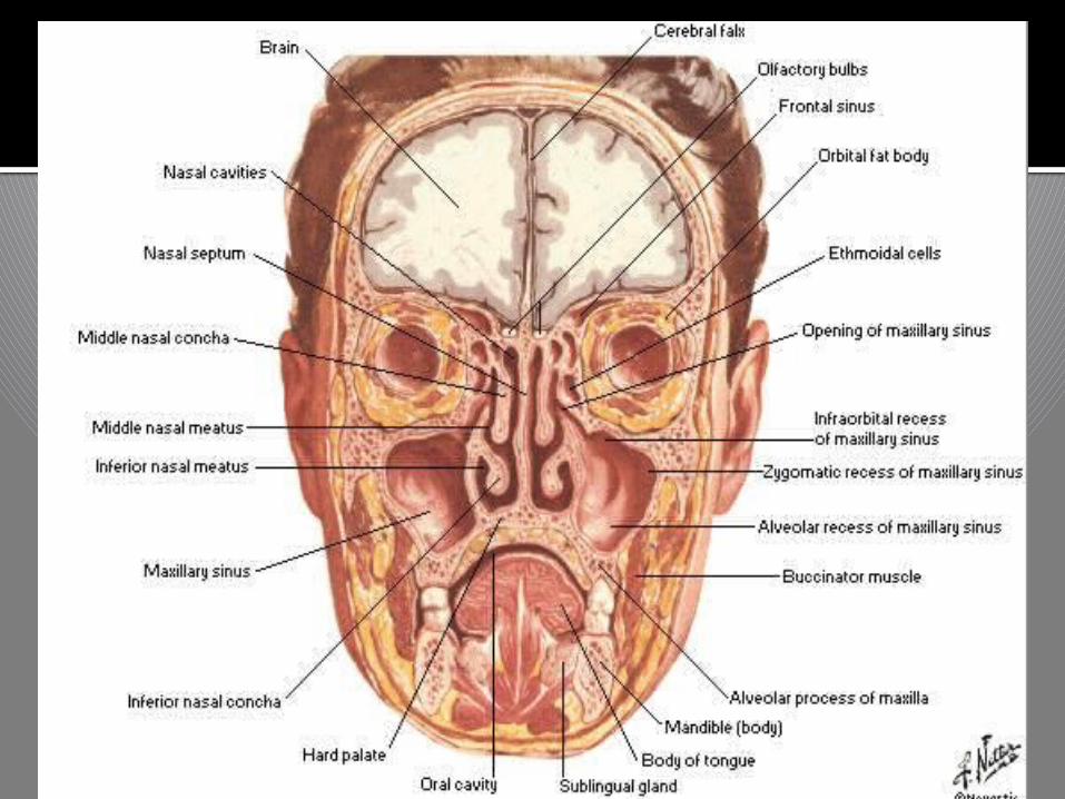
NOSE AND NASAL FOSSA
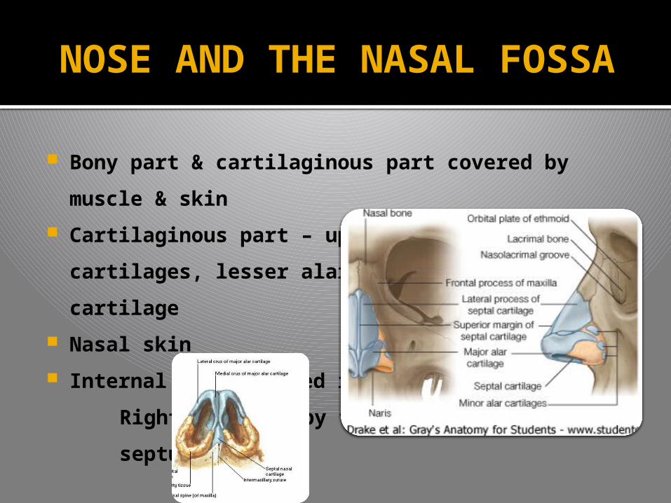
Bony part & cartilaginous part covered by
muscle & skin
Cartilaginous part – upper & lower lateral
cartilages, lesser alar cartilages & septal
cartilage
Nasal skin
Internal nose divided into the
Right and left by the nasal
septum
NOSE AND THE NASAL FOSSA

NOSE AND THE NASAL FOSSA
NASAL CAVITY PROPER
Roof – Nasal bone,
sphenoid & ethmoid
bone
Floor - Palatine
process of the maxilla
& Palatine bone
Medial wall
Lateral wall
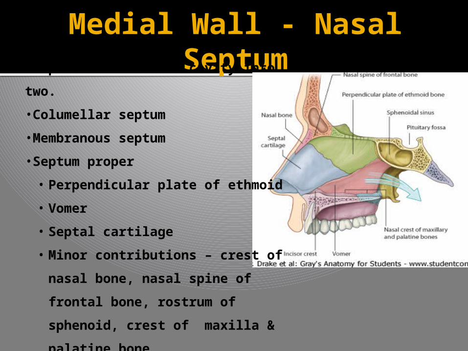
Medial Wall - Nasal Septum•Seperates the nasal cavity into
two.
•Columellar septum
•Membranous septum
•Septum proper
• Perpendicular plate of ethmoid
• Vomer
• Septal cartilage
• Minor contributions – crest of
nasal bone, nasal spine of
frontal bone, rostrum of
sphenoid, crest of maxilla &
palatine bone
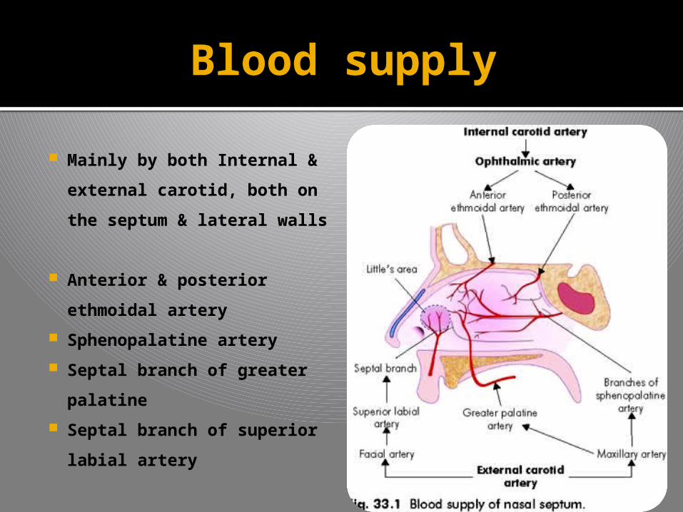
Blood supply
Mainly by both Internal &
external carotid, both on
the septum & lateral walls
Anterior & posterior
ethmoidal artery
Sphenopalatine artery
Septal branch of greater
palatine
Septal branch of superior
labial artery
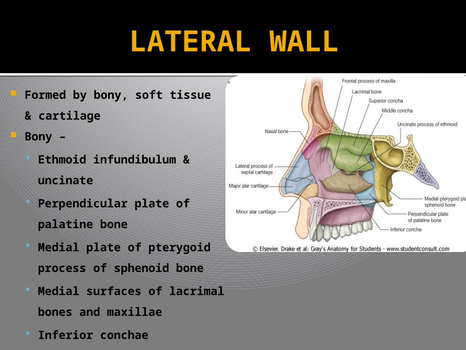
LATERAL WALL
Formed by bony, soft tissue
& cartilage
Bony –
Ethmoid infundibulum &
uncinate
Perpendicular plate of
palatine bone
Medial plate of pterygoid
process of sphenoid bone
Medial surfaces of lacrimal
bones and maxillae
Inferior conchae
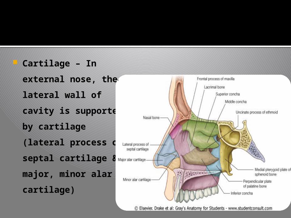
Cartilage – In
external nose, the
lateral wall of cavity
is supported by
cartilage (lateral
process of septal
cartilage & major,
minor alar cartilage)
Marked by three bony projections, they extend
medially across the nasal cavity separating the nasal
cavity into for air channels – the turbinates or conchae
Superior ,middle & inferior tubinates or conchae. The
conchae do not extend forwards into the external nose
The air space below and lateral to each turbinate is
called as meatus
Superior, middle & inferior meatus & sphenoethmoidal
recess
Middle Meatus – much significant
LATERAL WALL
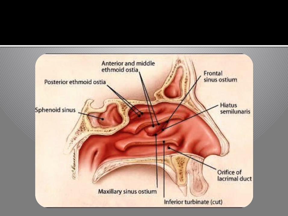
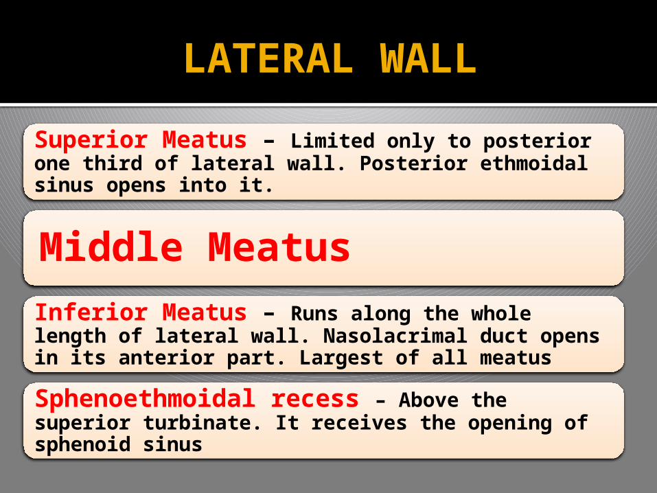
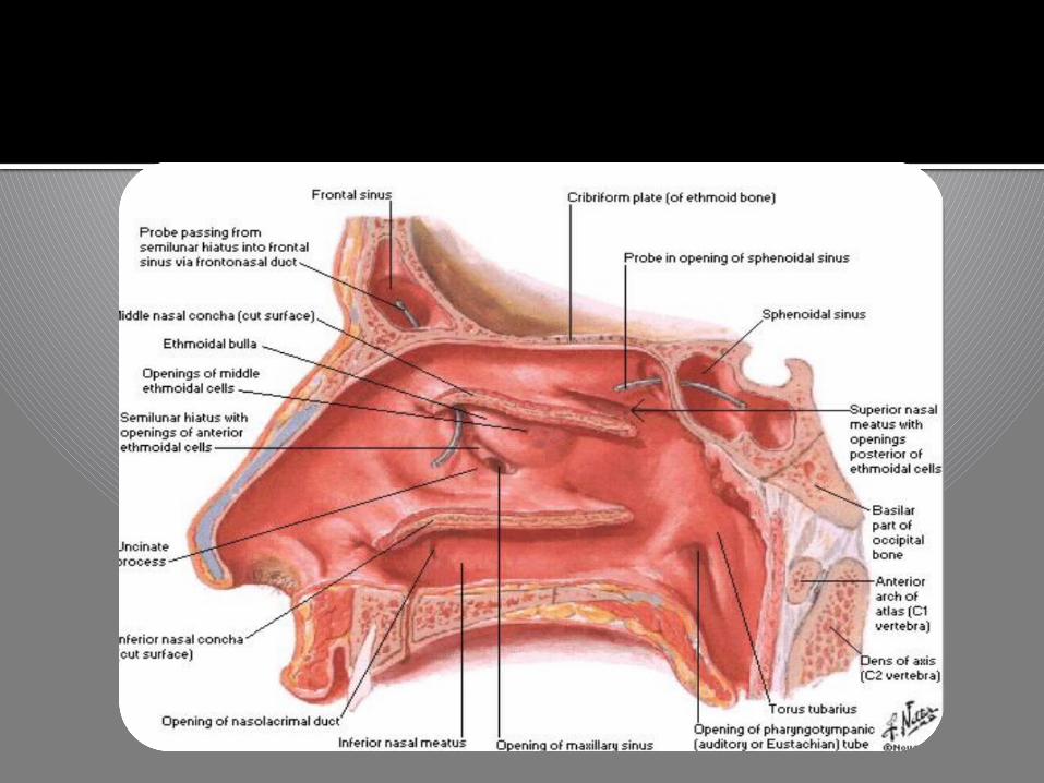
Superior Meatus – Limited only to posterior one third of lateral wall. Posterior ethmoidal sinus opens into it.
Middle Meatus
Inferior Meatus – Runs along the whole length of lateral wall. Nasolacrimal duct opens in its anterior part. Largest of all meatus
Sphenoethmoidal recess – Above the superior turbinate. It receives the opening of sphenoid sinus
LATERAL WALL
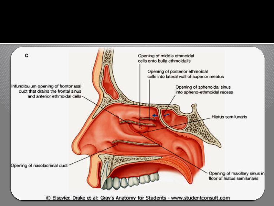
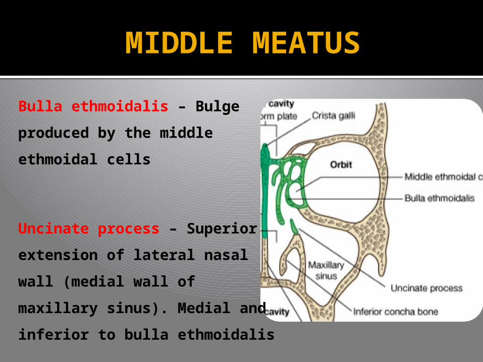
MIDDLE MEATUS
Bulla ethmoidalis – Bulge
produced by the middle
ethmoidal cells
Uncinate process – Superior
extension of lateral nasal wall
(medial wall of maxillary
sinus). Medial and inferior to
bulla ethmoidalis
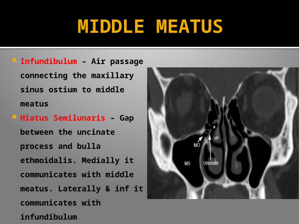
MIDDLE MEATUS
Infundibulum – Air passage
connecting the maxillary
sinus ostium to middle
meatus
Hiatus Semilunaris – Gap
between the uncinate
process and bulla
ethmoidalis. Medially it
communicates with middle
meatus. Laterally & inf it
communicates with
infundibulum
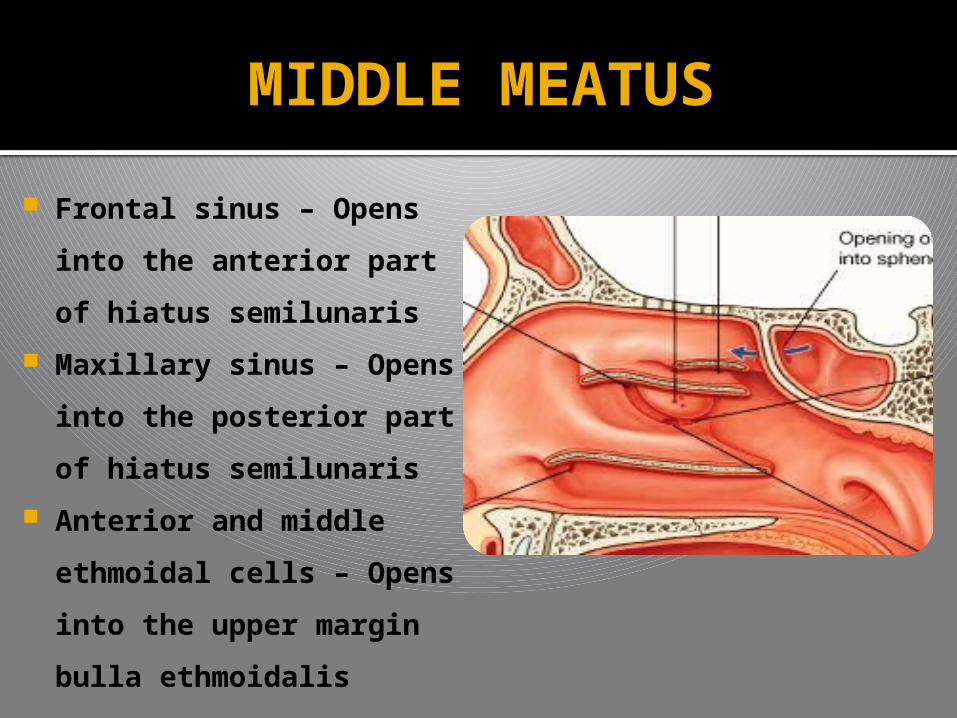
MIDDLE MEATUS
Frontal sinus – Opens
into the anterior part of
hiatus semilunaris
Maxillary sinus – Opens
into the posterior part
of hiatus semilunaris
Anterior and middle
ethmoidal cells – Opens
into the upper margin
bulla ethmoidalis
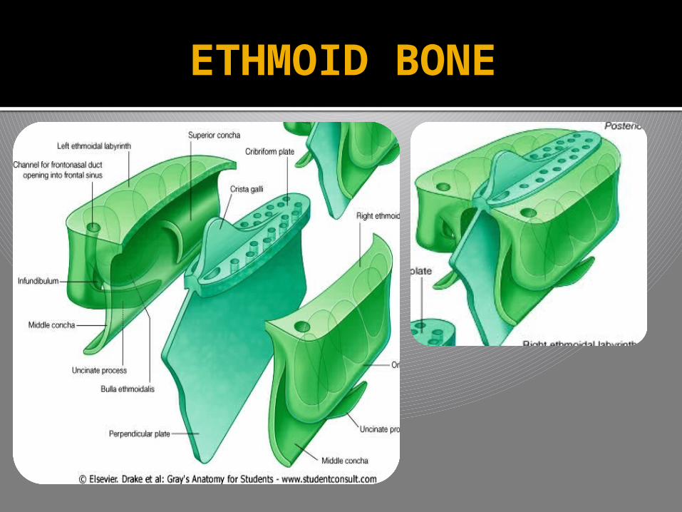
ETHMOID BONE
SINUSES

SINUSES
Air containing cavity in certain skull bones
Develop as a diverticula/outpouching from the
lat wall of nose & extend into Maxilla, Ethmoid,
sphenoid and frontal bones
Four sinuses – Maxillary, Frontal, Ethmoid (Ant
& Post) & Sphenoid
Some sinuses are well developed &
asymmetrical
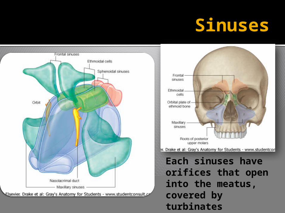
Sinuses
Each sinuses have orifices that open into the meatus, covered by turbinates


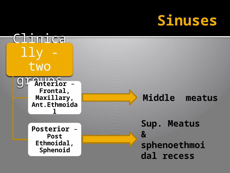
Clinically - two
groupsAnterior – Frontal,
Maxillary, Ant.Ethmoida
l
Posterior – Post
Ethmoidal, Sphenoid
Sinuses
Middle meatus
Sup. Meatus & sphenoethmoidal recess
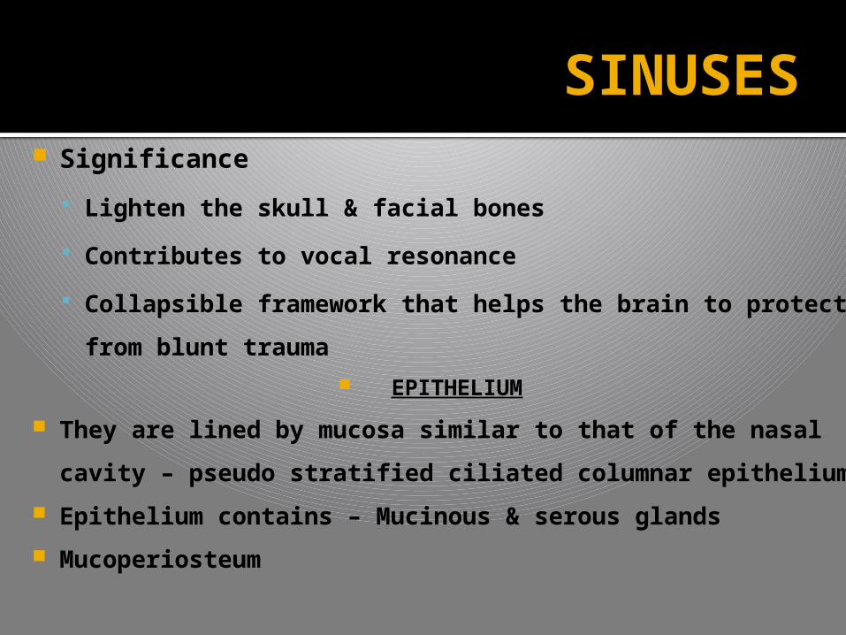
Significance
Lighten the skull & facial bones
Contributes to vocal resonance
Collapsible framework that helps the brain to
protect from blunt trauma
EPITHELIUM
They are lined by mucosa similar to that of the nasal
cavity – pseudo stratified ciliated columnar epithelium
Epithelium contains – Mucinous & serous glands
Mucoperiosteum
SINUSES
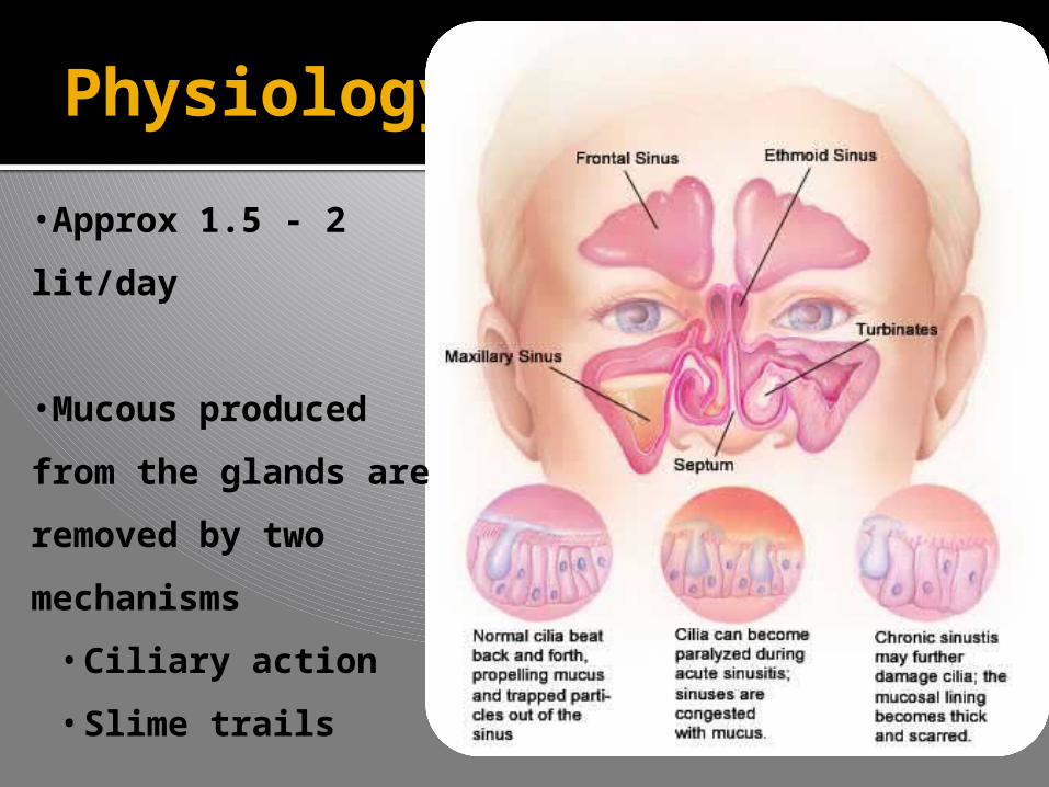
Physiology
•Approx 1.5 - 2
lit/day
•Mucous produced
from the glands are
removed by two
mechanisms
• Ciliary action
• Slime trails

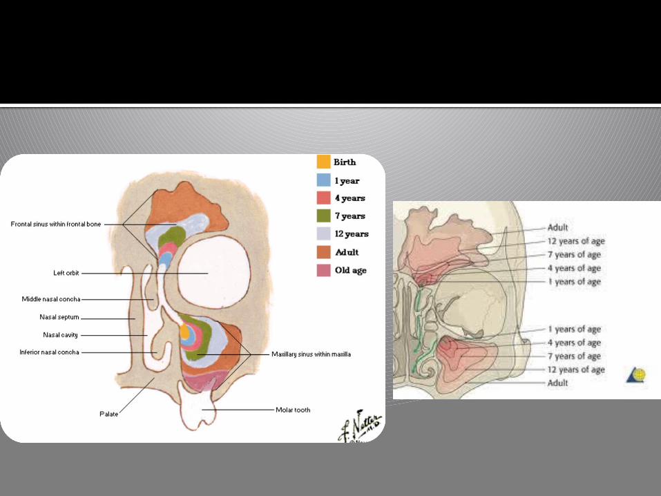
Sinuses Status at Birth
First Radiologi
cal evidence
Reaches Adult size by
Maxillary sinus
Present at birth
4-5 months after birth
15 years
Ethmoid sinus
Present at birth
1 year 12 years
Sphenoid sinus
Not Present 4 years 15 years – adult age
Frontal Sinus
Not Present 6 years Size increases
until teens
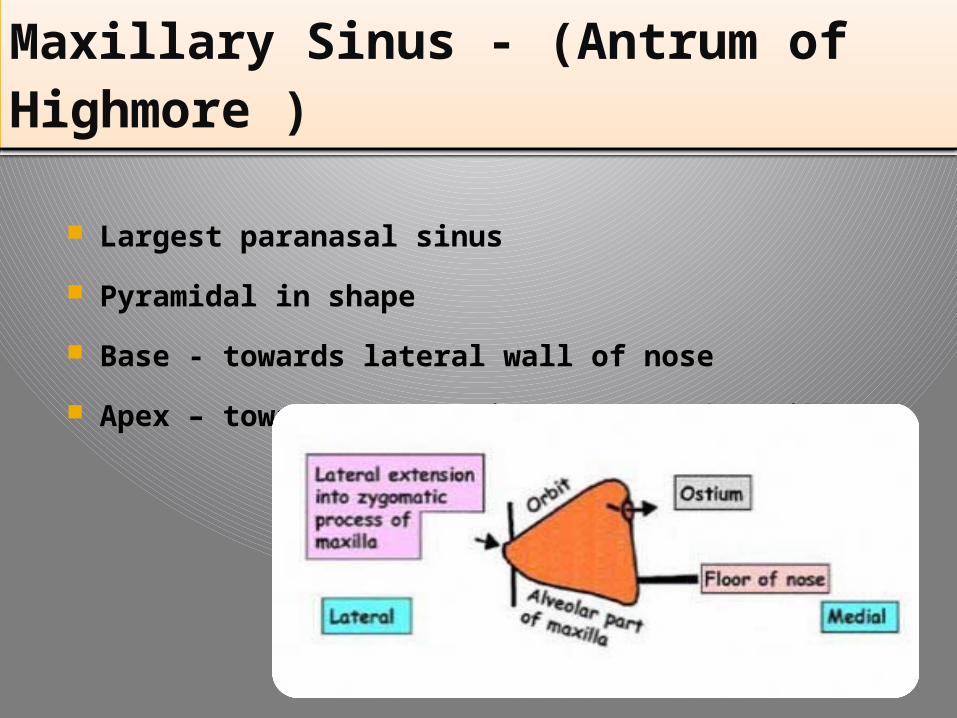
Maxillary Sinus - (Antrum of Highmore )
Largest paranasal sinus
Pyramidal in shape
Base - towards lateral wall of nose
Apex – towards zygomatic process of maxilla
Maxillary Sinus - (Antrum of Highmore )
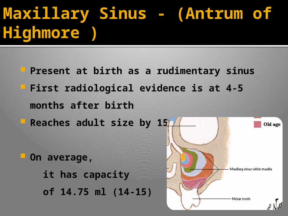
Present at birth as a rudimentary sinus
First radiological evidence is at 4-5 months
after birth
Reaches adult size by 15 years
On average,
it has capacity
of 14.75 ml (14-15)
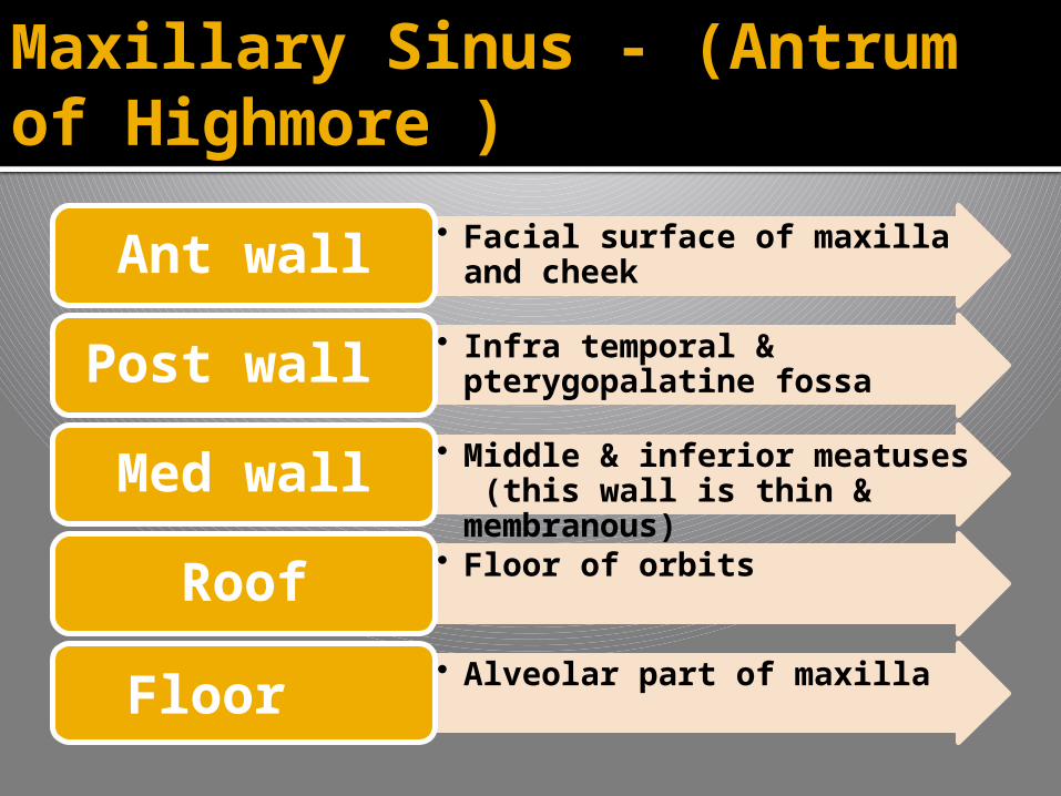
• Facial surface of maxilla and cheekAnt wall
• Infra temporal & pterygopalatine fossaPost wall
• Middle & inferior meatuses (this wall is thin & membranous)
Med wall• Floor of orbits
Roof• Alveolar part of maxilla
Floor
Maxillary Sinus - (Antrum of Highmore )
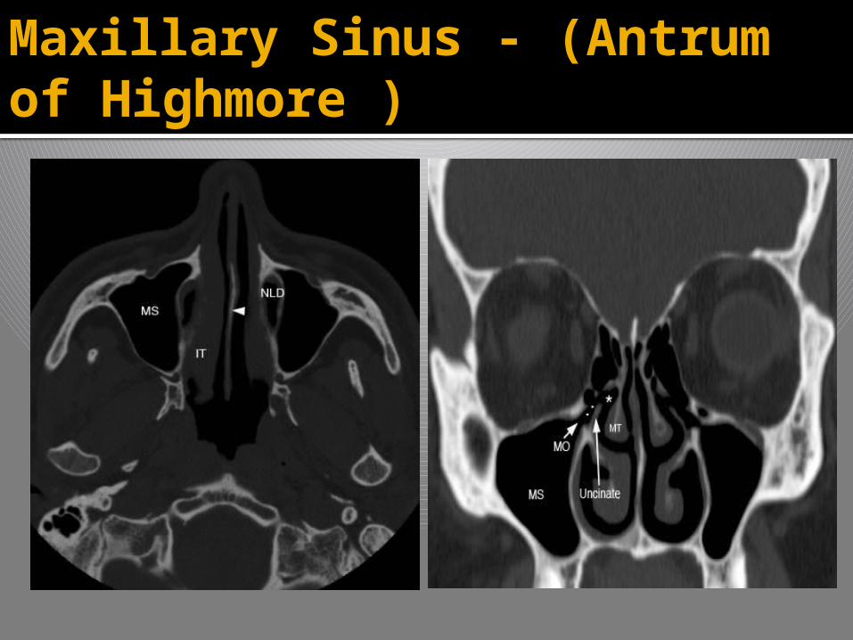
Maxillary Sinus - (Antrum of Highmore )
Maxillary Sinus - (Antrum of Highmore )
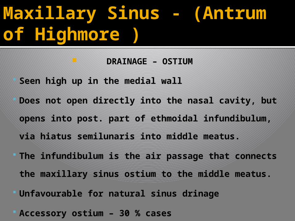
DRAINAGE – OSTIUM
Seen high up in the medial wall
Does not open directly into the nasal cavity, but
opens into post. part of ethmoidal infundibulum, via
hiatus semilunaris into middle meatus.
The infundibulum is the air passage that connects
the maxillary sinus ostium to the middle meatus.
Unfavourable for natural sinus drinage
Accessory ostium – 30 % cases
Maxillary Sinus - (Antrum of Highmore )
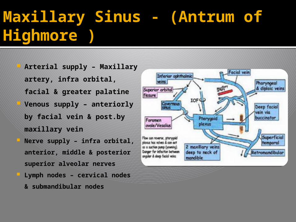
Arterial supply – Maxillary
artery, infra orbital, facial
& greater palatine
Venous supply – anteriorly
by facial vein & post.by
maxillary vein
Nerve supply – infra orbital,
anterior, middle & posterior
superior alveolar nerves
Lymph nodes – cervical nodes
& submandibular nodes

Maxillary Sinus - (Antrum of Highmore )
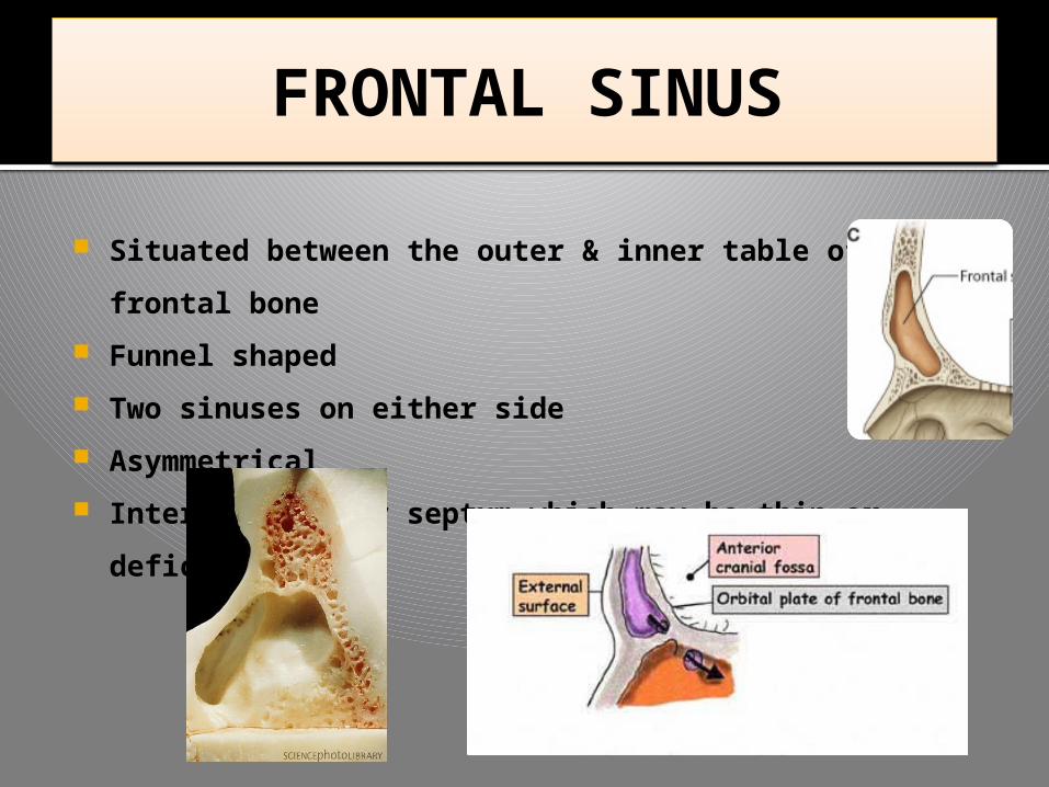
Situated between the outer & inner table of frontal
bone
Funnel shaped
Two sinuses on either side
Asymmetrical
Intervening bony septum which may be thin or
deficiency
FRONTAL SINUS
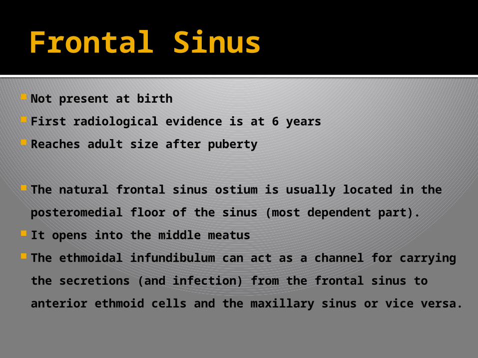
Not present at birth
First radiological evidence is at 6 years
Reaches adult size after puberty
The natural frontal sinus ostium is usually located in the
posteromedial floor of the sinus (most dependent part).
It opens into the middle meatus
The ethmoidal infundibulum can act as a channel for
carrying the secretions (and infection) from the frontal
sinus to anterior ethmoid cells and the maxillary sinus or
vice versa.
Frontal Sinus
Frontal Sinus
They develop from a variable site, their drainage
will be either via an ostium into the frontal
recess or via a nasofrontal duct into the anterior
infundibulum. The opening or duct can be
distorted by expansion of adjacent ethmoid cells
Boundaries
Ant wall – Skin over the forehead
Post wall - Meninges & the frontal lobe of brain
Inferior wall - orbit & its contents
Frontal Sinus
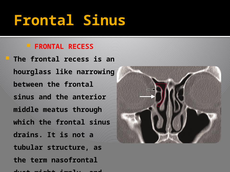
FRONTAL RECESS
The frontal recess is an
hourglass like narrowing
between the frontal
sinus and the anterior
middle meatus through
which the frontal sinus
drains. It is not a tubular
structure, as the term
nasofrontal duct might
imply, and therefore the
term recess is preferred.
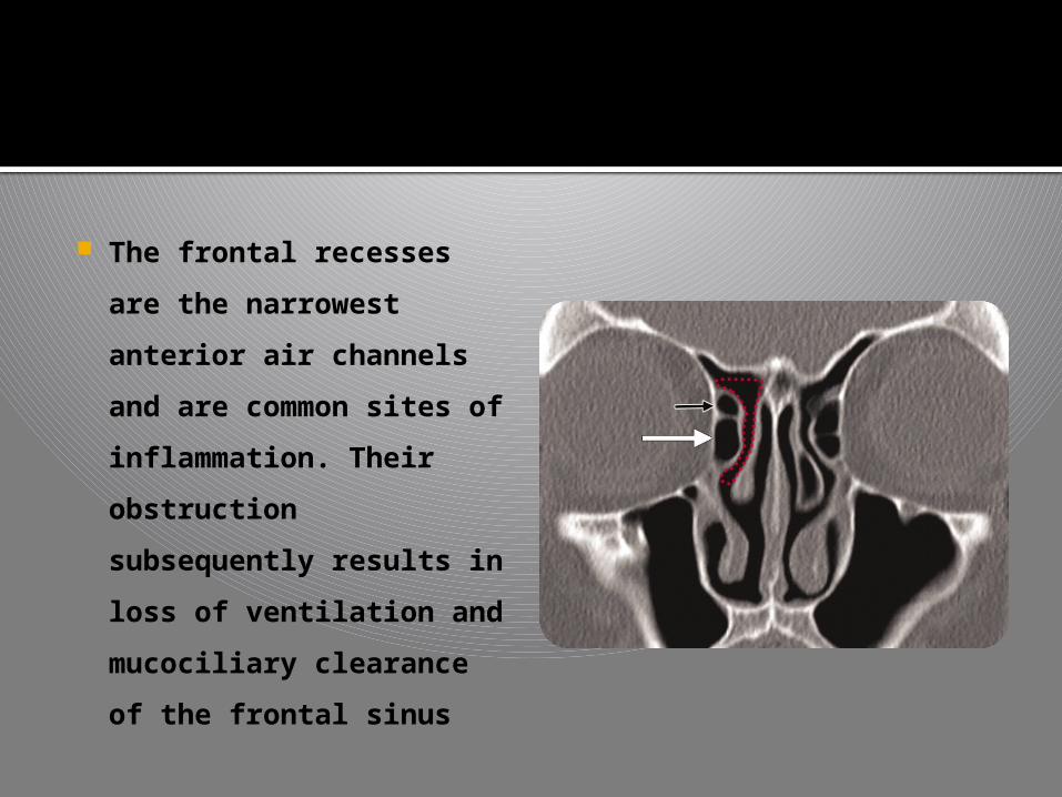
The frontal recesses are
the narrowest anterior
air channels and are
common sites of
inflammation. Their
obstruction
subsequently results in
loss of ventilation and
mucociliary clearance of
the frontal sinus
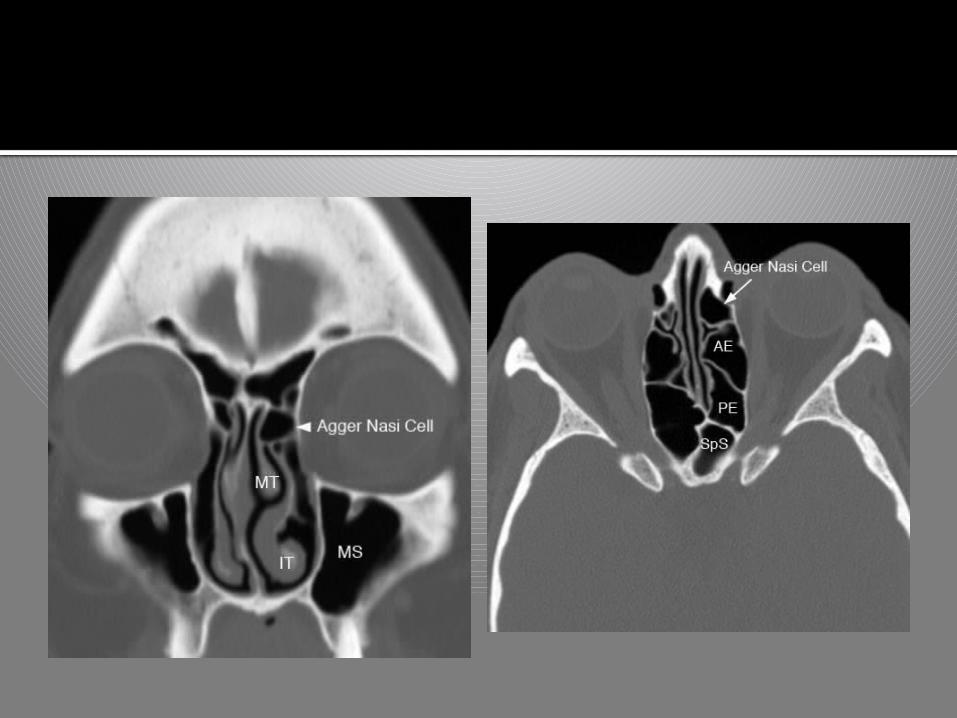
AGGER NASI CELL
Anterior, lateral, and inferior to the frontal
recess is the agger nasi cell. It is aerated and
represents the most anterior ethmoid air cell,
usually lying deep to the lacrimal bone.
It usually borders the primary ostium or floor
of the frontal sinus, and thus its size may
directly influence the patency of the frontal
recess and the anterior middle meatus.

The frontal sinus can pneumatize both the
vertical and the horizontal (orbital) plates of
the frontal bone. The deepest area of the
vertical portion of the sinus is near the midline
at the level of the supraorbital ridge, and the
medial sinus floor and the caudal anterior sinus
wall are thinnest in this area. As a result, the
sinus is best approached for a trephination at
this level
Frontal Sinus
There is a rich sinus venous plexus (Breschet’s
canals) that communicates with both the
diploic veins and the dural spaces.
Arterial supply – supra orbital & supra
trochlear
Venous supply – superior opthalmic vein
Lymph – Submandibular lymph node
Sensory innervation – supra orbital & supra
trochlear
Frontal Sinus
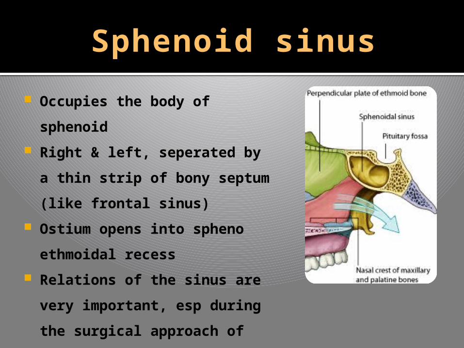
Sphenoid sinus
Occupies the body of
sphenoid
Right & left, seperated by a
thin strip of bony septum
(like frontal sinus)
Ostium opens into spheno
ethmoidal recess
Relations of the sinus are
very important, esp during
the surgical approach of
pituitary gland
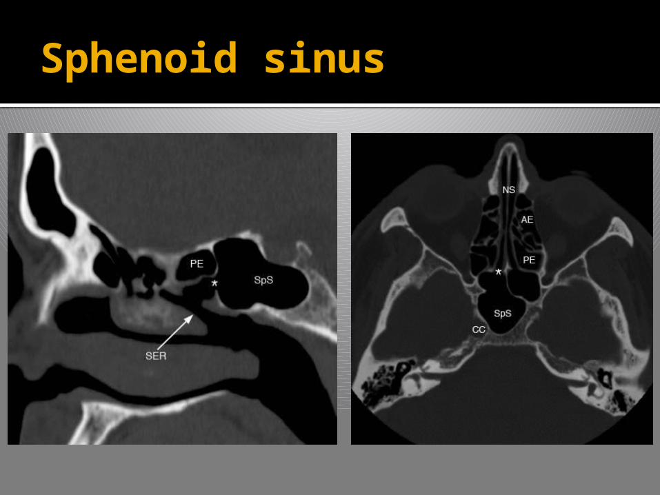
Sphenoid sinus

Relations –
Anterior part –
Roof – olfactory tract, optic chiasma
& frontal lobe
Lateral – optic nerve, internal
carotid artery & maxillary nerve
Posterior part
Roof – Pituitary gland in sella
turcica
Lateral – Cavernous sinus,ICA &
Cranial nerves III, IV, VI & all
divisions of V
Sphenoid sinus
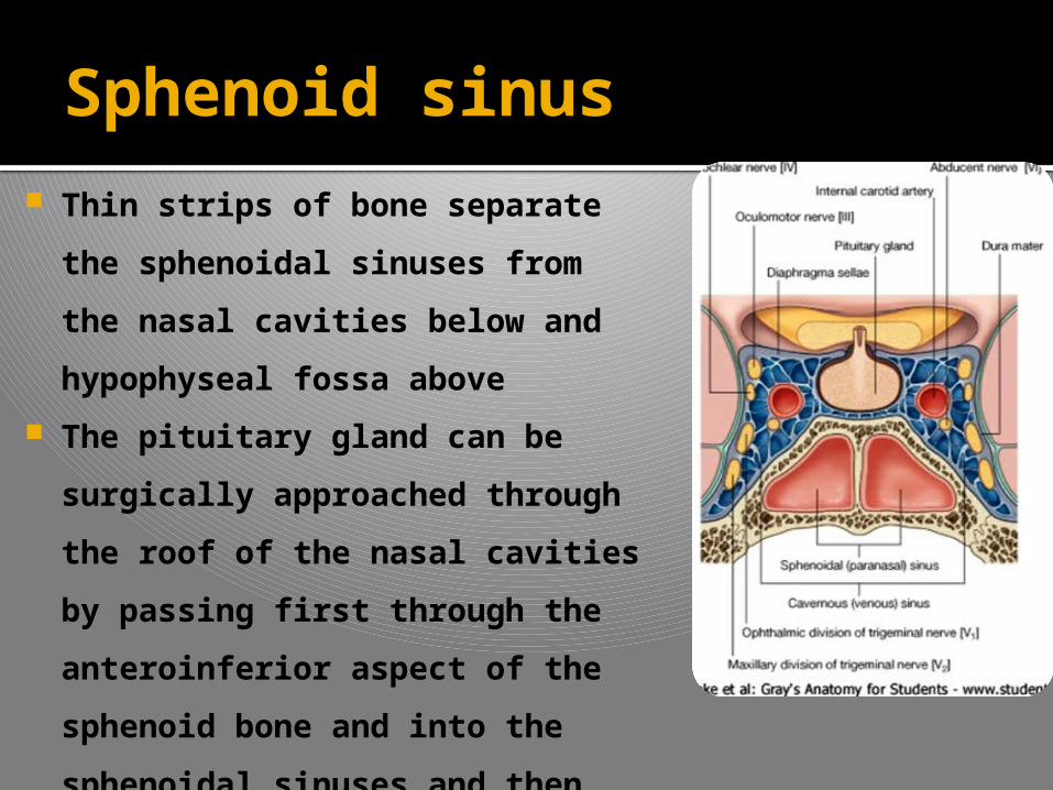
Thin strips of bone separate the
sphenoidal sinuses from the
nasal cavities below and
hypophyseal fossa above
The pituitary gland can be
surgically approached through
the roof of the nasal cavities by
passing first through the
anteroinferior aspect of the
sphenoid bone and into the
sphenoidal sinuses and then
through the top of the sphenoid
bone into the hypophyseal
fossa
Sphenoid sinus
ETHMOID SINUS
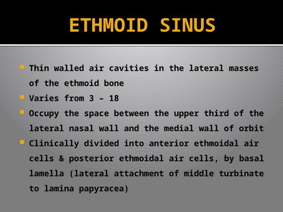
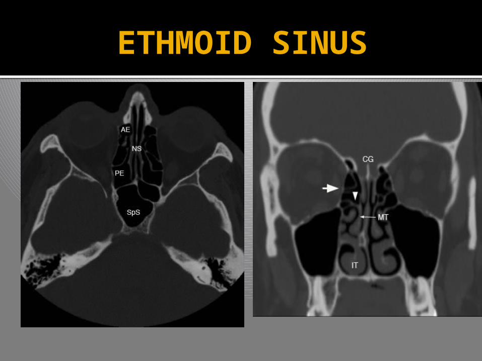
Thin walled air cavities in the lateral masses of
the ethmoid bone
Varies from 3 – 18
Occupy the space between the upper third of the
lateral nasal wall and the medial wall of orbit
Clinically divided into anterior ethmoidal air cells
& posterior ethmoidal air cells, by basal lamella
(lateral attachment of middle turbinate to
lamina papyracea)
ETHMOID SINUS
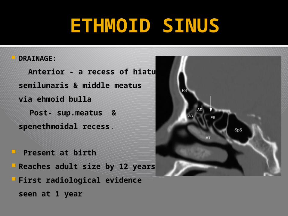
DRAINAGE:
Anterior - a recess of hiatus
semilunaris & middle meatus
via ehmoid bulla
Post- sup.meatus &
spenethmoidal recess.
Present at birth
Reaches adult size by 12 years
First radiological evidence
seen at 1 year
ETHMOID SINUS
Relations
Roof – formed by the anterior cranial fossa
Lateral wall - orbit
Medial wall – nasal cavity
Thin paper like bony part of the ethmoid separating
the air cells from the orbit, called lamina
papyracea, can be easily destroyed leading to
spread of ethmoidal infections into the orbit
Optic nerve forms a close relationship with the
posterior ethmoidal cells & is at risk during
ethmoidal surgery
ETHMOID SINUS
OSTEOMEATAL COMPLEX

OSTEO MEATAL COMPLEX
The osteomeatal complex is the key anatomic
area addressed by endoscopic sinus surgeons.
Blockage of the osteomeatal complex prevents
effective mucociliary clearance, thus leading to
a stagnation of secretions and therefore leading
to recurrent or chronic sinusitis.
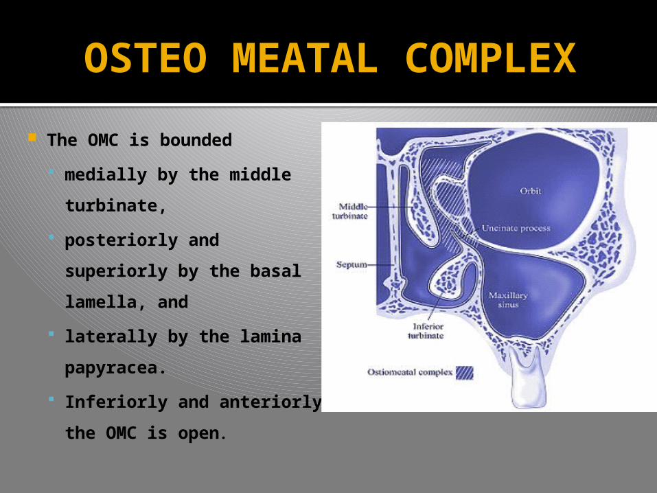
OSTEO MEATAL COMPLEX
The OMC is bounded
medially by the middle
turbinate,
posteriorly and
superiorly by the basal
lamella, and
laterally by the lamina
papyracea.
Inferiorly and anteriorly
the OMC is open.
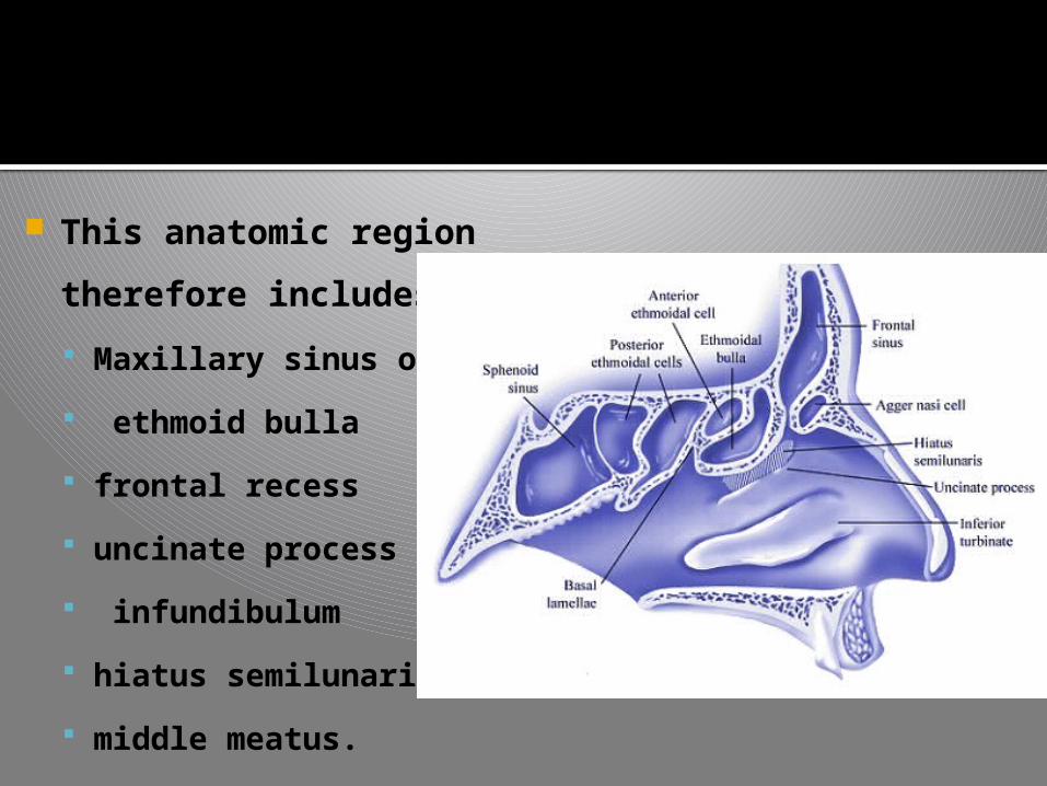
This anatomic region
therefore includes
Maxillary sinus ostium
ethmoid bulla
frontal recess
uncinate process
infundibulum
hiatus semilunaris
middle meatus.

OSTEO MEATAL COMPLEX

ANATOMICAL VARIANTS
Variations of Middle turbinate
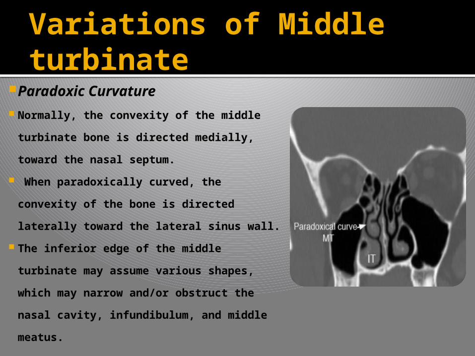
Paradoxic Curvature Normally, the convexity of the middle
turbinate bone is directed medially,
toward the nasal septum.
When paradoxically curved, the
convexity of the bone is directed
laterally toward the lateral sinus wall.
The inferior edge of the middle
turbinate may assume various shapes,
which may narrow and/or obstruct the
nasal cavity, infundibulum, and middle
meatus.
Variations of Middle turbinate
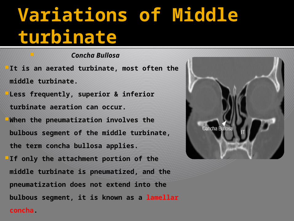
Concha Bullosa
It is an aerated turbinate, most often the
middle turbinate.
Less frequently, superior & inferior
turbinate aeration can occur.
When the pneumatization involves the
bulbous segment of the middle turbinate,
the term concha bullosa applies.
If only the attachment portion of the
middle turbinate is pneumatized, and the
pneumatization does not extend into the
bulbous segment, it is known as a
lamellar concha.
Other Variations Additional variations of the middle turbinate can
occur, including medial & lateral displacement, lateral
bending, L shape, and sagittal transverse clefts
Medial displacement – due to other middle meatal
structures (i.e., polypoid disease, pneumatized
uncinate process) encroaching upon the middle
turbinate.
Lateral displacement - due to the compression of the
turbinate toward the lateral nasal wall by a septal
spur or septal deviation.
Variations of Middle turbinate
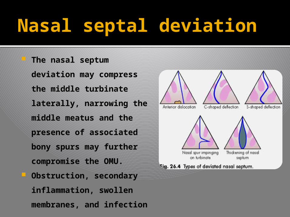
Nasal septal deviation
The nasal septum
deviation may compress
the middle turbinate
laterally, narrowing the
middle meatus and the
presence of associated
bony spurs may further
compromise the OMU.
Obstruction, secondary
inflammation, swollen
membranes, and
infection can occur

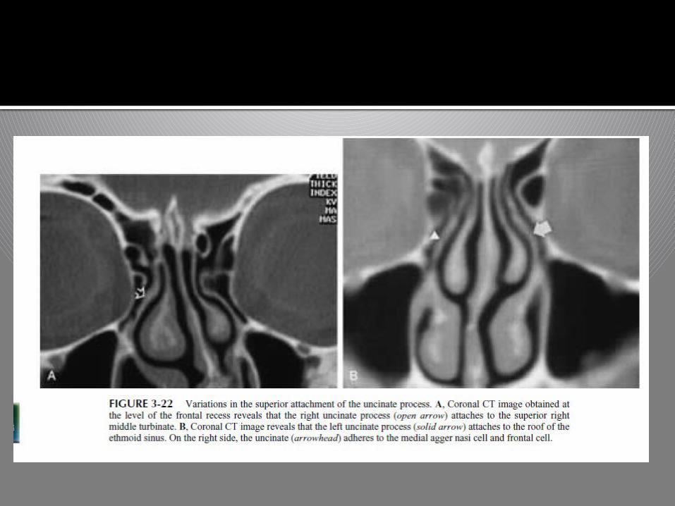
VARIATIONS OF UNCINATE PROCESS
DEVIATION
The course of the free edge of the uncinate
process may either extend slightly obliquely
toward the nasal septum, with the free edge
surrounding the inferoanterior surface of the
ethmoid bulla, or it extends more medially to
the medial surface of the ethmoid bulla. If the
free edge of the uncinate is deviated in a more
lateral direction, it may cause narrowing or
obstruction of the hiatus semilunaris and
infundibulum.
Attachment
Attachment to the lamina papyracea, the lateral
surface of the middle turbinate, or the fovea
ethmoidalis in the floor of the anterior cranial fossa
may occur.
If the uncinate process attaches to the ethmoidal
roof or middle turbinate, during uncinatectomy,
traction could inadvertently damage the ethmoid
roof and result in CSF rhinorrhea or other
intracranial complications.
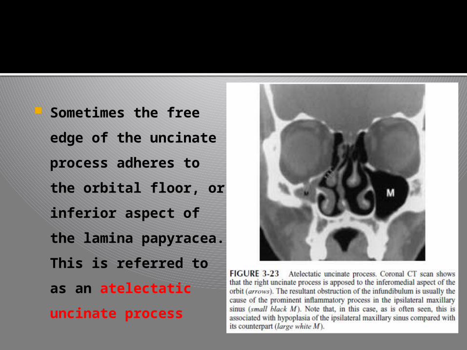
Sometimes the free
edge of the uncinate
process adheres to
the orbital floor, or
inferior aspect of the
lamina papyracea.
This is referred to as
an atelectatic
uncinate process
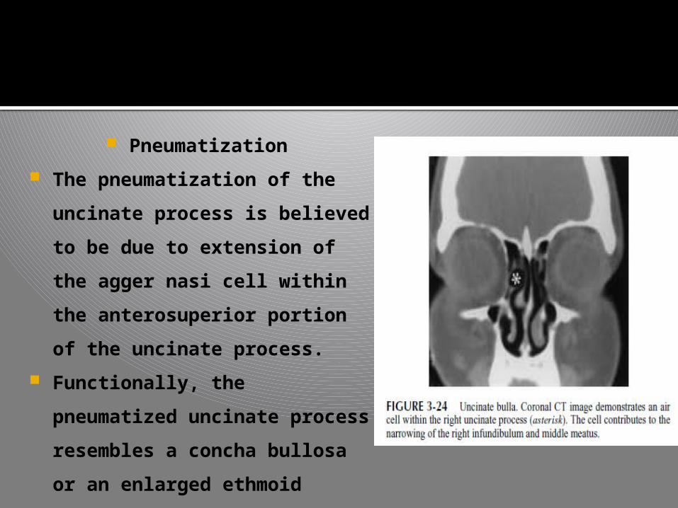
Pneumatization
The pneumatization of the
uncinate process is believed
to be due to extension of the
agger nasi cell within the
anterosuperior portion of the
uncinate process.
Functionally, the pneumatized
uncinate process resembles a
concha bullosa or an enlarged
ethmoid bulla.
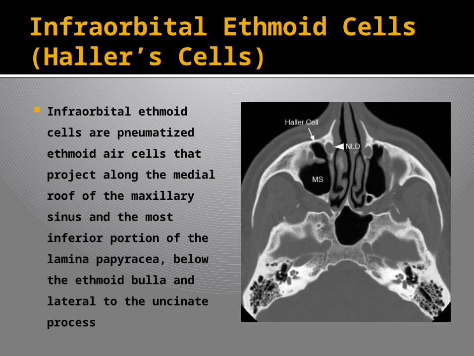
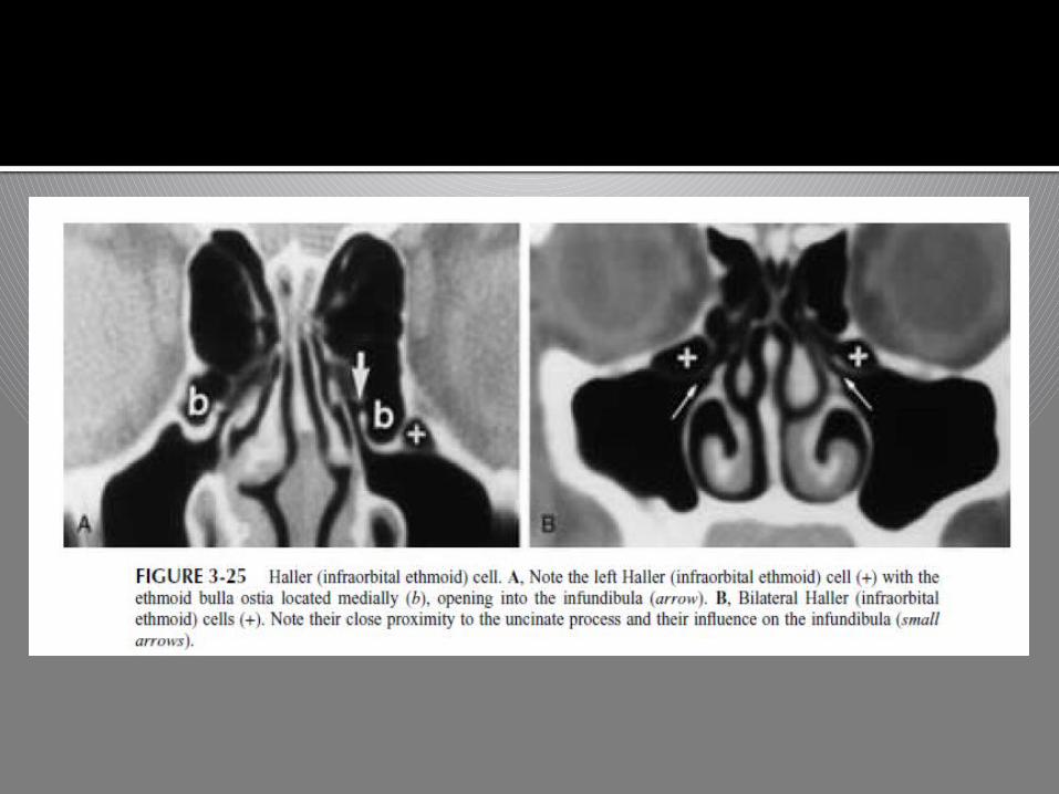
Infraorbital Ethmoid Cells (Haller’s Cells)
Infraorbital ethmoid cells
are pneumatized ethmoid
air cells that project
along the medial roof of
the maxillary sinus and
the most inferior portion
of the lamina papyracea,
below the ethmoid bulla
and lateral to the
uncinate process

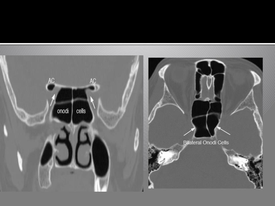
Onodi Cells
Two definitions of Onodi cells.
The first defines them as the most
posterior ethmoid cells, being
superolateral to the sphenoid sinus
and closely associated with the optic
nerve.
Another, more general description
defines Onodi cells as posterior
ethmoid cells extending into the
sphenoid bone, situated either
adjacent to or impinging upon the
optic nerve

Ethmoid Bulla Variations
Its appearance varies considerably, based on
the extent of pneumatization.
Extensive pneumatization may obstruct the
ostiomeatal complex.
Elongated ethmoid bullae are usually in a
superior to inferior direction rather than in an
anterior to posterior direction.
So, Relatively unlikely to obstruct the
ostiomeatal complex.
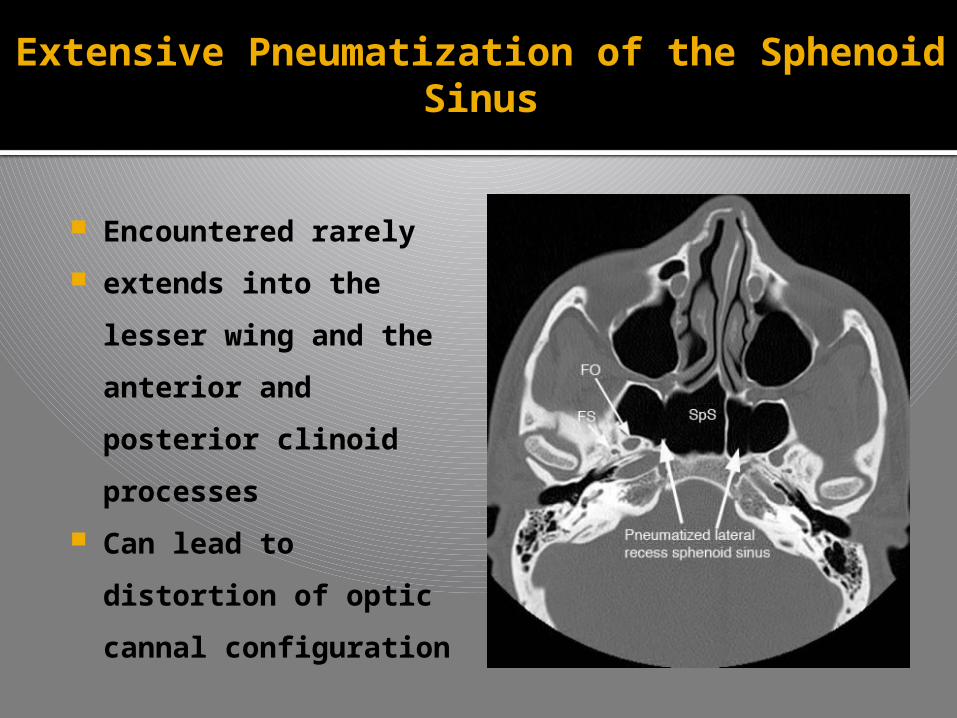
Extensive Pneumatization of the SphenoidSinus
Encountered rarely
extends into the
lesser wing and the
anterior and
posterior clinoid
processes
Can lead to
distortion of optic
cannal configuration
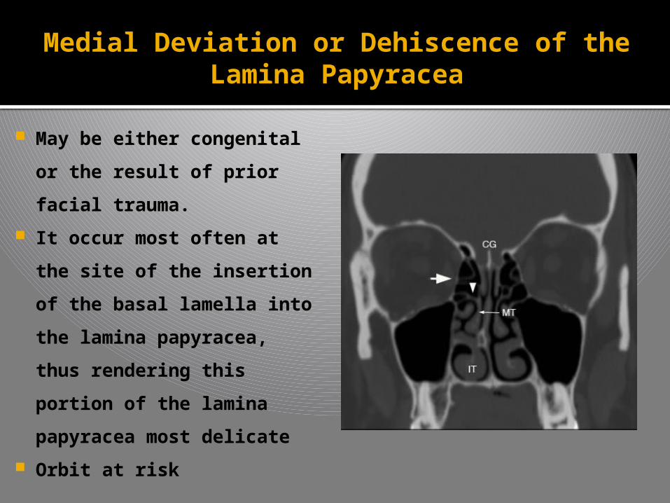
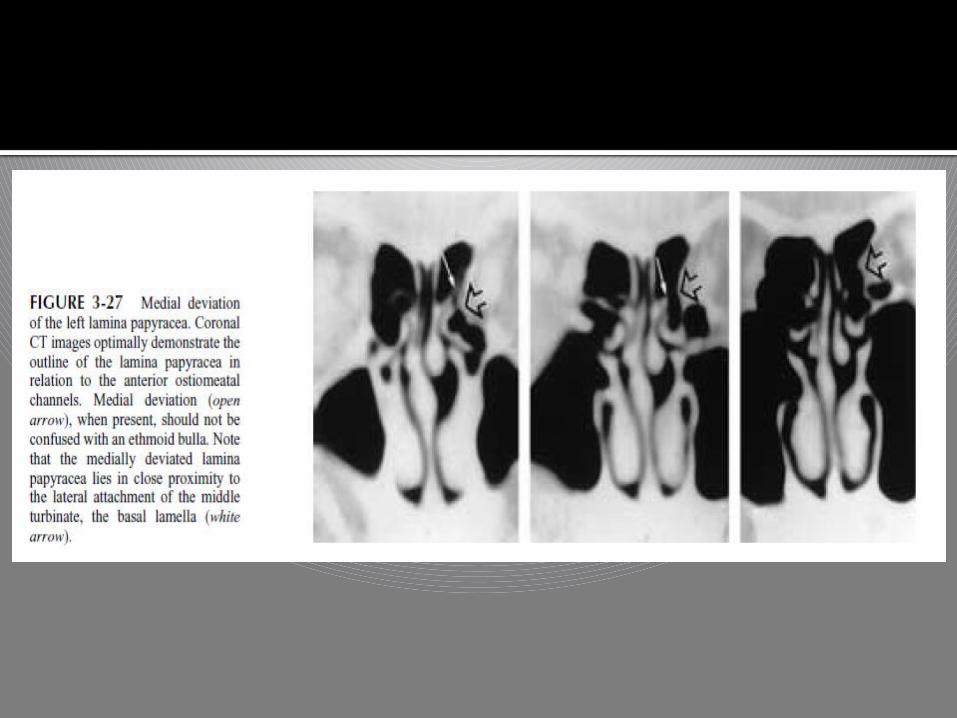
Medial Deviation or Dehiscence of theLamina Papyracea
May be either congenital
or the result of prior
facial trauma.
It occur most often at the
site of the insertion of the
basal lamella into the
lamina papyracea, thus
rendering this portion of
the lamina papyracea
most delicate
Orbit at risk
Aerated Crista Galli
When aeration of the normally bony crista galli
occurs the aerated cells may communicate with
the frontal recess, and obstruction of this
ostium.
To avoid unnecessary surgical extension into
the anterior cranial vault, it is important to
recognize an aerated crista galli and
differentiate it from an ethmoid air cell.
Posterior Nasal Septal Air Cell
Air cells are commonly found within the
posterosuperior portion of the nasal septum
and, when present, communicate with the
sphenoid sinus.
As a result, any inflammatory disease that
occurs within the paranasal sinuses may also
affect these cells
Asymmetry in Ethmoid Roof Height
It is important to note any asymmetry in
the height of the ethmoid roof.
Intracranial penetration during surgery is
more likely to occur on the side where the
position of the roof is lower
IMAGING MODALITIES
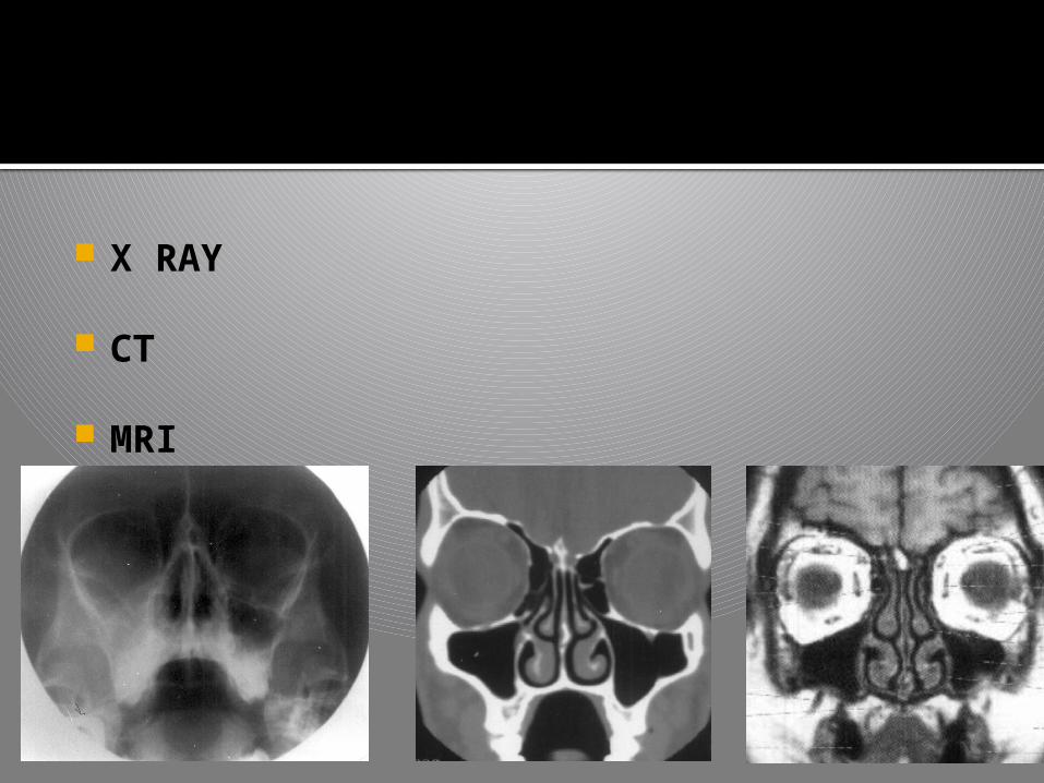
X RAY
CT
MRI

X ray – Water’s view & caldwell view
Ct – gold standard. Coronal & axial sections
MRI is predominantly used for pre and post
operative management of naso sinus malignancy
The chief disadvantage of MRI is its inability to
show the bony details of the sinuses, as both air
and bone give no signal
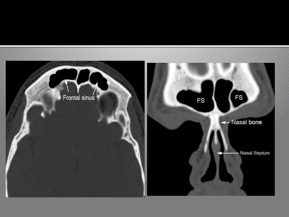
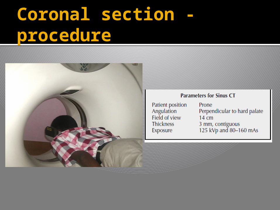
CT PROCEDURE & SECTIONS
CT PROCEDURE & SECTIONS
CT is currently the modality of choice in the
evaluation of the paranasal sinuses and adjacent
structures.
Its ability to optimally display bone, soft tissue, and
air provides an accurate depiction of both the
anatomy and the extent of disease in and around
the paranasal sinuses.
In contrast to standard radiographs, CT clearly
shows the fine bony anatomy of the osteomeatal
channels.
There are few pre requisites in few situations
a course of adequate medical therapy to eliminate
or diminish reversible mucosal inflammation.
pretreatment with a sympathomimetic nasal spray
15 minutes prior to scanning in order to reduce
nasal congestion (mucosal edema) and thus
improve the display of the fine bony architecture
and any irreversible mucosal disease
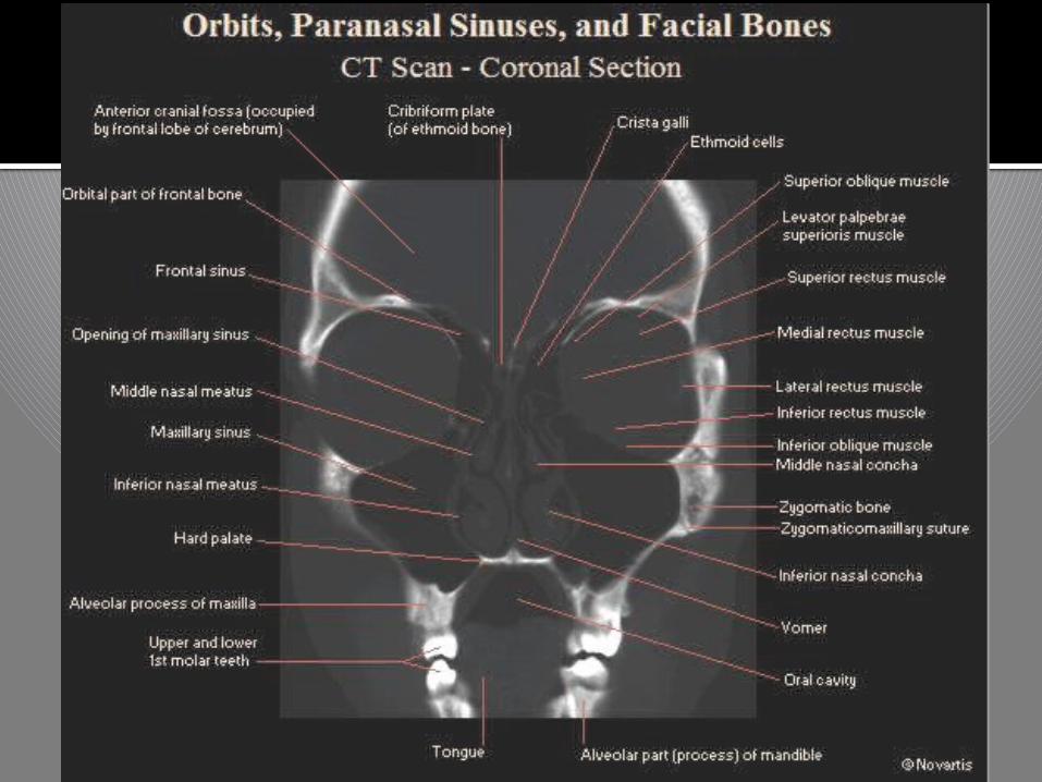
CT PROCEDURE & SECTIONS
Coronal & axial views
The coronal plane best shows the ostiomeatal
unit (OMU), shows the relationship of the brain
to the ethmoid roof.
Coronal plane should be the primary imaging
orientation for evaluation of the sinonasal tract
in all patients with inflammatory sinus disease
who are endoscopic surgical candidates
CT PROCEDURE & SECTIONS
Coronal section - procedure
Prone with chin
hyperextended
Gantry anglutaion-
perpendicular to hard palate
Section thickness-3mm
contigous
Table increment- 3-4
mmeach step
Kvp-125
Mas-80
Hanging head technique
Coronal section - procedure
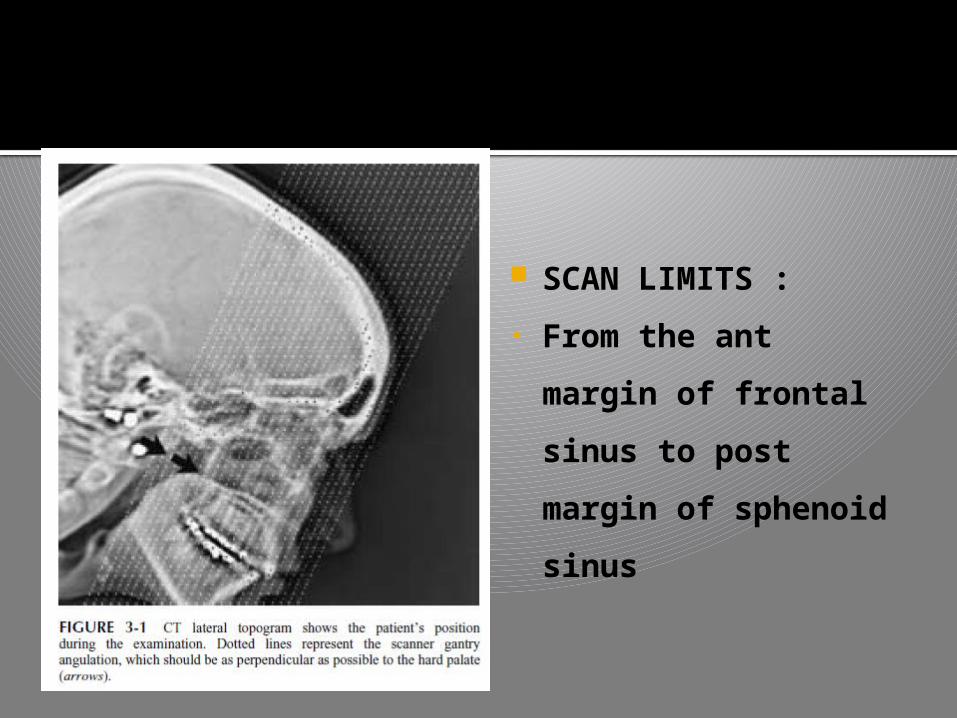
SCAN LIMITS :
• From the ant
margin of frontal
sinus to post
margin of sphenoid
sinus

Coronal section - procedure HEAD HANGING METHOD
Performed in the prone
position, so that any
remaining sinus secretions
do not obscure the OMU
In patients who cannot
tolerate prone positioning
(children, patients of
advanced age, etc.), the
hanging head technique
can sometimes be utilized.
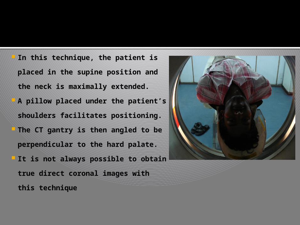
In this technique, the patient is
placed in the supine position and
the neck is maximally extended.
A pillow placed under the patient’s
shoulders facilitates positioning.
The CT gantry is then angled to be
perpendicular to the hard palate.
It is not always possible to obtain
true direct coronal images with this
technique
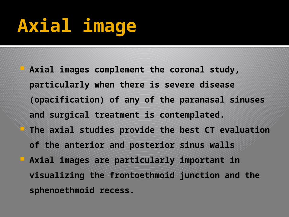
Axial image
Axial images complement the coronal study,
particularly when there is severe disease
(opacification) of any of the paranasal sinuses
and surgical treatment is contemplated.
The axial studies provide the best CT evaluation
of the anterior and posterior sinus walls
Axial images are particularly important in
visualizing the frontoethmoid junction and the
sphenoethmoid recess.
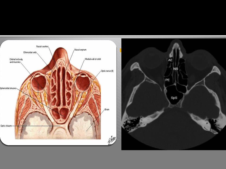
CT axial section of PNS - image
Whenever there is total opacification of the
frontal, maxillary, or sphenoid sinuses, a
complete axial and coronal CT examination
should be performed.
And also, if the patient has a suspected
neoplasm, a complete axial and coronal
examination need to be performed to provide the
most detailed analysis of the sinonasal cavities
and the adjacent skull base
Axial image
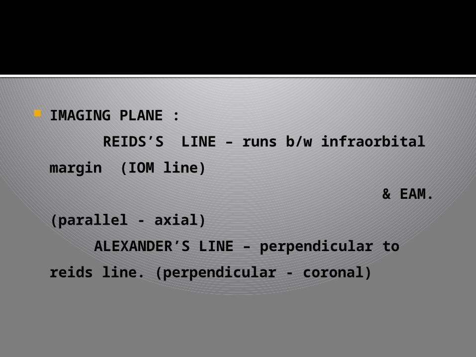
IMAGING PLANE :
REIDS’S LINE – runs b/w infraorbital margin
(IOM line)
& EAM. (parallel - axial)
ALEXANDER’S LINE – perpendicular to reids
line. (perpendicular - coronal)
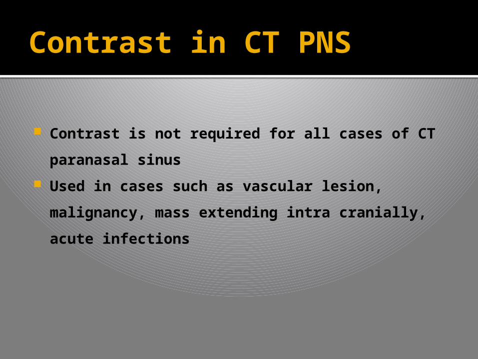
Contrast in CT PNS
Contrast is not required for all cases of CT
paranasal sinus
Used in cases such as vascular lesion,
malignancy, mass extending intra cranially,
acute infections

CHECK LIST
AGE OF THE PATIENT


THANK YOU

NEXT PRESENTATION
X RAY SHOULDER JOINTBY
DR.V.PRIYAON SATURDAY