crystal structures of kpc-2 -lactamase in complex with 3-nitrophenyl boronic...
TRANSCRIPT

Crystal Structures of KPC-2 �-Lactamase in Complex with3-Nitrophenyl Boronic Acid and the Penam Sulfone PSR-3-226
Wei Ke,a Christopher R. Bethel,f Krisztina M. Papp-Wallace,e,f Sundar Ram Reddy Pagadala,g,h Micheal Nottingham,g,h
Daniel Fernandez,g,h John D. Buynak,g,h Robert A. Bonomo,b,c,d,e,f and Focco van den Akkera
Departments of Biochemistry,a Pharmacology,b Molecular Biology and Microbiology,c and Medicinee and Center for Proteomics,d Case Western Reserve University,Cleveland, Ohio, USA; Research Service, Louis Stokes Cleveland VA Medical Center, Cleveland, Ohio, USAf; Department of Chemistry, Southern Methodist University, Dallas,Texas, USAg; and The SMU Center for Drug Discovery, Design, and Delivery at Dedman College, Dallas, Texas, USAh
Class A carbapenemases are a major threat to the potency of carbapenem antibiotics. A widespread carbapenemase, KPC-2, isnot easily inhibited by �-lactamase inhibitors (i.e., clavulanic acid, sulbactam, and tazobactam). To explore different mecha-nisms of inhibition of KPC-2, we determined the crystal structures of KPC-2 with two �-lactamase inhibitors that follow differ-ent inactivation pathways and kinetics. The first complex is that of a small boronic acid compound, 3-nitrophenyl boronic acid(3-NPBA), bound to KPC-2 with 1.62-Å resolution. 3-NPBA demonstrated a Km value of 1.0 � 0.1 �M (mean � standard error)for KPC-2 and blocks the active site by making a reversible covalent interaction with the catalytic S70 residue. The two boronhydroxyl atoms of 3-NPBA are positioned in the oxyanion hole and the deacylation water pocket, respectively. In addition, thearomatic ring of 3-NPBA provides an edge-to-face interaction with W105 in the active site. The structure of KPC-2 with the pe-nam sulfone PSR-3-226 was determined at 1.26-Å resolution. PSR-3-226 displayed a Km value of 3.8 � 0.4 �M for KPC-2, and theinactivation rate constant (kinact) was 0.034 � 0.003 s�1. When covalently bound to S70, PSR-3-226 forms a trans-enamine inter-mediate in the KPC-2 active site. The predominant active site interactions are generated via the carbonyl oxygen, which residesin the oxyanion hole, and the carboxyl moiety of PSR-3-226, which interacts with N132, N170, and E166. 3-NPBA and PSR-3-226are the first �-lactamase inhibitors to be trapped as an acyl-enzyme complex with KPC-2. The structural and inhibitory insightsgained here could aid in the design of potent KPC-2 inhibitors.
Carbapenemases pose a serious clinical threat to the “last-resortantibiotics,” the carbapenems (imipenem, meropenem, er-
tapenem, and doripenem) (22, 33). In particular, the Klebsiellapneumoniae carbapenemases (KPCs) are troublesome, as they areoften carried on a plasmid, leading to rapid dissemination (14, 22,30, 33). The first isolated member of the KPC family, KPC-2 �-lac-tamase, was identified in 1996 in North Carolina as part of theIntensive Care Antimicrobial Resistance Epidemiology (ICARE)Project (36). To date, 11 KPC variants have been described (http://www.lahey.org/studies/), with KPC-2 and KPC-3 being thedominant variants; both KPC-2 and KPC-3 are becoming en-demic in the United States, Greece, and Israel (35). KPCs have alsoemerged in China, South America, and many countries in Europe(35). Although Klebsiella pneumoniae is the predominant hostspecies for KPC �-lactamases, other Enterobacteriaceae, Pseu-domonas spp., and Acinetobacter spp. have recently been reportedto harbor blaKPC genes (6, 23, 27, 32, 35). These reports representa disturbing development in the spread of these carbapenemases.
Microbiological and biochemical studies have shown that KPCsare only weakly inhibited by clavulanic acid, sulbactam, and tazobac-tam (14, 18), spurring the need for development of inhibitors againstthis �-lactamase (7, 8, 25). A number of approaches exploiting dif-ferent inhibitor scaffolds are being undertaken to find inhibitors ofclass A �-lactamases (3). In this report, we focus on two of thesescaffolds: boronic acid compounds, which are used as probes to un-derstand structural interactions in class A and C �-lactamases and inKPC detection assays (2, 4, 9, 21), and 6-(unsubstituted)penam sul-fones, which have been shown to inhibit KPC-2 in the low micromo-lar range (18). We determined the structure of KPC-2 in complexwith 3-nitrophenyl boronic acid (3-NPBA) and with PSR-3-226 (Fig.1) and tested each compound’s activity against KPC-2 (26). PSR-3-
226 is a derivative of SA2-13 (Fig. 1) with a different C-2 moiety.Previously, SA2-13 was rationally designed to stabilize the inhibitorytrans-enamine intermediate (16). We present here the first structuresof �-lactamase inhibitors in complex with KPC-2. The knowledgeobtained from this structural analysis is a starting point for structure-based inhibitor optimization.
MATERIALS AND METHODSCloning. A minor truncation of the C terminus of KPC-2 resulted in im-proved crystal growth (25). We therefore engineered a similar truncatedKPC-2 construct by removing the last 4 KPC-2 amino acid residues by usingthe following primers: forward primer, 5=-GAATTCCATATGTCACTGTATCGCCGTCTAGTT-3=; reverse primer, 5=-CCGGAATTCTTAGCCCAATCCCTCGAG-3=. The gene encoding the truncated KPC-2 was PCRamplified using Pfx polymerase (Invitrogen) and the pBR322-catI-blaKPC-2 vector template, gel purified using a QIAquick gel extraction kit(Qiagen), and ligated into the NdeI and EcoRI restriction sites of thepET30a vector (Novagen) prior to transformation into Escherichia coliDH5� cells (36). After sequence confirmation, the plasmid was trans-formed into E. coli BL21(DE3) competent cells (Invitrogen) for large-scaleprotein expression.
Expression and purification. Six liters of lysogeny broth (LB) con-taining 30 �g/ml kanamycin was inoculated with 3% overnight culture,
Received 8 November 2011 Returned for modification 25 December 2011Accepted 4 February 2012
Published ahead of print 13 February 2012
Address correspondence to Focco van den Akker, [email protected].
Copyright © 2012, American Society for Microbiology. All Rights Reserved.
doi:10.1128/AAC.06099-11
0066-4804/12/$12.00 Antimicrobial Agents and Chemotherapy p. 2713–2718 aac.asm.org 2713
on July 12, 2018 by guesthttp://aac.asm
.org/D
ownloaded from

and cultures were grown at 37°C until the optical density at 600 nmreached 0.5 to 0.6. Subsequently, the temperature was lowered to 20°C,expression was induced with 0.4 mM isopropyl-�-D-thiogalactopyrano-side (IPTG; Sigma), and growth was allowed to continue overnight. Cellswere subsequently pelleted by centrifugation and stored at �80°C untilfurther use. Cell pellets were thawed and resuspended in 10 mM Trisbuffer (pH 7.0) followed by lysis using sonication. After centrifugation,the filtered supernatant was passed through a preequilibrated phenylbo-ronate affinity column as previously described (8). Briefly, a phenylboro-nate affinity column was preequilibrated with 10 mM Tris buffer (pH 7.0).The column was washed with 10 mM Tris buffer (pH 7.0)– 0.5 M NaClbefore eluting KPC-2 with 0.5 M boronate (pH 7.0)– 0.5 M NaCl. KPC-2was further purified using a Superdex 75 size exclusion column (GE Bio-sciences) equilibrated in 40 mM bis-Tris (pH 5.9). Sodium dodecyl sul-fate-polyacrylamide gel electrophoresis (SDS-PAGE) analysis indicatedthat the homogeneity of KPC-2 was over 95%. The protein was subse-quently concentrated to 22 mg/ml in 40 mM bis-Tris (pH 5.9). About 60mg of purified KPC-2 protein was obtained from a 6-liter culture.
Synthesis of PSR-3-226. PSR-3-226 was prepared as part of a large li-brary of more than 50 2=-substituted 6-(unsubstituted)penam sulfones,which were synthesized and screened for activity against 8 different �-lacta-mases (17). These data, including the synthesis and structure-activity rela-tionships (SARs), will be published separately. The synthetic scheme illustrat-ing the methodology utilized in this library design is illustrated in Fig. 2.
Crystallization and soaking experiments. Initial crystallization con-ditions were obtained using sparse matrix crystallization screens andtested under the previously published KPC-2 crystallization conditions(8, 25). The best crystals were grown from 20% polyethylene glycol (Mw,
6,000 Da) or polyethylene glycol 6000 (PEG 6000) in 100 mM potassiumthiocyanate (KSCN) and 100 mM citrate (pH 4) at 20°C by using thevapor diffusion sitting drop method with a drop size of 1 �l protein and a1 �l reservoir. To obtain an inhibitor-complexed KPC-2 structure, we didnot include citrate buffer in the soaking and freezing solutions, since ci-trate might compete with the inhibitor in the active site (25). The crystalswere therefore soaked overnight in a 3-NPBA soaking solution (contain-ing 50 mM 3-NPBA in 25% PEG 6000 and 100 mM Tris-Cl [pH 7.2]) andwere subsequently used for data collection. A similar soaking approachwas attempted to obtain a PSR-3-226:KPC-2 structure, but this approachwas not successful, as the crystals dissolved. To obtain the PSR-3-226-bound structure, KPC-2 crystals were soaked in a solution containing 50mM PSR-3-226 with 25% PEG 6000 and 100 mM citrate (pH 4.0) for 2 h45 min (longer soaking periods caused the crystal to visibly deteriorate).The soaked crystals were cryo-protected with 20% ethylene glycol in thecorresponding mother liquor inhibitor soaking solution and flash-frozenin liquid nitrogen prior to data collection.
Data collection and structure determinations. Data for the KPC-2:3-NPBA and the KPC-2:PSR-3-226 complexes were collected at the Stan-ford Synchrotron Radiation Lightsourse (SSRL) beamline BL11-1 andAdvanced Photon Source (APS) beamline 23-ID, respectively. Both datasets were processed using HKL2000 (15). The KPC-2 complex structureswere determined using molecular replacement with the program Phaser(11) with chain A of the truncated KPC-2 structure (PDB 3C5A) (25) asthe search model. Crystallographic refinement was performed usingREFMAC (12), and model building was done using COOT (5). ThePRODRG2 server (29) was used to obtain the parameter and topology filesfor the chemical structures built in the active site, including the 3-NPBA
FIG 1 Chemical structures of the inhibitors 3-NPBA, PSR-3-226, and SA2-13.
FIG 2 Synthesis of PSR-3-226. Reagents and conditions were as follows: (a) H2SO4, NaNO2, KBr, ethanol (EtOH), 6 to 8°C, 3.5 h; (b) peracetic acid,benzophenone hydrazone, KI, DCM/H2O, 0°C, 1 h; (c) Zn dust, AcOH/CH3CN, 0°C, 1 h; (d) MBT, toluene, 114°C, 2 h; (e) ClCH2CO2H, AgOAc, DCM, roomtemperature (RT), 4 h; (f) KMnO4, AcOH-H2O-acetone (5:1:1), RT, 1 h; (g) thiourea, EtOH, 70°C, 1 h; (h) DMP, DCM, RT, 1 h; (i) NaClO2, NaH2PO4,3-methyl-2-butene, tBuOH-THF (3:1), RT, 1 h; (j) oxalyl chloride, pyr, DCM, RT, 1 h; (k) HOCH2CONH2, pyr, DMAP; (l) (i) H2, cat. Pd and (ii) NaHCO3 or(i) TFA, anisole, �10°C and (ii) NaHCO3.
Ke et al.
2714 aac.asm.org Antimicrobial Agents and Chemotherapy
on July 12, 2018 by guesthttp://aac.asm
.org/D
ownloaded from
and the trans-enamine intermediate of PSR-3-226. Crystallographic re-finement was monitored using the program DDQ (31), and the finalmodel quality was assessed using PROCHECK (10). Data collection andrefinement statistics are shown in Table 1.
�-Lactamase inhibition assays. Steady-state kinetic parameters weredetermined with an Agilent 8453 diode array spectrophotometer at 25°Cin 10 mM PBS buffer using purified KPC-2. The kinetic parameters Vmax
and Km, calculated from initial steady-state velocities (v), were obtainedby using an iterative nonlinear least squares fit of the data to the Henri-Michaelis equation and utilizing Enzfitter (Biosoft Corporation), accord-ing to the following equation: v � (Vmax[S])/(Km � [S]), where v is theobserved velocity, Vmax is the maximum velocity, [S] is the substrate con-centration, and Km is the Michaelis constant determined for nitrocefin.
For the purposes of these analyses, 3-NPBA and PSR-3226 were re-garded as mechanism-based inhibitors. As a result, Km values of the in-hibitors were determined in competition assays using 100 �M nitrocefin(��482, 17,400 M�1 cm�1). Plots of 1/[S] versus activity values were linearand provided the y intercept/slope values used for Km determinations. Km
values were corrected by taking into account the nitrocefin affinity, ac-cording to the following equation: Km(corrected) � Km(observed)/{1 �[S]/Km(nitrocefin)}, where [S] is the concentration of nitrocefin (100�M) and Km(nitrocefin) is the Michaelis constant determined for nitro-cefin (for SHV-1, 20 �M; for KPC-2, 5 �M). The kinetic parameters werecalculated using data from experiments. The kinact values were determinedusing a fixed concentration of enzyme and nitrocefin and increasing in-
hibitor concentrations. The kobs values were determined using a nonlinearleast squares fit of the data employing Origin 7.5 software and the follow-ing equation: A � A0 � vf t � (v0 � vf)[1 � exp(�kobst)]/kobs, where A isthe absorbance, v0 (expressed as the variation of absorbance per unit oftime) is the initial velocity, vf is the final velocity, and t is time.
Next, kobs was plotted versus the inhibitor concentrations of the ex-periment and fit to determine kinact according to the following equation:kobs � (kinact[I]/(KI � [I]).
MIC determinations and disk diffusion assays. The susceptibilities ofKlebsiella pneumoniae possessing blaKPC-2 and E. coli DH10B pBR322-catIexpressing blaKPC-2 were assessed using Mueller-Hinton (MH) agar dilu-tion MICs according to a method described by the Clinical and Labora-tory Standards Institute (CLSI) (1). MICs were determined using a Steersreplicator, which delivered 10 �l of culture containing 104 CFU per spot.The cefotaxime concentration was varied from 0.06 mg/liter to 128 mg/liter, while 3-NPBA was maintained at 4 mg/liter. 3-NPBA and cefotaximewere purchased from Sigma (St. Louis, MO).
Disk diffusion assays were performed following CLSI guidelines (13).Disks containing 30 �g of cefotaxime were used alone (Becton Dickin-son). Ten micrograms of 3-NPBA, 10 �g of PSR-3-226, and 30 �g ofPSR-3-226 were resuspended in dimethyl sulfoxide (DMSO) and pipettedonto disks containing 30 �g of cefotaxime or blank (no inhibitor) disksand allowed to dry for 1 h; in addition, DMSO alone was used as a control.Colonies of E. coli DH10B that expressed pBR322-catI-blaKPC-2 were di-rectly resuspended into MH broth in an amount producing the equivalentof a 0.5 McFarland standard and were used to inoculate MH agar plates.The disks were carefully placed on each plate. The bacteria were grown at37°C for 18 h, and zone diameters were measured.
Protein Data Bank accession numbers. The coordinates and struc-ture factors for the KPC-2 complexes have been deposited with the Pro-tein Data Bank (PDBID for 3-NPBA:KPC-2, 3RXX; PDBID for PSR-3-226:KPC-2, 3RXW).
RESULTS AND DISCUSSIONMICs, kinetics, and the KPC-2:3-NPBA complex. In susceptibil-ity testing, 3-NPBA at 4 mg/liter decreased cefotaxime MICs from32 mg/liter to 4 mg/liter for K. pneumoniae and E. coli DH10Bstrains expressing blaKPC-2. For E. coli DH10B carrying pBR322-catI-blaKPC-2, zone diameters during disk diffusion assays in-creased from 20 mm for cefotaxime alone to 37 mm for cefo-taxime combined with 3-NPBA.
3-NPBA inhibited KPC-2 �-lactamase with a Km value of 1.0 �0.1 �M (mean � standard error) (Table 2).
The X-ray crystal structure of KPC-2:3-NPBA was determinedto 1.62-Å resolution. During the initial model-fitting stages, anunbiased omit Fo-Fc map contoured at 2.5� clearly revealed the3-NPBA ligand covalently attached to the O� atom of the catalyticS70 residues (Fig. 3A). Density for the nitro moiety of 3-NPBA issomewhat weaker than that for the rest of 3-NPBA, which is per-haps explained by the distance of this moiety from the boron-carbon bond of 3-NPBA. A relatively small rotational heterogene-ity around this bond would displace the more distant nitro group,yielding a weaker density for this moiety than for the rest of3-NPBA. In addition to 3-NPBA, 246 water molecules were in-
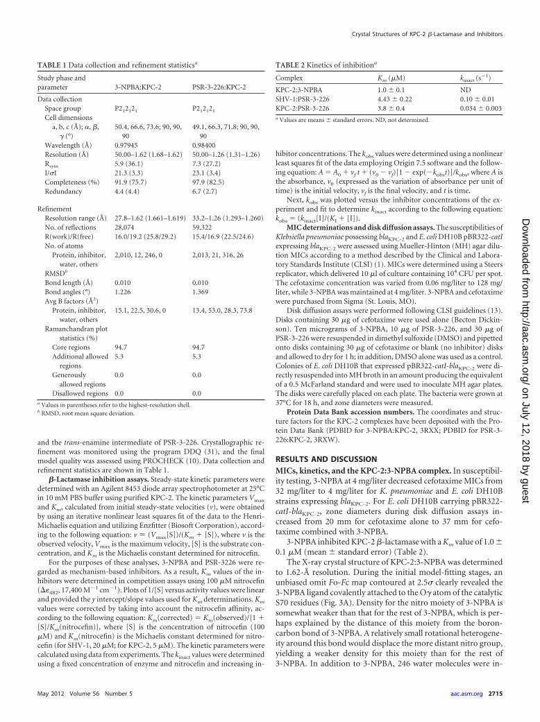
TABLE 1 Data collection and refinement statisticsa
Study phase andparameter 3-NPBA:KPC-2 PSR-3-226:KPC-2
Data collectionSpace group P212121 P212121
Cell dimensionsa, b, c (Å); �, �,
� (°)50.4, 66.6, 73.6; 90, 90,
9049.1, 66.3, 71.8; 90, 90,
90Wavelength (Å) 0.97945 0.98400Resolution (Å) 50.00–1.62 (1.68–1.62) 50.00–1.26 (1.31–1.26)Rsym 5.9 (36.1) 7.3 (27.2)I/�I 21.3 (3.3) 23.1 (3.4)Completeness (%) 91.9 (75.7) 97.9 (82.5)Redundancy 4.4 (4.4) 6.7 (2.7)
RefinementResolution range (Å) 27.8–1.62 (1.661–1.619) 33.2–1.26 (1.293–1.260)No. of reflections 28,074 59,322R(work)/R(free) 16.0/19.2 (25.8/29.2) 15.4/16.9 (22.5/24.6)No. of atoms
Protein, inhibitor,water, others
2,010, 12, 246, 0 2,013, 21, 316, 26
RMSDb
Bond length (Å) 0.010 0.010Bond angles (°) 1.226 1.369Avg B factors (Å2)
Protein, inhibitor,water, others
15.1, 22.5, 30.6, 0 13.4, 53.0, 28.3, 73.8
Ramanchandran plotstatistics (%)
Core regions 94.7 94.7Additional allowed
regions5.3 5.3
Generouslyallowed regions
0.0 0.0
Disallowed regions 0.0 0.0a Values in parentheses refer to the highest-resolution shell.b RMSD, root mean square deviation.
TABLE 2 Kinetics of inhibitiona
Complex Km (�M) kinact (s�1)
KPC-2:3-NPBA 1.0 � 0.1 NDSHV-1:PSR-3-226 4.43 � 0.22 0.10 � 0.01KPC-2:PSR-3-226 3.8 � 0.4 0.034 � 0.003a Values are means � standard errors. ND, not determined.
Crystal Structures of KPC-2 �-Lactamase and Inhibitors
May 2012 Volume 56 Number 5 aac.asm.org 2715
on July 12, 2018 by guesthttp://aac.asm
.org/D
ownloaded from

cluded in the model, and the final model refined to an R(work)value of 16.0% and an R(free) value of 19.2% (additional data andrefinement statistics are listed in Table 1).
The overall structure of KPC undergoes little change upon li-gand binding, as the 3-NPBA-bound KPC-2 structure was similarto that of the full-length KPC-2 and the C-terminally truncatedstructures, yielding a root mean square deviation (RMSD) for C�atoms of 0.574 and 0.226 Å, respectively.
In Fig. 4, the interactions of 3-NPBA with the KPC-2 active siteare illustrated. First, 3-NPBA forms a dative bond, via its boronatom, with the O� atom of S70. Second, both boronate oxygensform extensive interactions; one boronate oxygen hydrogenbonds with the backbone nitrogens of S70 and T237 and the car-bonyl oxygen of T237, whereas the other boronate oxygen inter-acts with the N170 and E166 side chains. Third, the CP5 carbonatom of the phenyl ring of 3-NPBA undergoes a 3.8-Å van derWaals interaction with W105 via an edge-to-face interaction, acommon interaction for aromatic moieties (1). Furthermore, the� ring electrons of the phenyl ring of 3-NPBA interact via cat-ion-� interactions with K73 (4.0 Å) and N132 (3.6 Å). Both typesof interactions with phenyl rings have been observed previously(24, 37). Finally, 3-NPBA allows an interaction with a water mol-ecule (Fig. 4A). The positions of the boronic acid oxygens of3-NPBA bound in the active site indicate that the inhibitor adoptsa conformation that is reminiscent of a deacylation transition stateinhibitor. Such a boronic acid transition state analog conforma-
tion was described previously for larger boronic acid transitionstate inhibitor (BATSI) compounds when bound to SHV-1 �-lac-tamase (9) (Fig. 4B). In this conformation, one of the boron hy-droxyl groups is positioned in the oxyanion hole and the other onedisplaces the deacylation water positioned normally between E166and N170. A previous structure analysis of 3-NPBA bound to theclass C �-lactamase AmpC (26) revealed a different orientationfor the ligand compared to the KPC-2:3-NPBA structure. The3-nitro moiety of 3-NPBA in the AmpC complex is in a positionthat would not be compatible within the KPC-2 structure, as itwould sterically clash with the KPC-2 N170 side chain. One com-mon feature, however, between the two 3-NPBA structures is thephenyl ring-Asn interaction, as Asn152 of AmpC is in a similarposition as Asn132 in KPC-2.
MICs, kinetics, and the KPC-2:PSR-3-226 complex. In diskdiffusion assays, 10 �g of PSR-3-226 combined with cefotaximedid not change zone sizes of E. coli DH10B pBR322-catI-blaKPC-2.When 30 �g of PSR-3-226 was combined with cefotaxime, anincrease in zone size from 16 mm to 19 mm was observed.
The measured affinities of PSR-3-226 for KPC-2 and SHV-1were similar, although it had a slightly lower Km for KPC-2 (Table2). The inactivation rate constant kinact is, however, about 3-foldlower for KPC-2 compared to SHV-1, likely pointing to KPC-2’sinherent reduced susceptibility to inhibition.
The X-ray crystal structure of the KPC-2:PSR-3-226 complexwas determined to 1.26-Å resolution. The unbiased omit Fo-Fc
FIG 4 3-NPBA bound to KPC-2. (A) Stereo view of interactions of 3-NPBA in the active site of KPC-2 �-lactamase. (B) Active site superposition of theKPC2:3-NPBA structure with the SHV-1:cefoperazone BATSI structure (PDB ID 3MKF). SHV-1:cefoperazone BATSI is shown in white, and the KPC-2:3-NPBAstructure is in black. Residues used for superpositioning include 70, 73, 105, 130, 132, 166, 170, and 234 to 237.
FIG 3 Unbiased omit Fo-Fc electron density maps contoured at 2.5�, depicting 3-NPBA in the active site of KPC-2 �-lactamase (A) or the trans-enamineintermediate of PSR-3-226 in the active site of KPC-2 �-lactamase (B). Citrate molecules in panel B were omitted for clarity. (C) Partially occupied citratemolecules in the PSR-3-226-bound KPC-2 structure.
Ke et al.
2716 aac.asm.org Antimicrobial Agents and Chemotherapy
on July 12, 2018 by guesthttp://aac.asm
.org/D
ownloaded from

map contoured at 2.5� revealed density for a ligand covalentlyattached to the O� atom of the catalytic S70 residue (Fig. 3B). Atrans-enamine intermediate was modeled into this density, andthe dihedral angle for this trans-enamine was refined to 179.9°.Clear density for the atoms of this intermediate was observed up toits CB atom, whereas the electron density for the rest of the tail ofthe compound was poor. This suggests that the tail region is flex-ible, in particular around the CA-CB bond, and these poorly re-solved atoms include the sulfone moiety, methyl group, andamide tail R2 linker. The apparent flexibility of this part of thecompound was also evident from the refined higher B factors ofthese tail atoms, which is consistent with their poorly resolveddensity. Occupancy for the PSR-3-226 ligand was varied andshowed that the best refinement results occurred with an occu-pancy of 0.7. Adjacent to PSR-3-226, a citrate molecule with twopartial occupancies was also modeled, of which 5 citrate atomshad strong density (Fig. 3C). After careful examination and ad-justment of the structure, we placed a water molecule with occu-pancy of 0.3 in the oxyanion hole and a deacylation water mole-cule with 0.5 occupancy, bridging E166 and N170, to account forthe extra density at these corresponding positions. In total, 316water molecules were included in refinement, and the final modelhad an R(work) of 15.4% and an R(free) of 16.9% (additionalrefinement statistics are listed in Table 1).
The overall protein conformation of KPC-2 when complexedto PSR-3-226 is similar to the uncomplexed full-length KPC-2 andthe C-terminally truncated KPC-2 structures, with an RMSD of0.591 and 0.158 Å, respectively. The active site undergoes littlechange upon ligand binding, except for residues W105 and S130,which have two alternative conformations that appeared to corre-late with the 0.7 occupancy of PSR-3-226 and alternate conforma-tions of citrate, respectively. The alternate W105 conformations,with 30 and 70% occupancies, are likely a result of avoiding stericclashes with the inhibitor, which has 70% occupancy. PSR-3-226forms a linear trans-enamine intermediate in the active site, withthe carbonyl oxygen being positioned in the oxyanion hole, as wasseen before for SA2-13 in SHV-1 (Fig. 5) and also in the R164mutants of SHV-1 (16, 28). Additional interactions are formed bythe C-3 carboxylate group of PSR-3-226, which is within hydro-gen bonding distance of N132, E166, and N170 (Fig. 5A). Such
interactions were also observed for the SHV-1:SA2-13 complex.Furthermore, a superpositioning of these two structures revealedthat substantial parts of SA2-13 and PSR-3-226 are in a similarconformation (Fig. 5B) (16). The interactions of the mostly dis-ordered tail region of PSR-3-226 are not further discussed here,due to the large uncertainty of their atomic positions and becauseof their poor electron densities. In addition to PSR-3-226, therewas also a partially ordered citrate molecule observed in the activesite. The position of the carboxylate of citrate interacting with theKPC-2 active site was localized in the general carboxylate bindingsite of serine �-lactamases, similar to where one of the other car-boxylates of citrate, bicine, and even BLIP residue D49 wereshown to interact in earlier-described KPC-2 structures (7, 8, 25).The presence of a carboxyl moiety, (i.e., that of citrate), situated inthe carboxyl binding pocket in close proximity to the covalentO�-carbonyl carbon bond, is reminiscent of SA2-13 bound toSHV-1, in which SA2-13’s own carboxyl linker is situated in thisposition. Although we assigned this density as a partially orderedcitrate, we did consider the possibility that this density repre-sented the amide linker of PSR-3-226 when in the trans-enamineintermediate conformation; this did not yield a satisfactory fitwith the electron density, likely due to the linker being two atomsshorter than SA2-13. Therefore, this density was modeled as ci-trate. Note that citrate was also present in the previous KPC-2crystal structure with one of its carboxyl moieties in a similarposition as in our structure (25). Interactions of the carboxyl moi-ety of citrate in the active site involved KPC-2 residues S130,adopting two different conformations, T235, T237, and K234.
We did attempt to obtain a KPC-2:SA2-13 complex structurevia soaking SA2-13 with KPC-2 crystals, but we were unsuccessful.This could have been due to steric repulsion, as superpositioningof the SA2-13:wtSHV-1 structure with that of the PSR-3-226-bound KPC-2 structure indicated that SA2-13 binding to KPC-2would be sterically hindered due to W105 in either of the twoobserved conformations, as extrapolated van der Waals distanceswould be less than 2.5 Å. We note that the region of the active site,encompassing residues W105 and P104, has previously been ob-served to have a key role in affecting substrate catalysis and inhi-bition efficacy in KPC-2 (19, 34).
In conclusion, 3-NPBA and PSR-3-226 are the first �-lacta-
FIG 5 PSR-3-226 bound to KPC-2. (A) Stereo view of interactions of PSR-3-226 in the active site of KPC-2. Citrate molecules were omitted for simplicity.Dashed black lines indicated hydrogen bonds. (B) Active site superposition of the KPC-2:PSR-3-226 structure with the SHV-1:SA2-13 structure (PDB ID 2H5S).KPC-2:PSR-3-226 is shown in black, while SHV-1:SA2-13 is white, with the protein shown in a transparent cartoon representation. Residues used for superpo-sition are the same as those used in Fig. 4.
Crystal Structures of KPC-2 �-Lactamase and Inhibitors
May 2012 Volume 56 Number 5 aac.asm.org 2717
on July 12, 2018 by guesthttp://aac.asm
.org/D
ownloaded from

mase inhibitors to be trapped as acyl-enzyme complexes withKPC-2. The KPC-2:PSR-3-226 structure presented here may serveas a good lead for further structure-based inhibitor optimizationbased on the penam sulfone inhibitor scaffold. The observation ofa trans-enamine conformation suggests that this mode of inhibi-tion has significant potential against KPC-2 �-lactamases, and thestructure provides insights into how to further stabilize this deacy-lation-resistant intermediate.
The KPC-2:3-NPBA structure is also a potential starting pointfor future drug design efforts to optimize boronic acid transitionstate inhibitors. Such small boronic acid compounds have clearpotential as inhibitors, because similar compounds, such as3-aminiphenylboronic acid, have a broad specificity for class Acarbapenemases (20). Further studies are under way to optimizethese types of inhibitors.
ACKNOWLEDGMENTS
J.D.B. is supported by the Robert A. Welch Foundation, grant N-0871.F.V.D.A. is supported by the National Institutes of Health (R01AI062968). The Veterans Affairs Merit Review Program, Geriatric Re-search Education and Clinical Care (GRECC), and the National Institutesof Health (RO1 AI063517-01) supported R.A.B. K.M.P.-W. is supportedby the Veterans Affairs Career Development Program.
The pBR322-catI-blaKPC-2 vector in Escherichia coli DH10B cells was akind gift of Fred Tenover, Centers for Disease Control and Prevention,Atlanta, GA. We thank the staff of Stanford Synchrotron Radiation Light-sourse beamline BL11-1 and Advanced Photon Source beamline 23-IDfor help with data collection.
REFERENCES1. Burley SK, Petsko GA. 1985. Aromatic-aromatic interaction: a mecha-
nism of protein structure stabilization. Science 229:23–28.2. Chen Y, Minasov G, Roth TA, Prati F, Shoichet BK. 2006. The deacy-
lation mechanism of AmpC beta-lactamase at ultrahigh resolution. J. Am.Chem. Soc. 128:2970 –2976.
3. Drawz SM, Bonomo RA. 2010. Three decades of beta-lactamase inhibi-tors. Clin. Microbiol. Rev. 23:160 –201.
4. Drawz SM, Taracila M, Caselli E, Prati F, Bonomo RA. 2011. Exploringsequence requirements for C/C carboxylate recognition in the Pseudomonasaeruginosa cephalosporinase: insights into plasticity of the AmpC beta-lactamase. Protein Sci. 20:941–958.
5. Emsley P, Cowtan K. 2004. Coot: model-building tools for moleculargraphics. Acta Crystallogr. D Biol. Crystallogr. 60:2126 –2132.
6. Halstead DC, et al. 2009. Klebsiella pneumoniae carbapenemase-producingEnterobacteriaceae, northeast Florida. South. Med. J. 102:680–687.
7. Hanes MS, Jude KM, Berger JM, Bonomo RA, Handel TM. 2009.Structural and biochemical characterization of the interaction betweenKPC-2 beta-lactamase and beta-lactamase inhibitor protein. Biochemis-try 48:9185–9193.
8. Ke W, Bethel CR, Thomson JM, Bonomo RA, van den Akker F. 2007.Crystal structure of KPC-2: insights into carbapenemase activity in class Abeta-lactamases. Biochemistry 46:5732–5740.
9. Ke W, et al. 2011. Novel insights into the mode of inhibition of class ASHV-1 beta-lactamases revealed by boronic acid transition state inhibi-tors. Antimicrob. Agents Chemother. 55:174 –183.
10. Laskowski RA, MacArthur MW, Moss DS, Thornton JM. 2001.PROCHECK: a program to check the stereochemical quality of proteinstructures. J. Appl. Crystallogr. 26:283–291.
11. McCoy AJ, et al. 2007. Phaser crystallographic software. J. Appl. Crystal-logr. 40:658 – 674.
12. Murshudov GN, Vagin AA, Dodson EJ. 1997. Refinement of macromo-lecular structures by the maximum-likelihood method. Acta Crystallogr.D Biol. Crystallogr. 53:240 –255.
13. National Committee for Clinical Laboratory Standards. 2005. Perfor-mance standards for antimicrobial susceptibility testing; 15th interna-tional supplement (M100-S15). National Committee for Clinical Labora-tory Standards, Wayne, PA.
14. Nordmann P, Cuzon G, Naas T. 2009. The real threat of Klebsiellapneumoniae carbapenemase-producing bacteria. Lancet Infect. Dis.9:228 –236.
15. Otwinowski Z, Minor W. 1997. Processing of X-ray diffraction datacollected in oscillation mode. Methods Enzymol. 276:307–326.
16. Padayatti PS, et al. 2006. Rational design of a beta-lactamase inhibitorachieved via stabilization of the trans-enamine intermediate: 1.28 Å crystalstructure of wt SHV-1 complex with a penam sulfone. J. Am. Chem. Soc.128:13235–13242.
17. Pagadala, SR, et al. 2011. Penicillin sulfones: an investigation of theeffect of the 2=-substituent, abstr MEDI-299. Abstr. Papers 242nd ACSNatl. Meet. Expo., Denver, CO. American Chemical Society, Washing-ton, DC.
18. Papp-Wallace KM, et al. 2010. Inhibitor resistance in the KPC-2 beta-lactamase, a preeminent property of this class A beta-lactamase. Antimi-crob. Agents Chemother. 54:890 – 897.
19. Papp-Wallace KM, et al. 2010. Elucidating the role of Trp105 in theKPC-2 beta-lactamase. Protein Sci. 19:1714 –1727.
20. Pasteran F, Mendez T, Guerriero L, Rapoport M, Corso A. 2009.Sensitive screening tests for suspected class A carbapenemase productionin species of Enterobacteriaceae. J. Clin. Microbiol. 47:1631–1639.
21. Pasteran F, et al. 2011. A simple test for the detection of KPC and metallo-beta-lactamase carbapenemase-producing Pseudomonas aeruginosa iso-lates with the use of meropenem disks supplemented with aminophenyl-boronic acid, dipicolinic acid and cloxacillin. Clin. Microbiol. Infect. 17:1438 –1441.
22. Patel G, Bonomo RA. 2011. Status report on carbapenemases: challengesand prospects. Expert Rev. Anti Infect. Ther. 9:555–570.
23. Perez F, et al. 2010. Carbapenem-resistant Acinetobacter baumannii andKlebsiella pneumoniae across a hospital system: impact of post-acute carefacilities on dissemination. J. Antimicrob. Chemother. 65:1807–1818.
24. Perutz MF, Fermi G, Abraham DJ, Poyart C, Bursaux E. 1986. Hemo-globin as a receptor of drugs and peptides: X-ray studies of the stereo-chemistry of binding. J. Am. Chem. Soc. 108:1064 –1078.
25. Petrella S, et al. 2008. Genetic and structural insights into the dissemina-tion potential of the extremely broad-spectrum class A beta-lactamaseKPC-2 identified in an Escherichia coli strain and an Enterobacter cloacaestrain isolated from the same patient in France. Antimicrob. Agents Che-mother. 52:3725–3736.
26. Powers RA, Shoichet BK. 2002. Structure-based approach for bindingsite identification on AmpC beta-lactamase. J. Med. Chem. 45:3222–3234.
27. Robledo IE, et al. 2010. Detection of KPC in Acinetobacter spp. in PuertoRico. Antimicrob. Agents Chemother. 54:1354 –1357.
28. Sampson JM, et al. 2011. Ligand-dependent disorder of the omega loopobserved in extended-spectrum SHV-type �-lactamase. Antimicrob.Agents Chemother. 55:2303–2309.
29. Schuttelkopf AW, van Aalten DM. 2004. PRODRG: a tool for high-throughput crystallography of protein-ligand complexes. Acta Crystal-logr. D Biol. Crystallogr. 60:1355–1363.
30. Smith ME, et al. 2003. Plasmid-mediated, carbapenem-hydrolysing beta-lactamase, KPC-2, in Klebsiella pneumoniae isolates. J. Antimicrob. Che-mother. 51:711–714.
31. van den Akker F, Hol WG. 1999. Difference density quality (DDQ): amethod to assess the global and local correctness of macromolecular crys-tal structures. Acta Crystallogr. D Biol. Crystallogr. 55:206 –218.
32. Villegas MV, et al. 2006. First detection of the plasmid-mediated class Acarbapenemase KPC-2 in clinical isolates of Klebsiella pneumoniae fromSouth America. Antimicrob. Agents Chemother. 50:2880 –2882.
33. Walsh TR. 2010. Emerging carbapenemases: a global perspective. Int. J.Antimicrob. Agents 36(Suppl. 3):S8 –S14.
34. Wolter DJ, et al. 2009. Phenotypic and enzymatic comparative analysis ofthe novel KPC variant KPC-5 and its evolutionary variants, KPC-2 andKPC-4. Antimicrob. Agents Chemother. 53:557–562.
35. Woodford N, Turton JF, Livermore DM. 2011. Multiresistant Gram-negative bacteria: the role of high-risk clones in the dissemination of an-tibiotic resistance. FEMS Microbiol. Rev. 35:736 –755.
36. Yigit H, et al. 2001. Novel carbapenem-hydrolyzing beta-lactamase,KPC-1, from a carbapenem-resistant strain of Klebsiella pneumoniae. An-timicrob. Agents Chemother. 45:1151–1161.
37. Zacharias N, Dougherty DA. 2002. Cation-pi interactions in ligand rec-ognition and catalysis. Trends Pharmacol. Sci. 23:281–287.
Ke et al.
2718 aac.asm.org Antimicrobial Agents and Chemotherapy
on July 12, 2018 by guesthttp://aac.asm
.org/D
ownloaded from