crystal structures of ca2+ calmodulin bound to nav c
TRANSCRIPT
Crystal structures of Ca2+–calmodulin bound to NaVC-terminal regions suggest role for EF-handdomain in binding and inactivationBernd R. Gardilla, Ricardo E. Rivera-Acevedoa,1, Ching-Chieh Tunga, and Filip Van Petegema,2
aDepartment of Biochemistry and Molecular Biology, Life Sciences Institute, The University of British Columbia, Vancouver, BC, Canada V6T 1Z3
Edited by Richard W. Aldrich, The University of Texas at Austin, Austin, TX, and approved April 16, 2019 (received for review October 29, 2018)
Voltage-gated sodium (NaV) and calcium channels (CaV) form tar-gets for calmodulin (CaM), which affects channel inactivationproperties. A major interaction site for CaM resides in the C-terminal (CT) region, consisting of an IQ domain downstream ofan EF-hand domain. We present a crystal structure of fully Ca2+-occupied CaM, bound to the CT of NaV1.5. The structure showsthat the C-terminal lobe binds to a site ∼90° rotated relative to aprevious site reported for an apoCaM complex with the NaV1.5 CTand for ternary complexes containing fibroblast growth factor ho-mologous factors (FHF). We show that the binding of FHFs forcesthe EF-hand domain in a conformation that does not allow bindingof the Ca2+-occupied C-lobe of CaM. These observations highlightthe central role of the EF-hand domain in modulating the bindingmode of CaM. The binding sites for Ca2+-free and Ca2+-occupiedCaM contain targets for mutations linked to long-QT syndrome, atype of inherited arrhythmia. The related NaV1.4 channel has beenshown to undergo Ca2+-dependent inactivation (CDI) akin to CaVs.We present a crystal structure of Ca2+/CaM bound to the NaV1.4 IQdomain, which shows a binding mode that would clash with theEF-hand domain. We postulate the relative reorientation of the EF-hand domain and the IQ domain as a possible conformationalswitch that underlies CDI.
sodium channel | inactivation | cardiac arrhythmia | long-QT syndrome |calcium-dependent inactivation
Voltage-gated sodium channels (NaVs) can rapidly depolarizean excitable cell by allowing the influx of extracellular Na+
ions (1). Mammalian NaVs typically assemble from differentsubunits. The principal component is the NaVα subunit, whichforms a 24-transmembrane (TM) helix membrane protein. Theseare organized in four homologous repeats (I–IV), connected bylong linkers (I–II linker, II–III linker, and III–IV linker) that areintrinsically disordered. In mammalian species, nine isoformsexist (NaV1.1–1.9), which differ in expression profile, pharmacology,and electrophysiological properties. The primary isoform expressedin cardiac muscle is NaV1.5. In contrast, the auxiliary NaVβ subunitencodes a smaller protein with an extracellular Ig-like domain, asingle TM helix, and a short cytosolic intracellular tail (2).Recent advances in cryo-EM have allowed reconstructions of
NaV1.4 from electric eels (3) and humans (4) and an Nav from theAmerican cockroach (5). In addition, high-resolution crystalstructures have been reported for the Ig domains of mammalianNaVβ2, β3, and β4 (6–9). Despite these advances, the bulk of thecytosolic region appeared invisible in the cryo-EM reconstructions,suggesting inherent flexibility relative to the TM region. With theexception of the cockroach NaV structure, no interpretable densitywas present downstream of the last TM segment.The C-terminal region (CT) of the NaVα subunits encode an
EF-hand–like domain, immediately downstream of the last TMsegment (10–12). EF-hand domains are frequently observed tobind Ca2+, but thus far all available crystal structures have beenunable to reveal Ca2+ density even at high Ca2+ concentrations(13). In the cryo-EM structure of the cockroach NaV, the EF-hand–like domain is found to interact with the III–IV linker (5).
NMR experiments suggest that this interaction also occurs inmammalian NaVs, and that it regulates the degree of channelinactivation (14). The EF-hand–like domain is followed by twoα-helices, one which latches on to the domain termed the “pre-IQ helix” and a second one containing an IQ motif. The IQ motifforms a binding site for both Ca2+-free and Ca2+-occupied cal-modulin (CaM), a ubiquitous Ca2+ sensor that endows manyproteins with Ca2+-dependent regulation.Several roles have been postulated for CaM binding to NaVs
and unfortunately there is disagreement on the precise functions(15). Multiple groups have reported a depolarizing shift in thesteady-state inactivation curves at elevated cytosolic Ca2+ levels(micromolar range) (12, 16–20). However, this was not recapit-ulated in an experiment with Ca2+ uncaging, and in the case ofone particular isoform, NaV1.4, Ca
2+/CaM was found to promoteinactivation in a manner similar to voltage-gated calcium chan-nels (CaV) (21). In several CaV isoforms, CaM causes a robustacceleration of current inactivation for several isoforms (CDI,Ca2+-dependent inactivation) (22–24). Since these CaV isoformsalso contain a CT encoding EF-hands and an IQ domain, the wayCaM interacts with and modulates these channels may be verysimilar. Indeed, it was found that chimeric CaV1.3 channels with theNaV1.4 CT also display CDI, further highlighting the similarities
Significance
Calmodulin (CaM) is a Ca2+-sensing protein that endows sev-eral voltage-gated sodium (NaV) and calcium (CaV) channelswith Ca2+-dependent inactivation (CDI). Although this phe-nomenon has been known to exist for decades, the exactmechanism remains at large. Several high-resolution structureshave captured complexes between Ca2+/CaM and NaV or CaV C-terminal IQ domains, but structures are scarce for a larger(Ca2+)4/CaM complex that also includes the upstream EF-handdomain, a domain that is critical for CDI. We show crystalstructures of the C-terminal domain of the cardiac Nav1.5 andthe IQ domain of Nav1.4, two NaVs that are differentially reg-ulated by CaM. The results uncover a role for the EF-hand do-main in dictating binding of CaM and suggest a conformationalswitch in CDI.
Author contributions: B.R.G., R.E.R.-A., and F.V.P. designed research; B.R.G., R.E.R.-A.,C.-C.T., and F.V.P. performed research; B.R.G., R.E.R.-A., C.-C.T., and F.V.P. analyzed data;and B.R.G. and F.V.P. wrote the paper.
The authors declare no conflict of interest.
This article is a PNAS Direct Submission.
Published under the PNAS license.
Data deposition: Crystal structures have been deposited in the Protein Data Bank, www.wwpdb.org (PDB ID codes 6MUD and 6MUE).1Present address: Department of Anesthesiology, Pharmacology, and Therapeutics, TheUniversity of British Columbia, Vancouver, BC, Canada V6T 1Z3.
2To whom correspondence should be addressed. Email: [email protected].
This article contains supporting information online at www.pnas.org/lookup/suppl/doi:10.1073/pnas.1818618116/-/DCSupplemental.
Published online May 9, 2019.
www.pnas.org/cgi/doi/10.1073/pnas.1818618116 PNAS | May 28, 2019 | vol. 116 | no. 22 | 10763–10772
BIOCH
EMISTR
Y
Dow
nloa
ded
by g
uest
on
Janu
ary
24, 2
022
between both channels (21). Despite these parallels, there is alsodivergence, as NaV CT fragments bind Ca2+-free CaM strongerthan Ca2+/CaM, opposite to most CaV1 and CaV2 channels. Inaddition, NaV CT fragments can bind FHF proteins, whereas CaVCTs do not (13, 25, 26). CaM has also been reported to interactwith the III–IV linker in NaV1.5 (16, 27), but so far this has not yetbeen described for CaVs.There is thus great interest in trying to understand how CaM
interacts with the CT of both NaV and CaV channels, in bothCa2+-free (apoCaM) and Ca2+-occupied (Ca2+/CaM) forms. Sofar, the IQ domain has remained invisible in all NaV and CaVcryo-EM reconstructions, and all structural insights have thuscome from crystallographic and NMR studies. For CaV channels,all high-resolution studies only contain the isolated IQ domainwith or without a pre-IQ helix, thus not showing the role of theEF-hand region (28–32). For NaV channels, however, crystalstructures containing the longer CT, encompassing EF-handsand IQ domain, are available. One report was for a ternarycomplex between CaM, the NaV1.5 CT, and FGF13, a fibroblastgrowth factor homologous factor (FHF) (25). Mg2+ was bound inthe Ca2+ binding sites, and thus the conformation of the lobes isthat of an apoCaM. We therefore refer to this structure as anapoCaM complex. The structure showed the apoC-lobe inter-acting with the N-terminal portion of the IQ domain, with theapoN-lobe not being involved in the interaction. Another reportalso captured the NaV1.5 CT in complex with apoCaM, but thistime in the absence of an FHF protein (33). This shows theapoC-lobe to interact with the IQ domain at the same site as inthe presence of the FHF protein. Additional interactions werealso observed between the apoN-lobe and the NaV1.5 EF-handdomain. The authors also noted a different relative orientationof the EF-hand domain to the IQ domain depending on thepresence of an FHF (33). An NMR study of the NaV1.5 IQdomain in complex with apoCaM also showed that the in-teraction is driven by the C-lobe, with a flexible N-lobe relative tothe IQ domain (11). Another NMR study also supports anapoCaM interaction with the IQ domain that is driven by the C-lobe (34). In addition, ternary complexes were solved for theNaV1.5 or NaV1.2 CT in complex with CaM and an FHF insaturating Ca2+ concentrations (13). Although reported as aCa2+/CaM complex of the NaV CT, the C-lobe did not displaythe open conformation observed for Ca2+/CaM, and this struc-
ture is in disagreement with NMR studies of isolated IQ domainsin complex with Ca2+/CaM lobes (35, 36). Therefore, a structureof Ca2+/CaM bound to the CT of either an NaV or CaV, con-taining both IQ domain and EF-hands, has not yet been reported.Here, we report a crystal structure of Ca2+/CaM bound to the
NaV1.5 CT. Although a previous structure was previouslyreported as a Ca2+/CaM complex (13), we show here that this is amisinterpretation and that the C-lobe was in the apo form. Ourdata show that the presence of the EF-hand is crucial in dictatingthe exact binding mode of CaM on the IQ domain, and thataltering the conformation of the EF-hand domain through aux-iliary proteins like FHFs can prevent Ca2+/CaM from binding tothe IQ domain. We also investigate the differences between Ca2+/CaM binding to different NaV isoforms, which hint at a possiblemechanism for CDI.
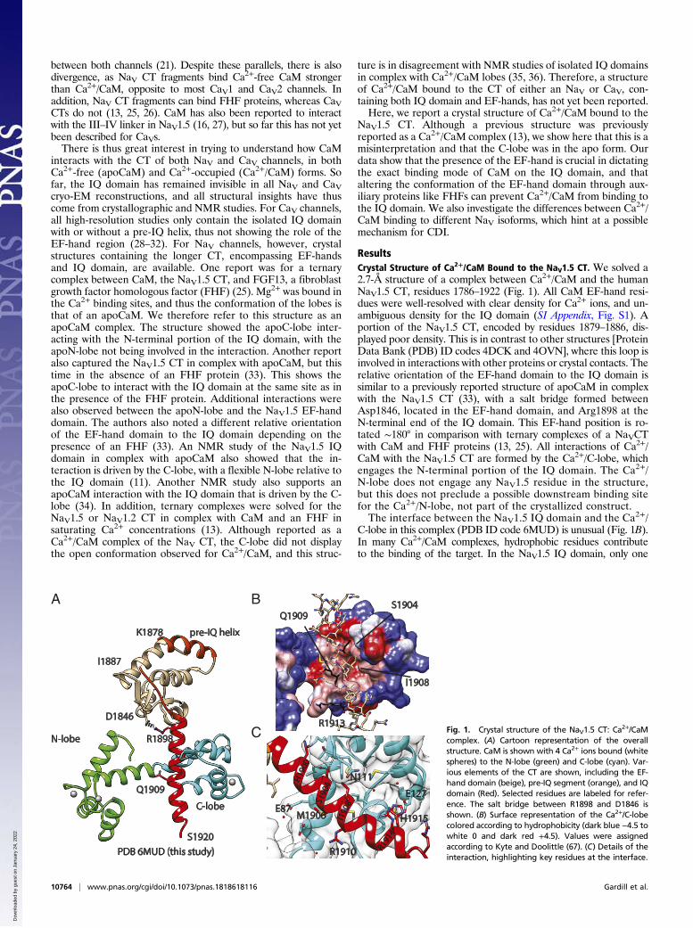
ResultsCrystal Structure of Ca2+/CaM Bound to the NaV1.5 CT. We solved a2.7-Å structure of a complex between Ca2+/CaM and the humanNaV1.5 CT, residues 1786–1922 (Fig. 1). All CaM EF-hand resi-dues were well-resolved with clear density for Ca2+ ions, and un-ambiguous density for the IQ domain (SI Appendix, Fig. S1). Aportion of the NaV1.5 CT, encoded by residues 1879–1886, dis-played poor density. This is in contrast to other structures [ProteinData Bank (PDB) ID codes 4DCK and 4OVN], where this loop isinvolved in interactions with other proteins or crystal contacts. Therelative orientation of the EF-hand domain to the IQ domain issimilar to a previously reported structure of apoCaM in complexwith the NaV1.5 CT (33), with a salt bridge formed betweenAsp1846, located in the EF-hand domain, and Arg1898 at theN-terminal end of the IQ domain. This EF-hand position is ro-tated ∼180° in comparison with ternary complexes of a NaVCTwith CaM and FHF proteins (13, 25). All interactions of Ca2+/CaM with the NaV1.5 CT are formed by the Ca2+/C-lobe, whichengages the N-terminal portion of the IQ domain. The Ca2+/N-lobe does not engage any NaV1.5 residue in the structure,but this does not preclude a possible downstream binding sitefor the Ca2+/N-lobe, not part of the crystallized construct.The interface between the NaV1.5 IQ domain and the Ca2+/
C-lobe in this complex (PDB ID code 6MUD) is unusual (Fig. 1B).In many Ca2+/CaM complexes, hydrophobic residues contributeto the binding of the target. In the NaV1.5 IQ domain, only one
I1887
K1878 pre-IQ helix
N-lobe
C-lobe
S1920
Q1909
D1846
R1898
H190
0S1
904
I190
8
R191
4
H1915
R1910
C
A B
E127
M1906
N111
E87
PDB 6MUD (this study)
Q1909S1904
R1913
I1908
Fig. 1. Crystal structure of the NaV1.5 CT: Ca2+/CaMcomplex. (A) Cartoon representation of the overallstructure. CaM is shown with 4 Ca2+ ions bound (whitespheres) to the N-lobe (green) and C-lobe (cyan). Var-ious elements of the CT are shown, including the EF-hand domain (beige), pre-IQ segment (orange), and IQdomain (Red). Selected residues are labeled for refer-ence. The salt bridge between R1898 and D1846 isshown. (B) Surface representation of the Ca2+/C-lobecolored according to hydrophobicity (dark blue −4.5 towhite 0 and dark red +4.5). Values were assignedaccording to Kyte and Doolittle (67). (C) Details of theinteraction, highlighting key residues at the interface.
10764 | www.pnas.org/cgi/doi/10.1073/pnas.1818618116 Gardill et al.
Dow
nloa
ded
by g
uest
on
Janu
ary
24, 2
022

Phe is present, which lies on the opposite side from the C-lobeinterface. Instead, the side chains of Val1903 and Val1907 makehydrophobic contacts with the C-lobe, whereas the pocket linedby C-lobe residues Met124, Phe141, and Met144 is only occupiedby a water molecule. A water molecule has been found at asimilar location in a high-resolution structure of Ca2+/CaMwithout peptide bound (PDB ID code 1EXR) (37). Other in-teractions of note involve IQ domain residues His1900, Ser1904,Met1906, Ile1908, and Arg1914 (Fig. 1C). Given the absence oftypical aromatic anchors, the interaction is relatively weak. In-deed, isothermal titration calorimetry (ITC) experiments titrat-ing C-lobe into the IQ domain under saturating Ca2+ concentrationsshowed an affinity (Kd) of ∼6 μM (16).These interactions are very different from the ones observed
for apoCaM with NaV1.5 CT (PDB ID code 4OVN), as the C-lobe is rotated around the IQ domain by ∼90° (Fig. 2). ApoCaMengages NaV1.5 residue Phe1912, whereas Ca2+/CaM does not.This likely underlies the large difference in affinity, as theapoCaM affinity for the NaV1.5 CT is an order of magnitudehigher than Ca2+/CaM (16).It has been suggested that the Ca2+/C-lobe also interacts with
the EF-hand domain in CaVs (38). In our structure, we observe avan der Waals interaction between EF-hand domain residueIle1833 and C-lobe residue Glu87, suggesting only a modestcontribution to the binding affinity. Investigation of the elec-trostatic surface potential suggests no major effect of electro-statics on the interaction between the Ca2+/C-lobe and EF-handdomain (SI Appendix, Fig. S2). In agreement with this, compar-ison of ITC data for binding of Ca2+/C-lobe to the NaV1.5 CT
(residues 1773–1924) showed an affinity not significantly differ-ent from the affinity to the individual IQ domain (16).
The Role of the EF-Hand Domain in Dictating the Binding Mode ofCaM. Previously, Wang et al. (13) published a crystal structurethat was proposed to represent a complex between Ca2+/CaMand the CT of NaV1.2 and NaV1.5. However, as noted by Hoveyet al. (36), a closer inspection of these structures (PDB ID codes4JPZ and 4JQ0) shows that the C-lobes display a semiopenconformation such as observed in complexes of apoCaM withmyosin V (39). Indeed, a direct superposition of the C-lobe withthe one in our current structure shows a very different confor-mation (Fig. 2A). Instead, the C-lobe conformation by Wanget al. (13) closely resembles the conformation found in a struc-ture of apoCaM in complex with the NaV1.5 CT. In contrast, theN-lobe in the complex by Wang et al. (13) displays the typicalopen conformation for a Ca2+-occupied lobe (Fig. 2 B and C).Irregularities in the difference density maps, as well as un-expected geometries for Ca2+-chelating residues in EF-hands3 and 4 for these structure (PDB ID codes 4JPZ and 4JQ0),were previously also reported by Hovey et al. (36). We furtherrefer to this structure containing a Ca2+/N-lobe and apoC-lobeconformation as a mixed CaM.How could saturating levels of Ca2+ (2 to 100 mM) used by
Wang et al. (13) result in a mixed CaM? Fig. 2D shows a directcomparison of the Ca2+/CaM and mixed CaM complexes withthe NaV1.5 CT. The view is based on superposing the IQ domain,showing the relative positions of the individual lobes using theIQ domain as reference. Importantly, the Ca2+/C-lobe binding
6MUD (this study) 4DCK (apoCaM)4JQ0 (mixed CaM)4OVN (apoCaM)
D
C I1908
Q1909
6MUD
4JQ04OVN4DCK
E
B
E
A
F
GH
A
B
C
D
E
F
G
H6MUD
4OVN4JQ0
6MUD4JQ0
F
E
H
G
Fig. 2. Structural comparison. (A) Comparisons ofthe C-lobe conformation from this study (PDB ID code6MUD, blue), a C-lobe from a previously proposedNaVCT:Ca
2+/CaM complex (PDB ID code 4JQ0, gray),and an apoC-lobe (PDB ID code 4OVN, magenta),which shows that the previously proposed Ca2+/C-lobefrom a NaV CT complex (13) is an apoC-lobe, not aCa2+/C-lobe. The letters in A–C correspond to the CaMhelices. (B) Comparisons of the N-lobe from this study(green) with the N-lobe from PDB ID code 4JQ0 (gray),confirming that the latter represents a Ca2+-occupiedN-lobe. (C) Superpositions showing the relative C-lobepositions found in this study (6MUD, cyan) and threeother previous CaM complex with the NaV1.5 CT. (D)Side-by-side comparison of the current structure (PDBID code 6MUD) and three other CaM complexes, in-cluding two apo-CaM complexes (PDB ID codes 4OVNand 4DCK) and one previously proposed Ca2+/CaMcomplex, which is a mixed CaM (PDB ID code 4JQ0).The structures are shown in the exact same view,based on a superposition of the sixth helix immedi-ately after the EF-hand domain. (E) Superposition ofthe Ca2+/CaM (PDB ID code 6MUD) and mixed CaM(PDB ID code 4JQ0) complexes with NaV1.5CT, basedon the sixth helix containing the IQ domain, using thesame colors as in D. This shows that the Ca2+/C-lobe(cyan) from the NaV1.5 CT: Ca2+/CaM structure wouldclash with the NaV1.5 EF-hand domain in the mixedCaM complex (PDB ID code 4JQ0). Therefore, bindingof the Ca2+/C-lobe to the IQ domain is prevented by theFHF, which utilizes reorientation of the EF-hand domainas a mechanism to prevent Ca2+/C-lobe binding.
Gardill et al. PNAS | May 28, 2019 | vol. 116 | no. 22 | 10765
BIOCH
EMISTR
Y
Dow
nloa
ded
by g
uest
on
Janu
ary
24, 2
022
site is on the opposite face of the IQ helix compared with theapoC-lobe site. The direct comparison reveals a crucial role forthe NaV1.5 EF-hand domain, which adopts a very different ori-entation relative to the IQ domain. The EF-hand domain, asobserved in the mixed-CaM complex, would clash with theCa2+/C-lobe in our structure (Fig. 2E).One crucial difference in the conditions used in this report and
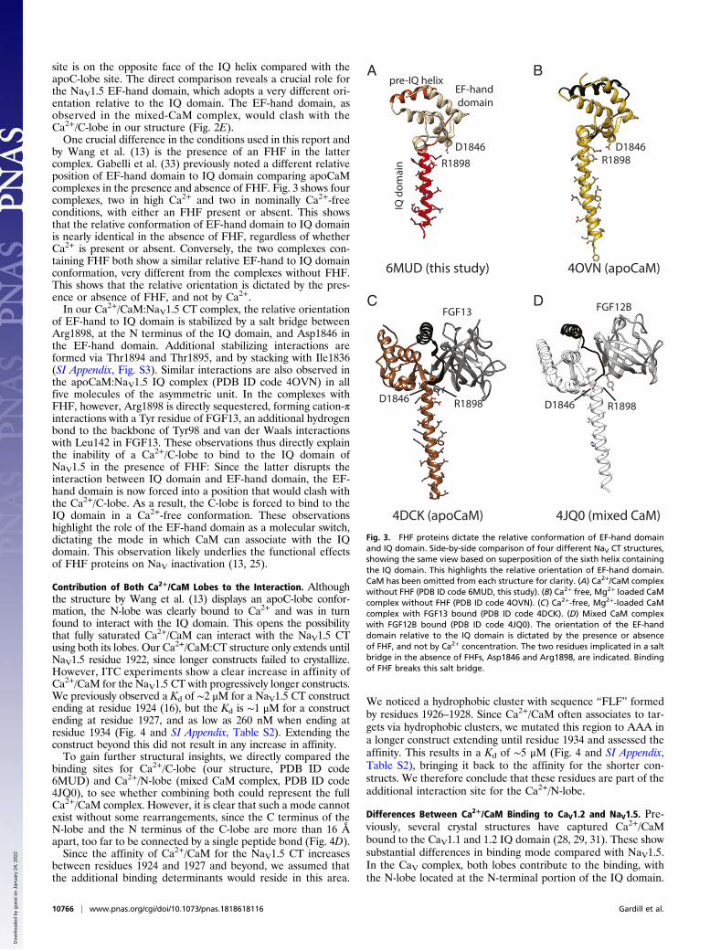
by Wang et al. (13) is the presence of an FHF in the lattercomplex. Gabelli et al. (33) previously noted a different relativeposition of EF-hand domain to IQ domain comparing apoCaMcomplexes in the presence and absence of FHF. Fig. 3 shows fourcomplexes, two in high Ca2+ and two in nominally Ca2+-freeconditions, with either an FHF present or absent. This showsthat the relative conformation of EF-hand domain to IQ domainis nearly identical in the absence of FHF, regardless of whetherCa2+ is present or absent. Conversely, the two complexes con-taining FHF both show a similar relative EF-hand to IQ domainconformation, very different from the complexes without FHF.This shows that the relative orientation is dictated by the pres-ence or absence of FHF, and not by Ca2+.In our Ca2+/CaM:NaV1.5 CT complex, the relative orientation
of EF-hand to IQ domain is stabilized by a salt bridge betweenArg1898, at the N terminus of the IQ domain, and Asp1846 inthe EF-hand domain. Additional stabilizing interactions areformed via Thr1894 and Thr1895, and by stacking with Ile1836(SI Appendix, Fig. S3). Similar interactions are also observed inthe apoCaM:NaV1.5 IQ complex (PDB ID code 4OVN) in allfive molecules of the asymmetric unit. In the complexes withFHF, however, Arg1898 is directly sequestered, forming cation-πinteractions with a Tyr residue of FGF13, an additional hydrogenbond to the backbone of Tyr98 and van der Waals interactionswith Leu142 in FGF13. These observations thus directly explainthe inability of a Ca2+/C-lobe to bind to the IQ domain ofNaV1.5 in the presence of FHF: Since the latter disrupts theinteraction between IQ domain and EF-hand domain, the EF-hand domain is now forced into a position that would clash withthe Ca2+/C-lobe. As a result, the C-lobe is forced to bind to theIQ domain in a Ca2+-free conformation. These observationshighlight the role of the EF-hand domain as a molecular switch,dictating the mode in which CaM can associate with the IQdomain. This observation likely underlies the functional effectsof FHF proteins on NaV inactivation (13, 25).
Contribution of Both Ca2+/CaM Lobes to the Interaction. Althoughthe structure by Wang et al. (13) displays an apoC-lobe confor-mation, the N-lobe was clearly bound to Ca2+ and was in turnfound to interact with the IQ domain. This opens the possibilitythat fully saturated Ca2+/CaM can interact with the NaV1.5 CTusing both its lobes. Our Ca2+/CaM:CT structure only extends untilNaV1.5 residue 1922, since longer constructs failed to crystallize.However, ITC experiments show a clear increase in affinity ofCa2+/CaM for the NaV1.5 CT with progressively longer constructs.We previously observed a Kd of ∼2 μM for a NaV1.5 CT constructending at residue 1924 (16), but the Kd is ∼1 μM for a constructending at residue 1927, and as low as 260 nM when ending atresidue 1934 (Fig. 4 and SI Appendix, Table S2). Extending theconstruct beyond this did not result in any increase in affinity.To gain further structural insights, we directly compared the
binding sites for Ca2+/C-lobe (our structure, PDB ID code6MUD) and Ca2+/N-lobe (mixed CaM complex, PDB ID code4JQ0), to see whether combining both could represent the fullCa2+/CaM complex. However, it is clear that such a mode cannotexist without some rearrangements, since the C terminus of theN-lobe and the N terminus of the C-lobe are more than 16 Åapart, too far to be connected by a single peptide bond (Fig. 4D).Since the affinity of Ca2+/CaM for the NaV1.5 CT increases
between residues 1924 and 1927 and beyond, we assumed thatthe additional binding determinants would reside in this area.
We noticed a hydrophobic cluster with sequence “FLF” formedby residues 1926–1928. Since Ca2+/CaM often associates to tar-gets via hydrophobic clusters, we mutated this region to AAA ina longer construct extending until residue 1934 and assessed theaffinity. This results in a Kd of ∼5 μM (Fig. 4 and SI Appendix,Table S2), bringing it back to the affinity for the shorter con-structs. We therefore conclude that these residues are part of theadditional interaction site for the Ca2+/N-lobe.
Differences Between Ca2+/CaM Binding to CaV1.2 and NaV1.5. Pre-viously, several crystal structures have captured Ca2+/CaMbound to the CaV1.1 and 1.2 IQ domain (28, 29, 31). These showsubstantial differences in binding mode compared with NaV1.5.In the CaV complex, both lobes contribute to the binding, withthe N-lobe located at the N-terminal portion of the IQ domain.
DC
BA
6MUD (this study)
4DCK (apoCaM) 4JQ0 (mixed CaM)
4OVN (apoCaM)
D1846R1898
D1846R1898
IQ d
omai
n
EF-hand domain
pre-IQ helix
D1846 R1898 D1846 R1898
FGF12BFGF13
Fig. 3. FHF proteins dictate the relative conformation of EF-hand domainand IQ domain. Side-by-side comparison of four different NaV CT structures,showing the same view based on superposition of the sixth helix containingthe IQ domain. This highlights the relative orientation of EF-hand domain.CaM has been omitted from each structure for clarity. (A) Ca2+/CaM complexwithout FHF (PDB ID code 6MUD, this study). (B) Ca2+ free, Mg2+ loaded CaMcomplex without FHF (PDB ID code 4OVN). (C) Ca2+-free, Mg2+-loaded CaMcomplex with FGF13 bound (PDB ID code 4DCK). (D) Mixed CaM complexwith FGF12B bound (PDB ID code 4JQ0). The orientation of the EF-handdomain relative to the IQ domain is dictated by the presence or absenceof FHF, and not by Ca2+ concentration. The two residues implicated in a saltbridge in the absence of FHFs, Asp1846 and Arg1898, are indicated. Bindingof FHF breaks this salt bridge.
10766 | www.pnas.org/cgi/doi/10.1073/pnas.1818618116 Gardill et al.
Dow
nloa
ded
by g
uest
on
Janu
ary
24, 2
022

The binding affinity here is also much higher, with a Kd in the lownanomolar range for the Ca2+/C-lobe alone (28, 40), and subpMfor full Ca2+/CaM (41). As the Ca2+/C-lobe binding affinity forthe NaV1.5 CT is three orders of magnitude lower, substantialdifferences at the interface with the IQ domain are expected.Indeed, the CaV1.2 IQ domain contributes three aromatic resi-dues to the interface with the Ca2+/Clobe, whereas the NaV1.5IQ domain contributes none (SI Appendix, Fig. S4). There is alsoa ∼90° rotation of the Ca2+/C-lobe relative to the IQ domain.
Disease Mutations in the NaV1.5 IQ Domain Can Affect Either Ca2+/CaM or apoCaM Binding. NaV1.5 is the target for disease mutationslinked to inherited arrhythmias, and four of these are locatedwithin the IQ domain, where they could interfere with eitherapoCaM and/or Ca2+/CaM binding. Fig. 5 highlights the muta-tions, which were previously shown to be involved in apoCaMbinding (33). E1901Q is at the N-terminal end of the IQ domainand has been involved in type-3 long-QT syndrome (LQT3) (42). Itwas shown to cause an increase in late sodium current up to 2.5%(43). However, this could be restored by increasing CaM expres-sion. A Ca2+ dependence of the steady-state inactivation curve hasnot been investigated. Glu1901 is involved in a salt bridge withLys95 in the apoCaM complex but not in any interactions withCa2+/CaM, as it is pointing to the solvent (Fig. 5 and SI Appendix,Fig. S5). Therefore, no effect is expected on Ca2+/CaM binding.
Two additional mutations, Q1909R (LQT3) (44) and R1913H(LQT3) (42), are also at interfaces with apoCaM, but not withCa2+/CaM (Fig. 5 and SI Appendix, Fig. S5). Both were pre-viously shown to cause an increase in late sodium current, andcoexpression of excess CaM restores this to wild-type levels (43).No inherent shift in steady-state inactivation (SSI) was observed.A separate report confirmed that Q1909R causes an increase inlate current, which could be normalized by increased cytosolicCa2+ (45). It thus seems that, for mutations that affect apoCaM,but not Ca2+/CaM binding, late currents could still be dampenedby either adding excess CaM (which would compensate for adecreased affinity) or by adding Ca2+, allowing the binding ofCa2+/CaM. In the structure of the CaV1.2 IQ domain:Ca2+/CaMcomplex, the equivalent Gln residue, which defines Q in the IQmotif, is involved in direct interactions with Ca2+/CaM (28, 29).To verify that that this residue is not involved in binding the Ca2+/C-lobe, we performed ITC experiments on the Q1909R mutant(SI Appendix, Fig. S5 and Table S2), which show a very similaraffinity (Kd 7 ± 2 μM versus 9 ± 1 μM for wild type), in agreementwith the structure.S1904L causes an increase in late sodium currents (46). No
inherent shift in SSI was observed. Rescue experiments increasingcytosolic Ca2+ or CaM expression have not been performed forthis mutant. In contrast to the above mutants, S1904L (LQT3)(47) is predicted to affect interactions with both apoCaM and
0.0
ces/lacµ
Molar Ratio Molar RatioMolar Ratio
kcal
/mol
e of
inje
ctan
t
kcal
/mol
e of
inje
ctan
t
kcal
/mol
e of
inje
ctan
t
ces/lacµ
ces/lacµ
Time (min) Time (min) Time (min)
0 0.5 1.0 1.5 2.0 0 0.5 1.0 1.5 2.0 0 0.5 1.0 1.5 2.0
0 10 20 30 40 50 60 0 10 20 30 40 50 60 0 10 20 30 40 50 60
-0.5
-1.5
-1.0
0.0
-0.5
-1.0
0.0
-0.5
-1.5
-1.0
0
-2
-4
0
-2
-4-6-8
-10
0-2-4-6-8
-10-12
Molar Ratio
kcal
/mol
e of
inje
ctan
tces/lacµ
Time (min)0 10 20 30 40 50 60
0.0
-0.5
-1.5
-1.0
0
-2
-4
-6
-8
A B C
E
NaV1.51773-1927
NaV1.51773-1934
NaV1.51773-1943
NaV1.51773-1934FLF/AAA
T79
D78
D
N-lobe
C-lobe
16.8 Å
0 0.5 1.0 1.5 2.0
F
1924 1927 1934 1943 1934FLF/AAA
0.01
0.1
1
Dis
soca
tion
cons
tant
( µM
)
10
Fig. 4. Analysis of additional binding determinants downstream of the IQ domain. All titrations shown are in saturating Ca2+ conditions. (A) ITC measurement of120 μM Nav1.51773–1927 titrated with 1.2 mM CaM. Kd = 1.02 ± 0.07 μM. (B) ITC measurement of 50 μM Nav1.51773–1934 titrated with 0.5 mM CaM. Kd = 0.26 ±0.06 μM. (C) ITC measurement of 60 μM Nav1.51773–1943 titrated with 0.6 mM CaM. Kd = 0.4 ± 0.2 μM. (D) Superposition based on the sixth helix containing the IQdomain to test a hypothetical combination of Ca2+/C-lobe position (this study, blue) and Ca2+/N-lobe (PDB ID code 4jq0, gray). The shown combination would leadto a clash around D78 and substantial distortion would have to occur to allow linking residues 78 and 79. (E) ITC measurement of 100 μM Nav1.51773–1934FLF1926AAA titrated with 1 mM CaM. Kd = 5 ± 1 μM. The mutation reduces binding affinity. (F) Bar graph showing Kd values for various constructs on a log-arithmic scale. The numbers below each bar represent the residue number of the C terminus of each construct. The value for the construct ending at 1924 wastaken from Sarhan et al. (16).
Gardill et al. PNAS | May 28, 2019 | vol. 116 | no. 22 | 10767
BIOCH
EMISTR
Y
Dow
nloa
ded
by g
uest
on
Janu
ary
24, 2
022

Ca2+/CaM: Ser1904 forms van der Waals packing interactionswith the apoC-lobe and it hydrogen bonds to the main chain ofresidues 109 and 110 in the Ca2+/C-lobe (Fig. 5). As it is also tightlypacked against the Ca2+/C-lobe (SI Appendix, Fig. S5), adding abulkier Leu residue is predicted to cause steric hindrance. Toverify the impact of the S1904L mutation on Ca2+/CaM, weperformed ITC experiments (SI Appendix, Fig. S5). As expected,the mutation has a significant impact on the affinity, yielding anisotherm that could not be fitted. In conclusion, the S1904Lmutant is unique among these four, as it affects both apoCaMand Ca2+/CaM binding.
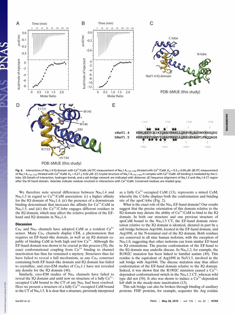
Isoform-Specific Differences Between NaV1.4 and NaV1.5 Hint at aMechanism for CDI. Ca2+-uncaging experiments have shown thatNaV1.4 currents display a Ca2+-sensitive inhibition, similar toCDI in CaV channels (21). Nav1.5, although a close homolog, didnot show this modulation. We therefore set out to characterizethe interaction between Ca2+/CaM and NaV1.4. We noticed theabsence of the “FLF” sequence, which increases the affinity forCa2+/CaM in NaV1.5. Instead, this sequence is replaced by“YMY.” To check whether NaV1.4 has additional binding de-terminants for Ca2+/CaM in this region, we compared peptidesspanning rNaV1.4 1716–1744 or rNaV1.4 1716–1753, respectively.The obtained Kd values (SI Appendix, Table S2) were comparablebetween long and short IQ domain. This is in direct contrast withNaV1.5, which contains an extra binding determinant.We determined a structure of the NaV1.4 IQ domain (rNaV1.4
1716–1744) in complex with Ca2+/CaM. Fig. 6 shows how theinteractions are driven by the C-lobe, with no contributions fromthe N-lobe. Interactions of note include salt bridges betweenNaV1.4 residues Arg1729 and Arg1733 with the Ca2+/C-lobe.Additional hydrogen bonds are formed by His1734 and Lys1726.Hydrophobic contacts are mostly contributed by Val1722 andIle1725. Similar to NaV1.5, no bulky hydrophobic NaV residueoccupies the deep hydrophobic pocket in the Ca2+/C-lobe. Of
note, a single Cys residue in the IQ domain is at a crystal contactwith a neighboring molecule in the asymmetric unit. Althoughmodeling a disulfide bond yields negative density, the possibilityexists for Cys-mediated cross-linking, which would affect thebinding mode of CaM. As many channels have displayed redoxsensitivity through cysteine modifications, it will be of interest tosee whether this applies to cysteine residues in the IQ domain.A superposition with the NaV1.5 CT:Ca2+/CaM structure
shows that the Ca2+/C-lobe binding site is very different, en-gaging another set of IQ domain residues (Fig. 7A). Althoughthe IQ domains are highly conserved, there are four differencesin the sequence, three of which are directly involved in interac-tions (Fig. 6E). Ser1904 in NaV1.5 makes two hydrogen bondswith the main chain of the Ca2+/C-lobe, but in NaV1.4 this isreplaced with a Cys, which cannot make such H-bonds.NaV1.5 residue Met1906, which makes van der Waals interac-tions with the Ca2+/C-lobe, is replaced by Ile, which makes dif-ferent van der Waals interactions. Val1907 in NaV1.5 does notform any interactions but is replaced by Lys in NaV1.4, whoseside chain forms a hydrogen bond with the Ca2+/C-lobe.Because the Ca2+/C-lobe occupies a different site on the IQ
domain in NaV1.4, we wondered whether it would be compatiblewith the position of the EF-hand domain. The salt bridge resi-dues, which determine the relative orientation of EF-hand to IQdomain, are conserved in NaV1.4, so the relative position, in theabsence of CaM, is likely the same as in NaV1.5. Fig. 7 B and Cshow that there would be a clash between the Ca2+/C-lobe andthe EF-hand domain in NaV1.4 in such an orientation. There-fore, for Ca2+/CaM to bind to its preferred site on the NaV1.4 IQdomain, the EF-hand domain has to be at a different positioncompared with NaV1.5. Potentially, such a movement of the EF-hand domain relative to the IQ domain, induced by binding ofCa2+ to CaM, may represent an allosteric switch through whichCa2+ binding results in CDI in NaV1.4 (21).
B
A
E1901E1901
S1904S1904
R1913R1913
Q1909
Q1909
T110
V108
K95
V92
E115
L113
E121
Fig. 5. Arrhythmia-associated mutations in the NaV1.5 IQ domain. Side-by-side comparisons of positions of Long-QT mutations in the Ca2+-free CaM (PDB4OVN, Left) and Ca2+/CaM complexes (PDB ID code 6MUD, current study, Right). Purple, Ca2+-free C-lobe; green, Ca2+/N-lobe; cyan, Ca2+/C-lobe. Hydrogenbonds are indicated via dotted lines. Positions for residues targeted by the E1901Q and S1904L mutations (A) and by the Q1909R and R1913H mutations (B).CaM residues involved in interactions with the wild-type residues are labeled. In the Ca2+/CaM complex, only S1904 is directly involved in interactions with theC-lobe. The same mutation sites, in respect to the Ca2+/C-lobe surface, are shown in SI Appendix, Fig. S5.
10768 | www.pnas.org/cgi/doi/10.1073/pnas.1818618116 Gardill et al.
Dow
nloa
ded
by g
uest
on
Janu
ary
24, 2
022
We therefore note several differences between NaV1.4 andNaV1.5 in regard to Ca2+/CaM association: (i) a higher affinityfor the IQ domain of NaV1.4, (ii) the presence of a downstreambinding determinant that increases the affinity for Ca2+/CaM inNaV1.5, and (iii) the Ca2+/C-lobe engages different residues inthe IQ domain, which may affect the relative position of the EF-hand and IQ domains in NaV1.4.
DiscussionCaV and NaV channels have adopted CaM as a resident Ca2+
sensor. Many CaV channels display CDI, a phenomenon thatrequires an EF-hand–like domain, as well as an IQ domain ca-pable of binding CaM in both high and low Ca2+. Although theEF-hand domain was shown to be crucial in this process (38), theexact conformational coupling from Ca2+ binding to channelinactivation has thus far remained a mystery. Structures thus farhave failed to reveal a full mechanism, as any CaV constructcontaining both EF-hand–like domain and IQ domain has failedto crystallize, and cryo-EM studies of CaV1.1 have not revealedany density for the IQ domain (48).Similarly, cryo-EM studies of NaV channels have failed to
reveal the IQ domain and until now no structure of a fully Ca2+-occupied CaM bound to the CT of any NaV had been resolved.Here we present a structure of a fully Ca2+-occupied CaM boundto the CT of NaV1.5. It is clear that a structure, previously interpreted
as a fully Ca2+-occupied CaM (13), represents a mixed CaM,whereby the C-lobe displays both the conformation and bindingsite of the apoC-lobe (Fig. 2).What is the exact role of the NaV EF-hand domain? Our results
suggest that the precise orientation of this domain relative to theIQ domain may dictate the ability of Ca2+/CaM to bind to the IQdomain. In both our structure and one previous structure ofapoCaM bound to the NaV1.5 CT, the EF-hand domain orien-tation relative to the IQ domain is identical, dictated in part by asalt bridge between Asp1846, located in the EF-hand domain, andArg1898, at the N-terminal end of the IQ domain. Both residuesare conserved in all nine human isoforms, with the exception ofNaV1.8, suggesting that other isoforms can form similar EF-handto IQ orientations. The precise conformation of the EF-hand tothe IQ domain may underlie disease. In NaV1.2, for example, theR1902C mutation has been linked to familial autism (49). Thisresidue is the equivalent of Arg1898 in NaV1.5, involved in thesalt bridge with Asp1846. The disease mutation may thus affectconformation of the EF-hand domain relative to the IQ domain.Indeed, it was shown that the R1902C mutation caused a Ca2+-dependent conformational switch in the NaV1.2 CT, whereas wildtype did not (50). It also was shown to induce a Ca2+-dependentleft shift in the steady-state inactivation (13).This salt bridge can also be broken through binding of auxiliary
proteins. FHF proteins, for example, sequester the Arg residue
1720 1730 1740 1750
| | | |rNaV1.4 KRKQEEVCAIKIQRAYRRHLLQRSVKQASYMYRHSQDGhNaV1.5 RRKHEEVSAMVIQRAFRRHLLQRSLKHASFLFRQQAGS
| | | |1900 1910 1920 1934
BA C
D
PDB: 6MUE (this study)
Molar Ratio
kcal
/mol
e of
inje
ctan
t
Time (min)
Molar Ratio
kcal
/mol
e of
inje
ctan
t
Time (min)
0 0.5 1.0 1.5 2.0 0 0.5 1.0 1.5 2.0
ces/lacµ
0.0
-0.1
-0.2
-0.3
ces/lacµ
0.0
-0.2
-0.4
0
-2
-4
-6
-8
0-2-4-6-8
-10-12
C-lobe
N-lobe
NaV1.4 IQ domain
E1721M145
V1722
K1726
I1725
R1729
R1733
H1734
E88
E85
M146
E
PDB: 6MUE (this study)
Fig. 6. Interactions of Nav1.4 IQ domain with Ca2+/CaM. (A) ITC measurement of Nav1.41716–1744 titrated with Ca2+/CaM. Kd = 0.3 ± 0.04 μM. (B) ITC measurementof Nav1.41716–1753 titrated with Ca2+/CaM. Kd = 0.27 ± 0.02 μM. (C) Crystal structure of Nav1.41716–1744 in complex with Ca2+/CaM. All binding is mediated by the C-lobe. (D) Details of interaction, hydrogen bonds, and a salt bridge network are indicated with distances. (E) Sequence alignment of Nav1.5 and rNav1.4 CT regionafter the EF-hand domain. Asterisks indicate residues involved in interactions with Ca2+/CaM. Conserved residues are shaded gray.
Gardill et al. PNAS | May 28, 2019 | vol. 116 | no. 22 | 10769
BIOCH
EMISTR
Y
Dow
nloa
ded
by g
uest
on
Janu
ary
24, 2
022

involved in the salt bridge, resulting in a ∼180° reorientation of theEF-hand domain relative to the IQ domain (Fig. 3) (13, 25), thuspreventing the Ca2+/C-lobe from binding to the site we observe inthis study (PDB ID code 6MUD).How do NaV isoforms differ in their ability to bind Ca2+/CaM?
A previous NMR structure was solved for the NaV1.2 IQ domainin complex with the individual Ca2+/C-lobe (36). Interestingly,the binding site here is different from the one in our NaV1.5 CTstructure (SI Appendix, Figs. S6 and S7). Similarly, we also solveda crystal structure of the NaV1.4 IQ domain in complex withCa2+/CaM. The binding site for the Ca2+/C-lobe differs fromboth the NaV1.2 and NaV1.5 binding sites, indicating that smallsubstitutions in the IQ domain can result in different bindingsites. The divergence between the isoforms is, however, expec-ted, given the differences in affinities and function. For example,Ca2+/CaM has been found to bind the NaV1.2 IQ domain with aKd ∼85 nM, roughly 25-fold stronger than to the NaV1.5 IQdomain (Kd ∼2 μM) (16). Of note, the relatively weak affinity ofCa2+/CaM for the NaV1.5 CT may suggest that a proportion ofthe channels is not bound to CaM under elevated Ca2+ condi-tions, unless it is bridged to other segments such as the III–IVlinker (16, 27).Using Ca2+-uncaging experiments, it was shown that NaV1.4,
an isoform expressed in skeletal muscle, displays a CDI-like be-havior reminiscent of CaV channels (21). However, NaV1.5 did notdisplay this phenomenon, despite the high degree of sequenceconservation in the CT region of both channels. Swapping the CTs
between both channels showed that this region is responsible forthe isoform-specific effect (21). We therefore set out to under-stand the intrinsic differences between NaV1.4 and NaV1.5.Through a crystal structure of the isolated NaV1.4 IQ domain
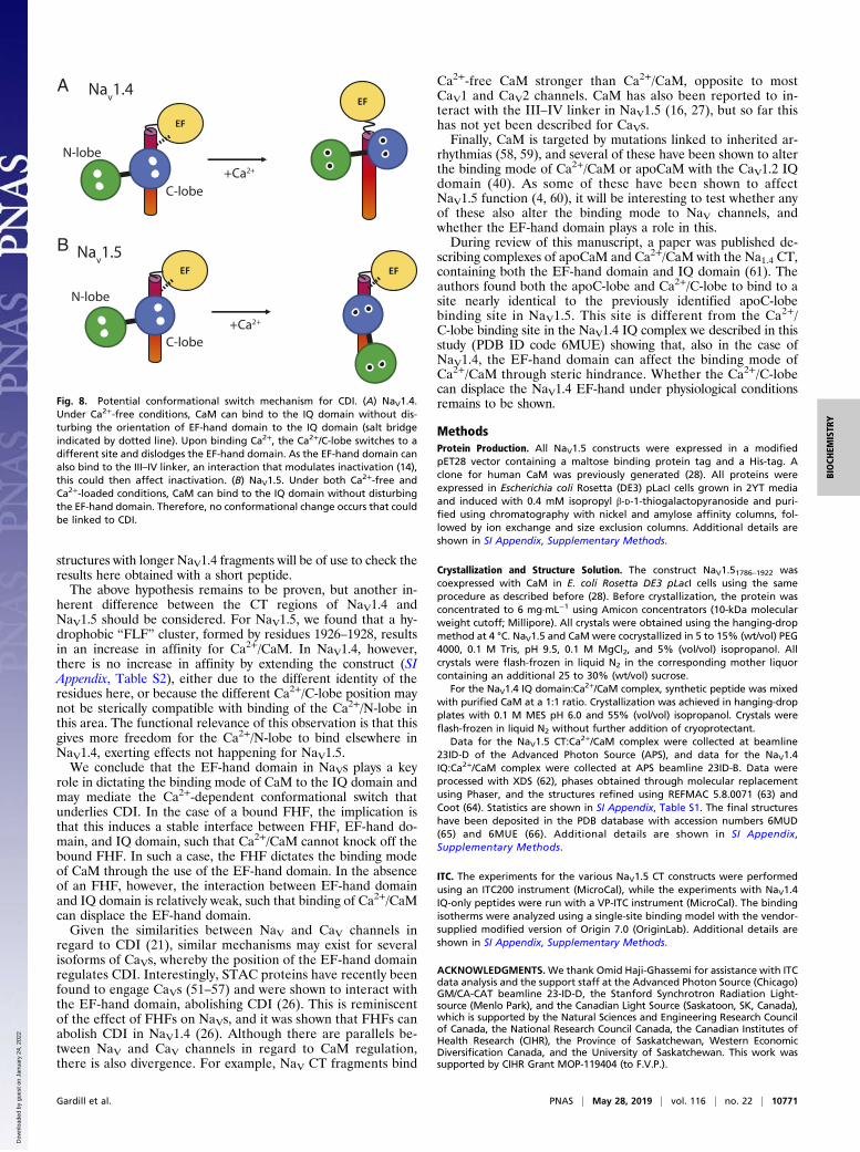
in complex with Ca2+/CaM, we noted that its binding modewould be incompatible with the position of the EF-hand domainas observed for NaV1.5. One possibility is that the NaV1.4 EF-hand domain forces Ca2+/CaM to bind at a different positionfrom the one we observe here or, alternatively, that the EF-handdomain adopts a different position when Ca2+/CaM is bound(Fig. 8A). The situation is different in NaV1.5, where bothapoCaM and Ca2+/CaM can bind to the IQ domain withoutsteric clashes with the EF-hand domain, as long as FHFs are ab-sent. As a result, Ca2+ binding to CaM would not produce aconformational rearrangement in NaV1.5, explaining the ab-sence of CDI for this channel (Fig. 8B). Interestingly, FHFs havebeen shown to abolish CDI in NaV1.4 (26), an effect that may beascribed to its inherent effect on the EF-hand orientation, lockingit into a position such that no Ca2+-dependent conformationalchange occurs. Similarly, STAC proteins, which can bind to twodifferent regions in CaV1 channels (26, 51–56), including the EF-hand domain (26), could affect the relative orientation betweenEF-hand and IQ domain. How this conformational change furtherresults in inactivation remains to be shown, but of note is theability of the EF-hand domain to interact with the III–IV linker,and that this interaction affects channel inactivation (14). Future
BA
C D
IleGln
6MUD Clobe
6MUEClobe
6MUD Clobe
6MUEClobe
6MUD Nlobe
6MUENlobe
6MUEClobe
6MUDEF-hand domain
6MUEClobe
4JQ0EF-hand domain
4JQ0FGF12B
Fig. 7. Differences in IQ motif binding between Nav1.5 and Nav1.4. The structures were superposed based on NaV CT helix 6, containing the IQ domain. (A)Superposition of the complexes NaV1.5 CT:Ca2+/CaM (PDB ID code 6MUD, this study) and NaV1.4 IQ domain:Ca2+/CaM (PDB ID code 6MUE, this study). N-lobeand C-lobe are shown in green and blue, respectively, with light colors for the NaV1.5 complex and dark colors for the NaV1.4 complex. NaV1.5 EF-handdomain (beige), NaV1.5 IQ domain (red), and NaV1.4 IQ domain (gray) are shown. Calcium ions are shown as white spheres. (B) Same superposition as in A butwith the view from the N terminus of the IQ domain toward the C terminus. The EF-hand domain has been omitted and only the C-lobes are shown. Thisindicates a ∼90° rotation of the C-lobe binding to NaV1.4 compared with the NaV1.5 IQ domain. The Ile and Gln residues of the “IQ” motif are shown in blackfor reference. (C) Same superposition as in A, but only showing the NaV1.5 CT (EF-hand domain in beige; IQ domain in red) and the C-lobe from theNaV1.4 complex. This shows that the C-lobe would clash with the EF-hand domain, suggesting that, for it to bind in this mode, the EF-hand domain inNaV1.4 would have to be displaced. (D) Superposition, based on helix 6, for the NaV1.5 CT: mixed-CaM complex with FGF12B (PDB ID code 4JQ0) and forthe NaV1.4 IQ: Ca2+/CaM complex (PDB ID code 6MUE, this study). Shown are the NaV1.5 CT (white) and FGF12B (yellow) from 4JQ0, and the C-lobe fromthe NaV1.4 complex. This shows that, also in the presence of an FHF, there would be clashes between the Ca2+/C-lobe and the EF-hand domain. In ad-dition, clashes would occur between Ca2+/Clobe and FGF12B.
10770 | www.pnas.org/cgi/doi/10.1073/pnas.1818618116 Gardill et al.
Dow
nloa
ded
by g
uest
on
Janu
ary
24, 2
022
structures with longer NaV1.4 fragments will be of use to check theresults here obtained with a short peptide.The above hypothesis remains to be proven, but another in-
herent difference between the CT regions of NaV1.4 andNaV1.5 should be considered. For NaV1.5, we found that a hy-drophobic “FLF” cluster, formed by residues 1926–1928, resultsin an increase in affinity for Ca2+/CaM. In NaV1.4, however,there is no increase in affinity by extending the construct (SIAppendix, Table S2), either due to the different identity of theresidues here, or because the different Ca2+/C-lobe position maynot be sterically compatible with binding of the Ca2+/N-lobe inthis area. The functional relevance of this observation is that thisgives more freedom for the Ca2+/N-lobe to bind elsewhere inNaV1.4, exerting effects not happening for NaV1.5.We conclude that the EF-hand domain in NaVs plays a key
role in dictating the binding mode of CaM to the IQ domain andmay mediate the Ca2+-dependent conformational switch thatunderlies CDI. In the case of a bound FHF, the implication isthat this induces a stable interface between FHF, EF-hand do-main, and IQ domain, such that Ca2+/CaM cannot knock off thebound FHF. In such a case, the FHF dictates the binding modeof CaM through the use of the EF-hand domain. In the absenceof an FHF, however, the interaction between EF-hand domainand IQ domain is relatively weak, such that binding of Ca2+/CaMcan displace the EF-hand domain.Given the similarities between NaV and CaV channels in
regard to CDI (21), similar mechanisms may exist for severalisoforms of CaVs, whereby the position of the EF-hand domainregulates CDI. Interestingly, STAC proteins have recently beenfound to engage CaVs (51–57) and were shown to interact withthe EF-hand domain, abolishing CDI (26). This is reminiscentof the effect of FHFs on NaVs, and it was shown that FHFs canabolish CDI in NaV1.4 (26). Although there are parallels be-tween NaV and CaV channels in regard to CaM regulation,there is also divergence. For example, NaV CT fragments bind
Ca2+-free CaM stronger than Ca2+/CaM, opposite to mostCaV1 and CaV2 channels. CaM has also been reported to in-teract with the III–IV linker in NaV1.5 (16, 27), but so far thishas not yet been described for CaVs.Finally, CaM is targeted by mutations linked to inherited ar-
rhythmias (58, 59), and several of these have been shown to alterthe binding mode of Ca2+/CaM or apoCaM with the CaV1.2 IQdomain (40). As some of these have been shown to affectNaV1.5 function (4, 60), it will be interesting to test whether anyof these also alter the binding mode to NaV channels, andwhether the EF-hand domain plays a role in this.During review of this manuscript, a paper was published de-
scribing complexes of apoCaM and Ca2+/CaM with the Na1.4 CT,containing both the EF-hand domain and IQ domain (61). Theauthors found both the apoC-lobe and Ca2+/C-lobe to bind to asite nearly identical to the previously identified apoC-lobebinding site in NaV1.5. This site is different from the Ca2+/C-lobe binding site in the NaV1.4 IQ complex we described in thisstudy (PDB ID code 6MUE) showing that, also in the case ofNaV1.4, the EF-hand domain can affect the binding mode ofCa2+/CaM through steric hindrance. Whether the Ca2+/C-lobecan displace the NaV1.4 EF-hand under physiological conditionsremains to be shown.
MethodsProtein Production. All NaV1.5 constructs were expressed in a modifiedpET28 vector containing a maltose binding protein tag and a His-tag. Aclone for human CaM was previously generated (28). All proteins wereexpressed in Escherichia coli Rosetta (DE3) pLacI cells grown in 2YT mediaand induced with 0.4 mM isopropyl β-D-1-thiogalactopyranoside and puri-fied using chromatography with nickel and amylose affinity columns, fol-lowed by ion exchange and size exclusion columns. Additional details areshown in SI Appendix, Supplementary Methods.
Crystallization and Structure Solution. The construct NaV1.51786–1922 wascoexpressed with CaM in E. coli Rosetta DE3 pLacI cells using the sameprocedure as described before (28). Before crystallization, the protein wasconcentrated to 6 mg·mL−1 using Amicon concentrators (10-kDa molecularweight cutoff; Millipore). All crystals were obtained using the hanging-dropmethod at 4 °C. NaV1.5 and CaM were cocrystallized in 5 to 15% (wt/vol) PEG4000, 0.1 M Tris, pH 9.5, 0.1 M MgCl2, and 5% (vol/vol) isopropanol. Allcrystals were flash-frozen in liquid N2 in the corresponding mother liquorcontaining an additional 25 to 30% (wt/vol) sucrose.
For the NaV1.4 IQ domain:Ca2+/CaM complex, synthetic peptide was mixedwith purified CaM at a 1:1 ratio. Crystallization was achieved in hanging-dropplates with 0.1 M MES pH 6.0 and 55% (vol/vol) isopropanol. Crystals wereflash-frozen in liquid N2 without further addition of cryoprotectant.
Data for the NaV1.5 CT:Ca2+/CaM complex were collected at beamline23ID-D of the Advanced Photon Source (APS), and data for the NaV1.4IQ:Ca2+/CaM complex were collected at APS beamline 23ID-B. Data wereprocessed with XDS (62), phases obtained through molecular replacementusing Phaser, and the structures refined using REFMAC 5.8.0071 (63) andCoot (64). Statistics are shown in SI Appendix, Table S1. The final structureshave been deposited in the PDB database with accession numbers 6MUD(65) and 6MUE (66). Additional details are shown in SI Appendix,Supplementary Methods.
ITC. The experiments for the various NaV1.5 CT constructs were performedusing an ITC200 instrument (MicroCal), while the experiments with NaV1.4IQ-only peptides were run with a VP-ITC instrument (MicroCal). The bindingisotherms were analyzed using a single-site binding model with the vendor-supplied modified version of Origin 7.0 (OriginLab). Additional details areshown in SI Appendix, Supplementary Methods.
ACKNOWLEDGMENTS.We thank Omid Haji-Ghassemi for assistance with ITCdata analysis and the support staff at the Advanced Photon Source (Chicago)GM/CA-CAT beamline 23-ID-D, the Stanford Synchrotron Radiation Light-source (Menlo Park), and the Canadian Light Source (Saskatoon, SK, Canada),which is supported by the Natural Sciences and Engineering Research Councilof Canada, the National Research Council Canada, the Canadian Institutes ofHealth Research (CIHR), the Province of Saskatchewan, Western EconomicDiversification Canada, and the University of Saskatchewan. This work wassupported by CIHR Grant MOP-119404 (to F.V.P.).
EF
EF
EF EF
A
B
Nav1.4
Nav1.5
N-lobe
N-lobe
C-lobe
C-lobe
+Ca2+
+Ca2+
Fig. 8. Potential conformational switch mechanism for CDI. (A) NaV1.4.Under Ca2+-free conditions, CaM can bind to the IQ domain without dis-turbing the orientation of EF-hand domain to the IQ domain (salt bridgeindicated by dotted line). Upon binding Ca2+, the Ca2+/C-lobe switches to adifferent site and dislodges the EF-hand domain. As the EF-hand domain canalso bind to the III–IV linker, an interaction that modulates inactivation (14),this could then affect inactivation. (B) NaV1.5. Under both Ca2+-free andCa2+-loaded conditions, CaM can bind to the IQ domain without disturbingthe EF-hand domain. Therefore, no conformational change occurs that couldbe linked to CDI.
Gardill et al. PNAS | May 28, 2019 | vol. 116 | no. 22 | 10771
BIOCH
EMISTR
Y
Dow
nloa
ded
by g
uest
on
Janu
ary
24, 2
022

1. Ahern CA, Payandeh J, Bosmans F, Chanda B (2016) The hitchhiker’s guide to thevoltage-gated sodium channel galaxy. J Gen Physiol 147:1–24.
2. Winters JJ, Isom LL (2016) Developmental and regulatory functions of Na(+) channelnon-pore-forming β subunits. Curr Top Membr 78:315–351.
3. Yan Z, et al. (2017) Structure of the Nav1.4-beta1 complex from electric eel. Cell 170:470–482.e11.
4. Yin G, et al. (2014) Arrhythmogenic calmodulinmutations disrupt intracellular cardiomyocyteCa2+ regulation by distinct mechanisms. J Am Heart Assoc 3:e000996.
5. Shen H, et al. (2017) Structure of a eukaryotic voltage-gated sodium channel at near-atomic resolution. Science 355:eaal4326.
6. Das S, Gilchrist J, Bosmans F, Van Petegem F (2016) Binary architecture of the Nav1.2-β2 signaling complex. eLife 5:e10960.
7. Gilchrist J, Das S, Van Petegem F, Bosmans F (2013) Crystallographic insights intosodium-channel modulation by the β4 subunit. Proc Natl Acad Sci USA 110:E5016–E5024.
8. Namadurai S, et al. (2014) Crystal structure and molecular imaging of the Nav channelβ3 subunit indicates a trimeric assembly. J Biol Chem 289:10797–10811.
9. Shimizu H, et al. (2016) Structure-based site-directed photo-crosslinking analyses ofmultimeric cell-adhesive interactions of voltage-gated sodium channel β subunits. SciRep 6:26618.
10. Miloushev VZ, et al. (2009) Solution structure of the NaV1.2 C-terminal EF-hand do-main. J Biol Chem 284:6446–6454.
11. Chagot B, Potet F, Balser JR, Chazin WJ (2009) Solution NMR structure of the C-terminal EF-hand domain of human cardiac sodium channel NaV1.5. J Biol Chem284:6436–6445.
12. Wingo TL, et al. (2004) An EF-hand in the sodium channel couples intracellular cal-cium to cardiac excitability. Nat Struct Mol Biol 11:219–225.
13. Wang C, et al. (2014) Structural analyses of Ca2+/CaM interaction with NaV channel C-termini reveal mechanisms of calcium-dependent regulation. Nat Commun 5:4896.
14. Gardill BR, et al. (2018) The voltage-gated sodium channel EF-hands form an interactionwith the III-IV linker that is disturbed by disease-causing mutations. Sci Rep 8:4483.
15. Van Petegem F, Lobo PA, Ahern CA (2012) Seeing the forest through the trees: To-wards a unified view on physiological calcium regulation of voltage-gated sodiumchannels. Biophys J 103:2243–2251.
16. Sarhan MF, Tung CC, Van Petegem F, Ahern CA (2012) Crystallographic basis forcalcium regulation of sodium channels. Proc Natl Acad Sci USA 109:3558–3563.
17. Sarhan MF, Van Petegem F, Ahern CA (2009) A double tyrosine motif in the cardiacsodium channel domain III-IV linker couples calcium-dependent calmodulin bindingto inactivation gating. J Biol Chem 284:33265–33274.
18. Potet F, et al. (2009) Functional interactions between distinct sodium channel cyto-plasmic domains through the action of calmodulin. J Biol Chem 284:8846–8854.
19. Biswas S, DiSilvestre D, Tian Y, Halperin VL, Tomaselli GF (2009) Calcium-mediateddual-mode regulation of cardiac sodium channel gating. Circ Res 104:870–878.
20. Tan HL, et al. (2002) A calcium sensor in the sodium channel modulates cardiac ex-citability. Nature 415:442–447.
21. Ben-Johny M, et al. (2014) Conservation of Ca2+/calmodulin regulation across Na andCa2+ channels. Cell 157:1657–1670.
22. Zühlke RD, Pitt GS, Deisseroth K, Tsien RW, Reuter H (1999) Calmodulin supports bothinactivation and facilitation of L-type calcium channels. Nature 399:159–162.
23. Ben-Johny M, Yue DT (2014) Calmodulin regulation (calmodulation) of voltage-gatedcalcium channels. J Gen Physiol 143:679–692.
24. Peterson BZ, DeMaria CD, Adelman JP, Yue DT (1999) Calmodulin is the Ca2+ sensorfor Ca2+ -dependent inactivation of L-type calcium channels. Neuron 22:549–558.
25. Wang C, Chung BC, Yan H, Lee SY, Pitt GS (2012) Crystal structure of the ternarycomplex of a NaV C-terminal domain, a fibroblast growth factor homologous factor,and calmodulin. Structure 20:1167–1176.
26. Niu J, et al. (2018) Allosteric regulators selectively prevent Ca2+-feedback of CaV andNaV channels. eLife 7:e35222.
27. Johnson CN, et al. (2018) A mechanism of calmodulin modulation of the humancardiac sodium channel. Structure 26:683–694.e3.
28. Van Petegem F, Chatelain FC, Minor DL, Jr (2005) Insights into voltage-gated calciumchannel regulation from the structure of the CaV1.2 IQ domain-Ca2+/calmodulincomplex. Nat Struct Mol Biol 12:1108–1115.
29. Fallon JL, Halling DB, Hamilton SL, Quiocho FA (2005) Structure of calmodulin boundto the hydrophobic IQ domain of the cardiac Ca(v)1.2 calcium channel. Structure 13:1881–1886.
30. Mori MX, Vander Kooi CW, Leahy DJ, Yue DT (2008) Crystal structure of the CaV2 IQdomain in complex with Ca2+/calmodulin: High-resolution mechanistic implicationsfor channel regulation by Ca2+. Structure 16:607–620.
31. Halling DB, et al. (2009) Determinants in CaV1 channels that regulate the Ca2+sensitivity of bound calmodulin. J Biol Chem 284:20041–20051.
32. Kim EY, et al. (2010) Multiple C-terminal tail Ca(2+)/CaMs regulate Ca(V)1.2 functionbut do not mediate channel dimerization. EMBO J 29:3924–3938.
33. Gabelli SB, et al. (2014) Regulation of the NaV1.5 cytoplasmic domain by calmodulin.Nat Commun 5:5126.
34. Isbell HM, Kilpatrick AM, Lin Z, Mahling R, Shea MA (2018) Backbone resonance as-signments of complexes of apo human calmodulin bound to IQ motif peptides ofvoltage-dependent sodium channels NaV1.1, NaV1.4 and NaV1.7. Biomol NMR Assign12:283–289.
35. Feldkamp MD, Yu L, Shea MA (2011) Structural and energetic determinants of apocalmodulin binding to the IQ motif of the Na(V)1.2 voltage-dependent sodiumchannel. Structure 19:733–747.
36. Hovey L, et al. (2017) Calcium triggers reversal of calmodulin on nested anti-parallelsites in the IQmotif of the neuronal voltage-dependent sodium channel NaV1.2. BiophysChem 224:1–19.
37. Wilson MA, Brunger AT (2000) The 1.0 a crystal structure of Ca(2+)-bound calmodulin:An analysis of disorder and implications for functionally relevant plasticity. J Mol Biol301:1237–1256.
38. Ben Johny M, Yang PS, Bazzazi H, Yue DT (2013) Dynamic switching of calmodulininteractions underlies Ca2+ regulation of CaV1.3 channels. Nat Commun 4:1717.
39. Houdusse A, et al. (2006) Crystal structure of apo-calmodulin bound to the first two IQmotifs of myosin V reveals essential recognition features. Proc Natl Acad Sci USA 103:19326–19331.
40. Wang K, et al. (2018) Arrhythmia mutations in calmodulin cause conformationalchanges that affect interactions with the cardiac voltage-gated calcium channel. ProcNatl Acad Sci USA 115:E10556–E10565.
41. Findeisen F, Rumpf CH, Minor DL, Jr (2013) Apo states of calmodulin and CaBP1 controlCaV1 voltage-gated calcium channel function through direct competition for the IQdomain. J Mol Biol 425:3217–3234.
42. Napolitano C, et al. (2005) Genetic testing in the long QT syndrome: Developmentand validation of an efficient approach to genotyping in clinical practice. JAMA 294:2975–2980.
43. Yan H, Wang C, Marx SO, Pitt GS (2017) Calmodulin limits pathogenic Na+ channelpersistent current. J Gen Physiol 149:277–293.
44. Tester DJ, Will ML, Haglund CM, AckermanMJ (2005) Compendium of cardiac channelmutations in 541 consecutive unrelated patients referred for long QT syndrome ge-netic testing. Heart Rhythm 2:507–517.
45. Abdelsayed M, et al. (2017) Differential calcium sensitivity in NaV 1.5 mixed syndromemutants. J Physiol 595:6165–6186.
46. Glaaser IW, et al. (2012) Perturbation of sodium channel structure by an inheritedlong QT syndrome mutation. Nat Commun 3:706.
47. Bankston JR, et al. (2007) A novel LQT-3 mutation disrupts an inactivation gatecomplex with distinct rate-dependent phenotypic consequences. Channels (Austin) 1:273–280.
48. Wu J, et al. (2016) Structure of the voltage-gated calcium channel Ca(v)1.1 at 3.6 Åresolution. Nature 537:191–196.
49. Weiss LA, et al. (2003) Sodium channels SCN1A, SCN2A and SCN3A in familial autism.Mol Psychiatry 8:186–194.
50. Kim J, et al. (2004) Calmodulin mediates Ca2+ sensitivity of sodium channels. J BiolChem 279:45004–45012.
51. Campiglio M, Flucher BE (2017) STAC3 stably interacts through its C1 domain withCaV1.1 in skeletal muscle triads. Sci Rep 7:41003.
52. Wong King Yuen SM, Campiglio M, Tung CC, Flucher BE, Van Petegem F (2017)Structural insights into binding of STAC proteins to voltage-gated calcium channels.Proc Natl Acad Sci USA 114:E9520–E9528.
53. Campiglio M, et al. (2018) STAC proteins associate to the IQ domain of CaV1.2 andinhibit calcium-dependent inactivation. Proc Natl Acad Sci USA 115:1376–1381.
54. Polster A, Perni S, Bichraoui H, Beam KG (2015) Stac adaptor proteins regulate traf-ficking and function of muscle and neuronal L-type Ca2+ channels. Proc Natl Acad SciUSA 112:602–606.
55. Polster A, et al. (2018) Stac proteins suppress Ca2+-dependent inactivation of neuronall-type Ca2+ channels. J Neurosci 38:9215–9227.
56. Polster A, Nelson BR, Papadopoulos S, Olson EN, Beam KG (2018) Stac proteins as-sociate with the critical domain for excitation-contraction coupling in the II-III loop ofCaV1.1. J Gen Physiol 150:613–624.
57. Polster A, Nelson BR, Olson EN, Beam KG (2016) Stac3 has a direct role in skeletalmuscle-type excitation-contraction coupling that is disrupted by a myopathy-causingmutation. Proc Natl Acad Sci USA 113:10986–10991.
58. Nyegaard M, et al. (2012) Mutations in calmodulin cause ventricular tachycardia andsudden cardiac death. Am J Hum Genet 91:703–712.
59. Crotti L, et al. (2013) Calmodulin mutations associated with recurrent cardiac arrest ininfants. Circulation 127:1009–1017.
60. Boczek NJ, et al. (2016) Spectrum and prevalence of CALM1-, CALM2-, and CALM3-encoded calmodulin variants in long QT syndrome and functional characterization ofa novel long QT syndrome-associated calmodulin missense variant, E141G. CircCardiovasc Genet 9:136–146.
61. Yoder JB, et al. (2019) Ca2+-dependent regulation of sodium channels NaV1.4 andNaV1.5 is controlled by the post-IQ motif. Nat Commun 10:1514.
62. Kabsch W (2010) Xds. Acta Crystallogr D Biol Crystallogr 66:125–132.63. Murshudov GN, et al. (2011) REFMAC5 for the refinement of macromolecular crystal
structures. Acta Crystallogr D Biol Crystallogr 67:355–367.64. Emsley P, Lohkamp B, Scott WG, Cowtan K (2010) Features and development of Coot.
Acta Crystallogr D Biol Crystallogr 66:486–501.65. Gardill BR, Tung CC, Van Petegem F (2018) Voltage-gated sodium channel NaV1.5 C-
terminal domain in complex with Ca2+/calmodulin. Protein Data Bank. Available athttps://www.rcsb.org/structure/6MUD. Deposited October 24, 2018.
66. Gardill BR, Tung CC, Van Petegem F (2018) Voltage-gated sodium channel NaV1.4 IQdomain in complex with Ca2+/calmodulin. Protein Data Bank. Available at https://www.rcsb.org/structure/6MUE. Deposited October 24, 2018.
67. Kyte J, Doolittle RF (1982) A simple method for displaying the hydropathic characterof a protein. J Mol Biol 157:105–132.
10772 | www.pnas.org/cgi/doi/10.1073/pnas.1818618116 Gardill et al.
Dow
nloa
ded
by g
uest
on
Janu
ary
24, 2
022