crystal structure of d-amino acid oxidase: a case of active site
TRANSCRIPT
Proc. Natl. Acad. Sci. USAVol. 93, pp. 7496-7501, July 1996Biochemistry
Crystal structure of D-amino acid oxidase: A case of active sitemirror-image convergent evolution with flavocytochrome b2
(density averaging/flavoenzyme/redox catalysis/stereospecificity)
ANDREA MATrEVI*t, MARIA ANTONIETTA VANONIt, FLAVIA TODONE*, MENICO RIzzI*, ALEX TEPLYAKOV§,ALESSANDRO CODA*¶, MARTINO BOLOGNESI*II, AND BRUNO CURTItl*Dipartimento di Genetica e Microbiologia, Universita di Pavia, Via Abbiategrasso 207, 27100 Pavia, Italy; tDipartimento di Fisiologia e Biochimica Generali,Universita di Milano, Via Celoria 26, 20133 Milan, Italy; §European Molecular Biology Laboratory Outstation, Deutsches Elektronen Synchrotron,22063 Hamburg, Germany; IlDipartimento di Fisica and IST Centro per le Biotecnologie Avanzate, Universita di Genova, Viale Benedetto XV 10,16132 Genoa, Italy; and ICentro Interuniversitario per lo Studio delle Macromolecole Informazionali
Communicated by Vincent Massey, University ofMichigan Medical School, Ann Arbor, MI, January 19, 1996 (received for review December 9, 1995)
ABSTRACT D-amino acid oxidase is the prototype of theFAD-dependent oxidases. It catalyses the oxidation of D-amino acids to the corresponding a-ketoacids. The reducingequivalents are transferred to molecular oxygen with produc-tion of hydrogen peroxide. We have solved the crystal struc-ture of the complex of D-amino acid oxidase with benzoate, acompetitive inhibitor of the substrate, by single isomorphousreplacement and eightfold averaging. Each monomer isformed by two domains with an overall topology similar to thatofp-hydroxybenzoate hydroxylase. The benzoate molecule laysparallel to the flavin ring and is held in position by a saltbridge with Arg-283. Analysis of the active site shows that noside chains are properly positioned to act as the postulatedbase required for the catalytic carboanion mechanism. On thecontrary, the benzoate binding mode suggests a direct transferof the substrate a-hydrogen to the flavin during the enzymereductive half-reaction. The active site ofD-amino acid oxidaseexhibits a striking similarity with that of flavocytochrome b2,a structurally unrelated FMN-dependent flavoenzyme. Theactive site groups of these two enzymes are in fact superim-posable once the mirror-image of the flavocytochrome b2active site is generated with respect to the flavin plane.Therefore, the catalytic sites of D-amino acid oxidase andflavocytochrome b2 appear to have converged to a highlysimilar but enantiomeric architecture in order to catalyzesimilar reactions (oxidation of a-amino acids or a-hydroxyacids), although with opposite stereochemistry.
Since the description of D-amino acid oxidase (EC 1.4.3.3;DAAO) activity in mammalian tissues by Krebs in 1935 (1),DAAO has been the subject of a number of biochemical,spectroscopic, and kinetic investigations, becoming the proto-type for the oxidase class of the flavin-containing enzymes [fora recent review, see ref. 2]. Its primary structure has beendetermined and its gene has been cloned (3, 4). Its kinetic andmechanistic properties have been studied in detail by a varietyof techniques, while information on the topology of the activesite and on its three-dimensional structure have only beenderived from chemical modification studies and site-directedmutagenesis of selected residues. Based on these approaches,a catalytic mechanism for DAAO has been proposed, althoughdefinitive evidence against alternative mechanisms has notbeen found (refs. 2 and 5 and references therein).The enzyme catalyzes the oxidation of D-a-amino acids into
the corresponding a-ketoacids. The reaction formally pro-ceeds according to the following scheme:
RCHNH2COOH + E-FAD -i.RC=NHCOOH+ E-FADH2 [1]
E-FADH2 + 02-*E-FAD + H202 [2]
RC=NHCOOH + H20- RCOCOOH + NH3 [3]
The reductive half reaction (Eq. 1), in which the noncovalentlybound FAD becomes reduced, is followed by the oxidative stepin which FAD is reoxidized by molecular oxygen, with therelease of hydrogen peroxide (Eq. 2). The imino acid productspontaneously hydrolyzes to the ketoacid in a nonenzymaticprocess (Eq. 3). DAAO displays a broad substrate specificity,with a preference for D-amino acids bearing hydrophobic sidechains up to four carbon-atoms long, followed by those car-rying polar and aromatic groups (6). The enzyme exhibits verylow activity toward basic amino acids and it does not oxidizethose with an acidic side chain. These are oxidatively deami-nated by a specific enzyme, D-aspartate oxidase, which shares50% sequence identity with DAAO (7). Both oxidases arehighly stereospecific, and unable to metabolize L-amino acids.The biological role of DAAO, a peroxisomal enzyme, is
controversial (2). The recent detection of significant quantitiesof D-amino acids in various mammalian tissues suggests thatDAAO is a detoxifying agent that removes the D-amino acidsderiving from either exogenous or endogenous sources (8).Peptides incorporating D-amino acids have been found notonly in prokaryotes, where they are common, but also inseveral vertebrates, in a few cases as neuropeptides producedby nerve cells (9). In this context, the presence of high levelsof DAAO activity in human cerebellum is in keeping with thedetection of D-amino acids in the human brain (10).We have now succeeded in solving the crystal structure of
pig kidney DAAO in complex with the competitive inhibitorbenzoate. The enzyme is a dimer comprising 2 x 347 aminoacids and a molecule of noncovalently bound FAD per subunit.The inhibitor has well-defined electron density which allowsthe identification and description of the active site.
MATERIALS AND METHODSThe methodology employed in.the crystallization and structuredetermination will be described in detail elsewhere. Briefly,DAAO isolated from pig kidneys (11) was crystallized by thehanging-drop method under conditions similar to those reportedby Bolognesi et al. (12). Protein solutions (50 mg protein/ml)containing 2 mM sodium benzoate were equilibrated by vapordiffusion against a reservoir containing 0.5 M ammonium succi-nate, 100 mM Tris HCl (pH 8.3) at 28°C. The crystals belong toorthorhombic space group C2221 with cell dimensions a = 328 A,b = 138 A, c = 201 A, and four dimers (296,000 Da) in theasymmetric unit. All data sets used for structure determinationwere collected at room temperature on a Raxis-Il imaging plate
Abbreviations: DAAO, D-amino acid oxidase; PHBH, p-hydroxyben-zoate hydroxylase; FCB, flavocytochrome b2; SIR, single isomorphusreplacement.Data deposition: The atomic coordinates have been deposited in theProtein Data Bank, Chemistry Department, Brookhaven NationalLaboratory, Upton, NY 11973 (reference 1KIF).tTo whom reprint requests should be addressed. e-mail: [email protected].
7496
The publication costs of this article were defrayed in part by page chargepayment. This article must therefore be hereby marked "advertisement" inaccordance with 18 U.S.C. §1734 solely to indicate this fact.
Proc. Natl. Acad. Sci. USA 93 (1996) 7497
system using CuKa radiation (Table 1). The 2.6 A native data setemployed in the refinement was collected at the X31 beam lineof the European Molecular Biology Laboratory/DeutschesElektronen Synchrotron (Hamburg). This data set was incom-plete at low resolution and therefore was merged with a dataset collected on the in-house Raxis-1I system (Table 1). Theimages were evaluated using MOSFLM (A. Leslie, personalcommunication), while the CCP4 suite (13) was used in datareduction. Only one heavy atom derivative was sufficient forstructure determination and it was obtained by soaking crys-tals in a saturated solution ofp-chloromercury benzoate. Theisomorphous difference Patterson map was interpreted usingSHELX-90 (14) and the heavy atom parameters were refinedusing the program MLPHARE (13). The structure solution was
based on the resulting single isomorphous replacement (SIR)map that allowed identification of the protein boundaries. Afragment of the electron density map was used as a searchprobe in a molecular replacement calculation that led to theidentification of the noncrystallographic symmetry operators.These calculations were carried out using programs ALMN (13)and GLRF (15). The SIR phases were refined and graduallyextended to 3.0 A by eightfold averaging and solvent flatteningusing the program package DEMON (16). The resulting map wasof excellent quality (Fig. 1), allowing us to trace the polypep-tide chain. An initial model was built using program o (17) andwas subjected to TNT (18) least-squares refinement to give a
crystallographic R-factor of 22.8% for all 119,237 measuredreflections up to 2.6 A (no a cut-off was applied; Table 2). Thefree R-factor (20) for 1000 reflections omitted from therefinement is 24.5%. In the least-squares refinement, the eightsubunits present in the asymmetric unit were constrained to beidentical, leading to a number of observations per number ofparameters ratio of about 12. A total of 270 ordered watermolecules were added at positions with density >lo- in the2Fo-Fc map and >3or in the Fo-Fc map. All these watermolecules are engaged in at least two hydrogen bonds with a
protein or a solvent atom. The final model has good stereo-chemistry (Table 2), and all residues are within energeticallyfavored regions of the Ramachandran plot (19).
RESULTS AND DISCUSSIONOverall Structure. The DAAO subunit is schematically
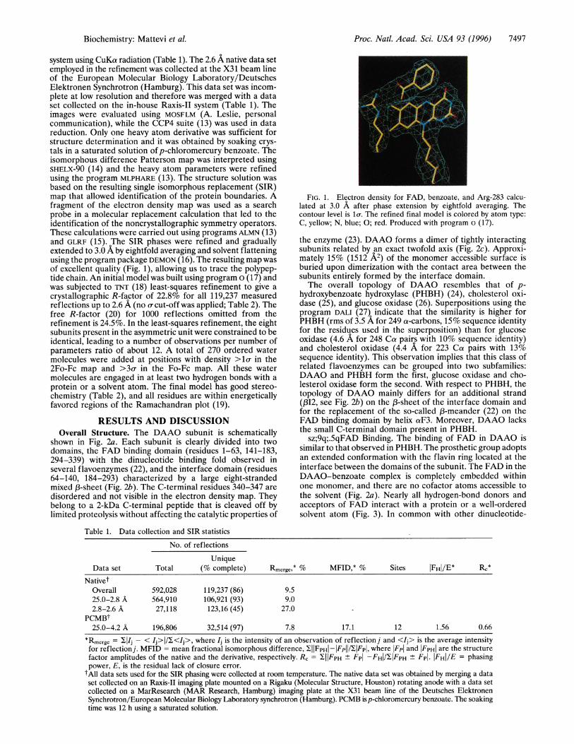
shown in Fig. 2a. Each subunit is clearly divided into twodomains, the FAD binding domain (residues 1-63, 141-183,294-339) with the dinucleotide binding fold observed inseveral flavoenzymes (22), and the interface domain (residues64-140, 184-293) characterized by a large eight-strandedmixed 13-sheet (Fig. 2b). The C-terminal residues 340-347 are
disordered and not visible in the electron density map. Theybelong to a 2-kDa C-terminal peptide that is cleaved off bylimited proteolysis without affecting the catalytic properties of
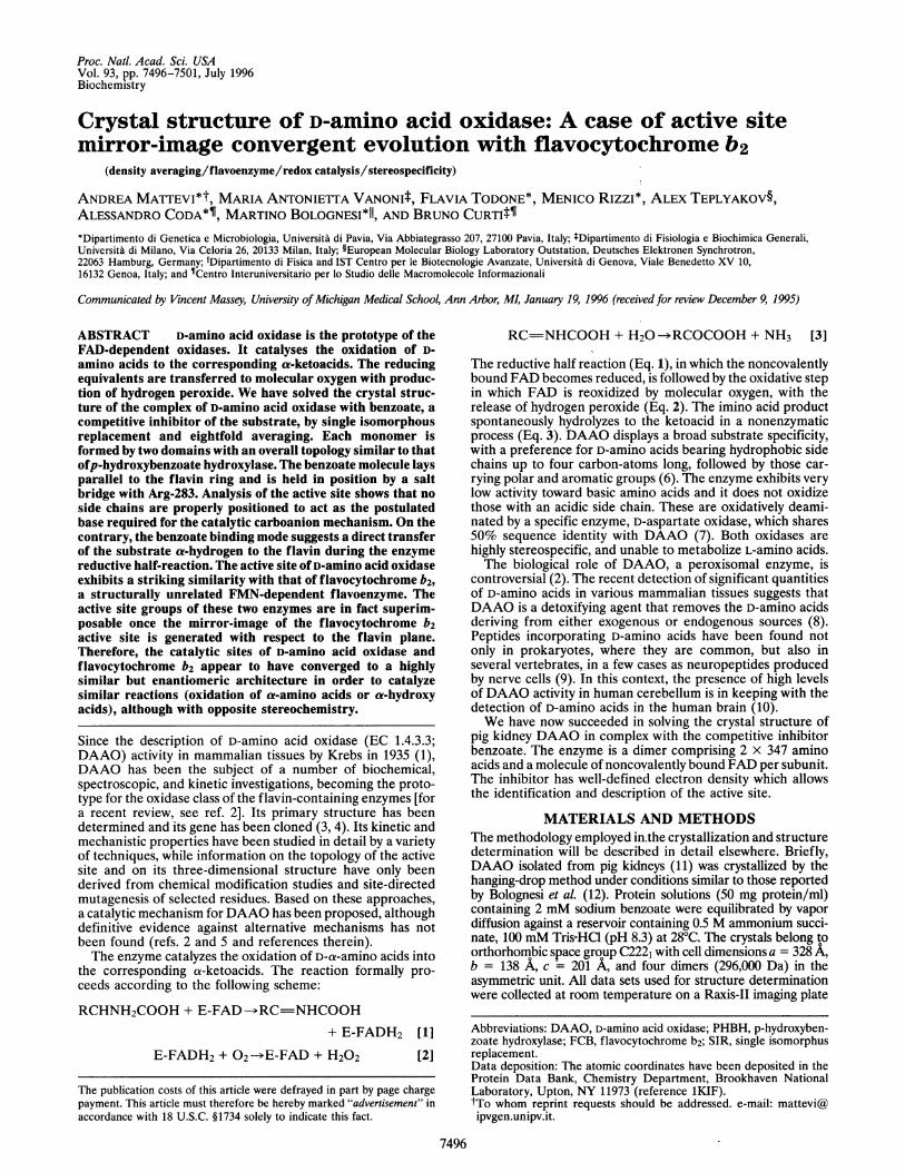
FIG. 1. Electron density for FAD, benzoate, and Arg-283 calcu-lated at 3.0 A after phase extension by eightfold averaging. Thecontour level is lo-. The refined final model is colored by atom type:C, yellow; N, blue; 0; red. Produced with program o (17).
the enzyme (23). DAAO forms a dimer of tightly interactingsubunits related by an exact twofold axis (Fig. 2c). Approxi-mately 15% (1512 A2) of the monomer accessible surface isburied upon dimerization with the contact area between thesubunits entirely formed by the interface domain.The overall topology of DAAO resembles that of p-
hydroxybenzoate hydroxylase (PHBH) (24), cholesterol oxi-dase (25), and glucose oxidase (26). Superpositions using theprogram DALI (27) indicate that the similarity is higher forPHBH (rms of 3.5 A for 249 a-carbons, 15% sequence identityfor the residues used in the superposition) than for glucoseoxidase (4.6 A for 248 Ca pairs with 10% sequence identity)and cholesterol oxidase (4.4 A for 223 Ca pairs with 13%sequence identity). This observation implies that this class ofrelated flavoenzymes can be grouped into two subfamilies:DAAO and PHBH form the first, glucose oxidase and cho-lesterol oxidase form the second. With respect to PHBH, thetopology of DAAO mainly differs for an additional strand(p12, see Fig. 2b) on the 13-sheet of the interface domain andfor the replacement of the so-called X3-meander (22) on theFAD binding domain by helix aF3. Moreover, DAAO lacksthe small C-terminal domain present in PHBH.
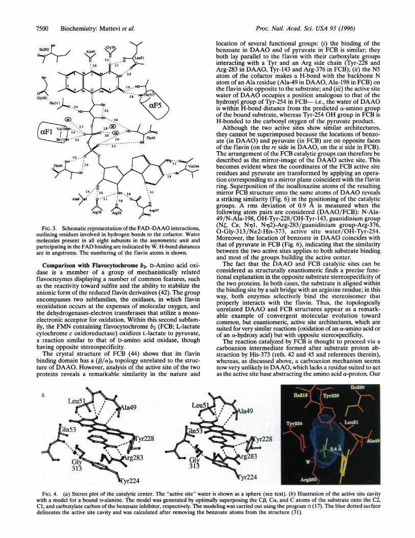
sz;9q;.5qFAD Binding. The binding of FAD in DAAO issimilar to that observed in PHBH. The prosthetic group adoptsan extended conformation with the flavin ring located at theinterface between the domains of the subunit. The FAD in theDAAO-benzoate complex is completely embedded withinone monomer, and there are no cofactor atoms accessible tothe solvent (Fig. 2a). Nearly all hydrogen-bond donors andacceptors of FAD interact with a protein or a well-orderedsolvent atom (Fig. 3). In common with other dinucleotide-
Table 1. Data collection and SIR statistics
No. of reflections
UniqueData set Total (% complete) Rmerge* % MFID,* % Sites IFH|/E* RC*
NativetOverall 592,028 119,237 (86) 9.525.0-2.8 A 564,910 106,921 (93) 9.02.8-2.6 A 27,118 123,16 (45) 27.0 -
PCMBt25.0-4.2 A 196,806 32,514 (97) 7.8 17.1 12 1.56 0.66
*Rmerge = IlIj - < Ij> / <Ij>, where Ij is the intensity of an observation of reflection j and <Ij> is the average intensityfor reflectionj. MFID = mean fractional isomorphous difference, IIIFPHI - IFpl /1lFpl, where lFpI and IFPHI are the structurefactor amplitudes of the native and the derivative, respectively. R, = XIIFpH ± Fpl -FHI/IIFPH ± Fp|. IFHI/E = phasingpower, E, is the residual lack of closure error.
tAll data sets used for the SIR phasing were collected at room temperature. The native data set was obtained by merging a dataset collected on an Raxis-II imaging plate mounted on a Rigaku (Molecular Structure, Houston) rotating anode with a data setcollected on a MarResearch (MAR Research, Hamburg) imaging plate at the X31 beam line of the Deutsches ElektronenSynchrotron/European Molecular Biology Laboratory synchrotron (Hamburg). PCMB isp-chloromercury benzoate. The soakingtime was 12 h using a saturated solution.
Biochemistry: Mattevi et al.
7498 Biochemistry: Mattevi et al.
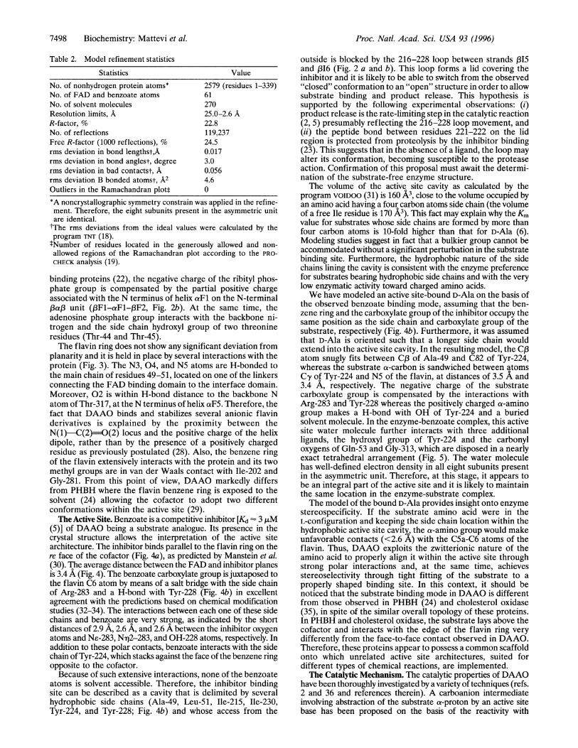
Table 2. Model refinement statistics
Statistics Value
No. of nonhydrogen protein atoms* 2579 (residues 1-339)No. of FAD and benzoate atoms 61No. of solvent molecules 270Resolution limits, A 25.0-2.6 AR-factor, % 22.8No. of reflections 119,237Free R-factor (1000 reflections), % 24.5rms deviation in bond lengthst,A 0.017rms deviation in bond anglest, degree 3.0rms deviation in bad contactst, A 0.056rms deviation B bonded atomst, A2 4.6Outliers in the Ramachandran plott 0
*A noncrystallographic symmetry constrain was applied in the refine-ment. Therefore, the eight subunits present in the asymmetric unitare identical.tThe rms deviations from the ideal values were calculated by theprogram TNT (18).tNumber of residues located in the generously allowed and non-allowed regions of the Ramachandran plot according to the PRO-CHECK analysis (19).
binding proteins (22), the negative charge of the ribityl phos-phate group is compensated by the partial positive chargeassociated with the N terminus of helix aFl on the N-terminalP3ap3 unit (,F1-FF1-al3F2, Fig. 2b). At the same time, theadenosine phosphate group interacts with the backbone ni-trogen and the side chain hydroxyl group of two threonineresidues (Thr-44 and Thr-45).The flavin ring does not show any significant deviation from
planarity and it is held in place by several interactions with theprotein (Fig. 3). The N3, 04, and N5 atoms are H-bonded tothe main chain of residues 49-51, located on one of the linkersconnecting the FAD binding domain to the interface domain.Moreover, 02 is within H-bond distance to the backbone Natom of Thr-317, at the N terminus of helix aF5. Therefore, thefact that DAAO binds and stabilizes several anionic flavinderivatives is explained by the proximity between theN(1)-C(2)=)0(2) locus and the positive charge of the helixdipole, rather than by the presence of a positively chargedresidue as previously postulated (28). Also, the benzene ringof the flavin extensively interacts with the protein and its twomethyl groups are in van der Waals contact with Ile-202 andGly-281. From this point of view, DAAO markedly differsfrom PHBH where the flavin benzene ring is exposed to thesolvent (24) allowing the cofactor to adopt two differentconformations within the active site (29).The Active Site. Benzoate is a competitive inhibitor [Kd 3 ,uM
(5)] of DAAO being a substrate analogue. Its presence in thecrystal structure allows the interpretation of the active sitearchitecture. The inhibitor binds parallel to the flavin ring on there face of the cofactor (Fig. 4a), as predicted by Manstein et al.(30). The average distance between the FAD and inhibitor planesis 3.4 A (Fig. 4). The benzoate carboxylate group is juxtaposed tothe flavin C6 atom by means of a salt bridge with the side chainof Arg-283 and a H-bond with Tyr-228 (Fig. 4b) in excellentagreement with the predictions based on chemical modificationstudies (32-34). The interactions between each one of these sidechains and benzoate are very strong, as indicated by the shortdistances of 2.9 A, 2.6 A, and 2.6 A between the inhibitor oxygenatoms and Ns-283, N-r2-283, and OH-228 atoms, respectively. Inaddition to these polar contacts, benzoate interacts with the sidechain of Tyr-224, which stacks against the face of the benzene ringopposite to the cofactor.
Because of such extensive interactions, none of the benzoateatoms is solvent accessible. Therefore, the inhibitor bindingsite can be described as a cavity that is delimited by severalhydrophobic side chains (Ala-49, Leu-51, Ile-215, Ile-230,Tyr-224, and Tyr-228; Fig. 4b) and whose access from the
Proc. Natl. Acad. Sci. USA 93 (1996)
outside is blocked by the 216-228 loop between strands (315and l316 (Fig. 2 a and b). This loop forms a lid covering theinhibitor and it is likely to be able to switch from the observed"closed" conformation to an "open" structure in order to allowsubstrate binding and product release. This hypothesis issupported by the following experimental observations: (i)product release is the rate-limiting step in the catalytic reaction(2, 5) presumably reflecting the 216-228 loop movement, and(ii) the peptide bond between residues 221-222 on the lidregion is protected from proteolysis by the inhibitor binding(23). This suggests that in the absence of a ligand, the loop mayalter its conformation, becoming susceptible to the proteaseaction. Confirmation of this proposal must await the determi-nation of the substrate-free enzyme structure.The volume of the active site cavity as calculated by the
program VOIDOO (31) is 160 A3, close to the volume occupied byan amino acid having a four carbon atoms side chain (the volumeof a free Ile residue is 170 A3). This fact may explain why the Kmvalue for substrates whose side chains are formed by more thanfour carbon atoms is 10-fold higher than that for D-Ala (6).Modeling studies suggest in fact that a bulkier group cannot beaccommodated without a significant perturbation in the substratebinding site. Furthermore, the hydrophobic nature of the sidechains lining the cavity is consistent with the enzyme preferencefor substrates bearing hydrophobic side chains and with the verylow enzymatic activity toward charged amino acids.We have modeled an active site-bound D-Ala on the basis of
the observed benzoate binding mode, assuming that the ben-zene ring and the carboxylate group of the inhibitor occupy thesame position as the side chain and carboxylate group of thesubstrate, respectively (Fig. 4b). Furthermore, it was assumedthat D-Ala is oriented such that a longer side chain wouldextend into the active site cavity. In the resulting model, the C(3atom snugly fits between Co3 of Ala-49 and C62 of Tyr-224,whereas the substrate a-carbon is sandwiched between atomsCy of Tyr-224 and N5 of the flavin, at distances of 3.5 A and3.4 A, respectively. The negative charge of the substratecarboxylate group is compensated by the interactions withArg-283 and Tyr-228 whereas the positively charged a-aminogroup makes a H-bond with OH of Tyr-224 and a buriedsolvent molecule. In the enzyme-benzoate complex, this activesite water molecule further interacts with three additionalligands, the hydroxyl group of Tyr-224 and the carbonyloxygens of Gln-53 and Gly-313, which are disposed in a nearlyexact tetrahedral arrangement (Fig. 5). The water moleculehas well-defined electron density in all eight subunits presentin the asymmetric unit. Therefore, at this stage, it appears tobe an integral part of the active site and it is likely to maintainthe same location in the enzyme-substrate complex.The model of the bound D-Ala provides insight onto enzyme
stereospecificity. If the substrate amino acid were in theL-configuration and keeping the side chain location within thehydrophobic active site cavity the a-amino group would makeunfavorable contacts (<2.6 X) with the C5a-C6 atoms of theflavin. Thus, DAAO exploits the zwitterionic nature of theamino acid to properly align it within the active site throughstrong polar interactions and, at the same time, achievesstereoselectivity through tight fitting of the substrate to aproperly shaped binding site. In this context, it should benoticed that the substrate binding mode in DAAO is differentfrom those observed in PHBH (24) and cholesterol oxidase(35), in spite of the similar overall topology of these proteins.In PHBH and cholesterol oxidase, the substrate lays above thecofactor and interacts with the edge of the flavin ring verydifferently from the face-to-face contact observed in DAAO.Therefore, these proteins appear to possess a common scaffoldonto which unrelated active site architectures, suited fordifferent types of chemical reactions, are implemented.The Catalytic Mechanism. The catalytic properties ofDAAO
have been thoroughly investigated by a variety of techniques (refs.2 and 36 and references therein). A carboanion intermediateinvolving abstraction of the substrate a-proton by an active sitebase has been proposed on the basis of the reactivity with
Proc. Natl. Acad. Sci. USA 93 (1996) 7499
a
C
b
!85 INTERFACEDOMAIN
FAD-BINDINGDOMAIN
chloro-amino acids and nitroalkanes (37). However, it has beenshown thatwhen the native FAD is replaced by 5-deaza-FAD, thea-hydrogen is directly transferred from the substrate to the C5atom of modified cofactor (38), suggesting that flavin reductionmay occur by direct transfer of the D-amino acid a-hydrogen.The structure ofDAAO in complex with benzoate reveals an
active site mainly consisting of hydrophobic residues(Fig. 4b). The only three side-chains within 5A from the predictedCa position of the substrate are Arg-283, Tyr-224, and Tyr-228.However, none of them seems properly positioned to act as thegeneral base abstracting the substrate a-proton to generate theproposed carboanion intermediate. Arg-283 and Tyr-228 interactwith the benzoate carboxylate group, whereas Tyr-224 is parallelto the benzoate ring and it forms an H-bond with the abovedescribed active site water molecule (Fig. 4a). In agreement withthese observations, substitution of either Tyr-224 or Tyr-228 withPhe causes only a 100-fold decrease in the rate of flavin reduction(39). Moreover, primary sequence alignment comparisons showthat Tyr224 is replaced by Ala in DAAO from Rhodotorulagracilis (40) and from Fusarium solani (41). Therefore, these dataseem to rule out the possibility of carboanion formation initiated
FICJ. 2. (a) MOLSCRIPT (21) stcrco diagralm ofthe DAAO subunit with bound benzoate. Theview is approximately along the molecular two-fold axis. The interface domain is colored red andthe FAD binding domaini is colored blue. Theletters N and C identify the N terminus and Cterminus of the protein, respcctivcly. Stick draw-ings of the FAD and benzoate are shown inyellow and green, respcctivcly. The ioop regioncomprising residues 216-228 is shlown in black.This loop forms a lid blocking the acccss to theactive site (see text). (b) Seconidairy structuretopology for DAAO. a-lilelices are shown ascylinders whereas a-strainds arc indicatted by ar-rows. The secondary structLre elements of theinterface domain and of the FAD-binding do-main are labelled by the letter I aind F, respec-tively. followed by their sequcntiail nuLmber. (c)The DAAO dimer viewed along the twofold axis.
by one of the active site side chains acting as general base. Theinhibitor-based model of the bound D-alanine shows instead thatthe substrate a-hydrogen oints exactly toward the flavin with aCa-N5 distance of 3.4 A (Fig. 4b). This arrangement closelyresembles the binding of the nicotinamide ring of pyridinenucleotides observed in several flavoenzymes (42). By analogywith these systems, a mechanism in which the flavin becomesreduced by direct transfer of the amino acid a-hydrogen to the N5atom can be proposed (Fig. 5). The reduction can occur either viaa direct hydride transfer as for the pyridine nucleotides or via aradical intermediate which does not need exact orbital overlap(42). In both cases, a proton must be released from the a-aminogroup of the substrate to produce the imino acid product. Sucha proton can be accepted by either the Tyr-224 hydroxyl group orthe active site water (Fig. 5), both groups being properly locatedto perform this task (Fig. 4). In this way, the reductive half-reaction is completed and the reduced FAD cofactor can donateits electrons to the final acceptor 02, presumably by forming aC4a hydroperoxide intermediate (43). In the absence of addi-tional structural information, no proposals for the mode ofreaction between the reduced flavin and oxygen can be putforward.
Biochemistry: Mattevi et aL
7500 Biochemistry: Mattevi et al.
0 I.'H I~~~~~~~~~~~~~~~~~~~~~
2.9. :2.6 . i. 2.6 '.31
Ala8/H j "3.1 ,30 ' s
/0-N AN\ sp37Arg38 Arg38 /.m
FIG. 3. Schematic representation of the FAD-DAAO interactions,outlining residues involved in hydrogen bonds to the cofactor. Watermolecules present in all eight subunits in the asymmetric unit andparticipating in the FAD binding are indicated by W. H-bond distancesare in angstroms. The numbering of the flavin atoms is shown.
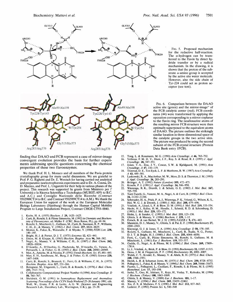
Comparison with Flavocytochrome b2. D-Amino acid oxi-dase is a member of a group of mechanistically relatedflavoenzymes displaying a number of common features, suchas the reactivity toward sulfite and the ability to stabilize theanionic form of the reduced flavin derivatives (42). The groupencompasses two subfamilies, the oxidases, in which flavinreoxidation occurs at the expenses of molecular oxygen, andthe dehydrogenases-electron transferases that utilize a mono-electronic acceptor for oxidation. Within this second subfam-ily, the FMN containing flavocytochrome b2 (FCB; L-lactatecytochrome c oxidoreductase) oxidizes L-lactate to pyruvate,a reaction similar to that of D-amino acid oxidase, thoughhaving opposite stereospecificity.The crystal structure of FCB (44) shows that its flavin
binding domain has a (f3/a)8 topology unrelated to the struc-ture of DAAO. However, analysis of the active site of the twoproteins reveals a remarkable similarity in the nature and
location of several functional groups: (i) the binding of thebenzoate in DAAO and of pyruvate in FCB is similar; theyboth lay parallel to the flavin with their carboxylate groupsinteracting with a Tyr and an Arg side chain (Tyr-228 andArg-283 in DAAO, Tyr-143 and Arg-376 in FCB); (ii) the N5atom of the cofactor makes .a H-bond with the backbone Natom of an Ala residue (Ala-49 in DAAO, Ala-198 in FCB) onthe flavin side opposite to the substrate; and (iii) the active sitewater of DAAO occupies a position analogous to that of thehydroxyl group of Tyr-254 in FCB- i.e., the water of DAAOis within H-bond distance from the predicted a-amino groupof the bound substrate, whereas Tyr-254 OH group in FCB isH-bonded to the carbonyl oxygen of the pyruvate product.Although the two active sites show similar architectures,
they cannot be superimposed because the locations of benzo-ate (in DAAO) and pyruvate (in FCB) are on opposite facesof the flavin (on the re side in DAAO, on the si side in FCB).The arrangement of the FCB catalytic groups can therefore bedescribed as the mirror-image of the DAAO active site. Thisbecomes evident when the coordinates of the FCB active siteresidues and pyruvate are transformed by applying an opera-tion corresponding to a mirror plane coincident with the flavinring. Superposition of the isoalloxazine atoms of the resultingmirror FCB structure onto the same atoms of DAAO revealsa striking similarity (Fig. 6) in the positioning of the catalyticgroups. A rms deviation of 0.9 A is measured when thefollowing atom pairs are considered (DAAO/FCB): N-Ala-49/N-Ala-198, OH-Tyr-228/OH-Tyr-143, guanidinium group(NC, Ce, Nq1, Ni12)-Arg-283/guanidinium group-Arg-376,O-Gly-313/Ne2-His-373, active site water/OH-Tyr-254.Moreover, the location of benzoate in DAAO coincides withthat of pyruvate in FCB (Fig. 6), indicating that the similaritybetween the two active sites applies to both substrate bindingand most of the groups building the active center.The fact that the DAAO and FCB catalytic sites can be
considered as structurally enantiomeric finds a precise func-tional explanation in the opposite substrate stereospecificity ofthe two proteins. In both cases, the substrate is aligned withinthe binding site by a salt bridge with an arginine residue; in thisway, both enzymes selectively bind the stereoisomer thatproperly interacts with the flavin. Thus, the topologicallyunrelated DAAO and FCB structures appear as a remark-able example of convergent molecular evolution towardcommon, but enantiomeric, active site architectures, which aresuited for very similar reactions (oxidation of an a-amino acid orof an a-hydroxy acid) but with opposite stereospecificity.The reaction catalyzed by FCB is thought to proceed via a
carboanion intermediate formed after substrate proton ab-straction by His-373 (refs. 42 and 45 and references therein),whereas, as discussed above, a carboanion mechanism seemsnow very unlikely in DAAO, which lacks a residue suited to actas the active site base abstracting the amino acid a-proton. Our
FIG. 4. (a) Stereo plot of the catalytic center. The "active site" water is shown as a sphere (see text). (b) Illustration of the active site cavitywith a model for a bound D-alanine. The model was generated by optimally superposing the Co, Ca, and C atoms of the substrate onto the C2,Cl, and carboxylate carbon of the benzoate inhibitor, respectively. The modeling was carried out using the program o (17). The blue dotted surfacedelineates the active site cavity and was calculated after removing the benzoate atoms from the structure (31).
Proc. Natl. Acad. Sci. USA 93 (1996)
Proc. Natl. Acad. Sci. USA 93 (1996) 7501
>GLN53
I
GLY313
0,>GLN53
0
ARG283 HK,(H
R ..
ON- NH' I-TYR2240
TYR228 H
2
e
R
FIG. 5. Proposed mechanismfor the reductive half-reaction.The a-hydrogen can be trans-ferred to the flavin by direct hy-dride transfer or by a radicalmechanism. In the drawing, it isshown that the proton of the sub-strate a-amino group is acceptedby the active site water molecule.However, also the side chain ofTyr-224 could act as proton ac-ceptor (see text).
FIG. 6. Comparison between the DAAOactive site (green) and the mirror-image" ofthe FCB catalytic center (red). FCB coordi-nates (44) were transformed by applying theoperation corresponding to a mirror coplanarto the flavin ring. The isoalloxazine atoms ofthe resulting mirror FCB structure were thenoptimally superposed to the equivalent atomsof DAAO. The picture outlines the strikinglysimilar location in three-dimensional space ofthe catalytic groups in the two active sites.The picture was produced by using the secondsubunit of the FCB crystal structure (ProteinData Bank entry 1FCB).
finding that DAAO and FCB represent a case of mirror-imageconvergent evolution provides the basis for further experi-ments addressing specific questions concerning the chemicalproperties of these two flavoenzymes.We thank Prof. H. L. Monaco and all members of the Pavia protein
crystallography group for many useful discussions. We are grateful toProf. P. G. Righetti and Dr. E. Wenisch for having carried out analyticaland preparative isoelectrophoresis experiments and to Dr. A. Cosma, Dr.D. Mazzeo, and Prof. L. Ungaretti for their help in various phases of theproject. This research was supported by grants from Ministero per 1'Universita e la Ricerca Scientifica e Tecnologica (MURST; 60% to B.C.and A.C.) and Consiglio Nazionale delle Ricerche (Contract9502988CT14 to B.C. and Contract 9502989CT14 to A.M.). We thank theEuropean Union for support of the work at the European MolecularBiology Laboratory (Hamburg) through the Human Capital MobilityProgram to Large Installations Project, Contract CHGE-CM93-0040.
1. Krebs, H. A. (1935) Biochem. J. 29, 1620-1625.2. Curti, B., Ronchi, S. & Pilone Simonetta, M. (1992) in Chemistry and Biochem-
istry of Flavoenzymes, ed. Muller F. (CRC, Boca Raton, FL), pp. 69-94.3. Ronchi, S., Minchiotti, L., Galliano, M., Curti, B., Swenson, R. P., Williams,
C. H., Jr., & Massey, V. (1982) J. Biol. Chem. 257, 8824-8829.4. Momoi, K., Fukui, K., Watanabe, F. & Miyake, Y. (1988) FEBS Lett. 238,
180-185.5. Bright, H. J. & Porter, D. J. T. (1975) Enzymes 12, 421-441.6. Dixon, M. & Kleppe, K. (1965) Biochim. Biophys. Acta 96, 368-382.7. Negri, A., Massey, V. & Williams, C. H., Jr. (1987) J. Biol. Chem. 262,
10026-10034.8. D'Aniello, A., D'Onofrio, G., Pischetola, M., D'Aniello, G., Vetere, A.,
Petrucelli, L. & Fisher, G. H. (1993) J. Biol. Chem. 268, 26941-26949.9. Mor, A., Amiche, M. & Nicolas, P. (1992) Trends Biochem. Sci. 17,481-485.
10. Man, E. H., Sandhouse, M., Burg, J. & Fisher, G. H. (1983) Science 220,1407-1408.
11. Curti, B., Ronchi, S., Branzoli, U., Ferri, G. & Williams, C. H., Jr. (1973)Biochim. Biophys. Acda 327, 231-266.
12. Bolognesi, M., Ungaretti, L., Curti, B. & Ronchi, S. (1978) J. Biol. Chem.253, 7513-7514.
13. Collaborative Computational Project Number 4 (1994)Acta Crystallogr. D50, 760-767.
14. Sheldrick, G. M. (1991) in Isomorphous Replacement and AnomalousScattering: Proceedings ofthe CCP4 Study Weekend 25-26 January 1991, eds.Wolf, W., Evans, P. R. & Leslie, A. G. W. (Science and EngineeringResearch Lab., Daresbury Lab., Warrington, U.K.), pp. 23-38.
15. Tong, L. & Rossmann, M. G. (1990) Acta Crystallogr. A 46, 783-792.16. Vellieux, F. M. D. V., Hunt, J. F., Roy, S. & Read, R. J. (1995) J. Appl.
Crystallogr. 28, 347-351.17. Jones, T. A., Zou, J. Y., Cowan, S. W. & Kjeldgaard, M. (1991) Acta
Crystallogr. A 47, 110-119.18. Tronrud, D. E., Ten Eyck, L. F. & Matthews, B. W. (1987) Acta Crystallogr.
A 43, 489-501.19. Laskowski, R. A., MacArthur, M. W., Moss, D. S. & Thornton, J. M. (1993)
J. Appl. Crystallogr. 26, 283-291.20. Briunger, A. T. (1992) Nature (London) 355, 472-475.21. Kraulis, P. J. (1991) J. Appl. Crystallogr. 24, 946-950.22. Wierenga, R. K., Drenth, J. & Schulz, G. E. (1983) J. Mol. Biol. 167,
725-739.23. Torri Tarelli, G., Vanoni, M. A., Negri, A. & Curti, B. (1990)J. Biol. Chem.
34, 21242-21246.24. Schreuder, H. A., Prick, P. A. J., Wierenga, P. K., Vriend, G., Wilson, K. S.,
Hol, W. G. J. & Drenth, J. (1989) J. Mol. Biol. 208, 679-696.25. Vrielink, A., Lloyd, L. F. & Blow, D. M. (1991) J. Mol. Biol. 219, 533-554.26. Hecht, H. J., Kalisz, H. M., Hendle, J., Schmid, R. D. & Schomburg, D.
(1993) J. Mol. Biol. 229, 153-172.27. Holm, L. & Sander, C. (1993) J. Mol. Biol. 233, 123-138.28. Ghisla, S. & Massey, V. (1986) Biochem. J. 239, 1-12.29. Entsch, B. & van Berkel, W. J. H. (1995) FASEB J. 9, 476-483.30. Manstein, D. J., Massey, V., Ghisla, S. & Pai, E. F. (1988) Biochemistry 27,
2300-2303.31. Kleywegt, G. J. & Jones, T. A. (1994) Acta Crystallogr. D 50, 178-185.32. Ronchi, S., Galliano, M., Minchiotti, L., Curti, B., Rudie, N. G., Porter,
D. J. T. & Bright, H. J. (1980) J. Biol. Chem. 255, 6044-6050.33. Ferti, C., Curti, B., Pilone Simonetta, M., Ronchi, S., Galliano, M. &
Minchiotti, L. (1981) Eur. J. Biochem. 119, 553-557.34. Gadda, G., Negri, A. & Pilone, M. S. (1994) J. Biol. Chem. 269, 17809-
17814.35. Li, J., Vrielink, A., Brick, P. & Blow, D. (1993) Biochemistry 32, 11507-11515.36. Denu, J. M. & Fitzpatrick, P. F. (1994) Biochemistry 33, 4001-4007.37. Walsh, C. T., Krodel, E., Massey, V. & Abels, R. H. (1973) J. Biol. Chem.
248, 1946-1951.38. Hersh, L. B. & Schuman Jorns, M. (1975) J. Biol. Chem. 250, 8728-8732.39. Pollegioni, L., Fukui, K. & Massey, V. (1994) J. Biol. Chem. 269, 31666-31673.40. Faotto, L., Pollegioni, L., Ceciliani, F., Ronchi, S. & Pilone, M. S. (1995)
Biotechnol. Lett. 17, 193-198.41. Isolai, T., Ono, H., Ishitani, Y., Kojo, H., Veday, Y., Kohsaka, M. (1994)
J. Biochem. (Tokyo) 108, 1063-1069.42. Ghisla, S. & Massey, V. (1989) Eur. J. Biochem. 181, 1-17.43. Massey, V. (1994) J. Biol. Chem. 269, 22459-22462.44. Xia, Z. X. & Mathews, F. S. (1990) J. Mol. Biol. 212, 837-863.45. Lederer, F. (1992) Protein Sci. 1, 540-548
Biochemistry: Mattevi et al.