crystal structure degradation substrate: a three-disulfide
TRANSCRIPT

Proc. Natl. Acad. Sci. USAVol. 90, pp. 4136-4140, May 1993Biochemistry
Crystal structure of a ubiquitin-dependent degradation substrate: Athree-disulfide form of lysozyme
(proteolysis/ubiquitination/speciflcity determinant/stability)
CHRISTOPHER P. HILL*, NANCY L. JOHNSTON, AND ROBERT E. COHENtDepartment of Chemistry and Biochemistry and the Molecular Biology Institute, University of California, Los Angeles, CA 90024-1570
Communicated by Howard K. Schachman, January 29, 1993 (received for review December 16, 1992)
ABSTRACT Covalent attachment of ubiquitin marks sub-strates for proteolysis, but features that identify ubiquitinationtargets such as chicken egg white lysozyme are poorly under-stood. Recognition of lysozyme first requires reduction ofCys-6 Cys-127, one of its four native disulfide bonds, andCys-6,Cys-127-carboxymethylated (6,127-rcm) lysozyme canmimic this three-disulfide intermediate. The 6,127-rcm form oflysozyme is known to retain a substantially native-like confor-mation in solution, and we demonstrate that it is this foldedstructure that is recognized for ubiquitination. Because nativelysozyme is not a substrate, differences between the native andthree-disulfide structures must include features responsible forselective ubiquitination. The 1.9-A resolution crystal structureof 6,127-rcm-lysozyme, reported here, affords a view of thisubiquitin-dependent degradation substrate. Two conformers of6,127-rcm-lysozyme were obtained in the crystal. These differuniquely from crystal forms of native lysozyme by displacementof the C-terminal residues. The structures suggest that localizedunfolding at the C terminus of three-disulfide lysozyme allowsthe complex ofE3a (ubiquitin-protein ligase) and E2 (ubiquitin-carrier protein) to bind to a surface that includes Lys-1 and theputative ubiquitination site Lys-13. From this we infer that theN-terminal and internal substrate recognition sites on theE3a-E2 complex are separated by -20 A.
Intracellular protein degradation is remarkable for its com-bination of extreme selectivity and the ability to accommo-date an enormous variety of substrates. Protein half-lives invivo span several orders of magnitude. Moreover, mutations,translational errors, mislocalization, and chemical damage allcan lead to polypeptides that are rapidly degraded (1-5). Howsuch proteins are distinguished from their long-lived coun-terparts is largely unknown. In eukaryotes, a major route forintracellular proteolysis involves covalent modification ofprotein lysine(s) with the protein ubiquitin (Ub) and subse-quent degradation by the 26S Ub-dependent protease com-plex (6-8). Specificity resides, at least in part, with theUb-protein ligase that marks a substrate for recognition bythe protease. This process is best understood for the E3aligase from rabbit reticulocytes and the related UBRI-encoded enzyme from yeast (9-13).One important E3a recognition determinant is the sub-
strate's N terminus, where only a subset of amino acids ispermissive for ubiquitination (9-12). A permissive N termi-nus is not suffi'cient, however, and the relative orientationsand distances that separate lysine ubiquitination sites fromN-terminal and other, as yet undiscovered, determinants alsoare likely to be critical for recognition (14-17). Progress inthis area has been hampered by the lack of Ub-dependentdegradation substrates of defined structure. Indeed, to obtaincompetent in vitro substrates, proteins generally must be
altered conformationally (16, 17). This generalization appliesto chicken egg white lysozyme, where we have found thatefficient ubiquitination requires prior reduction of one of thefour native disulfide bonds, C`ys-6 Cys-127. A reduced andcarboxymethylated derivative, 6,127-rcm-lysozyme, canmimic this three-disulfide degradation intermediate (17).Three-disulfide lysozyme offers a unique opportunity toprobe ubiquitination specificity because, unlike other E3asubstrates described thus far, it retains a substantially struc-tured, native-like conformation (17-19). We report here thex-ray crystal structure of 6,127-rcm-lysozyme determined toa resolution of 1.9 A. An experiment that relates this struc-ture to the substrate conformation recognized in solution byUb-protein ligase is presented, and the results are discussedin terms of structural determinants for ubiquitination.*
EXPERIMENTAL PROCEDURESProtein Crystallization. The 6,127-rcm derivative of
chicken egg white lysozyme (EC 3.2.1.17; Sigma, grade I)was prepared essentially as described (17), but with addi-tional chromatography on a PolyCAT A (The Nest Group,Southboro, MA) cation-exchange column. Stock solutionswere dialyzed against water and concentrated to 9 mg/ml inCentricon 10 microconcentrators (Amicon). Crystals weregrown by the hanging drop method under conditions similarto those used to obtain tetragonal crystals of unmodifiedlysozyme. The reservoir solution (1 ml) was 7% NaCl with 50mM sodium acetate at pH 3.8, and the hanging drops con-tained 10 ,ul of a 1:1 mixture of reservoir and protein stocksolutions. Typically, crystals appeared after 2 days and grewto 0.3 x 0.4 x 0.5 mm3 within a week. The crystals belongedto orthorhombic space group P212121 with cell dimensions a= 77.7 A, b = 81.3 A, and c = 38.0 A.Data Collection and Structure Determination. X-ray data
extending to 1.9-A resolution were collected from two crys-tals with a R-AXIS II imaging plate detector and a RigakuRU-200 rotating anode source (CuKa radiation at 50 kV and150 mA). Each frame, in which the crystal was oscillated by1.50, was acquired over 12 min; =35 frames were collectedfrom each crystal before there was significant deteriorationfrom radiation damage. Processing employed the MSC soft-ware package. The chicken egg white lysozyme structure of
Abbreviations: Ub, ubiquitin; 6,127-rcm, Cys-6,Cys-127-carboxy-methylated; rcmA and rcmB, the two molecules per asymmetric unitin 6,127-rcm-lysozyme crystals; E3, ubiquitin-protein ligase; E2,ubiquitin carrier or conjugating protein; (GlcNAc)3, N,N',N'-triacetylchitotriose.*Present address: Department of Biochemistry, University of Utah,Salt Lake City, UT 84132.tTo whom reprint requests should be addressed at: MolecularBiology Institute, University of California, 405 Hilgard Avenue,Los Angeles, CA 90024-1570.tCoordinates and diffraction data have been deposited in the ProteinData Bank, Chemistry Department, Brookhaven National Labora-tory, Upton, NY 11973 (reference lRCM) (20, 21).
4136
The publication costs of this article were defrayed in part by page chargepayment. This article must therefore be hereby marked "advertisement"in accordance with 18 U.S.C. §1734 solely to indicate this fact.
Dow
nloa
ded
by g
uest
on
Janu
ary
25, 2
022

Proc. Natl. Acad. Sci. USA 90 (1993) 4137
Strynadka and James (22) was used as a starting model for6,127-rcm-lysozyme structure determination. Water mole-cules and side chains with multiple conformations weredeleted from the starting model, as were the first 14 and last6 amino acids. The 6,127-rcm-lysozyme crystals are similar tothose of native lysozyme grown under similar conditions,although the latter are of the tetragonal space group P43212with cell dimensions of a = b = 79.24 A and c = 37.83 A (22).The P212121 space group of the 6,127-rcm-lysozyme is relatedto P43212 by one two-fold rotation axis. Consideration of thissymmetry relationship enabled two copies of the truncatednative lysozyme molecule to be placed within the 6,127-rcm-lysozyme asymmetric unit. Rigid-body refinement withXPLOR (23) gave an R factor of 26.7% against all 3.0- to 8.0-Adata. This solution was confirmed by rotation function and Rfactor searches using XPLOR. One cycle of simulated anneal-ing refinement using the "slow-cool" protocol was followedby cycles of positional and B factor refinement, with severalrounds of manual map-fitting using the program FRODO (24)implemented on an Evans and Sutherland ESV graphicsstation (Salt Lake City). The entire asymmetric unit wasinspected in difference maps, water molecules were added,and the protein model was rebuilt where appropriate. Datacollection and final refinement statistics are in Table 1.
Ub-Protein Conjugation. Ub-activating enzyme (El) andthe 14-kDa Ub carrier protein (E214K) were purified from calfthymus and rabbit reticulocytes, respectively (25, 26), andubiquitination assays were done essentially as described (17).Each 10-,u1 reaction mixture in 50 mM Tris HCl (pH 7.6)contained 2.5 mM dithiothreitol, 2 mM ATP, 5 mM MgCl2, 5mM phosphocreatine, 30 mU creatine phosphokinase, 3 mUinorganic pyrophosphatase, and a Ub-depleted rabbit retic-ulocyte extract (21 ,g of protein; see ref. 17) supplementedwith S ,g of bovine Ub (Sigma), 57 nM El, and 38 nM E214K.After a 5-min preincubation at 37°C with 2 ,uM Ub-aldehydeto prevent conjugate breakdown by endogenous Ub-proteinisopeptidases (16), 1251-labeled substrate (0.4 ,ug) was addedand the incubation was continued for 1 hr. Products werevisualized after SDS/polyacrylamide gel electrophoresis andautoradiography (17).
a ubiquitination assay, a few percent of 6,127-rcm-lysozymeis in an unfolded state (Fig. 1A). As with unmodified lyso-zyme (28), an oligosaccharide ligand can shift this equilibriumto stabilize the folded form of the 6,127-rcm derivative (Fig.1A), but such stabilization has no effect upon lysozymeubiquitination (Fig. 1B). This same result is obtained over awide range of lysozyme concentrations, where the substrateis well below the apparent Km of Ub-protein ligase (unpub-lished data). Thus, the relatively limited structural differ-ences between the folded conformations of native lysozymeand 6,127-rcm-lysozyme must include features used to dis-tinguish ubiquitination targets from other proteins.
Crystal Structure of 6,127-rcm-Lysozyme. The 6,127-rcm-lysozyme was crystallized into space group P212121, differentfrom that of native lysozyme crystals from which high-resolution structure coordinates are available. We were ableto solve the structure by molecular replacement with a model
'0a)'0
0
LL
1.0 T LI
0.8 0
0.6
0.4
0.2 0' 0
0. 0 0.*_- L2 5
B
66 -
35 45 55Temperature ( °C)
1 2
65 75
3 4 5 6
RESULTS AND DISCUSSIONThe Folded Form of 6,127-rcm-Lysozyme Is a Ubiquitination
Substrate. The 6,127-rcm derivative of lysozyme is thermo-dynamically less stable than unmodified lysozyme (18, 19),which might suggest that it is the unfolded protein that isrecognized by the Ub-protein ligase, E3a. We have foundthat this is not the case. At 37°C under conditions that mimic
Table 1. Data collection and refinement statisticsParameter
Resolution range, ANo. of reflectionsCompleteness, %Rsym*Rfactort (all data)No. of water molecules includedB factor
Molecule A average, A2Molecule B average, A2Solvent average, A2
rms deviations from ideal geometryBond distances, ABond angles, degreesDihedral angles, degrees
rms deviation of main-chain bond B values, A2
*Rsym = (1I1 - Iavl)/YIav.tRfactor = (Y|IIFobsI - IFcaIcI|)/I|FobsI.
Value
1.9-10.018,29593.80.0420.185133
24.723.936.3
0.0142.824.31.7
45 -36 -
29 -
24 -
20 -(GIcNAc)3, mM 0
... ..........
........
0.05 0.5 0 0.05 0.5
FIG. 1. Ubiquitination of lysozyme does not depend upon globalunfolding. (A) Thermal denaturation of 6,127-rcm-lysozyme in 50mM sodium 3-(N-morpholino)propanesulfonate buffer (pH 7.0) wasmonitored by the absorbance change at 280 nm (19), and the data arepresented as the fraction of protein in the unfolded state without (o)or with (e) 1.0 mM N,N',N"'-triacetylchitotriose [(GlcNAc)3]. Theligand stabilizes the protein, increasing its melting transition tem-perature from 47.6°C to 52.7°C. (B) Ub conjugation assays employed1251-labeled lysozyme (1 x 10⁵ cpm, lanes 1-3) or 6,127-rcm-lysozyme (1.4 x 10⁵ cpm, lanes 4-6) with 0, 0.05, and 0.5 mM(GlcNAc)3 as indicated; native lysozyme and 6,127-rcm-lysozymeunder these conditions are saturated by -0.05 mM (GlcNAc)3 (ref.27; unpublished data). Note that by inclusion of 2.5 mM dithiothreitolin these reactions, 1-2% of the native lysozyme is in the three-disulfide form (17). Conjugates (>20 kDa) were visualized afterSDS/polyacrylamide gel electrophoresis and autoradiography. Po-sitions of molecular mass markers (kDa) are to the left of the gel;bands corresponding to the 1251-labeled substrates (14 kDa) wereoverexposed and are not shown.
Biochemistry: Hill et al.
Dow
nloa
ded
by g
uest
on
Janu
ary
25, 2
022

Proc. Natl. Acad. Sci. USA 90 (1993)
FIG. 2. Comparisons of 6,127-rcm-lysozyme and native chicken egg white lysozyme crystal structures. The main-chain tracing from the1.75-A native lysozyme structure (22) is shown in green superimposed upon the two 6,127-rcm-lysozyme conformers (blue and red) determinedto 1.9 A. Where the structures coincide, they appear white; disulfide bonds are shown only for the native structure, and are in green. The Ctermini, N termini, and active site are indicated. The Cys-6CWy`s-127 disulfide, broken in the 6,127-rcm derivative, is identified by the arrow.The Lys-13 ubiquitination site (not shown here; see text) extends from the prominent helix (residues 5-15) that is linked via the C's-6 Cys-127disulfide to the C terminus. All three structures are nearly identical except for several residues at the C termini.
of native lysozyme that had been determined from crystalsbelonging to space group P43212 (22). The resultant 1.9-Aresolution atomic model for 6,127-rcm-lysozyme includestwo molecules per asymmetric unit and has been refined to anR factor of 0.185 (Table 1).
Overall, the conformations of 6,127-rcm-lysozyme andnative lysozyme are extremely similar, and significant devi-ations (i.e., displacements > 1.0 A) in the polypeptidebackbones are limited to only a few regions. Fig. 2 shows themain-chain polypeptide tracings for both of the 6,127-rcm-lysozyme molecules in the asymmetric unit, rcmA and rcmB,superimposed upon the 1.75-A resolution native lysozymestructure obtained from tetragonal crystals (22). Differencesare slight except at the C terminus and, to a lesser extent, fora short peptide segment centered about residue 71 (regions IIand IV in Fig. 3 Upper Left). Comparisons with the 1.5-Anative lysozyme structure obtained from triclinic crystals (29,30)§ revealed deviations for two additional regions centeredabout residues 47 and 101 (Fig. 3 Upper Right). For regionsI-III, similar main-chain deviations are observed between thenative lysozyme structures (Fig. 3 Lower Left, tetragonal vs.triclinic), whereas only regions I and IV differ between thetwo 6,127-rcm-lysozyme molecules (Fig. 3 Lower Right,rcmA vs. rcmB). Positional differences within the pairs ofnative or 6,127-rcm-lysozyme structures are unequivocalevidence of conformational mobility. Note, however, thatregion IV of the native lysozyme main chain appears fixed(Fig. 3 Lower Left). Thus, upon Cys-6 Cys-127 reduction andcarboxymethylation, only the C terminus is seen to deviatefrom the conformations accessible to lysozyme in the nativestate.Although Cy's-6 Cy's-127 disulfide cleavage frees lyso-
zyme's C terminus from its interaction with the body of theprotein, the position of the N-terminal peptide that encom-passes Cys-6 is essentially unchanged. An inventory of thecontacts in native lysozyme made by residues near the N andC termini shows that this might be expected. Other than theCys-6 Cys-127 disulfide itself, few interactions stabilize thenative conformation of lysozyme's five C-terminal residues,which contribute to only 4 hydrogen bonds as compared to 11for the first five (N-terminal) residues.
§Brookhaven Protein Data Bank reference 2LZT.
Destabilization of the native conformation at the C termi-nus of 6,127-rcm-lysozyme also must be due in part toelectrostatic repulsion of the side chain carboxylates ofcarboxymethylcysteines 6 and 127. In rcmA and rcmB,residue 127 is oriented to position its side chain into solventand away from the buried hydrophobic environment ofCys-127 in native lysozyme. These new conformations entaila redirection of the polypeptide backbone, as is evident inFig. 2. For Cys-6, however, positions of the main-chain andCp atoms are nearly identical for native lysozyme and 6,127-rcm lysozyme. Placement of the Cys-6 carboxymethyl moi-eties within the model was precluded by weak electrondensity, however, which indicates that this side chain is quitemobile.
Conformational Flexibility of the C-Terminal Residues. Theconformations of the C-terminal portions of rcmA and rcmBdiffer significantly from each other and from the nativestructure. These residues therefore are expected to be mobilein solution, and inspection of lattice contacts suggests thatthese motions may be much larger when free ofthe crystallineenvironment. As illustrated in Fig. 4, the last five residues of6,127-rcm-lysozyme have contacts to symmetry-related mol-ecules. These contacts are similar for rcmA and rcmB and, ineach case, are as extensive as the noncovalent intramolecularinteractions. Thus, the deviations of the 6,127-rcm-lysozymeC-terminal residues seen from the crystal structures mayseverely underestimate the true range of motion in solution.
In contrast, and despite the absence of restrictive latticecontacts (see Fig. 4), the conformations of the N-terminalresidues are essentially identical. Two-dimensional NMRmeasurements by Radford et al. (18) support our conclusionthat the N-terminal portion of 6,127-rcm-lysozyme is virtu-ally the same as that of the native protein. From an immu-nochemical study, however, it appeared that the 6,127-rcm-lysozyme N-terminal region may be distorted relative to thenative conformation (19). The immunochemical and struc-tural data can be reconciled if the N-terminal epitopes thatwere probed also included Cys-6 or distal portions of theantigen such as its C terminus.
Recognition by Ub-Protein Ligase. Although movement ofthe Leu-129 side chain was apparent from the NMR study of6,127-rcm-lysozyme, whether there was a more extensiverearrangement of the C terminus could not be determined
4138 Biochemistry: Hill et al.
Dow
nloa
ded
by g
uest
on
Janu
ary
25, 2
022

Proc. Natl. Acad. Sci. USA 90 (1993) 4139
6,127-RCM vs. Native (tetragonal)Ir
rcmA
4k
6,127-RCM vs. Native (triclinic)I, 1Iv 4
Aro j..C' 7j I'-'
2 - rcmB
4-
Native vs. Native4- (tetragonal II
vs. triclinic)2-
o I~~~~~~~~~~~~~~~~~~~~~~~~~~~~~~~~~~~~~~~~~~~~~~~~~~~~23i ~ ~ ~ ~ ~ ~~~~~~.........0 20 40 60 80 100
4.
2-
120 0
Residue number
MI IVI1 IlVrcmA
rcmB
6,127-RCM vs. 6,127-RCM(rcmA vs. rcmB)
1
2 0 4 0 6 0 8 0 1 00 120
FIG. 3. 6,127-rcm-lysozyme conformation differs uniquely from native lysozyme at its C terminus. Plots of the main-chain rms deviationsper residue are shown for all pairwise comparisons of the two 6,127-rcm-lysozyme forms (rcmA and rcmB) and two crystal forms of nativelysozyme [tetragonal (22) and triclinic (29, 30)]. For comparisons of 6,127-rcm vs. native forms (Upper Left and Upper Right), deviations forrcmA are plotted above the horizontal axis and, for rcmB, below the axis. Regions of significant displacement are labeled I-IV. Deviationsbetween the 6,127-rcm and native lysozymes at sites I, II, and III are also seen between the two native forms alone (Lower Left) and reflectconformational flexibility within the native protein. However, at the C-terminal five residues (region IV), both 6,127-rcm forms adoptconformations unavailable to native lysozyme.
(18). The crystal structure resolves this issidisplacement of the C-terminal segment frotion in the native protein as the key alteratthree-disulfide lysozyme a ubiquitination t;recognition by the Ub-protein ligase involideterminants, a permissive N-terminal amiinternal site that may include the ubiquitinat-(9-12, 15, 31); these correspond to the I"body" sites postulated by Reiss et al. (11accessibility of lysozyme's N terminus (Lys-the 6,127-rcm and native forms, we propose127 disulfide cleavage generates a ubiquitinatexposing elements of the second, body site d4site is unlikely to be within the conformaC-terminal region because neither carbox3Cys-127 nor deletion of Arg-128 and Letubiquitination of three-disulfide lysozymwork). Instead, as a consequence of the 4rangement, there is enhanced exposure of tieated by residues 5-15 and which contaipotential ubiquitination site. Evidence thamajor ubiquitination site has come from expLys-13 -* Arg mutant form oflysozyme. Unliprotein, the Arg-13 variant is ubiquitinatedvitro (unpublished).
Ubiquitination Determinants on Three-DistA view of the three-disulfide lysozyme surfathe N-terminal and putative internal recoi
A -
B *_ - - * __ - --
0 20 40 60 80Residue number
FIG. 4. Intermolecular contacts in the crystaland 6,127-rcm lysozymes. Intermolecular contameasured for four lysozyme structures, 6,127-rcrcmB (B)] and native [tetragonal (C) and triclinic (lupon lines representing the lysozyme primary sindicate residues <4.0 A away from a symmetmolecule in the crystal lattice.
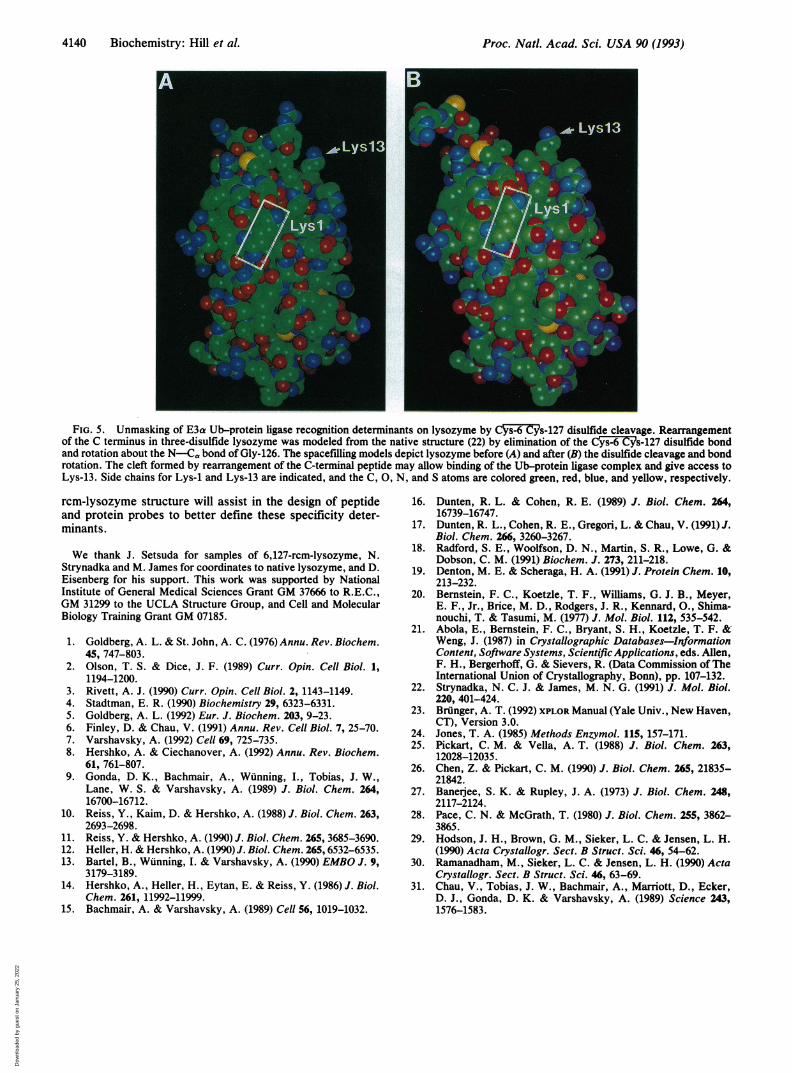
ae and points to nants is shown in Fig. 5. The model was constructed fromm its conforma- native lysozyme by removal of the Cys-6 Cys-127 disulfideion that renders bond and rotation about the N Ca bond of Gly-126 to swingarget. Substrate the C-terminal tetrapeptide into the solvent, as suggested byves at least two the 6,127-rcm structures. We propose that this conforma-ino acid and an tional change gives E3a or an E3a-E2 complex access toable Lys residue Lys-13 and allows simultaneous binding to the determinant atE3 "head" and the N terminus, Lys-1. This model, in which key binding0, 11). Because determinants are accessible only in the locally unfolded-1) is identical in molecule, can explain why native lysozyme was unable tothat C'ys-6 Cys- compete with the 6,127-rcm derivative in Ub-dependentLion substrate by degradation assays (17). Displacement of the C-terminaleterminant. This residues increases the accessibility of Lys-13 and adjacentLtionally altered residues, particularly Cys-6, Ala-9, and Ala-10, as well asymethylation of Asp-18 and Leu-25; any or all of these might contribute to aa-129 decreases surface for recognition by E3a or an E3aE2 complex.te (unpublished In yeast, a flexible polypeptide spacer between the NC-terminal rear- terminus and Ub attachment site was required for Ub-he a-helix delin- dependent degradation of modified forms of dihydrofolatens Lys-13 as a reductase and f3-galactosidase (15). The need for a minimumLt Lys-13 is the spacer length of >5 residues suggested that both determi-)eriments with a nants may be bound simultaneously to distinct sites on theike the wild-type Ub-protein ligase complex (15). Three-disulfide lysozymeonly poorly in differs from these other model substrates in that the recog-
nition determinants are within a comparatively rigid struc-ilfide Lysozyme. ture. Indeed, the crystallographic results presented heretce that presents together with NMR spectroscopy as a probe of solutiongnition determi- structure (18) show that in three-disulfide lysozyme the
main-chain atom positions are relatively fixed from residue 1n_ to residue 124. Thus, having demonstrated that E3a recog-
_ nizes the folded form of three-disulfide lysozyme rather thana transiently unfolded species, we can infer that a distance of
_ _ 20 A separates the N-terminal and internal sites (i.e., headand body sites) on the mammalian E3a Ub-protein ligase.¶
1 0 0 1 2 0 Other constraints upon substrate recognition are likely toinvolve residues in the vicinity of Lys-13, and E2 bound to
I lattice for native E3a also may participate in these interactions. The 6,127-.. _.. . .v. .
ct distances were.m [rcmA (A) andD)]. Superimposedequence, the barstry-related protein
$This distance applies only to the "Type I" N-terminal site thataccommodates the basic amino acid Lys, Arg, or His (10-12). Resultsfrom the in vivo experiments of Bachmair and Varshavsky (15) mayreflect proximity of the ubiquitination site to a "Type II" N-terminalsite (bulky hydrophobic amino acid) on the yeast enzyme.
4
2-
0
0
._
*5
2-
0
0
a)
C.0
._
(0
0 IV4
Biochemistry: Hill et al.
Dow
nloa
ded
by g
uest
on
Janu
ary
25, 2
022
Proc. Natl. Acad. Sci. USA 90 (1993)
FIG. 5. Unmasking of E3a Ub-protein ligase recognition determinants on lysozyme by Cys-6 Cy's-127 disulfide cleavage. Rearrangementof the C terminus in three-disulfide lysozyme was modeled from the native structure (22) by elimination of the C'ys-6 Cys-127 disulfide bondand rotation about the N-Ca bond of Gly-126. The spacefilling models depict lysozyme before (A) and after (B) the disulfide cleavage and bondrotation. The cleft formed by rearrangement of the C-terminal peptide may allow binding of the Ub-protein ligase complex and give access toLys-13. Side chains for Lys-1 and Lys-13 are indicated, and the C, 0, N, and S atoms are colored green, red, blue, and yellow, respectively.
rcm-lysozyme structure will assist in the design of peptideand protein probes to better define these specificity deter-minants.
We thank J. Setsuda for samples of 6,127-rcm-lysozyme, N.Strynadka and M. James for coordinates to native lysozyme, and D.Eisenberg for his support. This work was supported by NationalInstitute of General Medical Sciences Grant GM 37666 to R.E.C.,GM 31299 to the UCLA Structure Group, and Cell and MolecularBiology Training Grant GM 07185.
1. Goldberg, A. L. & St. John, A. C. (1976) Annu. Rev. Biochem.45, 747-803.
2. Olson, T. S. & Dice, J. F. (1989) Curr. Opin. Cell Biol. 1,1194-1200.
3. Rivett, A. J. (1990) Curr. Opin. Cell Biol. 2, 1143-1149.4. Stadtman, E. R. (1990) Biochemistry 29, 6323-6331.5. Goldberg, A. L. (1992) Eur. J. Biochem. 203, 9-23.6. Finley, D. & Chau, V. (1991) Annu. Rev. Cell Biol. 7, 25-70.7. Varshavsky, A. (1992) Cell 69, 725-735.8. Hershko, A. & Ciechanover, A. (1992) Annu. Rev. Biochem.
61, 761-807.9. Gonda, D. K., Bachmair, A., Wunning, I., Tobias, J. W.,
Lane, W. S. & Varshavsky, A. (1989) J. Biol. Chem. 264,16700-16712.
10. Reiss, Y., Kaim, D. & Hershko, A. (1988) J. Biol. Chem. 263,2693-2698.
11. Reiss, Y. & Hershko, A. (1990) J. Biol. Chem. 265, 3685-3690.12. Heller, H. & Hershko, A. (1990) J. Biol. Chem. 265, 6532-6535.13. Bartel, B., Wunning, I. & Varshavsky, A. (1990) EMBO J. 9,
3179-3189.14. Hershko, A., Heller, H., Eytan, E. & Reiss, Y. (1986) J. Biol.
Chem. 261, 11992-11999.15. Bachmair, A. & Varshavsky, A. (1989) Cell 56, 1019-1032.
16. Dunten, R. L. & Cohen, R. E. (1989) J. Biol. Chem. 264,16739-16747.
17. Dunten, R. L., Cohen, R. E., Gregori, L. & Chau, V. (1991) J.Biol. Chem. 266, 3260-3267.
18. Radford, S. E., Woolfson, D. N., Martin, S. R., Lowe, G. &Dobson, C. M. (1991) Biochem. J. 273, 211-218.
19. Denton, M. E. & Scheraga, H. A. (1991) J. Protein Chem. 10,213-232.
20. Bernstein, F. C., Koetzle, T. F., Williams, G. J. B., Meyer,E. F., Jr., Brice, M. D., Rodgers, J. R., Kennard, O., Shima-nouchi, T. & Tasumi, M. (1977) J. Mol. Biol. 112, 535-542.
21. Abola, E., Bernstein, F. C., Bryant, S. H., Koetzle, T. F. &Weng, J. (1987) in Crystallographic Databases-InformationContent, Software Systems, Scientific Applications, eds. Allen,F. H., Bergerhoff, G. & Sievers, R. (Data Commission of TheInternational Union of Crystallography, Bonn), pp. 107-132.
22. Strynadka, N. C. J. & James, M. N. G. (1991) J. Mol. Biol.220, 401-424.
23. Brunger, A. T. (1992) XPLOR Manual (Yale Univ., New Haven,CT), Version 3.0.
24. Jones, T. A. (1985) Methods Enzymol. 115, 157-171.25. Pickart, C. M. & Vella, A. T. (1988) J. Biol. Chem. 263,
12028-12035.26. Chen, Z. & Pickart, C. M. (1990) J. Biol. Chem. 265, 21835-
21842.27. Banerjee, S. K. & Rupley, J. A. (1973) J. Biol. Chem. 248,
2117-2124.28. Pace, C. N. & McGrath, T. (1980) J. Biol. Chem. 255, 3862-
3865.29. Hodson, J. H., Brown, G. M., Sieker, L. C. & Jensen, L. H.
(1990) Acta Crystallogr. Sect. B Struct. Sci. 46, 54-62.30. Ramanadham, M., Sieker, L. C. & Jensen, L. H. (1990) Acta
Crystallogr. Sect. B Struct. Sci. 46, 63-69.31. Chau, V., Tobias, J. W., Bachmair, A., Marriott, D., Ecker,
D. J., Gonda, D. K. & Varshavsky, A. (1989) Science 243,1576-1583.
4140 Biochemistry: Hill et al.
Dow
nloa
ded
by g
uest
on
Janu
ary
25, 2
022