cryotherapy of proliferative sickle retinopathy, triple ... · british journalofophthalmology,...
TRANSCRIPT

British Journal of Ophthalmology, 1979, 63, 97-101
Cryotherapy of proliferative sickle retinopathy, II:triple freeze-thaw cycleMICHAEL H. GOLDBAUM, ROBERT C. FLETCHER, LEE M. JAMPOL, ANDMORTON F. GOLDBERGFrom the Sickle Cell Eye Clinic, University of Illinois Eye and Ear Infirmary, Chicago
SUMMARY Complete closure of retinal neovascularisation due to proliferative sickle cell retinopathywas achieved in 28 'sea fans' (areas of neovascularisation) in 9 eyes of 9 patients by means of atriple freeze-thaw technique. However, 2 eyes developed subsequent rhegmatogenous retinaldetachments, presumably related to vitreous traction on necrotic retina. We therefore recommendphotocoagulation as the best treatment of proliferative sickle cell retinopathy. If opacities in themedia prevent photocoagulation, a single freeze-thaw cycle is preferable.
Persons with sickle cell anaemia (haemoglobin SS),sickle cell B thalassaemia (haemoglobin S-Thal),and especially sickle cell haemoglobin C disease(haemoglobin SC) develop vasoproliferation fromthe retina into the vitreous. Left untreated, theseareas of neovascularisation (sea fans) may progress tovitreous haemorrhage, tractional retinal detachmentor rhegmatogenous retinal detachment (Goldberg,1971). Photocoagulation with the xenon arc orargon laser has been used to destroy neovasculartissue (Goldberg and Acacio, 1973; Goldbaum etal., 1977). For successful photocoagulation themedia must be sufficiently clear over the feedingvessels to allow the photocoagulation beam toform a sharp image in the retina. Vitreous haemor-rhage or other opacities in the media can preventsuccessful photocoagulation. Furthermore, photo-coagulation has known complications such ashaemorrhage (Goldbaum et al., 1977), choroidalischaemia (Goldbaum et al., 1976), and choroido-vitreal neovascularisation (Galinos et al., 1975;Goldbaum et al., 1977). A previous paper from ourclinic relates our experiences with a single freeze-thaw cycle of transconjunctival cryopexy in thetreatment of proliferative sickle retinopathy (Leeet al., 1975). Thirteen sea fans in 10 eyes of 6 patientswere treated. Four of these 13 sea fans (31 %) werestill patent, though markedly attenuated, aftertreatment. None of the attenuated sea fans has bledin the 3 years since the cryopexy.
Address for reprints: Dr Lee M. Jampol, Sickle Cell EyeClinic, Illinois Eye and Ear Infirmary, 1855 West TaylorStreet, Chicago, Illinois 60612, USA
Several authors have reported successful treatmentof retinoblastomas and vascular tumours, such asretinal angiomatosis, with repeated freeze-thawtechniques (Hale and Christensen, 1968; Amoilsand Smith, 1969; Welch, 1970; Watzke, 1973;Watzke, 1974). With this in mind an attempt wasmade to improve our previous cryotherapeuticresults in proliferative sickle retinopathy by employ-ing a triple freeze-thaw cycle.
Materials and methods
Between February 1974 and July 1974 every thirdeye of the patients seen at the Sickle Cell Eye Clinicof the University of Illinois Eye and Ear Infirmarythat required treatment for proliferative sickle cellretinopathy and any eye in which sea fans could beidentified but were partially obscured by vitreoushaemorrhage were selected for triple freeze-thawcycle cryotherapy. A total of 28 sea fans in 9 eyes of9 patients were treated. The patients' ages rangedfrom 16 to 53 years, with a mean of 30 years.Seven patients had haemoglobin SC, 1 had haemo-globin SS, and I had haemoglobin AS (all confirmedby quantitative haemoglobin electrophoresis).
Before treatment each patient received an eyeexamination with measurement of best correctedvisual acuity, slit-lamp examination, and a detailedretinal drawing with indirect ophthalmoscopy andscleral depression. Fluorescein angiography con-firmed the patency of the sea fans to be treated. Ineyes in which vitreous haemorrhage made identifi-cation of a sea fan difficult or uncertain, fluoresceinangioscopy with a Wratten 47 blue filter on an
97
on 7 August 2018 by guest. P
rotected by copyright.http://bjo.bm
j.com/
Br J O
phthalmol: first published as 10.1136/bjo.63.2.97 on 1 F
ebruary 1979. Dow
nloaded from

Michael H. Goldbatum, Robert C. Fletcher, Lee M. Jampol, and Morton F. Goldberg
indirect ophthalmoscope was performed to locatethe telltale green vitreous cloud of fluorescein over-lying each sea fan.Each treatment was performed either with a
carbon dioxide probe or a fluorocarbon probe, each
A
B
C --Fig. 1 Technique of triple freeze-thaw cryopexy. For a
small sea fan (A) a single site was treated. For a some-
what larger lesion (B) the feeding vessels were treatedfirst, followed by freezing of the lesion itself. For a largesea fan (C) the feeding vessels were treated and thenmultiple applications were made to cover the entire lesion
pv-~~~~4~-
-*-i_;- 1
Fig. 2 Endpoint of treatment corresponded toincorporation of vessels within the iceball with a frosted'appearance to the vessels
adjusted for maximum cooling. The intense cryo-pexy was painful with topical anaesthesia. Hence1% lignocaine was injected subconjunctivally in thequadrant to be treated, or a retrobulbar injectionwas given. The cryoprobe was applied to the globedirectly over the site of the smaller sea fans, whichwere then treated with a triple freeze-thaw thattotally encompassed the area of neovascularisation.Larger sea fans were first treated with a triple freeze-thaw cycle to the feeding vessels. The cryoprobewas then moved to adjacent sites until the entire seafan had been covered (Fig. 1).Each transconjunctival cryopexy lesion was
monitored by indirect ophthalmoscopy. The probewas positioned to indent the desired area. The iceballwas allowed to grow until the treated vessels wereincorporated. The end point was a hazy appearanceof the vessels to be treated within the iceball as ifthey were being viewed through frosted glass (Fig.2). The process was repeated two additional timesat each site of application. It is important to notethat, when the vessels to be treated stood outbrilliantly in relief against the white background ofthe iceball, the iceball had not yet reached thevessels and the end point had not yet been achieved.When vitreous haemorrhage was present and theview of the sea fan was obscured, the end pointwas more difficult to identify and had to be esti-mated.
Twenty-four or 48 hours after treatment and againseveral weeks later fluorescein angiography was
98
on 7 August 2018 by guest. P
rotected by copyright.http://bjo.bm
j.com/
Br J O
phthalmol: first published as 10.1136/bjo.63.2.97 on 1 F
ebruary 1979. Dow
nloaded from
C ryotheracpy of proiiresative sickle retinopathy
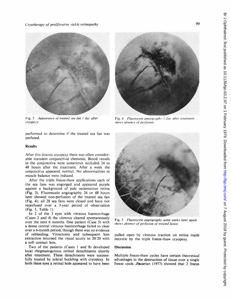
Fig. 3 Appearantce of treated sea fin I day aftercryopexv!
performed to determine if the treated sea fan wasperfused.
Results
After this intense cryopexy there was often consider-able transient conjunctival chemosis. Blood vesselsin the conjunctiva were sometimes occluded 24 to48 hours after the treatment. After a week theconjunctiva appeared normal. No abnormalities inmuscle balance were induced.
After the triple freeze-thaw applications each ofthe sea fans was engorged and appeared purpleagainst a background of pale oedematous retina(Fig. 3). Fluorescein angiography 24 or 48 hourslater showed non-perfusion of the treated sea fan(Fig. 4); all 28 sea fans were closed and have notreperfused over a 3-year period of observation(Fig. 5, Table 1).
In 2 of the 3 eyes with vitreous haemorrhage(Cases 2 and 4) the vitreous cleared spontaneouslyover the next 6 months. One patient (Case 5) witha dense central vitreous haemorrhage failed to clearover a 6-month period, though there was no evidenceof rebleeding. Vitrectomy and subsequent lensextraction returned the visual acuity to 20,T20 witha soft contact lens.Two of the patients (Cases I and 8) developed
local rhegmatogenous retinal detachments shortlyafter treatment. These detachments were success-fully treated by scleral buckling with cryopexy. Inboth these eyes a retinal hole appeared to have been
Fig. 4 Fluor-escein anqiographiv 1 daY after treatozienitshows absence of'perfiusion
Fig. 5 Fluorescein angiography some weeks later againshows absence of perfusion of treated lesiont
pulled open by vitreous traction on retina madenecrotic by the triple freeze-thaw cryopexy.
Discussion
Multiple freeze-thaw cycles have certain theoreticaladvantages in the destruction of tissue over a singlefreeze cycle.-Zcarian (1977) showed that 2 freeze
99
on 7 August 2018 by guest. P
rotected by copyright.http://bjo.bm
j.com/
Br J O
phthalmol: first published as 10.1136/bjo.63.2.97 on 1 F
ebruary 1979. Dow
nloaded from

100 Michael H. Goldbaum, Robert C. Fletcher, Lee M. Jampol, and Mortonl F. Goldberg
Table 1 Triple freeze-thaw cryotherapy ofproliferative sickle retinopathy
Patient Haemoglobintype
I SC
Age at timeof treatment(years)20
Lyetreated
R
Site ofsea fan(clock hours)03000800
Pre-op. Finalvisual acuity visual acuity
20/30 20/40
Comments
Developed retinal detachment; successfullyrepaired
2 SC 24 R 07000800083009001030
3 SC 25 L 0200030011001200
4 SC 26 R 02000300090010001130
5 SC 31 R 03001200
6 SC 34 L 02000330
7 SC 53 R 03001200
8 SS 16 R 0800
9 AS 39 L 02000300100011001130
CF 3 ft 20/25-3 Vitreous haemorrhage preventedphotocoagulation
20/25
LP withprojection
20/25
20/25
HM 20/20
20/25
20/30
20/30
20/25
20/25
Vitreous haemorrhage preventedphotocoagulation
Vitreous haemorrhage preventedphotocoagulation. Vitrectomy and lensextraction were subsequently performed
20/40
20/25
20/50
Developed retinal detachment; successfullyrepaired
cycles are far more lethal to suspended HeLa cellsthan a single cycle, and he concluded that a periodof incomplete thaw permitted 10% more cells toremain viable than did a complete thaw betweenfreezes. In experimental retinal cryopexy lesions inalbino rabbits Amoils (1969) noted that a triplefreeze-thaw cycle potentiated vascular damage andaccelerated and intensified the exudation, stasis, andthrombosis present in such lesions. He showed thatwith a triple freeze-thaw technique the vascularstasis became marked within 20 minutes and in-volved larger choroidal veins and arteries through-out the lesion. Petechiae, presumably secondary tominute ruptures of the capillary walls, were notedon the surface of retinal angiomata and retino-blastomas treated with a triple freeze-thaw cycle,but in no case did bleeding extend into the vitreous.
In the current study complete closure of 28 seafans was achieved with a triple freeze-thaw cycle.Unfortunately 2 of the 9 eyes treated by this methoddeveloped retinal detachments. From our experiencewith photocoagulation, single freeze-thaw cryopexy,and triple freeze-thaw cryopexy we consider thatphotocoagulation is the safest and preferred treat-ment for closing the circulation to a sea fan. We
have discontinued triple freeze-thaw cryopexybecause of the occurrence of the 2 retinal detach-ments. The vitreous traction that is almost in-variably present with proliferative sickle retinopathymakes triple freeze-thaw cryopexy too risky. Whenvitreous or lenticular opacities prevent photo-coagulation, but allow identification and partialvisualisation of sea fans, single freeze-thaw cryopexyis an acceptable alternative to photocoagulation.Though single freeze-thaw cryopexy is not 100%effective in closing treated sea fans, none of theattenuated sea fans that were not totally closed hasbled after treatment (Lee et al., 1975).
We would like to thank Drs George Asdourian, KrishanNagpal, Dimitri Patrianakos, and Chang-Bok Lee for theirassistance in the care of these patients.
This study was supported in part by contract 72-2956B andgrant PHS HL15168 from the National Heart and LungInstitute, National Institutes of Health.
References
Amoils, S. P. (1969). Early cryo-surgical chorioretinalmicrocirculatory changes. Archives of Ophthalmology, 82,220-228.
Amoils, S. P., and Smith, T. R. (1969). Cryotherapy of angio-
on 7 August 2018 by guest. P
rotected by copyright.http://bjo.bm
j.com/
Br J O
phthalmol: first published as 10.1136/bjo.63.2.97 on 1 F
ebruary 1979. Dow
nloaded from

Cryotherapy ofproliferative sickle retinopathy
matosis retinae. Archives of Ophthalmology, 81, 689-691.Galinos, S. O., Asdourian, G. K., Woolf, M. B., Goldberg,M. F., and Busse, B. J. (1975). Choroido-vitreal neovascu-larization after argon laser photocoagulation. Archives ofOphthalmology, 93, 524-530.
Goldbaum, M. H., Galinos, S. O., Apple, D. J., Asdourian,G. K., Nagpal, K., Jampol, L., Woolf, M. B., and Busse,B. (1976). Acute choroidal ischemia as a complication ofphotocoagulation. Archives of Ophthalmology, 94, 1025-1035.
Goldbaum, M. H., Goldberg, M. F., Nagpal, K.,Asdourian, G. K., and Galinos. S. 0. (1977). Proliferativesickle retinopathy. In Current Diagnosis and Managementof Chorioretinal Diseases, 1st edn., pp. 132-145. Edited byF. A. L'Esperance. Mosby: St. Louis.
Goldberg, M. F. (1971). Natural history of untreatedproliferative sickle retinopathy. Archives of Ophthalmology,85, 428-437.
Goldberg, M. F., and Acacio, 1. (1973). Argon laser photo-coagulation of proliferative sickle retinopathy. Archives ofOphthalmology, 90, 35-44.
Hale, P. N., and Christensen, R. E. (1968). Cryotherapy ofretinoblastoma. Transactions of the Pacific Coast Oto-ophthalmologic Society, 49, 197-211.
Lee, C-B., Woolf, M. B., Galinos, S. O., Goldbaum, M. H.,Stevens, T. S., and Goldberg, M. F. (1975). Cryotherapyof proliferative sickle retinopathy. Part 1. Single freeze-thaw cycle. Annals of Ophthalmology, 7, 1299-1308.
Watzke, R. C. (1973). Cryotherapy for retinal angiomatosis:a clinico-pathologic report. Documenta Ophthalmologica,35, 405-411.
Watzke, R. C. (1974). Cryotherapy for retinal angiomatosis,a clinico-pathologic report. Archives of Ophthalmology, 92,399-401.
Welch. R. B. (1970). Von Hippel-Lindau disease: therecognition and treatment of early angiomatosis retinaeand the use of cryosurgery as an adjunct to therapy.Transactions of the American Ophthalmological Society,68, 367-424.
Zacarian, S. A. (1977). The observation of freeze-thawcycles upon cancer cell suspensions. Journal of Dermato-logical Surgery and Oncology, 3, 173-174.
101
on 7 August 2018 by guest. P
rotected by copyright.http://bjo.bm
j.com/
Br J O
phthalmol: first published as 10.1136/bjo.63.2.97 on 1 F
ebruary 1979. Dow
nloaded from