cryoinjury_nature protocols
TRANSCRIPT
©20
16N
atu
re A
mer
ica,
Inc.
All
rig
hts
res
erve
d.
protocol
542 | VOL.11 NO.3 | 2016 | nature protocols
IntroDuctIonAdult mammalian hearts show little evidence of cellular regenera-tion; however, research on growing mouse, rat, pig and human hearts has revealed active mechanisms of cellular proliferation1–6. Since the introduction of mouse models of cardiac injury7–11, it has become possible to investigate to what degree growing mammals are capable of cardiac regeneration. Myocardial injury models in neonatal mice also provide a useful tool for research on the mechanisms of heart failure in human infants and could be helpful in identifying and evaluating new therapies12.
Development of the protocolResearch on the mechanisms of cardiac regeneration in mammals has greatly benefited from the use of injury models for neonatal mouse hearts. However, different groups have seen apparently contradictory results despite using a similar method. For exam-ple, using a model of amputation of the neonatal mouse heart, some researchers have observed full regeneration8, others have reported partial regeneration13, and Anderson et al.14 report that no regeneration occurs after amputation of a large piece of myocardium. Similarly, using the model of ligation of the left anterior descending coronary artery (LAD) to create a myocar-dial infarction in neonatal mice, along with other researchers, we have found scarless repair below the ligation site9,10, whereas others have found scar formation and aneurysms15. In adult mice, multiple reports have shown that cardiac cryoinjury induces scar formation11,16,17, whereas one report indicated scarless repair18. We and others have shown that cryoinjury in neonatal mouse hearts resulted in scar formation11,19,20, whereas another report indicated scarless repair21. Cardiac cryoinjury performed on adult MRL/MpJ+/+ (Jackson Laboratory) mice has been reported to heal within 60 d post injury (d.p.i.) with restored myocardial function and structure22. However, the technique used in this report was a trans-diaphragmatic right ventricular cryoinjury, in which the cardiac injury cannot be visually confirmed22.
This report has been convincingly refuted by another study that shows that C57BL/6 and MRL mice both exhibit scar formation following cryoinjury that was induced through the open chest and visually confirmed17. Furthermore, myocardial cryoinjury in adult zebrafish resulted in collagen deposition, which was completely resorbed over the course of 60–90 d23–26. The extent of collagen deposition was greater and the duration until complete resorp-tion longer than after apical amputation in adult zebrafish27, suggesting that the regenerative response may vary between different injury types. The extent to which neonatal mouse hearts can regenerate has been difficult to ascertain with the avail-able methods, and several recent publications28,29 suggest that the conflicting results are attributable to technical challenges associated with these two models.
We present a protocol for a different model for studying cardiac regeneration in neonatal mice, by inducing heart muscle cell death by cryoinjury. We recognize the technical complexity of perform-ing cryoinjury procedures and how technical differences may lead to varying results. Therefore, we hope that the protocol provided here will improve reproducibility. To resolve and to account for technical differences, we have summarized relevant cardiac cry-oinjury papers in mice (Table 1). Variables that may potentially affect the tissue response after cardiac cryoinjury are summarized (Table 2, Experimental design).
Applications of the cryoinjury protocolThe neonatal mouse cardiac cryoinjury model has multiple appli-cations. First, the model enables comparative research with other neonatal mouse models7 and between species, with zebrafish24, which do show myocardial regeneration after cryoinjury23,25,26. The different mechanisms activated in zebrafish and mice may hold the key for understanding how the ability to regenerate myo-cardium is regulated. Second, it provides an animal model for testing interventions that stimulate mechanisms of cardiac protec-tion and regeneration, meaning that the cryoinjury model enables
A cryoinjury model in neonatal mice for cardiac translational and regeneration researchBrian D Polizzotti1,2, Balakrishnan Ganapathy1,3, Bernhard J Haubner4,5, Josef M Penninger4 & Bernhard Kühn1–3
1Department of Cardiology, Boston Children’s Hospital, Boston, Massachusetts, USA. 2Department of Pediatrics, Harvard Medical School, Boston, Massachusetts, USA. 3Department of Pediatrics, University of Pittsburgh, and Richard King Mellon Institute for Pediatric Research and Division of Pediatric Cardiology, Children’s Hospital of Pittsburgh of University of Pittsburgh Medical Center (UPMC), Pittsburgh, Pennsylvania, USA. 4Institute of Molecular Biotechnology of the Austrian Academy of Sciences, Vienna, Austria. 5Present address: Department of Cardiology, Medical University of Innsbruck, Innsbruck, Austria. Correspondence should be addressed to B.K. ([email protected]).
Published online 18 February 2015; doi:10.1038/nprot.2016.031
the introduction of injury models for neonatal mouse hearts has accelerated research on the mechanisms of cardiac regeneration in mammals. However, some existing models, such as apical resection and ligation of the left anterior descending artery, produce variable results, which may be due to technical difficulties associated with these methods. Here we present an alternative model for the study of cardiac regeneration in neonatal mice in which cryoinjury is used to induce heart injury. this model yields a reproducible injury size, does not induce known mechanisms of cardiac regeneration and leads to a sustained reduction of cardiac function. this protocol uses reusable cryoprobes that can be assembled in 5 min, with the entire procedure taking 15 min per pup. the subsequent heart collection and fixation takes 2 d to complete. cryoinjury results in a myocardial scar, and the size of injury can be scaled by the use of different cryoprobes (0.5 and 1.5 mm). cryoinjury models are medically relevant to diseases in human infants with heart disease. In summary, the myocardial cryoinjury model in neonatal mice described here is a useful tool for cardiac translational and regeneration research.
©20
16N
atu
re A
mer
ica,
Inc.
All
rig
hts
res
erve
d.
protocol
nature protocols | VOL.11 NO.3 | 2016 | 543
testing of molecular interventions to stimulate regeneration. Third, cardiac cryoinjury could be used to determine whether the specific mouse strain can influence cardiac regeneration. Fourth, the cryoinjury model decreases cardiomyocyte cell cycle activity and induces scar formation19,20. Decreased cardiomyocyte cell cycling19 and increased scar formation are also present in patients with pediatric heart disease30–32. Thus, the ability to model these myocardial changes should enable a new line of translational heart failure research toward pediatric-specific disease mecha-nisms and therapies12.
Advantages and adaptations of this cryoinjury protocolMyocardial injury in neonatal mice can also be induced with apical resection and ligation of the LAD8–10; however, cryoin-jury differs in multiple aspects. Compared with the amputation model, cryoinjury does not involve cutting the myocardium. As a result, blood loss is negligible, which increases survivability and the reproducibility of the injury. In addition, this enables an adap-tation of our original protocol19, which consists of scaling the injury size, without alteration of the principal outcome—i.e., scar formation. Scaling the size of the cryoinjury may be advantageous for experimentation in strains that differ in the size of pups. The method of LAD ligation requires visualization of an extremely small vessel. This leads to some operators using microsurgical loupes or dissecting microscopes. Even with these magnifying tools, threading thin surgical suture material around the LAD and tying a secure knot is extremely challenging because of the small size of the neonatal heart. The cryoinjury model does not require identification and manipulation of the LAD, thereby reducing the technical challenge for researchers.
The cryoinjury model enables comparisons with the other neona-tal mouse heart injury models that exhibit features of regeneration— i.e., amputation and LAD ligation. Because the comparison between regenerating and nonregenerating neonatal models is direct, it should be possible to relate the molecular differences to myocardial regeneration33.
Although it is very useful for basic regeneration research, the amputation model is criticized as being nonphysiologic because there is no type of myocardial disease in human patients that involves a traumatic amputation of a piece of myocardium. Similarly, although ischemic injury is common in adults with coronary artery disease, in infants and children it is restricted to rare disorders, including anomalous left coronary artery from the pulmonary artery (ALCAPA, a congenital malformation of the origin of the left coronary artery) and Kawasaki disease, an acquired disease of the coronary artery. Although the precise
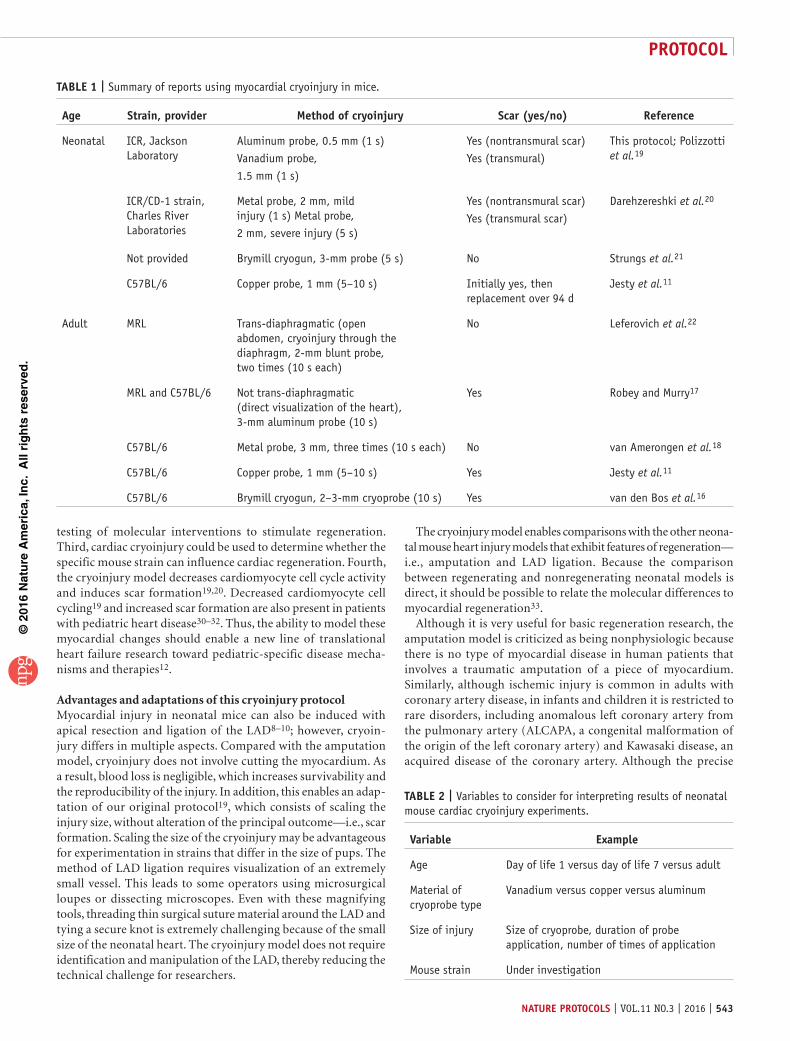
table 1 | Summary of reports using myocardial cryoinjury in mice.
age strain, provider Method of cryoinjury scar (yes/no) reference
Neonatal ICR, Jackson Laboratory
Aluminum probe, 0.5 mm (1 s) Vanadium probe, 1.5 mm (1 s)
Yes (nontransmural scar) Yes (transmural)
This protocol; Polizzotti et al.19
ICR/CD-1 strain, Charles River Laboratories
Metal probe, 2 mm, mild injury (1 s) Metal probe, 2 mm, severe injury (5 s)
Yes (nontransmural scar) Yes (transmural scar)
Darehzereshki et al.20
Not provided Brymill cryogun, 3-mm probe (5 s) No Strungs et al.21
C57BL/6 Copper probe, 1 mm (5–10 s) Initially yes, then replacement over 94 d
Jesty et al.11
Adult MRL Trans-diaphragmatic (open abdomen, cryoinjury through the diaphragm, 2-mm blunt probe, two times (10 s each)
No Leferovich et al.22
MRL and C57BL/6 Not trans-diaphragmatic (direct visualization of the heart), 3-mm aluminum probe (10 s)
Yes Robey and Murry17
C57BL/6 Metal probe, 3 mm, three times (10 s each) No van Amerongen et al.18
C57BL/6 Copper probe, 1 mm (5–10 s) Yes Jesty et al.11
C57BL/6 Brymill cryogun, 2–3-mm cryoprobe (10 s) Yes van den Bos et al.16
table 2 | Variables to consider for interpreting results of neonatal mouse cardiac cryoinjury experiments.
Variable example
Age Day of life 1 versus day of life 7 versus adult
Material of cryoprobe type
Vanadium versus copper versus aluminum
Size of injury Size of cryoprobe, duration of probe application, number of times of application
Mouse strain Under investigation
©20
16N
atu
re A
mer
ica,
Inc.
All
rig
hts
res
erve
d.
protocol
544 | VOL.11 NO.3 | 2016 | nature protocols
mechanism of inducing tissue damage with cryoinjury does not exist in human heart disease, the induced cellular changes, such as myocardial cell death, inflammation, scar formation and decrease in cycling, are clinically relevant. For example, scar for-mation30–32 and decreased cardiomyocyte cycling19 are reported in patients with heart disease such as tetralogy of Fallot with pulmonary stenosis.
Experimental designBecause of the multiple technical challenges associated with sur-gical manipulations of neonatal mouse hearts, we recommend using a structured and didactic approach to learn this surgery. It is advisable to have a team of two experienced researchers working together during the steps of inducing cryoinjury. One researcher can perform the surgery while the other researcher can anesthetize the pups and revive them after surgery. This neonatal cryoinjury protocol can be learned by following a recommended sequence of steps during a dedicated period of training. The suggested sequence of steps and metric for evaluating progress have been provided (Table 3). Potential variables that may determine the tissue response after cardiac cryoinjury experiments are provided (Table 2). Researcher 1 developed and optimized this protocol. To validate the proposed didactic steps (Table 3) of this protocol, we assessed the learning curve of researchers 2 and 3, who had no prior experience with mouse surgery (Supplementary Fig. 1).
Below we discuss parts of the protocol, which have been explained in further detail so the individual steps can be adapted according to experimental conditions.Mouse strain and age. In our laboratory, we have predominantly used the ICR mouse strain for cryoinjury experiments because of the fact that the dams have better nursing behavior (lead-ing to decreased cannibalism), pups are bigger in size (easier to perform surgery) and the litter size is consistently larger. These advantages may lead to better survival rates. We have also used this cryoinjury protocol in mice up to 7 d old (postnatal day 7 (P7)). Although we believe that the surgery itself may be easier in slightly larger pups, the duration of hypothermia-anesthesia may have to be optimized. Because of the inherent variability between animals, we recommend an experimental design that allows for at
least six mice per data point. A corresponding number of sham- operated pups (chest opened and closed) should be included in every experiment.
Assembly of cryoprobes (Equipment setup). The size and type of metal used to fabricate the tip cryoprobe is an important parameter. Several companies manufacture cryoprobes fabricated from various metals and with continuous liquid nitrogen feeds. Alternatively, custom-made probes can be made using metal fila-ments that can be purchased from a local hardware store. Probes of varying diameters are easily fabricated by grinding stock fila-ments to the desired diameter. It is important to note that metals with low thermal conductivities will remain colder for a longer period of time, and they are capable of generating a more severe injury. We used a vanadium probe because of its low thermal conductivity (κ = 30.7 W m−1 K−1) compared with aluminum (237 W m−1 K−1) and copper (κ = 401 W m−1 K−1) filaments.
Anesthesia by hypothermia (Steps 5 and 6). We commonly use the thumb of a disposable nitrile glove as a protective barrier to prevent direct contact between the pup skin and the ice-water slurry. P1 pups are remarkably resilient to hypothermia, and they can remain in a hypothermic state for at least 15 min before mor-tality rates begin to rise. Pups older than P4 typically experience much higher mortality rates. The key parameter to maximizing survivability is to minimize the duration of hypothermia. Once the pup is anesthetized, the surgeon should be able to perform the complete thoracotomy (including skin closure) in ~5–8 min. Complications commonly occur during exteriorizing the heart, application of the cryoprobe and suturing the chest wall and skin. These complications prolong the duration of hypothermia and can lead to increased mortality.
Exteriorizing the heart (Step 9). Exteriorizing the heart is a cru-cial step that allows visualization of the left ventricular surface and reproducible application of the cryoprobe. Common com-plications associated with exteriorizing the heart are dissection of the wrong intercostal space, lung injury and heart laceration. To effectively exteriorize the heart, one must first determine the
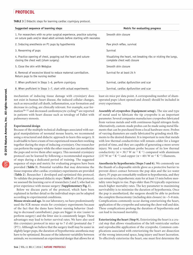
table 3 | Didactic steps for learning cardiac cryoinjury protocol.
suggested sequence of learning steps Metric for evaluating progress
1. For researchers with no prior surgical experience, practice suturing on suture pads and/or dead adult animals before starting with neonates
Smooth skin closure
2. Inducing anesthesia on P1 pups by hypothermia Paw pinch reflex, survival
3. Rewarming of pups Survival
4. Practice opening of chest, popping out the heart and suture- closing the chest wall (sham surgery)
Visualizing the heart, not breaking ribs or nicking the lungs, complete chest wall closure
5. Close the skin with Webglue Smooth skin closure
6. Removal of excessive blood to reduce maternal cannibalism. Return pups to the nursing mother
Survival for at least 24 h
7. When proficient in Steps 1–6, perform cryoinjury Survival, cardiac dysfunction and scar
8. When proficient in Steps 1–7, start with actual experiments Survival, cardiac dysfunction and scar
©20
16N
atu
re A
mer
ica,
Inc.
All
rig
hts
res
erve
d.
protocol
nature protocols | VOL.11 NO.3 | 2016 | 545
MaterIalsREAGENTS
Mouse strain—ICR (Taconic) ! cautIon All animal experiments must be performed in accordance with all relevant institutional and governmen-tal ethics guidelines and regulations. This protocol was approved by the Institutional Animal Care and Use Committee of Boston Children’s Hospital and the University of Pittsburgh.Sodium heparin (5,000 USP units per ml)Sodium phosphate dibasic (Na2HPO4; Sigma-Aldrich, cat. no. 255793)Sodium phosphate dibasic (NaH2PO4; Sigma-Aldrich, cat. no. S3139)Formaldehyde (Sigma-Aldrich, cat. no. F8775)Liquid nitrogen ! cautIon Liquid nitrogen is extremely cold. Do not touch it. Wear appropriate personal protection equipment.PBS, 1×Cardioplegia solution: 2.5 M KCl (74.6 g +1,000 ml water) = 50× stock phosphate-buffered saline (PBS2−) / 50 mM KCl (4 °C): 500 ml PBS2− + 10 ml 2.5 M KCl. Precool on ice or 4 °C.Bupivacaine (0.25%, wt/vol)
EQUIPMENTMicrodissecting spring scissors (angular; Roboz, cat. no. RS-5658)Microdissecting scissors (curved; Roboz, cat. no. RS-5913)Angled forceps (Fine Science Tools, cat. no. 11251-35)Microneedle holder (Roboz, cat. no. RS 6437)Graefe forceps (straight; Roboz, cat. no. RS-5112)Dewar vessel for liquid nitrogenHalsey microneedle holder (Fine Science Tools, cat. no. FST-12500-12)Sterile glovesNylon sutures, 8-0 (Ethicon, cat. no. 1716G)Insulin syringe, 3/10 cc (Becton Dickinson and Company, cat. no. 309301)Dissecting microscope or surgical loops, 10× (Design for Vision)Gaymar T pumps with heated water blanket, model TP-500
•
•••••
••
•
••••••••••••
Q-tipsPolydrapeGlass Petri dish filled with ice as operating tableSterile gauzeWebglue (Webster Veterinary, cat. no. 07-8566128)Styrofoam platformSurgical tapeHeating lampBucket filled with an ice-water bathBetadine swabsBead sterilizerLight sourceVanadium probe (made from screwdriver; WIHA, Sp 353 SW, 1.5 mm screwdriver)Aluminum probe (hardware store, 1.0 mm filament ground down to 0.5 mm using a Dremel tool equipped with a grinder bit)
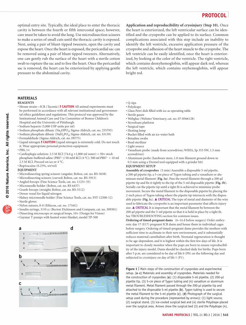
EQUIPMENT SETUPAssembly of cryoprobes (5 min) Assemble a disposable 5-ml pipette, a 200-µl pipette tip, a 5-cm piece of Tygon tubing and a vanadium or alu-minum metal filament (Fig. 1a). Pass the metal filament through a 200-µl pipette tip and fix it tightly to the tip of the 5-ml disposable pipette (Fig. 1b). Serially cut the pipette tip until a tight fit is achieved to minimize probe movement. Secure the metal filament to the disposable pipette by placing the 2-inch piece of Tygon tubing where the pipette tip intersects with the dispos-able pipette (Fig. 1c). crItIcal The type of metal and diameter of the wire used to fabricate the cryoprobe is an important parameter that affects injury size. crItIcal It is important that the metal filament be between the 200-µl pipette and the 5-ml pipette so that it is held in place by a tight fit. See TROUBLESHOOTING section for common issues.Ordering of timed-pregnant dams (6–14 d before surgery) Order embry-onic day 17 (E17) pregnant ICR dams and house them in individual cages before surgery. Ordering of timed-pregnant dams provides the mothers with sufficient time to acclimate to their new environment, and it substantially reduces maternal cannibalism after birth. Neonatal regeneration is thought to be age-dependent, and it is highest within the first few days of life. It is important to closely monitor when the pups are born to ensure reproducibil-ity of the injury model. Dams should be checked daily for births. Pups born after 5 p.m. are considered to be day of life 0 (P0) on the following day and subjected to cryoinjury on day of life 1 (P1).
•••••••••••••
•
optimal entry site. Typically, the ideal place to enter the thoracic cavity is between the fourth or fifth intercostal space; however, care must be taken to avoid the lung. Use microdissection scissors to make a series of small cuts until the thoracic cavity is exposed. Next, using a pair of blunt-tipped tweezers, open the cavity and expose the heart. Once the heart is exposed, the pericardial sac can be removed using a pair of blunt tipped tweezers. Alternatively, one can gently rub the surface of the heart with a sterile cotton swab to rupture the sac and to free the heart. Once the pericardial sac is removed, the heart can be exteriorized by applying gentle pressure to the abdominal cavity.
Application and reproducibility of cryoinjury (Step 10). Once the heart is exteriorized, the left ventricular surface can be iden-tified and the cryoprobe can be applied to its surface. Common complications associated with this step include an inability to identify the left ventricle, excessive application pressure of the cryoprobe and adhesion of the heart muscle to the cryoprobe. The left ventricle can be easily identified, once the heart is exterior-ized, by looking at the color of the ventricle. The right ventricle, which contains deoxyhemoglobin, will appear dark red, whereas the left ventricle, which contains oxyhemoglobin, will appear bright red.
a
2 1
13
1
2
3
4
3
4
4 2
b
c
d
1
2
3
4Figure 1 | Main steps of the construction of cryoprobes and experimental setup. (a–c) Materials and assembly of cryoprobes. Materials needed for the construction of cryoprobes (a): (1) disposable 5-ml pipette, (2) 200-µl pipette tip, (3) 5-cm piece of Tygon tubing and (4) vanadium or aluminum metal filament. Metal filament passed through the 200-µl pipette tip and attached to the disposable 5-ml pipette (b). Tygon tubing is used to secure the metal filament to the 5-ml pipette (c). (d) Photograph of the surgical setup used during the procedure (represented by arrows): (1) light source, (2) surgical stand, (3) ice-cooled surgical bed and (4) sterile Polydrape placed over the surgical area. Arrows show the surgical bed (3) and the Polydrape (4).

©20
16N
atu
re A
mer
ica,
Inc.
All
rig
hts
res
erve
d.
protocol
546 | VOL.11 NO.3 | 2016 | nature protocols
proceDuresurgery ● tIMInG 15 min per mouse pup1| Prepare bupivacaine working solution (0.1% (wt/vol) in sterile saline).! cautIon Follow your institutional guidelines regarding administration of analgesics preoperatively and postoperatively. Although it is known that neonatal mice lack centralized pain reflexes, your institution may require administration of analgesics.
2| Precool the cryoprobe by immersing it in liquid nitrogen for at least 10 min before the first surgery. crItIcal step Remove the probe only to induce injury. It is important to maintain adequate liquid nitrogen levels in the Dewar.
3| Remove half of the pups from the nursing mother, and place them on a heated water blanket set to 37 °C. Cover the pups with bedding from the mother’s nest. crItIcal step Removing all the pups from the mother increases her stress level, and it may lead to increased cannibalism. Leave half of the litter with the mother at all times.
4| If analgesics are required, administer them now. Some institutions may require administration of 20 µl of 0.1% bupivacaine (2 mg kg−1 body weight) subcutaneously to the thoracic region.
5| Place the neonatal pup into a protective sleeve and anesthetize it by placing the pup into an ice-water bath for ~3–5 min.! cautIon Hypothermia-anesthesia must be performed in accordance with all relevant institutional and governmental ethics guidelines and regulations. This protocol was approved by the Institutional Animal Care and Use Committee of Boston Children’s Hospital and University of Pittsburgh. crItIcal step The sleeve provides a physical barrier between the ice-water and the pup skin, which is required to prevent frostbite. crItIcal step Check the pup frequently while on ice until there is no paw reflex. Prolonged exposure to hypothermia can lead to increased mortality. Hypothermia-anesthesia is challenging in pups older than P4. It is important to note that the anesthesia has to remain consistent in depth and duration to prevent the pups from waking up during the course of surgery. The exact duration of hypothermia needed to induce anesthesia may vary between strains, and hence the researchers should titrate the depth of anesthesia (table 3). However, once optimal durations of hypothermia have been established, this duration should not be changed.? troublesHootInG
6| Remove the pup from the ice-water bath and dry it using a sterile gauze pad. Place the pup in the surgical area (Fig. 1d) in the supine position and tape the arms, legs and tail to immobilize the pup. crItIcal step To maintain a hypothermic state during surgery, place the pup on top of a cold surface such as an ice pack or a chilled Petri dish.? troublesHootInG
7| Sterilize the surgical area by gently wiping it with a Betadine swab (Fig. 2a).
8| Make a transverse skin incision across the chest using a pair of micro-scissors, and then separate the skin from the underlying muscle using blunt dissection (Fig. 2b–d). crItIcal step Carefully dissect the skin from the underlying muscle using a blunt-tipped tool such as a Halsey microneedle holder. Skin tears complicate wound closure and may increase maternal cannibalism. crItIcal step Care should be taken to minimize the length of the incision site. Larger incisions greatly enhance the visibility of the rib cage, which will help with selecting the point of entry. However, larger incisions may require additional time to suture, may lead to increased sternal scar formation that can complicate ultrasound sound-based functional studies, and may increase maternal cannibalism.
9| Perform lateral thoracotomy (microdissecting spring scissors) by making a small incision at the fourth or fifth intercostal space (Fig. 2e,f). Separate the intercostal muscles using Graefe forceps (Fig. 2g) and carefully remove the pericardial sac. Exteriorize the heart by gently pressing on the abdomen (Fig. 2h). crItIcal step Selecting the correct entry site is crucial to the success of the procedure. Select the intercostal space that provides the best access to the heart while minimizing lung injury. Visualization of the entry site with surgical loops or a dissecting microscope may greatly enhance accuracy and reproducibility of the surgery. crItIcal step Removal of the pericardial sac is crucial for exteriorization, but care must be taken not to lacerate the heart.? troublesHootInG
©20
16N
atu
re A
mer
ica,
Inc.
All
rig
hts
res
erve
d.
protocol
nature protocols | VOL.11 NO.3 | 2016 | 547
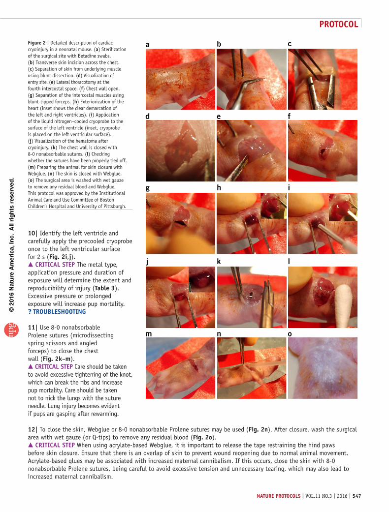
10| Identify the left ventricle and carefully apply the precooled cryoprobe once to the left ventricular surface for 2 s (Fig. 2i,j). crItIcal step The metal type, application pressure and duration of exposure will determine the extent and reproducibility of injury (table 3). Excessive pressure or prolonged exposure will increase pup mortality.? troublesHootInG
11| Use 8-0 nonabsorbable Prolene sutures (microdissecting spring scissors and angled forceps) to close the chest wall (Fig. 2k–m). crItIcal step Care should be taken to avoid excessive tightening of the knot, which can break the ribs and increase pup mortality. Care should be taken not to nick the lungs with the suture needle. Lung injury becomes evident if pups are gasping after rewarming.
12| To close the skin, Webglue or 8-0 nonabsorbable Prolene sutures may be used (Fig. 2n). After closure, wash the surgical area with wet gauze (or Q-tips) to remove any residual blood (Fig. 2o). crItIcal step When using acrylate-based Webglue, it is important to release the tape restraining the hind paws before skin closure. Ensure that there is an overlap of skin to prevent wound reopening due to normal animal movement. Acrylate-based glues may be associated with increased maternal cannibalism. If this occurs, close the skin with 8-0 nonabsorbable Prolene sutures, being careful to avoid excessive tension and unnecessary tearing, which may also lead to increased maternal cannibalism.
a b c
d e f
g h i
j k l
m n o
Figure 2 | Detailed description of cardiac cryoinjury in a neonatal mouse. (a) Sterilization of the surgical site with Betadine swabs. (b) Transverse skin incision across the chest. (c) Separation of skin from underlying muscle using blunt dissection. (d) Visualization of entry site. (e) Lateral thoracotomy at the fourth intercostal space. (f) Chest wall open. (g) Separation of the intercostal muscles using blunt-tipped forceps. (h) Exteriorization of the heart (inset shows the clear demarcation of the left and right ventricles). (i) Application of the liquid nitrogen–cooled cryoprobe to the surface of the left ventricle (inset, cryoprobe is placed on the left ventricular surface). (j) Visualization of the hematoma after cryoinjury. (k) The chest wall is closed with 8-0 nonabsorbable sutures. (l) Checking whether the sutures have been properly tied off. (m) Preparing the animal for skin closure with Webglue. (n) The skin is closed with Webglue. (o) The surgical area is washed with wet gauze to remove any residual blood and Webglue. This protocol was approved by the Institutional Animal Care and Use Committee of Boston Children’s Hospital and University of Pittsburgh.
©20
16N
atu
re A
mer
ica,
Inc.
All
rig
hts
res
erve
d.
protocol
548 | VOL.11 NO.3 | 2016 | nature protocols
13| Rapidly warm the pup by placing it in your hands and warming it under a heat lamp for several minutes. crItIcal step Exposure to excessive heat can increase neonatal mortality. crItIcal step Sterilize the surgical instruments in a dry bead sterilizer for the next surgery.
14| Return the pup to the heating blanket with the other pups and cover it with bedding from the mother’s cage. crItIcal step Use a cotton swab soaked with sterile saline to remove all traces of blood from the wound area immedi-ately after surgery. Any remnants of blood around the injury may lead to increased maternal cannibalism.
15| Repeat Steps 4–16 for all the pups on the heating blanket.
16| Once all the pups have fully recovered from the surgery, swap them with the uninjured pups in the mother’s cage. crItIcal step Placing pups after surgery with a mother that is nursing pups without prior surgery will result in maternal cannibalism. The remaining healthy pups must be removed first before the injured pups can be returned to their mother.
17| Repeat Steps 8–18 for the remaining pups in the litter. Once the pups are fully recovered from surgery, return them to their injured littermates in their mother’s den. Leave pups with their mother until the chosen time point for heart collection and fixation, at which point proceed to the next step. crItIcal step A typical litter of ICR mice yields 12–16 pups, which we break up into two groups with 6–8 pups each. Higher 24-h survivability is achieved when >10 pups are placed with a single mother after surgery. Adjusting the litter size is suggested. crItIcal step Follow your institutional guidelines regarding the use and administration of analgesics after recovery from surgery.
Heart collection and fixation ● tIMInG 2 d, variable timing after injury18| Prepare a working solution of cardioplegia solution (PBS2− with 50 mM KCl) in PBS and place it on ice.
19| For neonatal hearts (P1–P7), place 1 ml of 3.7% (vol/vol) formaldehyde into a microcentrifuge tube. For older animals, place 5 ml of 3.7% formaldehyde into a 15-ml conical tube.
20| To perform the resection, gather dissection scope or surgical loops, Graefe forceps, one curved forceps, one curved microdissecting scissors and a microdissecting spring scissors.
21| Administer 50 µl of heparin (5,000 USP units per ml) to the mouse via i.p. injection 10 min before resecting the heart.
22| Euthanize the mouse using a technique approved by your institution. Carbon dioxide should be avoided because of its negative effects on the heart. crItIcal step Mouse pups (P0–P7) may be euthanized by decapitation, whereas mice older than P7 can be euthanized by isoflurane and cervical dislocation. It is the researcher’s responsibility to follow institutional guidelines regarding accepted forms of euthanasia.
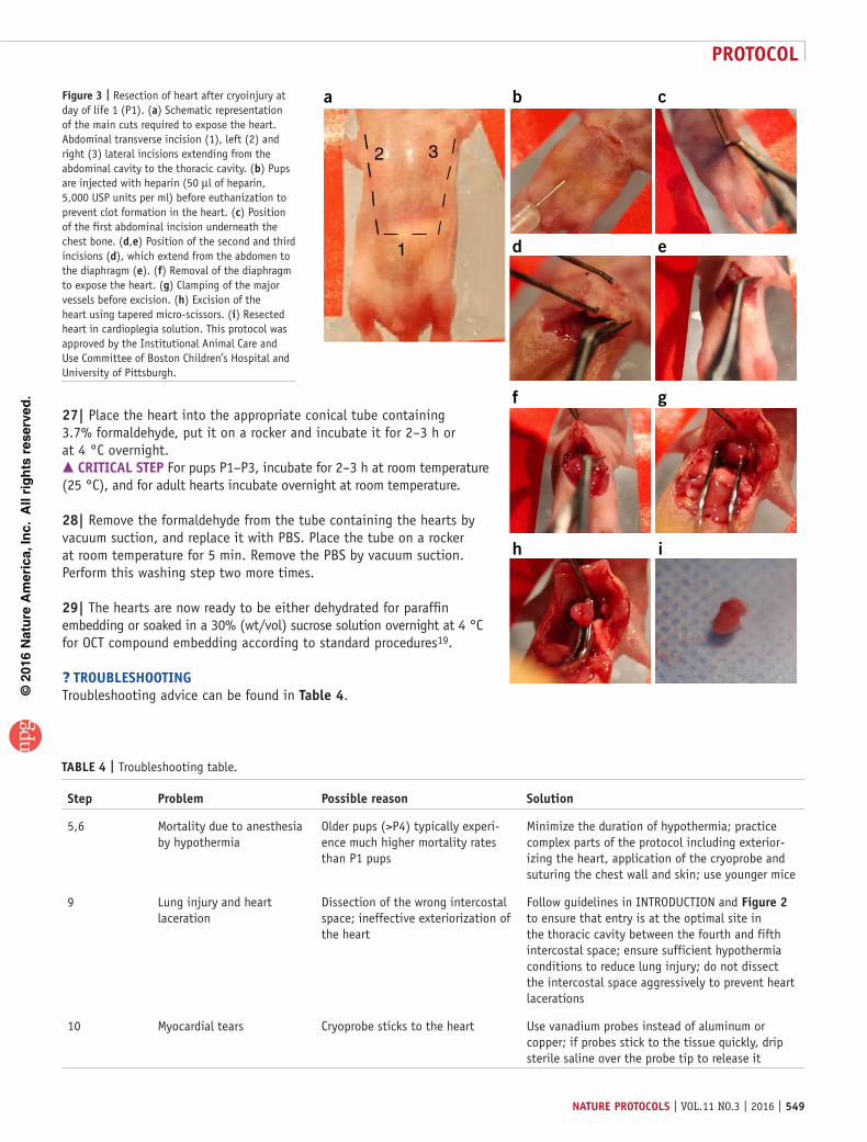
23| Place the mouse in the supine position and tape the arms, legs and tail to immobilize the mouse. Using the microdis-section spring scissors, make a transverse incision across the entire abdomen and extend vertically until the diaphragm is exposed (Fig. 3a–d).
24| Place the Graefe forceps in your nondominant hand, and lift the chest wall by the sternum to expose the diaphragm (Fig. 3e). Place curved forceps in your dominant hand, and carefully dissect the diaphragm from the anterior chest wall to expose the thoracic cavity (Fig. 3f).
25| Place the curved forceps in your nondominant hand and position it under the base of the heart; pinch down on the main vessels and carefully lift the heart (Fig. 3g). By using your dominant hand, place the curved microdissecting scissors under the tweezers and cut the vessels to free the heart (Fig. 3h). Place the heart in the ice-cold cardioplegia solution (Fig. 3i). crItIcal step Make sure that the heart does not adhere to the rib cage before removing it from the thoracic cavity. If adhesions are present, carefully dissect the heart free under a dissection microscope using fine-tipped tweezers and microdissection scissors. When in doubt, leave adhesions attached to the heart. They can be resected after fixation.
26| Place the curved forceps in your dominant hand, and gently squeeze excess blood from the heart.
©20
16N
atu
re A
mer
ica,
Inc.
All
rig
hts
res
erve
d.
protocol
nature protocols | VOL.11 NO.3 | 2016 | 549
a b c
d e
f g
h i
32
1
Figure 3 | Resection of heart after cryoinjury at day of life 1 (P1). (a) Schematic representation of the main cuts required to expose the heart. Abdominal transverse incision (1), left (2) and right (3) lateral incisions extending from the abdominal cavity to the thoracic cavity. (b) Pups are injected with heparin (50 µl of heparin, 5,000 USP units per ml) before euthanization to prevent clot formation in the heart. (c) Position of the first abdominal incision underneath the chest bone. (d,e) Position of the second and third incisions (d), which extend from the abdomen to the diaphragm (e). (f) Removal of the diaphragm to expose the heart. (g) Clamping of the major vessels before excision. (h) Excision of the heart using tapered micro-scissors. (i) Resected heart in cardioplegia solution. This protocol was approved by the Institutional Animal Care and Use Committee of Boston Children’s Hospital and University of Pittsburgh.
27| Place the heart into the appropriate conical tube containing 3.7% formaldehyde, put it on a rocker and incubate it for 2–3 h or at 4 °C overnight. crItIcal step For pups P1–P3, incubate for 2–3 h at room temperature (25 °C), and for adult hearts incubate overnight at room temperature.
28| Remove the formaldehyde from the tube containing the hearts by vacuum suction, and replace it with PBS. Place the tube on a rocker at room temperature for 5 min. Remove the PBS by vacuum suction. Perform this washing step two more times.
29| The hearts are now ready to be either dehydrated for paraffin embedding or soaked in a 30% (wt/vol) sucrose solution overnight at 4 °C for OCT compound embedding according to standard procedures19.
? troublesHootInGTroubleshooting advice can be found in table 4.
table 4 | Troubleshooting table.
step problem possible reason solution
5,6 Mortality due to anesthesia by hypothermia
Older pups (>P4) typically experi-ence much higher mortality rates than P1 pups
Minimize the duration of hypothermia; practice complex parts of the protocol including exterior-izing the heart, application of the cryoprobe and suturing the chest wall and skin; use younger mice
9 Lung injury and heart laceration
Dissection of the wrong intercostal space; ineffective exteriorization of the heart
Follow guidelines in INTRODUCTION and Figure 2 to ensure that entry is at the optimal site in the thoracic cavity between the fourth and fifth intercostal space; ensure sufficient hypothermia conditions to reduce lung injury; do not dissect the intercostal space aggressively to prevent heart lacerations
10 Myocardial tears Cryoprobe sticks to the heart Use vanadium probes instead of aluminum or copper; if probes stick to the tissue quickly, drip sterile saline over the probe tip to release it

©20
16N
atu
re A
mer
ica,
Inc.
All
rig
hts
res
erve
d.
protocol
550 | VOL.11 NO.3 | 2016 | nature protocols
● tIMInGEquipment setup, assembly of cryoprobes: 5 min per probeEquipment setup, ordering of timed-pregnant dams: 6–7 d before surgerySteps 1–17, surgery: 15 min per mouse pupSteps 18–29, heart collection and fixation: 2 d, 1–30 d after injury
antIcIpateD resultsWe have previously published results obtained with the large (1.5 mm) cryoprobe (Fig. 4a, left and right)19, which show that cryoinjury induces myocardial cell death in neonatal mice. Cryoinjury leads to the formation of a hematoma at the application site with both a 0.5-mm probe and a 1.5-mm probe (Fig. 4a, middle and right). Cryoinjury with the 0.5-mm probe leads to a much smaller hematoma size (Fig. 4a, middle), whereas the 1.5-mm probe damages ~15–20% of the left ventricle (Fig. 4a, right)19. Quantifica-tion of hematoma size after application of the 1.5-mm cryoprobe showed damage to 17.5 ± 8.6% (n = 5, mean ± s.d.) of the surface of the heart19. We use whole-organ staining with triphenyltetrazolium chloride (TTC) to demarcate the injury zone on the heart surface (Fig. 4b). Sections after TTC staining show that the degree of penetration of injury is higher with the large cryoprobe compared with the 0.5-mm probe (Fig. 4c, middle and right). Using TUNEL (Fig. 5), we estimated the myocardial volume damaged by the 1.5-mm probe to be ~0.58 ± 0.02 mm3 (n =5), which corresponds to ~18% of the myocardium19.
Echocardiography revealed that myocardial dysfunction was evident within 2 d.p.i. (ejection fraction after 1.5-mm probe 55.1% (n = 13) versus 84.4% after sham injury, n = 19) (ref. 19). At 30 d.p.i., the ejection fraction was 45.7 ± 3% for cryoinjured animals (large probe) versus 57.9 ± 3.5% for shams (n = 13, P < 0.05, analysis of variance (ANOVA))19. Staining with acid fuchsin orange G (AFOG) revealed fibrin deposition at 1 d.p.i. and substantial fibrosis at 7 d.p.i., which matured into a transmural scar after application of the 1.5-mm probe (i.e., the entire myocardial wall thickness is composed of scar) at 30 d.p.i. (~3% of the total myocardium)19,20. Cryoinjury with the 0.5-mm probe resulted in a nontransmural scar at 30 d.p.i. (supplementary Fig. 2), ~2% of the myocardium. Note that transmural scars were present only after application of the 1.5-mm probe but not after application of the 0.5-mm probe (Fig. 6). We have previously published results obtained with the large (1.5 mm) cryoprobe (Fig. 6, lower right two images for the 7 and 30 d.p.i. time points)19.
Sham Cryo 0.5 mm Cryo 1.5 mma
b
c
LV
RV
LARA
LV
LARA
LV
RV
LA
RA
Figure 4 | Cryoinjury in neonatal mice induces tissue damage. Mice underwent sham surgery or cryoinjury on day of life 1 (P1) with either a 0.5-mm probe or a 1.5-mm probe, and their hearts were resected the next day (P2). (a) Hematoma is seen at the site of injury. (b) Vital staining with 1% (wt/vol) TTC (in PBS2−, pH 7.4) at 37 °C for 20 min and then fixation in 10% (vol/vol) formaldehyde overnight shows the injury zone (indicated with white arrow) in whole hearts. (c) Vital staining with TTC shows that the degree of penetration of injury varies with probe size in sliced heart sections (indicated by white dotted lines). Scale bars, 1 mm. Note the scalability of the injury size with different probes. LA, left atrium; LV, left ventricle; RA, right atrium; RV, right ventricle. Portions of (left and right images) were previously published in Polizzotti, B.D. et al., Sci. Transl. Med. 7, 281ra245 (2015) (ref. 19). Reprinted with permission from AAAS.
Sham Cryo 0.5 mm Cryo 1.5 mm
TU
NE
L, H
oech
st
Figure 5 | Cryoinjury in neonatal mice induces apoptosis in myocardial cells. Cryoinjury was performed on day of life 1 (P1) with either a 0.5-mm probe or a 1.5-mm probe, and hearts were resected the next day (P2). The scalability of injury is shown with myocardial cell death, as visualized by TUNEL staining (green; ApopTag Red in situ apoptosis detection kit, EMD Millipore) and DNA staining with Hoechst (blue). Scale bars, 1 mm.

©20
16N
atu
re A
mer
ica,
Inc.
All
rig
hts
res
erve
d.
protocol
nature protocols | VOL.11 NO.3 | 2016 | 551
Cardiomyocyte proliferation was previously reported to contribute to neonatal heart regeneration following apical resection and myocardial infarction8,9,34,35. Staining with an anti–phospho-histone H3 antibody revealed that cryoinjury inhibited the endogenous proliferation of car-diomyocytes within the injury and border zones at 1, 4 and 7 d.p.i.19, suggesting that neonatal mouse hearts do not regenerate after cryoinjury to the same degree, as reported after amputation and LAD ligation injury. The decreased local cardiomyocyte cell cycle activity is consistent with the unchanged global cardiomyocyte cell cycle activity after cryoinjury reported by Lien and colleagues20. In conclusion, this protocol describes a useful model for cardiac translational and regeneration researchers.
Sha
m
30 dpi7 dpi1 dpi
Cry
o 0.
5 m
mC
ryo
1.5
mm
Figure 6 | Cryoinjury induces scar formation in neonatal mice. Mice underwent sham surgery or cryoinjury on day of life 1 with either a 0.5-mm probe or a 1.5-mm probe. Time series of AFOG-stained sections shows fibrin deposition at 1 d post injury (d.p.i., orange staining, black arrows). Scar (blue) is formed within 7 d.p.i. (black arrowheads) and still present 30 d later. Note that transmural scars were present only after 1.5-mm cryoinjury (lower right) but not after the 0.5-mm probe. Scale bars, 1 mm. Photomicrographs in the lower right two images (1.5-mm probe) were previously published in Polizzotti, B.D. et al., Sci. Transl. Med. 7, 281ra245 (2015) (ref. 19). Reprinted with permission from AAAS. Staining is carried out as described by Polizzotti et al.19.
Note: Any Supplementary Information and Source Data files are available in the online version of the paper.
acknowleDGMents We thank H. Sadek and M. Ahmad (University of Texas Southwestern Medical Center) for training in mouse surgery, and members of the Kühn laboratory for helpful suggestions and discussions. We thank M.A. Missinato, University of Pittsburgh, for sharing her cryoinjury learning curve experience. We apologize to researchers whose relevant work could not be discussed or referenced because of the limitations of the scope of this paper. This research was supported by the Department of Cardiology and the Translational Research Program at Boston Children’s Hospital and US National Institutes of Health (NIH) grants R01HL106302 and K08HL085143 (to B.K.). B.D.P. was supported by the Office of Faculty Development (Boston Children’s Hospital) and by grant no. T32HL007572 from the NIH. B.G. and B.K. are supported by the Richard King Mellon Institute for Pediatric Research (Children’s Hospital of Pittsburgh of UPMC). B.G., B.J.H., J.M.P. and B.K. were supported by Transatlantic Network of Excellence grants by the Fondation Leducq (no. 15CVD03 to B.G. and B.K.).
autHor contrIbutIons B.D.P., B.G., B.J.H. and B.K. designed the research. B.D.P., B.G. and B.J.H. performed and analyzed the experiments. B.J.H. and J.M.P. provided data for Figures 4a,c and 5. B.D.P., B.G. and B.K. wrote the manuscript, and all authors edited the manuscript.
coMpetInG FInancIal Interests The authors declare no competing financial interests.
Reprints and permissions information is available online at http://www.nature.com/reprints/index.html.
1. Naqvi, N. et al. A proliferative burst during preadolescence establishes the final cardiomyocyte number. Cell 157, 795–807 (2014).
2. Wulfsohn, D., Nyengaard, J.R. & Tang, Y. Postnatal growth of cardiomyocytes in the left ventricle of the rat. Anat. Rec. A Discov. Mol. Cell. Evol. Biol. 277, 236–247 (2004).
3. Li, F., Wang, X., Capasso, J.M. & Gerdes, A.M. Rapid transition of cardiac myocytes from hyperplasia to hypertrophy during postnatal development. J. Mol. Cell. Cardiol. 28, 1737–1746 (1996).
4. Mollova, M. et al. Cardiomyocyte proliferation contributes to heart growth in young humans. Proc. Natl. Acad. Sci. USA 110, 1446–1451 (2013).
5. Bergmann, O. et al. Evidence for cardiomyocyte renewal in humans. Science 324, 98–102 (2009).
6. Beinlich, C.J., Rissinger, C.J. & Morgan, H.E. Mechanisms of rapid growth in the neonatal pig heart. J. Mol. Cell. Cardiol. 27, 273–281 (1995).
7. Mahmoud, A.I., Porrello, E.R., Kimura, W., Olson, E.N. & Sadek, H.A. Surgical models for cardiac regeneration in neonatal mice. Nat. Protoc. 9, 305–311 (2014).
8. Porrello, E.R. et al. Transient regenerative potential of the neonatal mouse heart. Science 331, 1078–1080 (2011).
9. Porrello, E.R. et al. Regulation of neonatal and adult mammalian heart regeneration by the miR-15 family. Proc. Natl. Acad. Sci. USA 110, 187–192 (2013).
10. Haubner, B.J. et al. Complete cardiac regeneration in a mouse model of myocardial infarction. Aging 4, 966–977 (2012).
11. Jesty, S.A. et al. c-kit+ precursors support postinfarction myogenesis in the neonatal, but not adult, heart. Proc. Natl. Acad. Sci. USA 109, 13380–13385 (2012).
12. Burns, K.M. et al. New mechanistic and therapeutic targets for pediatric heart failure: report from a National Heart, Lung, and Blood Institute Working Group. Circulation 130, 79–86 (2014).
13. Bryant, D.M. et al. A systematic analysis of neonatal mouse heart regeneration after apical resection. J. Mol. Cell. Cardiol. 79, 315–318 (2015).
14. Andersen, D.C., Ganesalingam, S., Jensen, C.H. & Sheikh, S.P. Do neonatal mouse hearts regenerate following heart apex resection? Stem Cell Rep. 2, 406–413 (2014).
15. Konfino, T., Landa, N., Ben-Mordechai, T. & Leor, J. The type of injury dictates the mode of repair in neonatal and adult heart. J. Am. Heart Assoc. 4, e001320 (2015).
16. van den Bos, E.J., Mees, B.M., de Waard, M.C., de Crom, R. & Duncker, D.J. A novel model of cryoinjury-induced myocardial infarction in the mouse: a comparison with coronary artery ligation. Am. J. Physiol. Heart Circ. Physiol. 289, H1291–H1300 (2005).
17. Robey, T.E. & Murry, C.E. Absence of regeneration in the MRL/MpJ mouse heart following infarction or cryoinjury. Cardiovasc. Pathol. 17, 6–13 (2008).
18. van Amerongen, M.J., Harmsen, M.C., van Rooijen, N., Petersen, A.H. & van Luyn, M.J. Macrophage depletion impairs wound healing and increases left ventricular remodeling after myocardial injury in mice. Am. J. Pathol. 170, 818–829 (2007).
©20
16N
atu
re A
mer
ica,
Inc.
All
rig
hts
res
erve
d.
protocol
552 | VOL.11 NO.3 | 2016 | nature protocols
19. Polizzotti, B.D. et al. Neuregulin stimulation of cardiomyocyte regeneration in mice and human myocardium reveals a therapeutic window. Sci. Transl. Med. 7, 281ra245 (2015).
20. Darehzereshki, A. et al. Differential regenerative capacity of neonatal mouse hearts after cryoinjury. Dev. Biol. 399, 91–99 (2015).
21. Strungs, E.G. et al. Cryoinjury models of the adult and neonatal mouse heart for studies of scarring and regeneration. Methods Mol. Biol. 1037, 343–353 (2013).
22. Leferovich, J.M. et al. Heart regeneration in adult MRL mice. Proc. Natl. Acad. Sci. USA 98, 9830–9835 (2001).
23. Gonzalez-Rosa, J.M., Martin, V., Peralta, M., Torres, M. & Mercader, N. Extensive scar formation and regression during heart regeneration after cryoinjury in zebrafish. Development 138, 1663–1674 (2011).
24. Gonzalez-Rosa, J.M. & Mercader, N. Cryoinjury as a myocardial infarction model for the study of cardiac regeneration in the zebrafish. Nat. Protoc. 7, 782–788 (2012).
25. Schnabel, K., Wu, C.C., Kurth, T. & Weidinger, G. Regeneration of cryoinjury induced necrotic heart lesions in zebrafish is associated with epicardial activation and cardiomyocyte proliferation. PLoS ONE 6, e18503 (2011).
26. Chablais, F., Veit, J., Rainer, G. & Jazwinska, A. The zebrafish heart regenerates after cryoinjury-induced myocardial infarction. BMC Dev. Biol. 11, 21 (2011).
27. Poss, K.D., Wilson, L.G. & Keating, M.T. Heart regeneration in zebrafish. Science 298, 2188–2190 (2002).
28. Sadek, H.A. et al. Multi-investigator letter on reproducibility of neonatal heart regeneration following apical resection. Stem Cell Rep. 3, 1 (2014).
29. Sen, S. & Sadek, H.A. Neonatal heart regeneration: mounting support and need for technical standards. J. Am. Heart Assoc. 4, e001727 (2015).
30. Wald, R.M. et al. Effects of regional dysfunction and late gadolinium enhancement on global right ventricular function and exercise capacity in patients with repaired tetralogy of Fallot. Circulation 119, 1370–1377 (2009).
31. Babu-Narayan, S.V. et al. Late gadolinium enhancement cardiovascular magnetic resonance of the systemic right ventricle in adults with previous atrial redirection surgery for transposition of the great arteries. Circulation 111, 2091–2098 (2005).
32. Babu-Narayan, S.V. et al. Ventricular fibrosis suggested by cardiovascular magnetic resonance in adults with repaired tetralogy of Fallot and its relationship to adverse markers of clinical outcome. Circulation 113, 405–413 (2006).
33. Xin, M., Olson, E.N. & Bassel-Duby, R. Mending broken hearts: cardiac development as a basis for adult heart regeneration and repair. Nat. Rev. Mol. Cell Biol. 14, 529–541 (2013).
34. Mahmoud, A.I. et al. Meis1 regulates postnatal cardiomyocyte cell cycle arrest. Nature 497, 249–253 (2013).
35. Xin, M. et al. Hippo pathway effector Yap promotes cardiac regeneration. Proc. Natl. Acad. Sci. USA 110, 13839–13844 (2013).