cronicon · the traction generates tension on the skeletal and surrounding soft tissue ... included...
TRANSCRIPT

CroniconO P E N A C C E S S EC DENTAL SCIENCE
Case Report
Teledontics and Telegnathic Surgery for Treatment of Obstructive Sleep Apnea Syndrome Utilizing Surgically Assisted Mandibular Expansion (SAME)
Joseph Yousefian1* and Michael Brown2
1Orthodontist Specialist, Bellevue, Washington, USA2Physiatrist Specialist, Bellevue, Washington, USA
*Corresponding Author: Joseph Yousefian, Orthodontist Specialist, Bellevue, Washington, USA.
Citation: Joseph Yousefian and Michael Brown. “Teledontics and Telegnathic Surgery for Treatment of Obstructive Sleep Apnea Syndrome Utilizing Surgically Assisted Mandibular Expansion (SAME)”. EC Dental Science 14.1 (2017): 31-39.
Received: August 22, 2017; Published: September 12, 2017
Abstract
Medical and dental clinicians are faced with the effects of obstructive sleep apnea (OSA), obstructive sleep apnea syndrome (OSAS) and other forms of sleep disordered breathing (SDB) on the oral and systemic health of their patients ever day in clinical practice.
This article describes a comprehensive and interdisciplinary treatment, which was able to successfully address patient’s den-tofacial aesthetic concerns and resolve other health issues including OSAS, high blood pressure, developing diabetes and improve outcomes of other comorbidities.
Moreover, a new and simplified teledontic and telegnathic surgical techniques and principles utilizing surgically assisted man-dibular expansion (SAME) for care of patients with OSA will be introduced describing a clinical case presentation and discussion.
Keywords: Obstructive Sleep Apnea Syndrome; Sleep Disordered Breathing; Interdisciplinary; High Blood Pressure; Diabetes; Cancer; Teledontics; Telegnathic Surgery; Pharyngorofacial
Introduction
OSA and especially OSAS are the more severe forms of SDB. It can be a debilitating, potentially life-threatening, condition. Potential major comorbidities associated with OSAS include tooth grinding, temporomandibular disorders, facial deformities, ADHD [1], gastro-esophageal reflux disease, premature aging, depression, hypertension, sexual impotence, Alzheimer disease, metabolic syndrome, diabe-tes, obesity, increase cancer risk [2,3], heart disease and stroke [4,5].
Many of the etiologies that contribute to these disorders are unrecognized by clinicians dealing with these patients and one can miss an opportunity to recognize and screen for potential reversible causes of these sleep disturbances.
These recognizable etiologic factors involve oral, dental, temporomandibular joint, pharyngeal and facial bone structures which are referenced hereinafter as pharyngorofacial disorders (POFD). Having a basic understanding of the POFD that can be at the root cause of sleep disorders and SDB may allow physicians to quickly identify underlining structural and functional pathology that is having an unrec-ognized profound effect on the oral and systemic health.
The data published by American Academy of Sleep Medicine on September 2014, estimated an increase in prevalence of OSA to 26 percent amongst adults between the ages of 30 and 70 years and gave much credit to the rising obesity epidemic [6]. Although Franklin., et al. investigated 400 females from a population based random sample of 10,000 Swedish females and found OSA (apnea-hypopnea index

32
Teledontics and Telegnathic Surgery for Treatment of Obstructive Sleep Apnea Syndrome Utilizing Surgically Assisted Mandibular Expansion (SAME)
Citation: Joseph Yousefian and Michael Brown. “Teledontics and Telegnathic Surgery for Treatment of Obstructive Sleep Apnea Syndrome Utilizing Surgically Assisted Mandibular Expansion (SAME)”. EC Dental Science 14.1 (2017): 31-39.
AHI ≥ 5) in 50% of females aged 20 - 70 years. Severe sleep apnea (AHI ≥ 30) was scored in 14% of females aged 55 - 70 years with normal body mass index (BMI), and just in 31% of obese females with a BMI of > 30 kg·m−2 [7]. Asian men with severe OSAS have less tendency to be obese yet may present with significant posterior airway restriction leading to OSA [8].
Besides obesity there are other abnormalities in anatomy and neuromuscular function of upper airway, which clinicians need to recognize that are more important risk factors [9-11]. POFD including hypoplastic maxilla and mandible, are common findings in OSA and needs to be recognized by clinicians. POFD and its effects on metabolism and other systems continues to be underestimated in the development and severity of OSAS [11,12].
The field of dentistry and its involvement with the POF environment, provides the dental practitioner with an opportunity in screening for the presence of OSAS as a complex health condition or participation in its treatment [13].
In the recent orthodontic literature, there has been a significant emphasis on the impact of constricted dental arches along with the retrognathic position of the jaws in development and treatment of OSA [13,14].
Traditionally, orthodontist considered the mandibular basal bone width dimension unchangeable. Based on this, treatment-planning decisions in the transverse dimension have conventionally been based on the alteration only in mandibular arch width and form.
In early the nineteen nineties, Mandibular Symphyseal Distraction Osteogenesis (MSDO) was introduced by Guerrero, providing a new paradigm to overcome this dilemma.15 Distraction Osteogenesis (DO) was introduced in the beginning of the 20th century and popular-ized by Russian orthopedic surgeon Ilizarov in the 1960s [16,17]. DO was originally developed for dramatic limb lengthening by Ilizarov [18]. Several investigators have adapted these advances to the facial skeleton to change the anterior-posterior position of the jaws with good success [19,20].
DO is the biologic process of new bone formation between bone segments that are gradually separated by incremental traction [16]. The traction generates tension on the skeletal and surrounding soft tissue structures, which stimulates new bone formation parallel to the vector of distraction [17].
Adapting the Ilizarov DO protocol to the mandibular symphysis can regenerate bone, thereby adding dimension to the innate basal bone in mandible. The most important benefit that the expansion with SAME has over functional appliances is a change in the basal bone not just a change in the alveolar bone, thereby producing a change in the functional matrix and tone of buccal musculature. As a result, the expansion should have better long-term stability [21,22].
Mandibular symphyseal distraction osteogenesis (MSDO) was introduced by Guerrero, providing a new paradigm for patients whose treatment alternatives and results were previously limited [15,23-25]. MSDO has been attempted with different techniques. Overall, each of these methods has produced transverse dimensional changes but with questionable long-term stability especially with regard to health of TM joints and gum recession in adjacent teeth. Many of these questions are not answered until further long-term stability studies can be completed.
The case presented in this paper demonstrates the effective participation of the orthodontist as a member of an interdisciplinary den-tal/medical team collaborating in the treatment of SDB and OSAS by treatment of POFD. This article also demonstrates the effectiveness of teledontics and telegnathic surgery utilizing SAME procedure in the treatment of OSAS [12].
Case Report
A 58-year-old male patient presented for potential Invisalign treatment to improve his non-aesthetic smile. In the past he had received multidisciplinary oral care provided by a number of reputable dental specialists in the area. A review of the patient’s medical history in-dicated a healthy life style including healthy diet, routine exercise with no history of obesity, smoking or alcohol abuse.

33
Teledontics and Telegnathic Surgery for Treatment of Obstructive Sleep Apnea Syndrome Utilizing Surgically Assisted Mandibular Expansion (SAME)
Citation: Joseph Yousefian and Michael Brown. “Teledontics and Telegnathic Surgery for Treatment of Obstructive Sleep Apnea Syndrome Utilizing Surgically Assisted Mandibular Expansion (SAME)”. EC Dental Science 14.1 (2017): 31-39.
The patient reported a history of severe OSAS (an apnea/hypopnea index AHI of 53) diagnosed by sleep specialist and polysomnogra-phy at sleep center. His initial clinical symptoms included high blood pressure, excessive daytime sleepiness (EDS) affecting his job perfor-mance, and falling asleep during driving. For treatment of his OSAS he was using continuous positive airway pressure device (CPAP) with H2O pressure of 18 cm. Although his CPAP compliance effort was good but it was ineffective in reducing his symptoms due to air leakage around the facial mask. As result he was taking Provigil medication as wakefulness promoting agent.
The patient had not proceeded with the previously proposed surgical protocol for treatment of his OSAS. Phase one of this protocol included hyoid suspension, midline glossectomy, nasal valve stabilization and septoplasty followed by phase two consisting of maxillo-mandibular advancement surgery.
A clinical examination showed that the patient had a Class III skeletal and dental relationship with a moderate retrognathic position of the maxilla and retrusive position of the dentition in mandible (Figure 1). His oral hygiene was excellent. There was no presence of decay or gingival inflammation, but he had generalized type one periodontitis, with horizontal bone loss and gingival recession.
Figure 1: Pretreatment extraoral and intraoral photographs.
He was missing teeth 1, 2, 16, 23 and 32; teeth 3, 30 and 31 had been replaced with implant-supported restorations. Tooth #23 was extracted at childhood.
The anterior crossbite as patient’s main aesthetic dissatisfaction with his smile never has been addressed properly.
Based on the complexity of the patient’s oral and medical health issues including the clinical evaluation of his narrow oropharyngeal airway, the general dentist referred the patient to a new interdisciplinary dental/medical team. The result was the development of a new, holistically oriented treatment.

34
Teledontics and Telegnathic Surgery for Treatment of Obstructive Sleep Apnea Syndrome Utilizing Surgically Assisted Mandibular Expansion (SAME)
Citation: Joseph Yousefian and Michael Brown. “Teledontics and Telegnathic Surgery for Treatment of Obstructive Sleep Apnea Syndrome Utilizing Surgically Assisted Mandibular Expansion (SAME)”. EC Dental Science 14.1 (2017): 31-39.
Treatment began with a combination of teledontic and telegnathic surgery for correction of the maxillary/mandibular vertical, sagittal and transverse deficiencies. When orthodontic and orthognathic surgery procedures are used for the expansion and advancement of the maxillofacial components for treatment of OSA, they are referred to as teledontics and telegnathic surgery [12,26].
These treatment protocols exclude the procedures and techniques, which can retract or constrict the jaws or dentition impacting the upper airway negatively.
An 8 mm SAME as outpatient technique was performed to create a recipient site for future replacement of missing tooth #23 by an implant-supported restoration.
During this stage of treatment, patient reported a recent diagnosis of elevated blood sugar and prostate cancer. The interdisciplinary medical/dental team based on the severity of patient’s OSAS and his intolerance of CPAP recommended on postponing the surgical proto-col for treatment of prostate cancer until after the second stage surgery for treatment of his OSA.
The second stage of telegnathic surgery was performed to provide definitive OSA relief. The procedure included a 10 mm maxillary advancement, a 6 mm maxillary expansion, a 5 mm mandibular advancement combined with counterclockwise rotation of the maxillo-mandibular complex. Patient proceeded with the surgery and treatment protocol for his prostate cancer three months later with complete remission. The postsurgical orthodontic treatment was completed within 15 months. The implant for the future replacement of missing tooth #23 was installed. The patient received partial connective tissue grafting to restore the excessive gingival recession.
Treatment Results
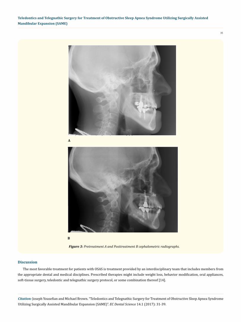
A well intercuspated Class I molar and canine relationship was attained. General dentist provided a comprehensive equilibration followed by the restoration of the implant replacing missing tooth # 23. A balanced facial profile with improved chin protrusion was ob-tained (Figure 2) but, most importantly, the patient reported a significant improvement in the night sleep and daytime level of alertness. Later tests demonstrated that his high blood pressure and high blood sugar normalized. His spouse reported almost no disturbances during sleep, and the postsurgical polysomnography performed at the sleep lab by his sleep specialist showed an AHI of 2.1 (below 5 is normal). An increase in the retropalatal, retroglossal, and hypoglossal airway spaces concomitant with maxillary and mandibular ad-vancement was evident on the cephalometric radiograph (Figure 3A and 3B).
Figure 2: Posttreatment extraoral and intraoral photographs.
35
Teledontics and Telegnathic Surgery for Treatment of Obstructive Sleep Apnea Syndrome Utilizing Surgically Assisted Mandibular Expansion (SAME)
Citation: Joseph Yousefian and Michael Brown. “Teledontics and Telegnathic Surgery for Treatment of Obstructive Sleep Apnea Syndrome Utilizing Surgically Assisted Mandibular Expansion (SAME)”. EC Dental Science 14.1 (2017): 31-39.
A
B
Figure 3: Pretreatment A and Posttreatment B cephalometric radiographs.
Discussion
The most favorable treatment for patients with OSAS is treatment provided by an interdisciplinary team that includes members from the appropriate dental and medical disciplines. Prescribed therapies might include weight loss, behavior modification, oral appliances, soft-tissue surgery, teledontic and telegnathic surgery protocol, or some combination thereof [14].

36
Teledontics and Telegnathic Surgery for Treatment of Obstructive Sleep Apnea Syndrome Utilizing Surgically Assisted Mandibular Expansion (SAME)
Citation: Joseph Yousefian and Michael Brown. “Teledontics and Telegnathic Surgery for Treatment of Obstructive Sleep Apnea Syndrome Utilizing Surgically Assisted Mandibular Expansion (SAME)”. EC Dental Science 14.1 (2017): 31-39.
In majority of telegnathic cases only the anteroposterior jaw dimension is addressed, and due to the complexity in incorporating the transverse dimension, this opportunity is overlooked. Based on this concern, usually a maxillomandibular advancement of 10 mm has been considered as one of the most effective surgical treatments for OSA [27,28]. However, not all patients diagnosed with OSA are af-fected by severe sagittal discrepancies of maxillary or mandibular skeletal structures. Therefor a routine cookbook approach of a 10 mm bimaxillary advancement just to treat OSA could create a very unaesthetic facial result for this category of patients. For this reason, the literature describes few attempts to incorporate a transverse expansion in addition to sagittal skeletal corrections for treatment of OSA patients [13,14,29].
There is some literature that discuss the role of increased transverse dimension by means of MSDO in resolving dentoalveolar crowd-ing in treatment of OSA patients [14]. However, there has been no known previous description of the important role of SAME in the devel-opment of implant site for replacement of missing teeth and its subsequent effectiveness in the treatment of OSA.
A nonsurgical treatment approach, including the extraction of one lower incisor or two lower first bicuspids, would have addressed the orthodontic aspects of this patient’s malocclusion. However, correction of the malocclusion was only one of the objectives of treat-ment. The need for treatment of the patient’s OSAS, which was a more health-threatening condition, overshadowed the need for treatment of the patient’s malocclusion. Tooth extraction could also could cause deterioration in the facial and dental esthetic by retracting the lower lip and magnifying the chin protrusion [30-34]. Ideally, a treatment plan involving expansion of the oral environment in all three dimen-sions would be a more effective treatment for these patients.
SAME as outpatient protocol produced a proper recipient site for the installation of an implant to replace tooth #23 [35]. Also com-bined with the subsequent bimaxillary advancement surgery as telegnathic surgical protocol provided adequate oral volume to accom-modate the tongue thus opening the oropharynx [13].
Maxillomandibular counterclockwise rotation and advancement by expansion of the posterior nasopharyngeal openings also augment the nasal cavity. A nasal cavity volume increase should reduce the resistance to nasal airflow [36]. If the airway is considered a simple tube, as the radius of the tube increases, the resistance to flow decreases exponentially to the fourth power (resistance = 8Lη/πr4) [37]. Therefore, even small increases in the diameter of the tube (the nasal cavity) can dramatically decrease the resistance to nasal air flow [37].
Physicians and dentists managing these patients should become aware of an emerging paradigm shift in modern orthodontic concepts and practice. Children and adults can commonly present with airway pathology requiring treatment protocols that are significantly dif-ferent from traditional orthodontic treatment such as the use of headgear, functional appliances such as the herpes appliance or extrac-tion of teeth to straighten teeth. Improvement of the whole POF complex requires more detailed management than just the correction of crossbite or a narrow plate in order to impact on the upper airway and improve OSA [38,39].
In a society where CPAP compliance has been historically low, physicians, dentists, orthodontists and maxillofacial surgeons trained in management of POFD may also play an important role in prevention and treatment of OSAS. An understanding of the related POFD as epigenetic factors contributing in the development of OSAS that can take advantage of a “teledontic and telegnathic” treatment could play a relevant part of OSAS management in the future.
While oral surgeons are moving away from “orthognathic” to a “telegnathic” approach, which reflects the forward positioning of the maxilla and mandible for the treatment of OSAS patients, orthodontists with an understanding of a “teledontic” method will be effective members of interdisciplinary team for further expansion of the POF complex by restoration of missing teeth and avoiding extraction [12,26].

37
Teledontics and Telegnathic Surgery for Treatment of Obstructive Sleep Apnea Syndrome Utilizing Surgically Assisted Mandibular Expansion (SAME)
Citation: Joseph Yousefian and Michael Brown. “Teledontics and Telegnathic Surgery for Treatment of Obstructive Sleep Apnea Syndrome Utilizing Surgically Assisted Mandibular Expansion (SAME)”. EC Dental Science 14.1 (2017): 31-39.
Bibliography
1. Youssef NA., et al. “Is obstructive sleep apnea associated with ADHD?” Annals of Clinical Psychiatry 23.3 (2011): 213-224.
2. Martinez-Garcia MA C-RF. “Obstructive sleep apnea is associated with cancer mortality in younger patients”. Sleep Medicine 15.7 (2014): 742-748.
3. Chen JC HJ. “Sleep apnea increased incidence of primary central nervous system cancers: a nationwide cohort study”. Sleep Medicine 15.7 (2014): 749-754.
4. Young T and Palta M. “The occurance of sleep-disordered breathing among middle-aged adults”. New England Journal of Medicine 328.17 (1993): 1230-1235.
5. Jonas DE., et al. “Screening for Obstructive Sleep Apnea in Adults: Evidence Report and Systematic Review for the US Preventive Ser-vices Task Force”. Journal of the American Medical Association 317.4 (2017): 415-433.
6. Medicine AAoS (2014).
7. Franklin KA., et al. “Sleep apnoea is a common occurrence in females”. European Respiratory Journal 41.3 (2013): 619-615.
8. Li KK., et al. “Obstructive sleep apnea syndrome: a comparison between Far-East Asian and white men”. Laryngoscope 110.10 (2000): 1689-1693.
9. Khan Y and Heckmatt J. “Obstructive apneas in Duchenne muscular dystrophy”. Thorax 49.2 (1994): 157-161.
10. Kotagal S., et al. “Sleep abnormalities in patients with severe cerebral palsy”. Developmental Medicine and Child Neurology 36.4 (1994): 304-311.
11. Joosten SA., et al. “Impact of Weight Loss Management in OSA”. Chest 3692.17 (2017): 30157-3015.
12. Yousefian J., et al. “Correction of Severe Obstructive Sleep Apnea with Interdisciplinary treatment”. Dentistry Today 34.8 (2015): 80-88.
13. Yousefian J and Moghadam B. “The role of contemporary orthodontics in the diagnosis and treatment of sleep-disordered breathing”. First edition. USA: Roth Williams Legacy Foundation (2013).
14. Conley S and Legan H. “Correction of severe obstructive sleep apnea with bimaxillary transverse distraction osteogenesis and maxil-lomandibular advancement”. American Journal of Orthodontics and Dentofacial Orthopedics 129.2 (2006): 283-292.
15. Guerrero CA. “Rapid mandibular expansion”. Revista Venezolana de Orthodontia (1990): 1-9.
16. Ilizarov GA. “The tension-stress effect on the genesis and growth of tissues: Part 1. The influence of stability of fixation and soft tissue preservation”. Clinical Orthopaedics and Related Research 238 (1989): 249-281.
17. Ilizarov GA. “The tension-stress effect on the genesis and growth of tissues: Part 2. The influence of the rate and frequency of distrac-tion”. Clinical Orthopaedics and Related Research 239 (1989): 263-285.
18. Ilizarov GA. “Clinical application of the tension-stress effect for limb lengthening”. Clinical Orthopaedics and Related Research 250 (1990): 8-26.
19. Toth BA KJ., et al. “Distraction osteogenesis and its application to the midface and bony orbit in craniosynostosis syndromes”. Journal of Craniofacial Surgery 9.2 (1998): 100-113.

38
Teledontics and Telegnathic Surgery for Treatment of Obstructive Sleep Apnea Syndrome Utilizing Surgically Assisted Mandibular Expansion (SAME)
Citation: Joseph Yousefian and Michael Brown. “Teledontics and Telegnathic Surgery for Treatment of Obstructive Sleep Apnea Syndrome Utilizing Surgically Assisted Mandibular Expansion (SAME)”. EC Dental Science 14.1 (2017): 31-39.
20. Molina F OF. “Mandibular elongation and remodeling by distraction: a farewell to major osteotomies”. Plastic and Reconstructive Surgery 96.4 (1995): 825-842.
21. Haluk E MS. “Long-term skeletal effects of mandibular symphyseal distraction osteogenesis. An implant study”. European Journal of Orthodontics 27.5 (2005): 512-517.
22. Bell W GM., et al. “Intra-oral widening and lengthening of the mandible in baboons by distraction osteogenesis”. Journal of Oral and Maxillofacial Surgery 57.5 (1999): 548-562.
23. Del Santo M GC., et al. “Long-term skeletal and dental effects of mandibular symphyseal distraction osteogenesis”. American Journal of Orthodontics and Dentofacial Orthopedics 118.5 (2000): 485-493.
24. Guererero C BW., et al. “Mandibular widening by intraoral distraction osteogenesis”. British Journal of Oral and Maxillofacial Surgery 35.6 (1997): 383-392.
25. Contasti G GC., et al. “Mandibular widening by distraction osteogenesis”. Journal of Clinical Orthodontics 35.3 (2001): 165-173.
26. Prinsell J. “Telegnatic maxillomandibular advancement surgery for OSA”. Dental Sleep Practice (2015): 6-9.
27. Waite A SS. “Maxillomandibular advancement (a cure for obstructive sleep apnea)”. Oral and Maxillofacial Surgery Clinics of North America (1995): 327-336.
28. Hochban W BU and Peter J. “Surgical treatment of obstructive sleep apnea by maxillomandibular advancement”. Sleep 17.7 (1994): 624-629.
29. Pirelli P SM and Guilleminault C. “Rapid maxillary expansion in children with obstructive sleep apnea syndrome”. Sleep 27.4 (2004): 761-766.
30. Yousefian J., et al. “A New Look at the Treatment of Class II Division 2 Malocclusion”. American Journal of Orthodontics and Dentofacial Orthopedics 130.6 (2006): 771-778.
31. Sarver D. “The importance of incisor positioning in the esthetic smile: the smile arc”. American Journal of Orthodontics and Dentofacial Orthopedics 120.2 (2001): 98-111.
32. Sarver D AM. “Dynamic smile visualization and quantification: Part 2 (Smile analysis and treatment strategies)”. American Journal of Orthodontics and Dentofacial Orthopedics 124.2 (2003): 116-127.
33. Peck S PL. “Selected aspects of the art and science of facial esthetics”. Seminars in Orthodontics 1.2 (1995): 105-126.
34. Zachrisson BU. “Esthetic factors involved in anterior tooth display and the smile, vertical dimension”. Journal of Clinical Orthodontics 32.7 (1998): 432-445.
35. Yousefian J. “A simple technique for mandibular symphyseal distraction osteogenesis”. Journal of Clinical Orthodontics 44.12 (2010): 731-737.
36. Kunkel M HW. “The influence of maxillary osteotomy on nasal airway patency and geometry”. Mund-, Kiefer- Und Gesichtschirurgie 1.4 (1997): 94-98.
37. Courtiss E and Goldwyn R. “The effects of nasal surgery on airflow”. Plastic and Reconstructive Surgery 72.1 (1983): 9-21.
38. Yousefian J and Moghadam B. “The role of contemporary orthodontics in the diagnosis and treatment of sleep-disordered breathing”. 2nd edition. USA: Roth Williams Legacy Foundation (2015).

39
Teledontics and Telegnathic Surgery for Treatment of Obstructive Sleep Apnea Syndrome Utilizing Surgically Assisted Mandibular Expansion (SAME)
Citation: Joseph Yousefian and Michael Brown. “Teledontics and Telegnathic Surgery for Treatment of Obstructive Sleep Apnea Syndrome Utilizing Surgically Assisted Mandibular Expansion (SAME)”. EC Dental Science 14.1 (2017): 31-39.
39. Yousefian J. “Correction of Severe Obstructive Sleep Apnea Syndrome with Interdisciplinary Medical and Dental Treatment Planning”. Journal of Dental Sleep Medicine 3.3 (2016): 89-91.
Volume 14 Issue 1 September 2017© All rights reserved by Joseph Yousefian and Michael Brown.