crohn vs tb 2015 03 22 tnisgcon
TRANSCRIPT
Differentiating Crohn’s disease from tuberculosis
B.S. Ramakrishna
Institute of GastroenterologySRM Institutes for Medical Science
Vadapalani, Chennai 600 026
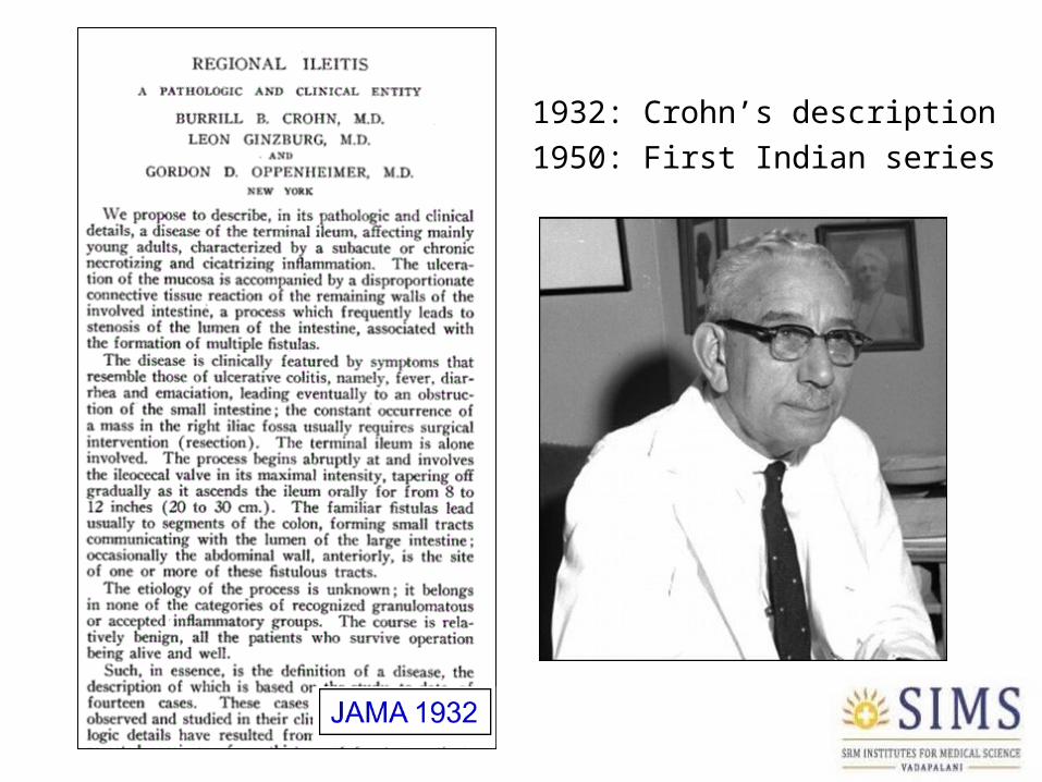
1932: Crohn’s description 1950: First Indian series
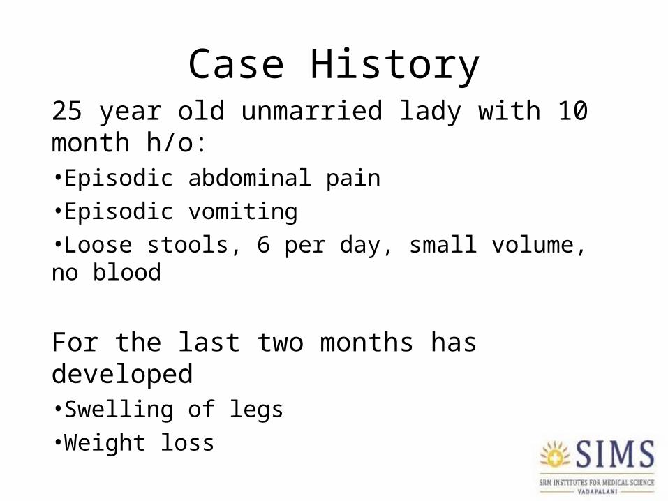
Case History25 year old unmarried lady with 10 month h/o:•Episodic abdominal pain•Episodic vomiting•Loose stools, 6 per day, small volume, no blood
For the last two months has developed•Swelling of legs•Weight loss
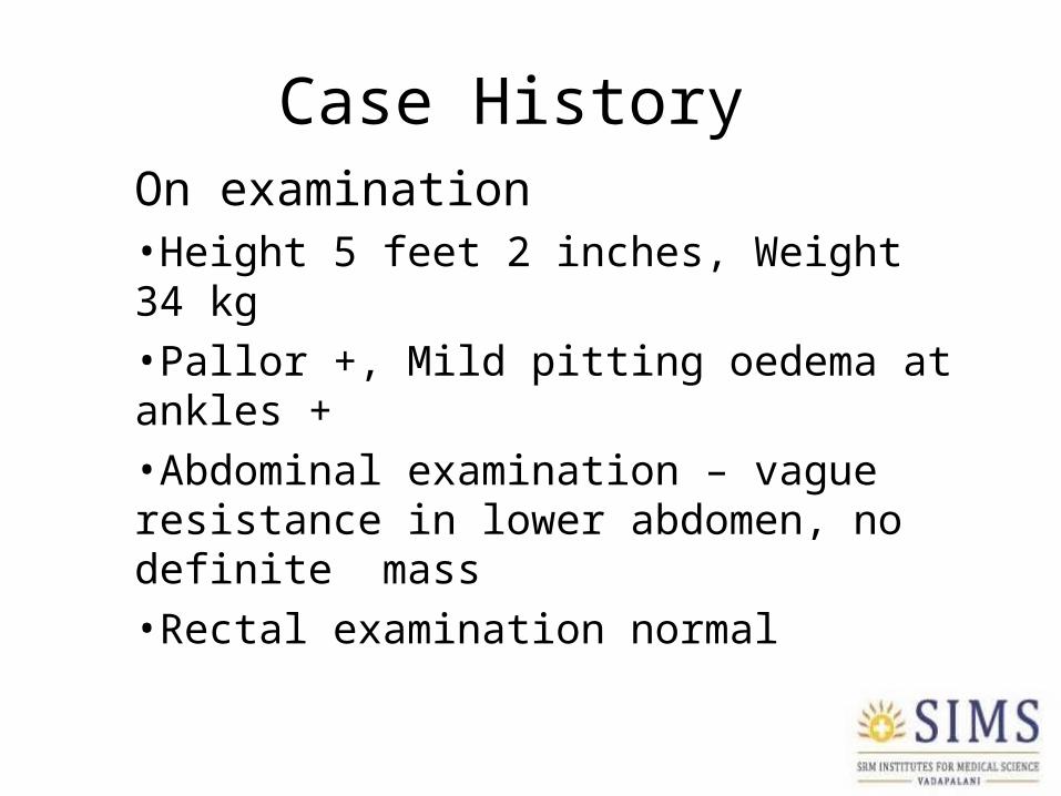
Case History On examination•Height 5 feet 2 inches, Weight 34 kg•Pallor +, Mild pitting oedema at ankles +•Abdominal examination – vague resistance in lower abdomen, no definite mass•Rectal examination normal
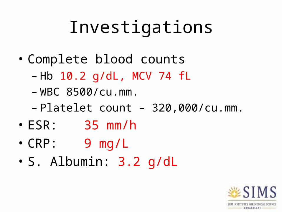
Investigations
• Complete blood counts– Hb 10.2 g/dL, MCV 74 fL– WBC 8500/cu.mm.– Platelet count – 320,000/cu.mm.
• ESR: 35 mm/h• CRP: 9 mg/L• S. Albumin: 3.2 g/dL
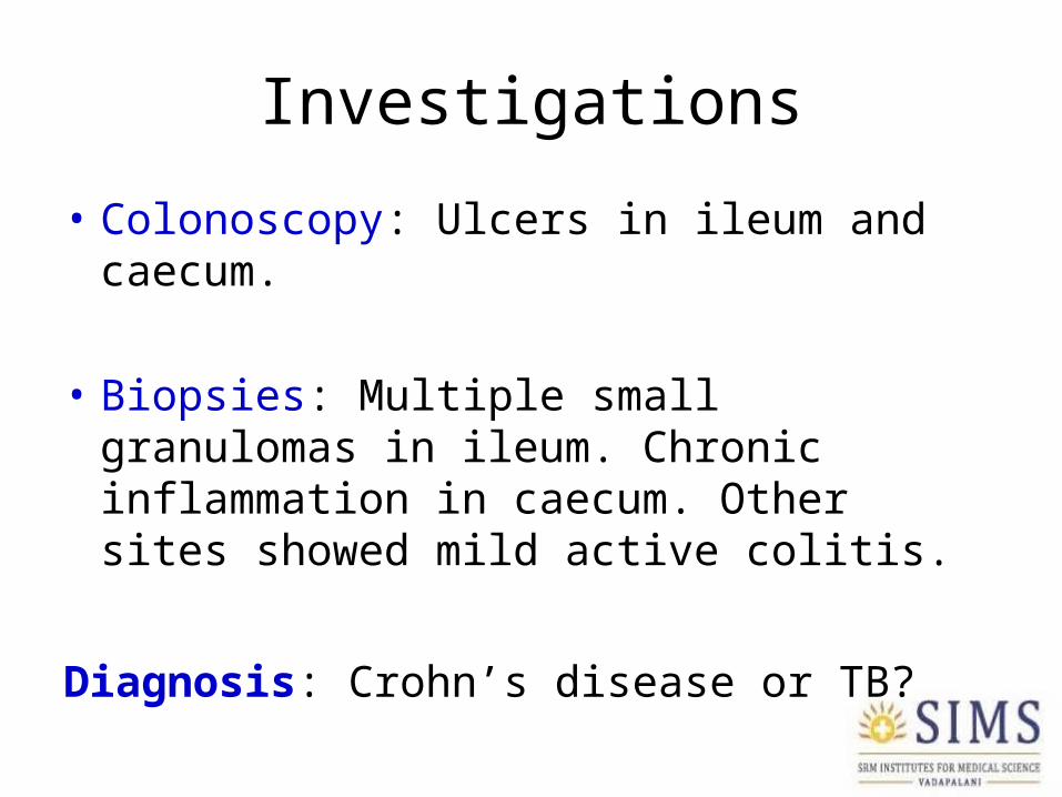
Investigations
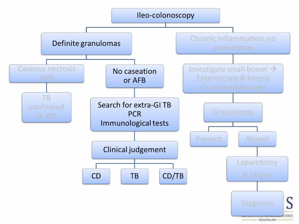
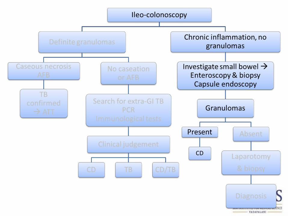
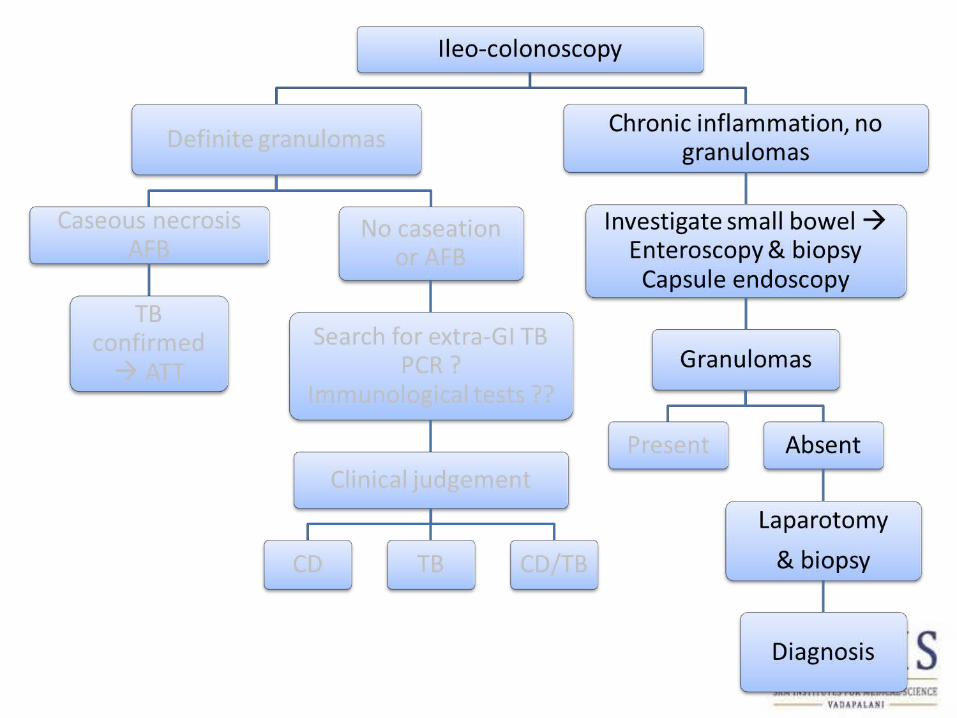
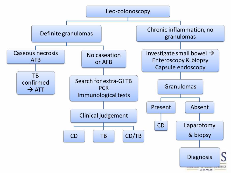
• Colonoscopy: Ulcers in ileum and caecum.
• Biopsies: Multiple small granulomas in ileum. Chronic inflammation in caecum. Other sites showed mild active colitis.
Diagnosis: Crohn’s disease or TB?
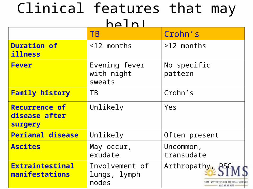
Clinical features that may help!TB Crohn’s
Duration of illness <12 months >12 months
Fever Evening fever with night sweats
No specific pattern
Family history TB Crohn’s
Recurrence of disease after surgery
Unlikely Yes
Perianal disease Unlikely Often present
Ascites May occur, exudate Uncommon, transudate
Extraintestinal manifestations
Involvement of lungs, lymph nodes
Arthropathy, PSC
SEROLOGY/IMMUNOLOGY
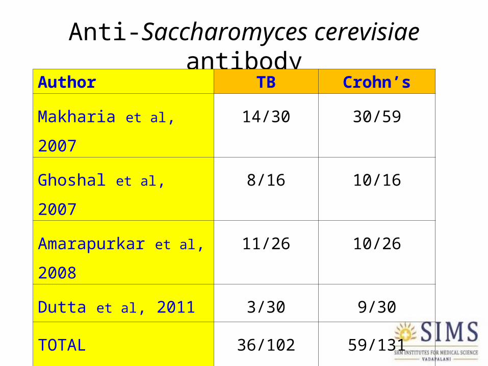
Anti-Saccharomyces cerevisiae antibodyAuthor TB Crohn’s
Makharia et al, 2007 14/30 30/59
Ghoshal et al, 2007 8/16 10/16
Amarapurkar et al, 2008 11/26 10/26
Dutta et al, 2011 3/30 9/30
TOTAL 36/102
(35.2%)
59/131
(45.0%)

TB ELISA
• Meta-analysis of 68 studies– Poor sensitivity
• Sensitivity higher in smear positive patients
– Unreliable specificity• Specificity higher when healthy individuals used for
comparisonSteingart et al. PLoS Med 2007
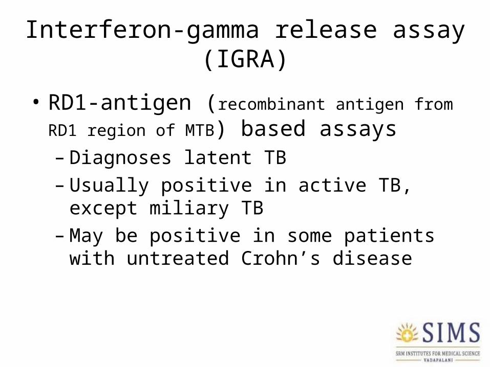
Interferon-gamma release assay (IGRA)
• RD1-antigen (recombinant antigen from RD1 region of
MTB) based assays – Diagnoses latent TB– Usually positive in active TB, except miliary TB– May be positive in some patients with untreated
Crohn’s disease

Tuberculin (PPD) skin test
• PPD reactivity rate in Crohn’s disease in India & other TB endemic countries not known
• PPD testing advised by US FDA prior to infliximab therapy in order to prevent TB reactivation

RADIOLOGY

Ileocaecal TB•Straightening of IC junction •Narrow caecum•Nodules •Transverse ulcers
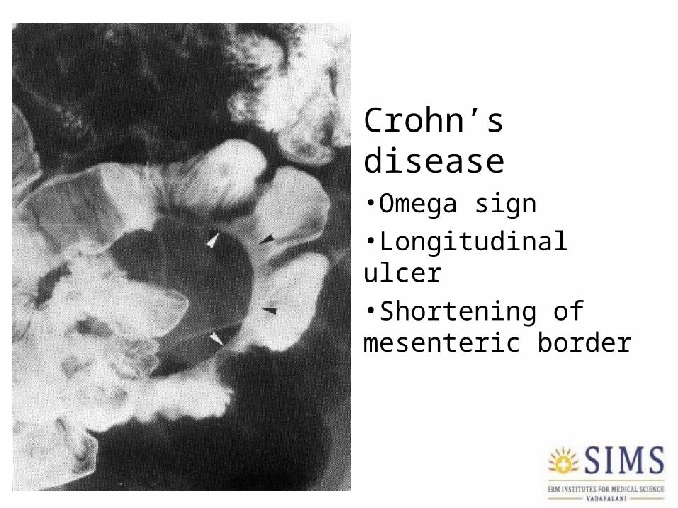
Crohn’s disease•Omega sign•Longitudinal ulcer•Shortening of mesenteric border
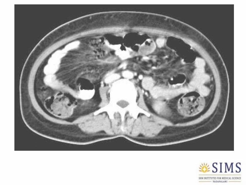

CD versus TB: CECT abdomen
TB Crohn’s
Wall thickening Absent or asymmetric
Concentric
Target sign Absent Present in 50%
Lymph nodes Central necrosis in 1/3
No central necrosis
Bowel displacement Due to lymph nodes Fibrofatty change
Makanjuola et al, AJR 1998

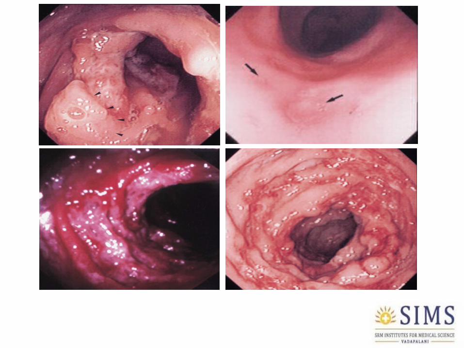
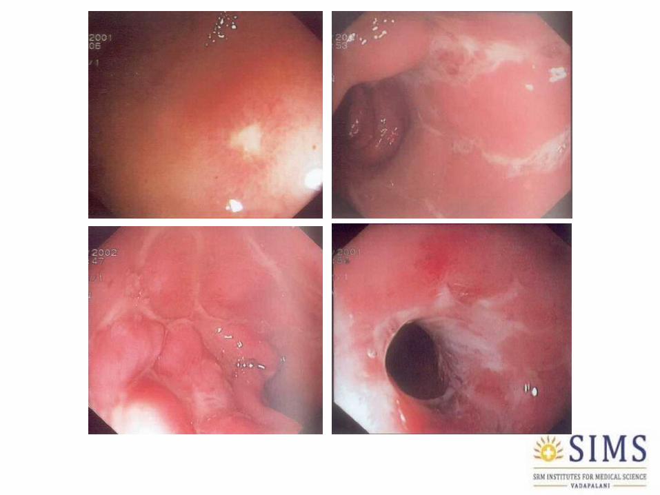
ENDOSCOPY
Colonoscopy in TB• Superficial well defined ulcers with irregular
margins• Nodular mucosa• Deformed IC valve with ulcers• Ileocecal involvement • Skip lesions in 10%• Segmental involvement in 26%• Pancolitis in 4%
Shah S et al, Gut 1992
Colonoscopy in Crohn’s disease• Aphthous ulcers• Deep irregular ulcers• Longitudinal ulcers• Cobblestones• Pseudopolyps• Mucosal bridging• Discontinuous involvement (Skip lesions)• Luminal narrowing• Fistulas

HISTOPATHOLOGY
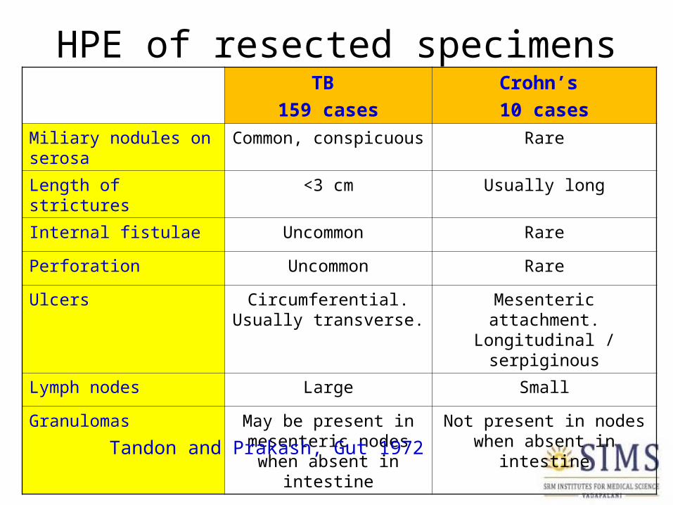
HPE of resected specimensTB
159 cases
Crohn’s
10 cases
Miliary nodules on serosa
Common, conspicuous Rare
Length of strictures <3 cm Usually long
Internal fistulae Uncommon Rare
Perforation Uncommon Rare
Ulcers Circumferential. Usually transverse.
Mesenteric attachment. Longitudinal / serpiginous
Lymph nodes Large Small
Granulomas May be present in mesenteric nodes when
absent in intestine
Not present in nodes when absent in intestine
Tandon and Prakash, Gut 1972

Mucosal biopsy
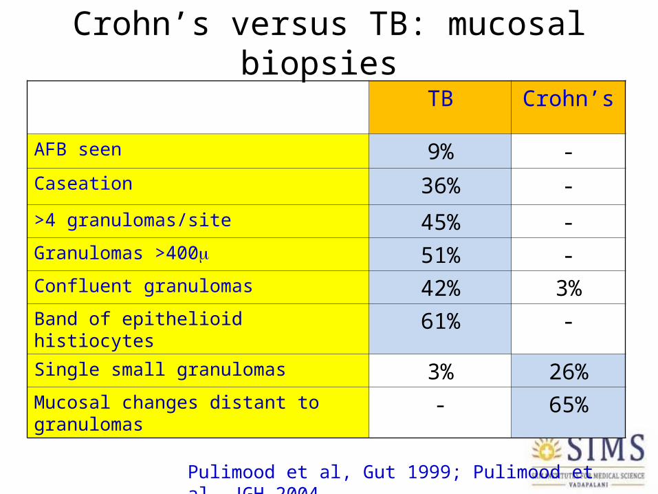
Crohn’s versus TB: mucosal biopsies
TB Crohn’s
AFB seen 9% -Caseation 36% -
>4 granulomas/site 45% -Granulomas >400 51% -Confluent granulomas 42% 3%Band of epithelioid histiocytes 61% -Single small granulomas 3% 26%Mucosal changes distant to granulomas - 65%
Pulimood et al, Gut 1999; Pulimood et al, JGH 2004
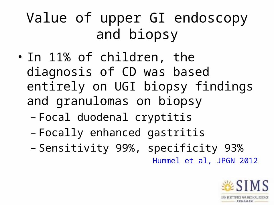
Value of upper GI endoscopy and biopsy
• In 11% of children, the diagnosis of CD was based entirely on UGI biopsy findings and granulomas on biopsy– Focal duodenal cryptitis– Focally enhanced gastritis– Sensitivity 99%, specificity 93%
Hummel et al, JPGN 2012

MICROBIOLOGY

Confirmation of TB on biopsy
Test % positive AuthorsCaseous necrosis 23-36% Lee 2004, Pulimood
2005
AFB smear of biopsy 5-10%AFB culture of biopsy 7-40% Bhargava 1985, Lee
2004, Khan 2006
AFB culture of surgical tissue
70% Veeragandham 1996

TB PCRTB Crohn’s
Moatter et al 1998 8/12 -
Gan et al 2002 25/39 0/30
Amarapurkar et al 2004 17/26 0/26
Pulimood et al 2008 6/20 1/20
Balamurugan et al 2008 21/26 5/46

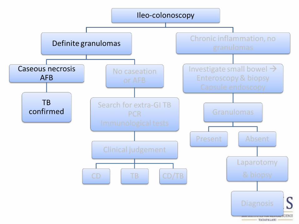
TB versus Crohn’s Search for extraintestinal tuberculosis
• Search for peripheral lymphadenopathy FNAC, culture, PCR, and biopsy
• Search for pulmonary lesion on chest x-ray, and do induced sputum for AFB (x3), sputum PCR, bronchoalveolar lavage if necessary
• Search for ascites, aspirate and send for cells, protein, culture, PCR

Diagnosis of ITB
• Histology + Culture diagnostic in 60% Bhargava DK et al, 1992
• Histology + Culture + Extra-intestinal TB diagnostic in 56% of 225 patients Lee et al 2004

Diagnosis of ITB
• Histology + Culture diagnostic in 60% Bhargava DK et al, 1992
• Histology + Culture + Extra-intestinal TB diagnostic in 56% of 225 patients Lee et al 2004
• Histology + culture diagnostic in 80%; addition of stool TB PCR diagnosed 100% of 26 ITB patients Balamurugan et al, 2011
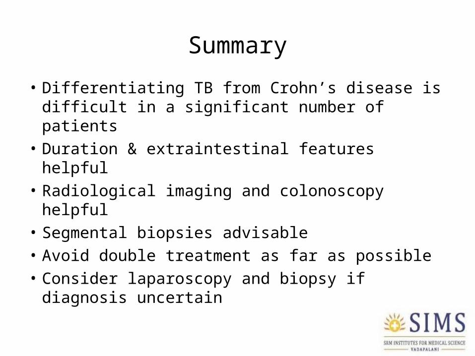
Summary
• Differentiating TB from Crohn’s disease is difficult in a significant number of patients
• Duration & extraintestinal features helpful• Radiological imaging and colonoscopy helpful• Segmental biopsies advisable• Avoid double treatment as far as possible• Consider laparoscopy and biopsy if diagnosis
uncertain