critical care onboarding for the non-critical care nurse · critical care onboarding for the...
TRANSCRIPT

Critical Care Onboarding for the Non-Critical Care Nurse
Welcome! Thank you for being here!Ground Rules
• Please refrain from using the hand raise function, instead place any questions in the chat box
• Questions will be addressed at the end of each set of slides
Revised 12.19.19
Critical Care Academy
Pulmonary System
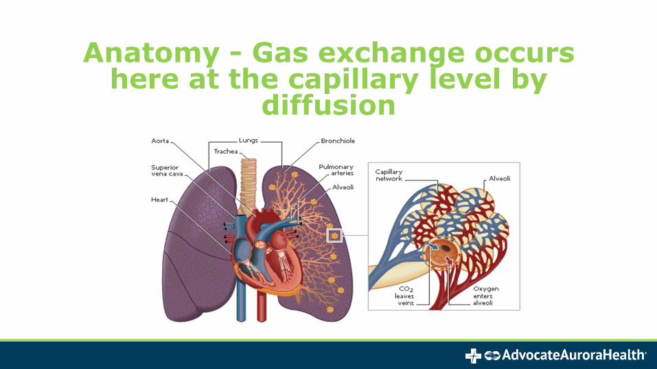
Anatomy - Gas exchange occurs here at the capillary level by
diffusion

Pleura
• Parietal Pleura: outer layer that lines the inside of the thoracic cage
• Visceral Pleura: covers the lung
• The two layers are separated by a very small amount of pleural fluid

Alveolar Ventilation
• CO2 diffuses 20 x easier than O2
• Blood shunts to where it can take part in gas exchange
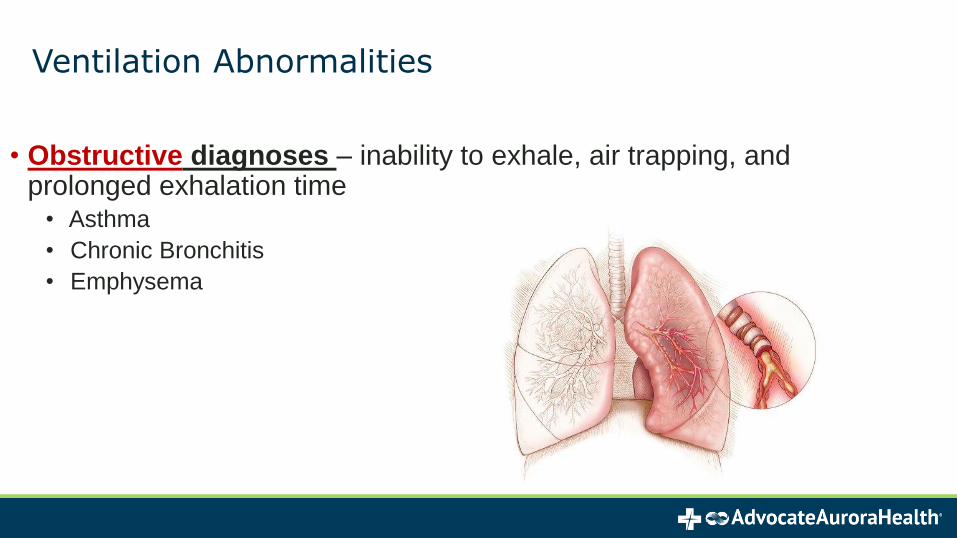
Ventilation Abnormalities
• Obstructive diagnoses – inability to exhale, air trapping, and prolonged exhalation time
• Asthma
• Chronic Bronchitis
• Emphysema
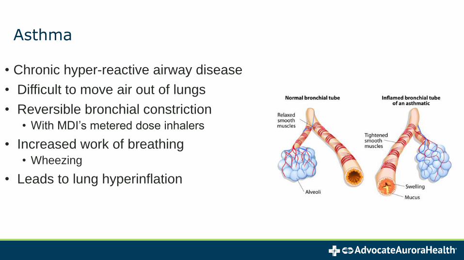
Asthma
• Chronic hyper-reactive airway disease
• Difficult to move air out of lungs
• Reversible bronchial constriction• With MDI’s metered dose inhalers
• Increased work of breathing• Wheezing
• Leads to lung hyperinflation

Pneumonia
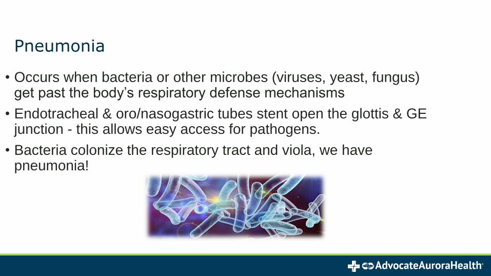
Pneumonia
• Occurs when bacteria or other microbes (viruses, yeast, fungus) get past the body’s respiratory defense mechanisms
• Endotracheal & oro/nasogastric tubes stent open the glottis & GE junction - this allows easy access for pathogens.
• Bacteria colonize the respiratory tract and viola, we have pneumonia!

Pneumonia
Alveolar inflammation due to infection
• Categories:• Community Acquired (CAP)
• Hospital Acquired (HAP)
• Ventilator Associated (VAP)
• Etiologies:• Inhalation of causative pathogen
• Aspiration
• Hematogenous spread
• Chronic colonization (ex: COPD)
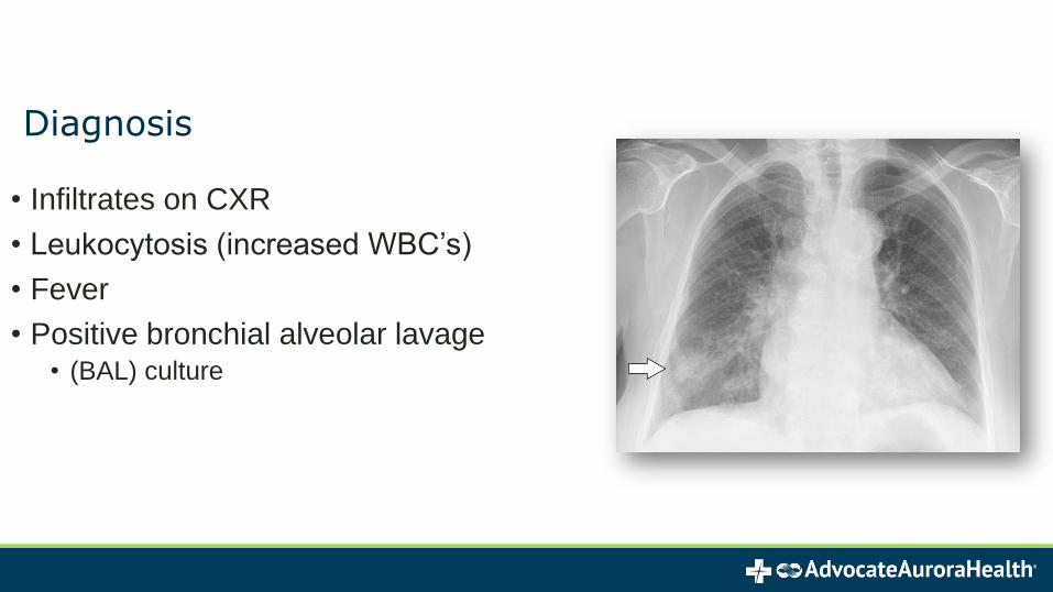
Diagnosis
• Infiltrates on CXR
• Leukocytosis (increased WBC’s)
• Fever
• Positive bronchial alveolar lavage• (BAL) culture
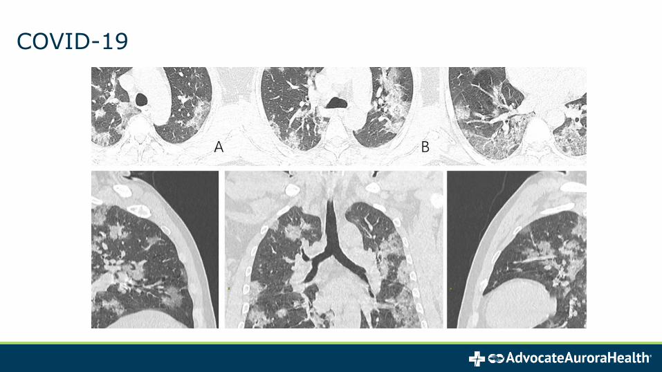
COVID-19
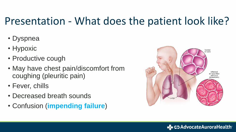
• Dyspnea
• Hypoxic
• Productive cough
• May have chest pain/discomfort from coughing (pleuritic pain)
• Fever, chills
• Decreased breath sounds
• Confusion (impending failure)
Presentation - What does the patient look like?

Treatment
• Broad-spectrum antibiotics to treat suspected pathogens
• Narrow antibiotic therapy when culture & sensitivity results acquired
• Hydration
• Oxygen
• Mechanical ventilation, if necessary
Nursing Considerations
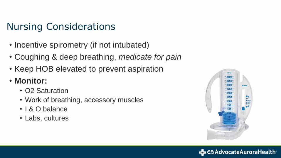
• Incentive spirometry (if not intubated)
• Coughing & deep breathing, medicate for pain
• Keep HOB elevated to prevent aspiration
• Monitor:• O2 Saturation
• Work of breathing, accessory muscles
• I & O balance
• Labs, cultures
Respiratory Failure
• Respiratory system unable to perform one or both of its main functions:
• Oxygenation
• Removal of carbon dioxide
• Adequacy of gas exchange determined by:• Balance between pulmonary ventilation & capillary blood flow
• Characterized by ABG abnormalities• Hypoxemia: PaO2 < 55 mmHg
• Hypercarbia: pCO2 > 50 mmHg

Acute Respiratory Distress Syndrome
• Complex clinical syndrome, rather than a single disease process
• Causes are many & diverse• Direct or indirect injury to the lungs
• Acute in onset (4 – 48 hours)
• Diagnosis largely based on clinical presentation
*There is no one test is truly indicative of ARDS!

Pathophysiology of ARDS
Early Stage: first 12 hours• Acute increased shortness of
breath (dyspnea)
• Acute respiratory rate (tachypnea)
Stage II: 24 hours• Respiratory distress
• Crackles
• Cyanosis
• Dry cough & pleuritic pain
Stage III: 2 – 10 days
– Respiratory failure/Hypoxemia
– Hemodynamic instability
– Decreased lung volume &
compliance
Stage IV: >10 days
– Persistent infiltrates / SEPSIS
– S/S of multi-organ failure
– Possible pneumothorax
– Fibrosis of interstitial wall of
lung

ARDS Severity
Severity ----------- PaO2/FiO2----------MortalityMild-----------------200 – 300-------------27%Moderate-----------100 – 200-------------32%Severe--------------< 100-----------------45%
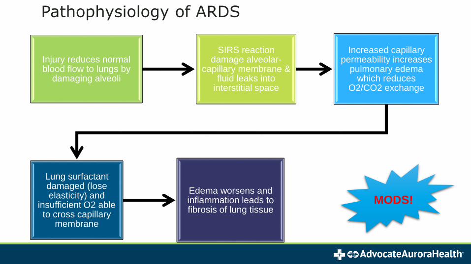
Injury reduces normal blood flow to lungs by
damaging alveoli
SIRS reaction damage alveolar-
capillary membrane & fluid leaks into
interstitial space
Increased capillary permeability increases
pulmonary edema which reduces
O2/CO2 exchange
Lung surfactant damaged (lose elasticity) and
insufficient O2 able to cross capillary
membrane
Edema worsens and inflammation leads to fibrosis of lung tissue
Pathophysiology of ARDS
MODS!
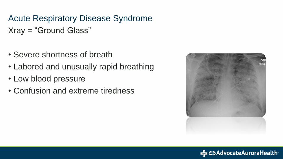
Acute Respiratory Disease Syndrome
Xray = “Ground Glass”
• Severe shortness of breath
• Labored and unusually rapid breathing
• Low blood pressure
• Confusion and extreme tiredness

Nursing Considerations
Monitor:
• Breath sounds, O2 sats, & airway pressures
• Vitals and ECG for any dysrhythmias (r/t hypoxia)
• I & O’s
• Lactate levels
• Replace electrolytes as needed
• Reposition Q2 hours w/HOB >30 degrees – Think “LUNG UP”
• S/S of pneumothorax d/t high PEEP pressures
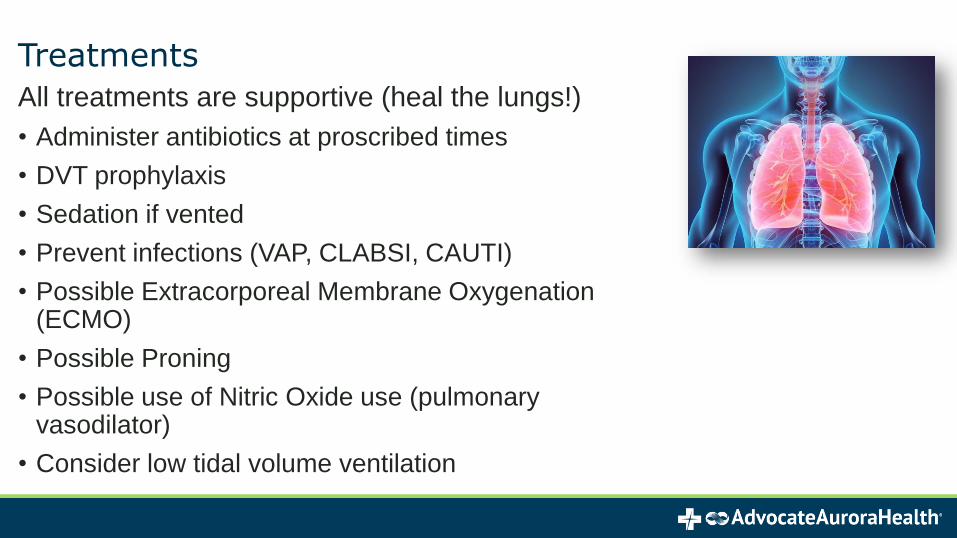
Treatments
All treatments are supportive (heal the lungs!)
• Administer antibiotics at proscribed times
• DVT prophylaxis
• Sedation if vented
• Prevent infections (VAP, CLABSI, CAUTI)
• Possible Extracorporeal Membrane Oxygenation (ECMO)
• Possible Proning
• Possible use of Nitric Oxide use (pulmonary vasodilator)
• Consider low tidal volume ventilation

Illinois: Continuous Lateral Rotation Therapy (CLRT)
• A setting on some ICU beds which allow for the patient to be automatically rotated to get the “lung up”
• Best practice is to rotate to get the patients lungs alternately “up in the air” at least 80%
• 60% rotation acceptable, but the more we rotate up, the better the oxygenation
• Can manually turn patients and it is recommended to turn immobile patients EVERY 2 hours to alternate sides and elevate heels off the bed
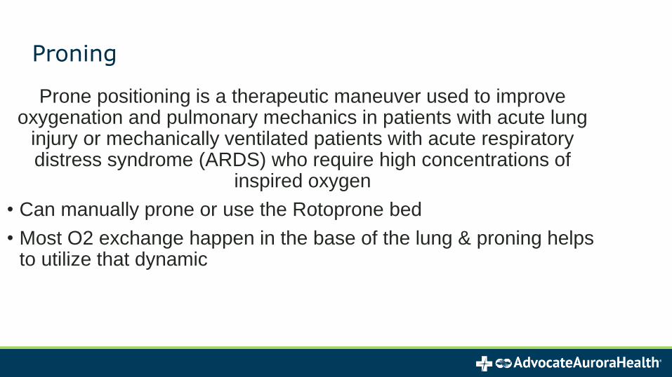
Proning
Prone positioning is a therapeutic maneuver used to improve oxygenation and pulmonary mechanics in patients with acute lung
injury or mechanically ventilated patients with acute respiratory distress syndrome (ARDS) who require high concentrations of
inspired oxygen
• Can manually prone or use the Rotoprone bed
• Most O2 exchange happen in the base of the lung & proning helps to utilize that dynamic


Manual Prone
Proning Nursing Considerations
• Endotracheal tubes must be double taped. When patient is in supine position RT and RN to re-tape and re-position ETT at least once in a 24-hour period.
• Capnography must be observed on all prone patients to detect adverse tube placement and patient decline
• Tube feeding should be stopped at least one-hour prior• Eyes lubricated and taped lids closed
Proning Nursing Considerations
• Provide sedation/pain medication as ordered• Provide neuromuscular blockers for ventilator dys-synchrony if
ordered (BIS monitoring if available)• Respiratory therapy must be present to maintain endotracheal
tube placement when turning prone and supine.• Following lift strap positioning, always confirm metal on metal
within the device prior to lift utilization.
Proning Nursing Considerations
• Patients will be placed prone for at least 16 hours and supine for 2 hours unless specified in the order. If patient responds, proningcan extend to 24 hours with 2 to 4 hours supine. Reprone per physician order.
• Respiratory therapist and RN to reposition the patient’s head every two hours when prone.
• Place patient in swimmers’ crawl position alternating sides every two hours, perform micromovements of high-risk areas every hour and prn.
• Assess device positioning to minimize pressure points.

Pneumothorax
• Air trapped between lung and chest wall
• Requires chest tube
• Can occur with or without tension
Emergent needle decompression if no chest tube immediately available!

Tension Pneumothorax
Tracheal
deviation
AWAY
from affected
side
• Life Threatening
EMERGENCY
• Immediate Needle
decompression
• Chest tube
insertion
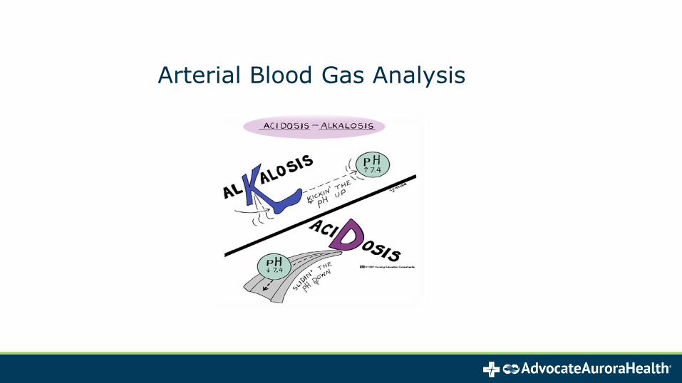
Arterial Blood Gas Analysis
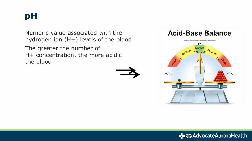
pH
Numeric value associated with the hydrogen ion (H+) levels of the blood
The greater the number of H+ concentration, the more acidic the blood

Terminology
CO2 is a respiratory acid
• PaCO2 assess adequacy of ventilation
HCO3 is the metabolic base
• Shows if the kidneys are producing or excreting bicarbonate.
Acidosis: pH < 7.35
Alkalosis: pH > 7.45
Acid-Base Balance
As the body pH heads towards acidosis, the body has 3 defense mechanisms which go into effect
Buffers (proteins, phosphates)
• Quick response
Increasing alveolar ventilation
• Decreases CO2
Increasing H+ (hydrogen ion) excretion
• also causes bicarbonate re-absorption


Respiratory Acidosis & Common Causes
• Respiratory failure
• Neurologic depression
– Due to disease, medications,
etc.
• Inadequate respiratory rate or
tidal volume on mechanical
ventilation

Metabolic Acidosis & Common Causes
H+ ACCUMULATIONShock
(lactic acidosis)
ESRD (uremia)
DKA
Medications
Hyperchloremia
LOSS OF HCO3
Diarrhea
High volume NGT drainage
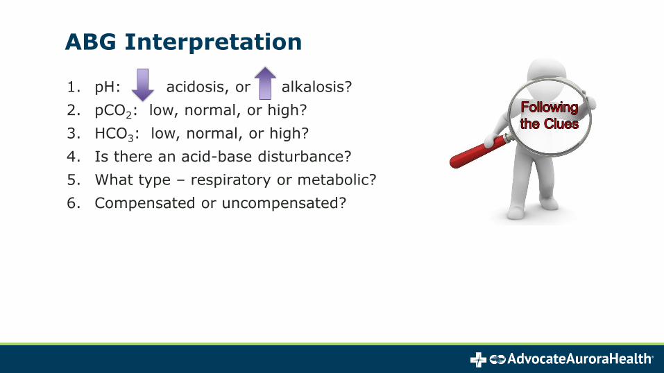
ABG Interpretation
1. pH: acidosis, or alkalosis?
2. pCO2: low, normal, or high?
3. HCO3: low, normal, or high?
4. Is there an acid-base disturbance?
5. What type – respiratory or metabolic?
6. Compensated or uncompensated?
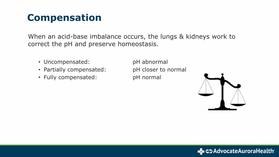
Compensation
When an acid-base imbalance occurs, the lungs & kidneys work to correct the pH and preserve homeostasis.
• Uncompensated: pH abnormal
• Partially compensated: pH closer to normal
• Fully compensated: pH normal

Acute Respiratory Failure
• Will have elevated CO2 and the pH will never be normal
• The pH will always be low since carbon dioxide is an acid
• Ex: Hypercapneic patients may also be tachypneic, but the respirations will be very shallow (guppy breathing) and the CO2 will not be expelled
• Where CO2 is low, the ph will be high – alkalotic
• Ex: A patient who is tachypneic from anxiety & blowing off all their CO2

Name that Blood Gas?
Low Normal High
pH
7.35 – 7.45
7.40
pCO2
35-45
40
HCO3
22-26
24
Normal ABG

Name that Blood Gas?
Low Normal High
pH
7.35 – 7.45
7.25
pCO2
35-45
60
HCO3
22-26
24
Respiratory
Acidosis
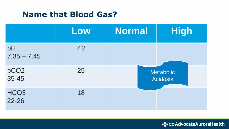
Name that Blood Gas?
Low Normal High
pH
7.35 – 7.45
7.2
pCO2
35-45
25
HCO3
22-26
18
Metabolic
Acidosis

Airway Devices & Management

Oxygen Delivery Systems
Nasal Cannula
Hi-Flo Nasal Cannula
Aerosol Mask
Non-Rebreather Mask
Venturi Mask
• CPAP
• BPAP
• Intubation
• Tracheostomy
– Trach Collar
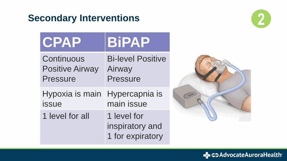
Secondary Interventions 2
CPAP BiPAPContinuous
Positive Airway
Pressure
Bi-level Positive
Airway
Pressure
Hypoxia is main
issue
Hypercapnia is
main issue
1 level for all 1 level for
inspiratory and
1 for expiratory

INTUBATION / Endotracheal Tubes
• Used to establish or protect an airway
• Can be inserted nasally or orally
• Will be cuffed
• Various sizes
• Provides continuous ventilatory assistance
➢What is the first nursing assessment you would do after your patient is intubated?

Intubation
When would you intubate?
When primary and secondary efforts of intervention have failed
What are Indications for intubation?
• Inability to maintain airway patency
• Inability to protect the airway against aspiration
• Ventilatory compromise
• Failure to adequately oxygenate pulmonary capillary blood
• Anticipation of a deteriorating course

Rapid Sequence Intubation (RSI)
What is the Nurse’s role in intubation prep?
oKnow the airway placement plan
oPosition placement supine and at the head of the bed
oPreoxygenate
oHave suction at bedside
oSedate & possibly paralyze: ALWAYS sedate prior to paralyzing
oEducate patient and family

ETT vs NTT intubations
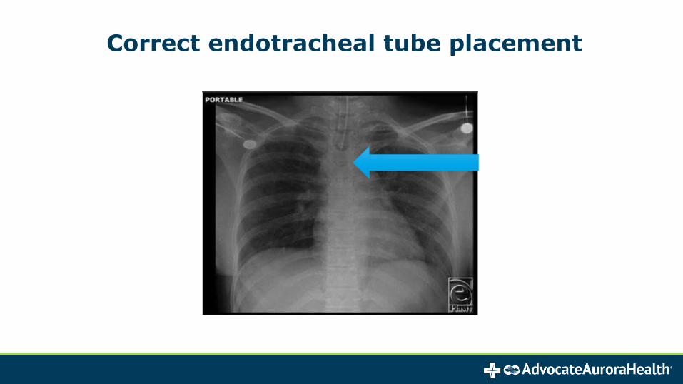
Correct endotracheal tube placement
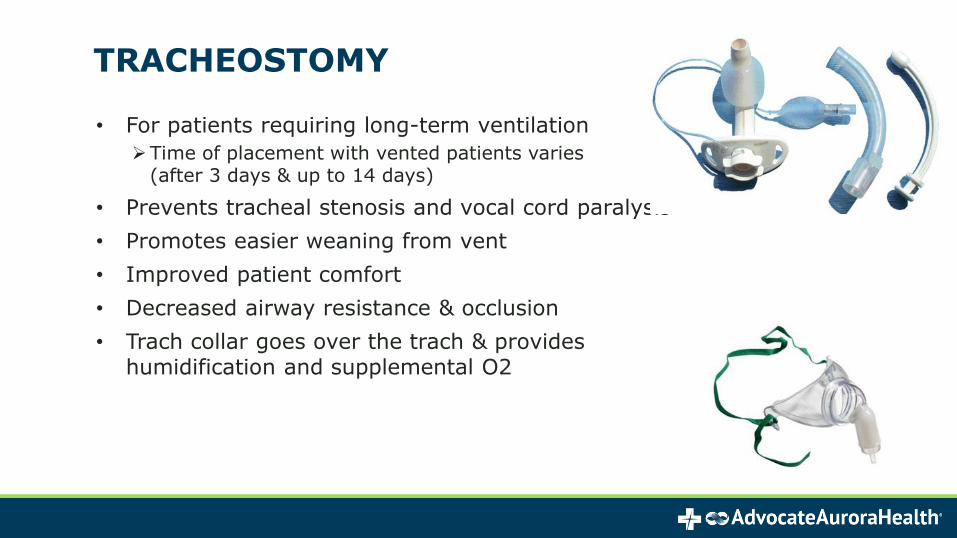
TRACHEOSTOMY
• For patients requiring long-term ventilation
➢Time of placement with vented patients varies (after 3 days & up to 14 days)
• Prevents tracheal stenosis and vocal cord paralysis
• Promotes easier weaning from vent
• Improved patient comfort
• Decreased airway resistance & occlusion
• Trach collar goes over the trach & provides humidification and supplemental O2

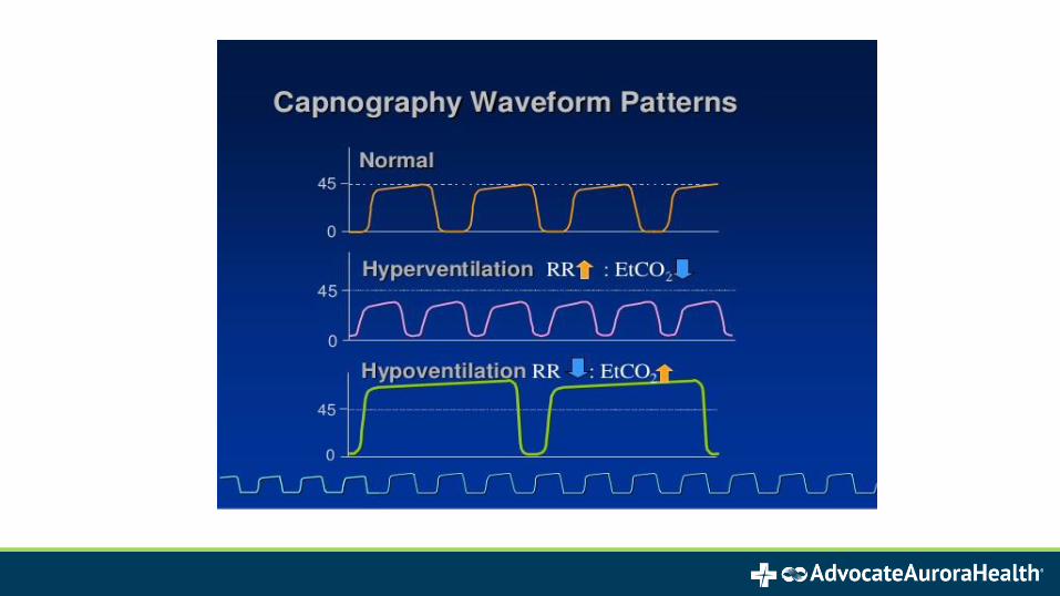
ETCO2 (Capnography) Monitoring
• Measures amounts of CO2 in exhaled breath
• Measures VENTILATION – “Are you breathing!”
• Faster indicator of breathing trouble than pulse oximetry
• Can be used with NC and ETT/Vents
*Think respiratory failure when ETCO2 is high!
*Think perfusion, metabolic or psychological problem when ETCO2 is low!

Ventilator Basics & Spontaneous Breathing
Trial (SBT)
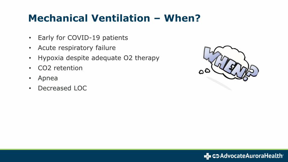
Mechanical Ventilation – When?
• Early for COVID-19 patients
• Acute respiratory failure
• Hypoxia despite adequate O2 therapy
• CO2 retention
• Apnea
• Decreased LOC
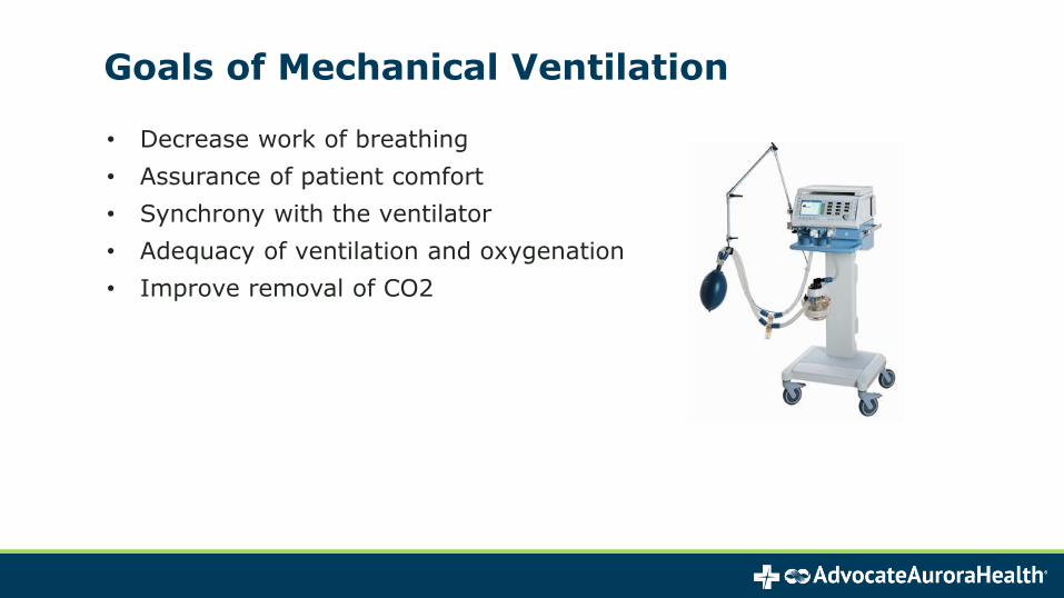
Goals of Mechanical Ventilation
• Decrease work of breathing
• Assurance of patient comfort
• Synchrony with the ventilator
• Adequacy of ventilation and oxygenation
• Improve removal of CO2

Common Modes Used
• AC/VC: Assist Control/Volume Control Ventilation
• SIMV: Synchronized Intermittent Mandatory Ventilation
• PRVC: Pressure Regulated Volume Control Ventilation
• PC: Pressure Control Ventilation
• PS: Pressure Support Ventilation

Basic Ventilator Settings
• Mode: AC, SIMV, PVC
• Rate
• Tidal Volume: 6-8 ml/kg of IBW (ideal)
• FiO2: 21-100%
• Respiratory Rate: minimum rate set
• PEEP: physiologic 3-5 mmHg
• Pressure Support: available on some modes
PEEP (Positive End Expiratory Pressure)
• What is considered Critical PEEP?
• Can you break the circuit with a patient on high PEEP?
• What about if you have to go on a road trip?
• Suctioning/sputum specimens on patients with high PEEP
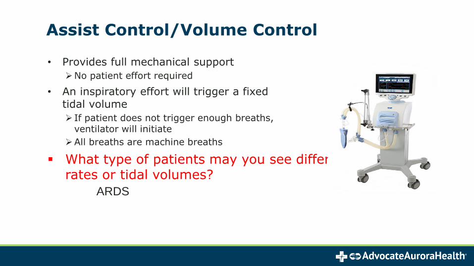
Assist Control/Volume Control
• Provides full mechanical support
➢No patient effort required
• An inspiratory effort will trigger a fixed tidal volume
➢ If patient does not trigger enough breaths, ventilator will initiate
➢All breaths are machine breaths
▪ What type of patients may you see different respiratory rates or tidal volumes?
ARDS
Synchronized Intermittent Mandatory Ventilation (SIMV)
• Set respiratory rate by ventilator, and patient can take additional breaths over the set rate
• Delivers preset rate and tidal volume
• Allows patient to take spontaneous breaths over the set rate where the TV is determined by the patient
• Pressure support is set to provide to support the unassisted ventilator breaths
Pressure Control
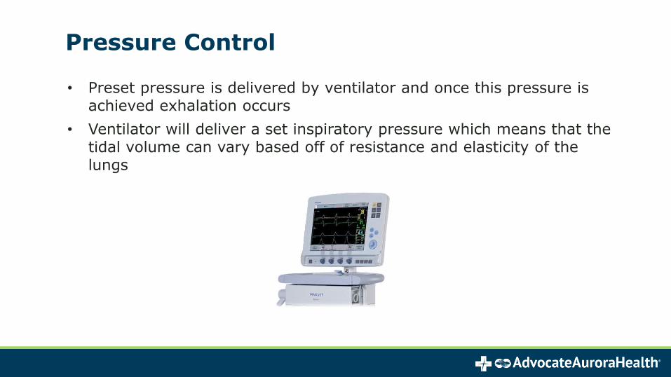
• Preset pressure is delivered by ventilator and once this pressure is achieved exhalation occurs
• Ventilator will deliver a set inspiratory pressure which means that the tidal volume can vary based off of resistance and elasticity of the lungs

Pressure Support Ventilation (PSV)
• Patient receives an increase in airway pressure during inspiration
to augment spontaneous TV
• Patient triggered mode only
• Rate, TV, inspiratory flow rate are determined by the patient
• Cannot be used in AC mode
• Frequently used during weaning to reduce WOB
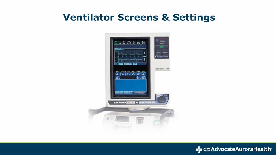
Ventilator Screens & Settings

Ventilator Screens & Settings

Ventilator Alarms
High PIP
(High Pressure)
• Pressure of delivered breath exceeds set parameter
• Impeding delivery of breath, increased pressure is needed
• Secretions
Low PIP
(Low Pressure)
• Pressure of delivered breath does not meet set parameter
• Usually an asynchronous patient
• Flow hungry
• Circuit Disconnection
Circuit Occlusion
• Obstruction in the circuit that prevents a breath from being delivered
• Water in the circuit
• Filters plugged with secretions or medications

Scenario: You walk into a patient's room, vent is alarming High Pressure, what do you do?
Look at the patient
• Coughing? No intervention needed, watch for cough to subside
• Patient biting ETT? Place a bite block
• Secretions? Suction
• Listen for breath sounds bilaterally
• Order a CXR
Look at the machine
• Kinked circuit? Undo kink
• Water accumulation in circuit: Call RT
Nursing Assessments
Monitor• Respiratory rate
• Heart rate
• Blood pressure
• Breath sounds
• Oxygen saturation
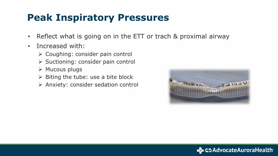
Peak Inspiratory Pressures
• Reflect what is going on in the ETT or trach & proximal airway
• Increased with:
➢ Coughing: consider pain control
➢ Suctioning: consider pain control
➢ Mucous plugs
➢ Biting the tube: use a bite block
➢ Anxiety: consider sedation control
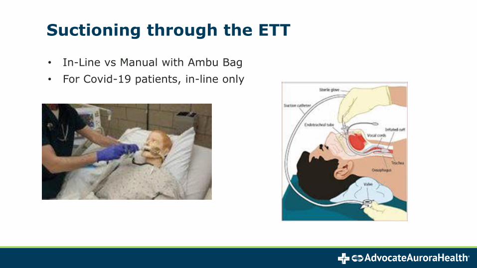
Suctioning through the ETT
• In-Line vs Manual with Ambu Bag
• For Covid-19 patients, in-line only
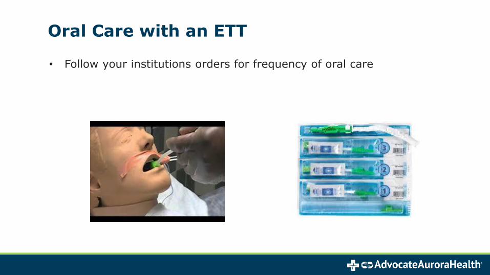
Oral Care with an ETT
• Follow your institutions orders for frequency of oral care
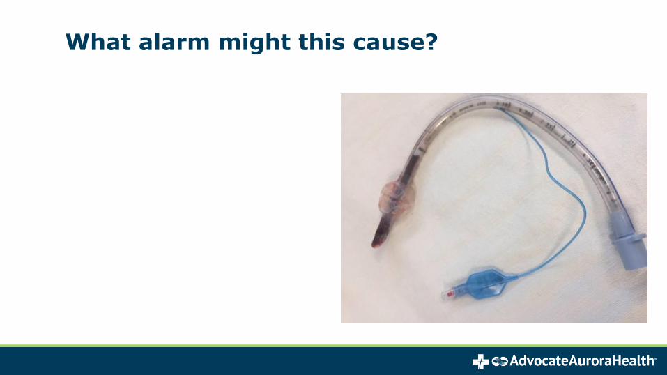
What alarm might this cause?

Oxygenation
• PEEP recruits non-functional alveoli.
• Increasing FiO2 or PEEP will increase oxygenation
➢What is an undesired risk of using too much PEEP?
➢If you are suctioning your patient and the heart rate drops dramatically, what would you do?

Which concerns you the most?
• PaO2 of 68 mmHg
• Ppeak of 46 cmH2O
• AC mode
• Pplat of 42 cmH2O
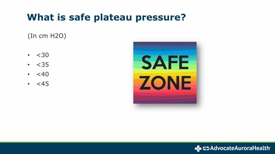
What is safe plateau pressure?
(In cm H2O)
• <30
• <35
• <40
• <45

Plateau Pressure/ Alveolar Pressure
• Pressures measured after a “breath hold”
• Specifically reflects alveolar pressure
• Alveolar pressures consistently greater >30 cm H2O significantly increase the risk of ventilator inducted lung injury via volume trauma.
• RT only measures these pressures
So what do you recommend to reduce this patients plateau pressure?
A. Decrease PEEP
B. Decrease RR
C. Decrease FIO2
D. Decrease TV
Why Reduce Tidal Volume?
• The therapeutic intervention, to date, which has demonstrated a reduction in mortality for ARDS patients, is a low TV strategy
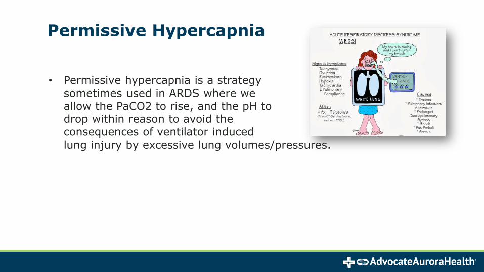
Permissive Hypercapnia
• Permissive hypercapnia is a strategy sometimes used in ARDS where we allow the PaCO2 to rise, and the pH to drop within reason to avoid the consequences of ventilator induced lung injury by excessive lung volumes/pressures.
Spontaneous Breathing Trial (SBT)
✓A goal for most patients on mechanical ventilation is to be weaned off from the ventilator
✓The weaning process is highly dependent on the patient's pathology, but the final common method to ventilator independence always includes at least one trial of spontaneous breathing
Most common CPAP settings for SBT:
PEEP 5, Pressure Support 7, FIO2 40%
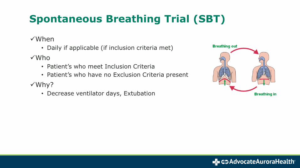
Spontaneous Breathing Trial (SBT)
✓When
• Daily if applicable (if inclusion criteria met)
✓Who
• Patient’s who meet Inclusion Criteria
• Patient’s who have no Exclusion Criteria present
✓Why?
• Decrease ventilator days, Extubation
SBT Inclusion
All ventilated adult patients will have a daily spontaneous breathing trial (SBT) to determine readiness for extubation as outlined in your facility’s protocol unless they meet exclusion criteria.
If a patient is excluded with the assessment, discuss plan of care with physician.
SBT Exclusion Examples (not all inclusive)
• PEEP > 5 cmH2O
• FIO2 > 50%
• Long term ventilator dependence
• Upper airway obstruction/edema
• ICP monitoring
• Neuromuscular blockade
• Open chest
• Severe agitation/withdrawal
• Unstable hemodynamics requiring vasopressors
• Deep sedation to facilitate mechanical ventilation
• On-going seizures

SBT considerations
✓Hold tube feedings 2 hours prior to initiating weaning (consult physician for dextrose containing fluids to replace glucose source if necessary)
✓Sedation Vacation initiated as per unit protocol

SBT Tolerance
✓If the patient remains hemodynamically stable**as indicated** for 30-120 minutes on SBT settings
✓**Heart rate does not change more than 20 beats/minute; systolic blood pressure does not change more than 20 mmHg; diastolic blood pressure < 90, no change in mental status, and SpO2 is ≥ 92%**
SBT tolerance
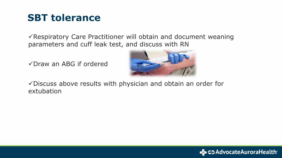
✓Respiratory Care Practitioner will obtain and document weaning parameters and cuff leak test, and discuss with RN
✓Draw an ABG if ordered
✓Discuss above results with physician and obtain an order for extubation
SBT not tolerated
✓If RSBI > 105 and/or hemodynamically unstable, resume ventilator settings prior to SBT
✓Discuss further plan of care with physician
✓Resume tube feedings unless another breathing trial will be completed later in the same day
✓Documentation by RT and RN
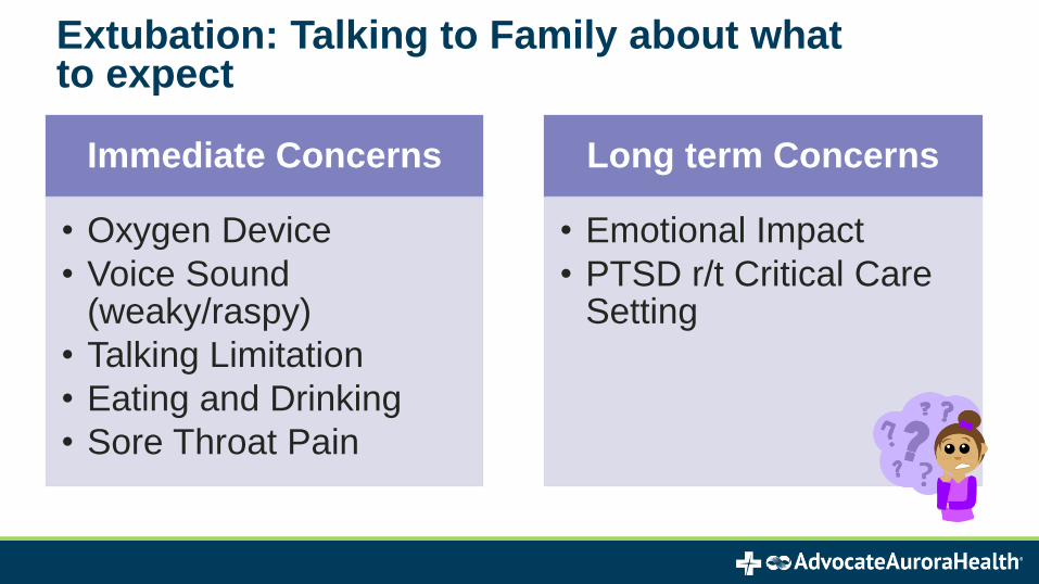
Extubation: Talking to Family about what to expect
Immediate Concerns
• Oxygen Device
• Voice Sound (weaky/raspy)
• Talking Limitation
• Eating and Drinking
• Sore Throat Pain
Long term Concerns
• Emotional Impact
• PTSD r/t Critical Care Setting
Extubation
Courtesy of: www.heathercairncross.com
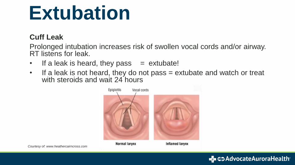
Cuff Leak
Prolonged intubation increases risk of swollen vocal cords and/or airway. RT listens for leak.
• If a leak is heard, they pass = extubate!
• If a leak is not heard, they do not pass = extubate and watch or treat with steroids and wait 24 hours


Extubation
Post Extubation Success
• Continue to monitor for signs of airway swelling for several hours
(breath sounds, voice)
• A bedside swallow exam may be performed an hour after extubation
• Patient could potentially be transferred to the floor as soon as the next
day
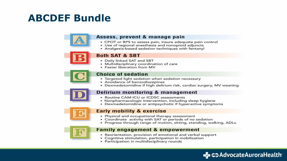
ABCDEF Bundle
Sedation Agents and Richmond Agitation-
Sedation Scale (RASS) Scoring
Alleviation of Pain
✓It is recommended that analgesia be given first before sedation is used in
mechanically ventilated patients in the ICU
✓The Behavioral Pain Scale (BPS) is
one of the most valid and reliable
pain scales for ICU patients who
cannot communicate
Remember to address pain first!

Behavioral Pain Scale (BPS)*Patients who cannot rate their pain on a number scale!
Fentanyl is the First Line Medication for Intubated Patients
✓Fentanyl 25 mcg IV push q 15 min prn
-for BPS score greater than or equal to 5
-or a RASS score of greater than or equal to +1
✓Fentanyl IV infusion: titrate by 25 mcg/hr every 30 min until pain goal is reached (maintain BPS <5)
- Prior to increasing the drip rate bolus with 25 mcg of Fentanyl IV push first!
✓After pain is addressed, the patient may be given a sedative agent if needed
Monitoring the Depth of SedationRASS SCALE
✓The RASS (Richmond Agitation-Sedation Scale) is one of the most valid assessment tools for measuring the quality and depth of sedation
✓A RASS score of -1 to 0 is the accepted range for all sedated patients unless there is a contraindication or a doctor’s order to the contrary
✓Current review shows that there has been over sedation in the past with RASS scores of -2

Richmond Agitation Sedation Scale; RASS
+4 Combative Overly combative, violent, immediate danger to staff
+3 Very Agitated Pulls or removes tube(s) or catheter(s); aggressive
+2 Agitated Frequent non-purposeful movement, fights ventilator
+1 Restless Anxious but movements not aggressive or vigorous
0 Alert and calm
<<<Verbal Stimulation>>>
-1 Drowsy Not fully alert, but movements not aggressive or vigorous
-2 Light sedation Briefly awakens with eye contact to voice (<10 seconds)
-3 Moderate sedation Movement or eye opening to voice (but no eye contact)
<<<Physical Stimulation>>>
-4 Deep Sedation No response to voice, but movement or eye opening to physical stimulation
-5 Unarousable No response to voice or physical stimulation

The Goal
✓The goal is to have patients follow commands without exhibiting agitation
✓ The RASS score should be done & documented every 4 hours and PRN
✓ If after titration the goal is unable to be achieved, discuss plan with the physician.
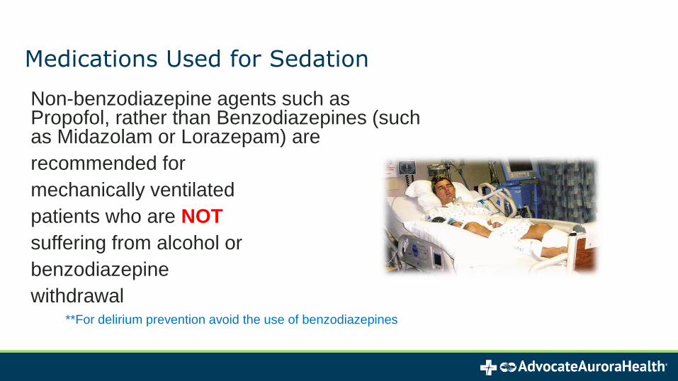
Medications Used for Sedation
Non-benzodiazepine agents such as Propofol, rather than Benzodiazepines (such as Midazolam or Lorazepam) are
recommended for
mechanically ventilated
patients who are NOT
suffering from alcohol or
benzodiazepine
withdrawal **For delirium prevention avoid the use of benzodiazepines

Acknowledgements:
We would like to thank the following contributors for the development of this course content:
• Angeline L. Brooker MSN, RN Clinical Practice Specialist, MICU/MSDU Advocate Christ Medical Center
• Sandra Ziembo MSN, RN, CCRNClinical Development Specialist, Critical Care, Lutheran General Hospital
• Jennifer Spagnolia BSN, MS, RN, CCRNStaff Nurse, MICU/MSDU Advocate Christ Medical Center
• Lukasz Halon BSN, RNStaff Nurse, MICU/MSDU Advocate Christ Medical Center
• Paul Hoffmann MHA, RRTRespiratory Manager Lutheran General Hospital
References
Barr, J., et al. (2013). Clinical practice guidelines for the management of pain, agitation and delirium in adult patients inthe intensive care unit, Critical Care Medicine, 41, 263-306.
Delirium. (n.d.) Medical Dictionary for the Health Professions and Nursing. (2012).
Morton, P. G., & Fontaine, D. K. (2013). Critical care nursing: A holistic approach (10th ed.). Philadelphia, PA: Wolters Kluwer Health/Lippincott Williams & Wilkins.
Siegel, MD., M. D., and R. C. Hyzy, MD. “Mechanical Ventilation of Adults in Acute Respiratory Distress Syndrome.” UpToDate, 2018, www.uptodate.com/contents/mechanical-ventilation-of-adults-in-acute-respiratory-distress-syndrome?sectionName=LOW%2BTIDAL%2BVOLUME%2BVENTILATION.
Wesley, E. E. & Vanderbilt University. (2014). Confusion Assessment Method for the ICU (CAM-ICU), The Complete Training Manual. Revised Edition: March 2014. http://www.icudelirium.org/delirium/monitoring.html
Zarbano, C., & Professional Education Systems, Inc. (2016). CCRN prep class. Lecture, Eau Clair, Wisconsin.

December 2019
Critical Care Academy
Renal System

Objectives
• Describe basic functions and structures of the renal system
• Identify and classify pre-renal and intra-renal injuries
• Discuss associated interventions and management related to
renal injuries
• Identify the importance of electrolyte and fluid balance in renal
injuries
• Identify different types Dialysis and accesses
Renal Physiology
• Rid body of waste.
– Endogenous
– Exogenous
• Maintain homeostasis by regulating fluid, electrolyte,
and acid-base balance.
• Absorption and excretion of molecules and
hormones.

Renal Anatomy

Renal Anatomy
• Nephrons
• Approx. 1 million per kidney
• After age 40, decreases by 10% every 10 years
• Corpuscle
• Glomerulus
• Bowman’s capsule
• Tubules
• Proximal convoluted tubule
• Nephron loop (Loop of Henle)
• Distal convoluted tubule

Assessing Kidney Function
• Serum creatinine
• Creatinine clearance
• Glomerular filtration rate (GFR)
– Estimated
– Best indicator of overall kidney function
– Varies based on age, sex, and ethnicity
• Serum blood urea nitrogen (BUN)
– Normal BUN to Creatinine ratio is 10:1This Photo by Unknown Author is licensed under CC BY

Assessing Kidney Function• Urinalysis
• Urine specific gravity
• Urine osmolality
• Fractional Excretion of NA (FeNA+)
• Other studies
– Ultrasound
– CT/MRI
– Arteriography/Venography
– Biopsy This Photo by Unknown Author is licensed under CC BY-NC-ND
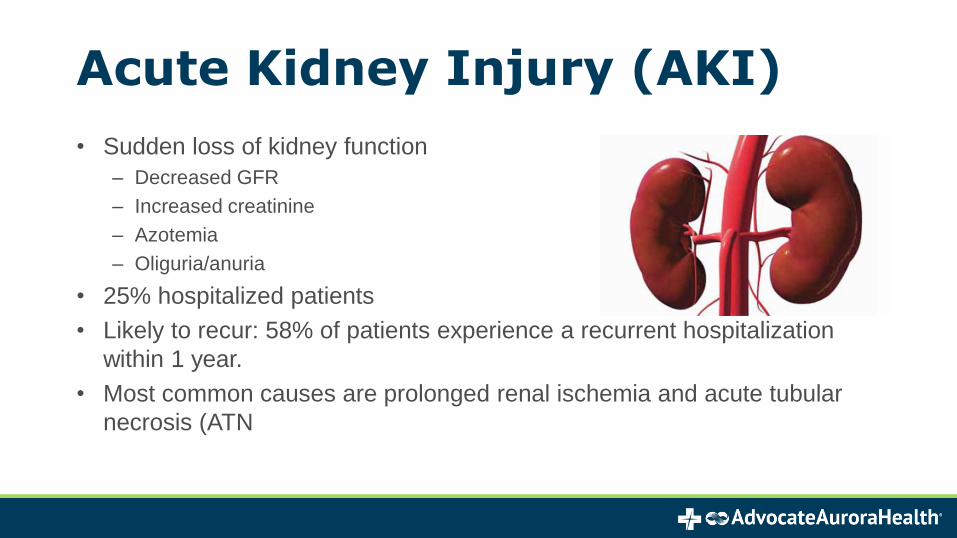
Acute Kidney Injury (AKI)
• Sudden loss of kidney function
– Decreased GFR
– Increased creatinine
– Azotemia
– Oliguria/anuria
• 25% hospitalized patients
• Likely to recur: 58% of patients experience a recurrent hospitalization
within 1 year.
• Most common causes are prolonged renal ischemia and acute tubular
necrosis (ATN
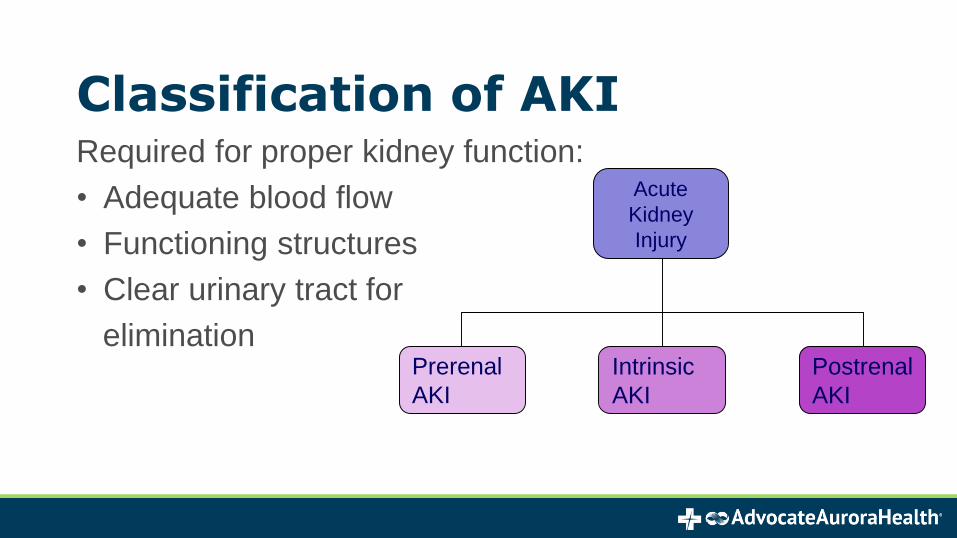
Classification of AKIRequired for proper kidney function:
• Adequate blood flow
• Functioning structures
• Clear urinary tract for
eliminationPrerenal
AKI
Postrenal
AKI
Intrinsic
AKI
Acute
Kidney
Injury
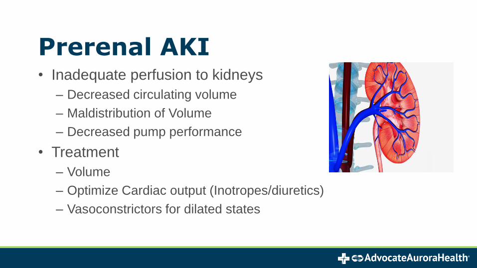
Prerenal AKI• Inadequate perfusion to kidneys
– Decreased circulating volume
– Maldistribution of Volume
– Decreased pump performance
• Treatment
– Volume
– Optimize Cardiac output (Inotropes/diuretics)
– Vasoconstrictors for dilated states

Intrinsic (Intrarenal) AKI• Direct damage to kidneys and their structures
• Etiologies
– Vasoconstriction
– Toxicity
– Vascular disease
– Inflammation
– Infection
– Transplant rejection
• Looks and acts like organ failure

Stages of AKI
• Initiation• Time of injury
• Oliguric• Urine output < 400mL/day• Patients with non-oliguric AKI have the best chance of
recovery.
• Diuretic• Excrete waste but cannot concentrate urine
• Recovery• Filtration increase

Managing complications: Electrolytes• Hyperkalemia
• Metabolic Acidosis
• Anion Gap
• Uremia (High BUN)
• Hematologic/Immunologic
• Watch for arrhythmias!!
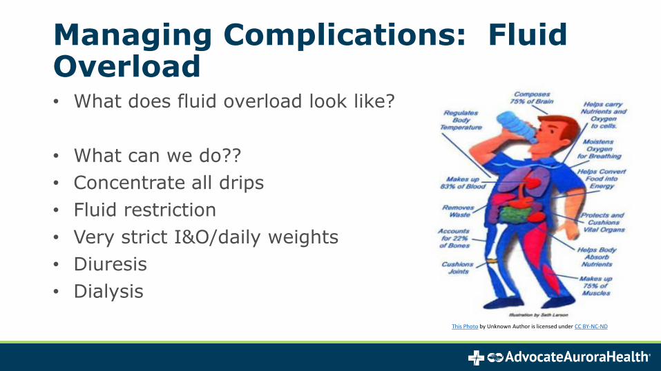
Managing Complications: Fluid Overload• What does fluid overload look like?
• What can we do??
• Concentrate all drips
• Fluid restriction
• Very strict I&O/daily weights
• Diuresis
• Dialysis
This Photo by Unknown Author is licensed under CC BY-NC-ND

Dialysis
• Hemodialysis
• Continuous Renal Replacement Therapy (CRRT)
• Peritoneal Dialysis
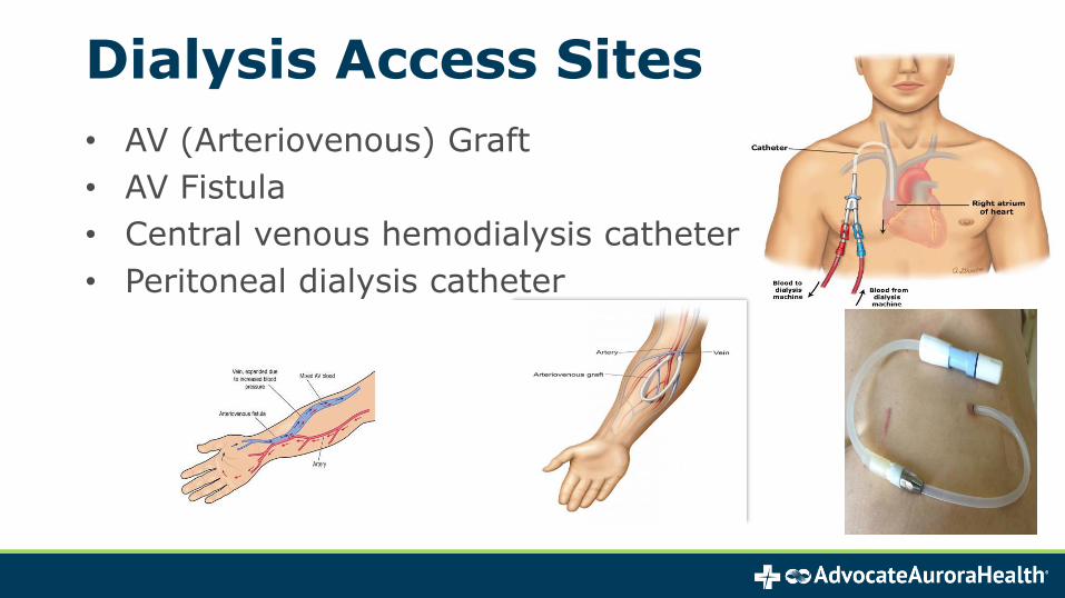
Dialysis Access Sites
• AV (Arteriovenous) Graft
• AV Fistula
• Central venous hemodialysis catheter
• Peritoneal dialysis catheter
References: Hoppe, Kay. (2015). Critical Care Nurse Certification Review Course. Lecture presented at Aurora Academy.
Juarez, P. (2010). Introduction to Critical Care Nursing Course. Lecture presented at Advocate Health Care.
Kidney Disease: Improving Global Outcomes (KDIGO) Acute Kidney Injury Work Group. KDIGO Clinical Practice Guideline for Acute Kidney Injury. Kidney inter., Suppl. 2012; 2: 1–138.
Kidney Disease: Improving Global Outcomes (KDIGO) CKD Work Group. KDIGO 2012 Clinical Practice Guideline for the Evaluation and Management of Chronic Kidney Disease. Kidney inter., Suppl. 2013; 3: 1–150.
Murugan, R., & Kellum, J. A. (2011). Acute kidney injury: what's the prognosis?. Nature reviews. Nephrology, 7(4), 209–217. doi:10.1038/nrneph.2011.13
Siew, D. (2015). The growth of acute kidney injury: a rising tide or just closer attention to detail?. Kidney International. 87 (1): 46–61.
Zarbano, C., & Professional Education Systems, Inc. (2016). CCRN prep class. Lecture, Eau Clair, Wisconsin.

December 2019
Critical Care AcademyCardiovascular System

ObjectivesAt the completion of this program, the learner will be able to:
• Identify critical anatomy, physiology, and pathophysiology of the presented cardiovascular system.
• Discuss significant assessment and diagnostic findings relevant to the cardiac critical care environment.
• Recognize and differentiate clinical presentation of commonly seen cardiac conditions in critical care related to Covid-19 patients.
• Evaluate nursing interventions for cardiovascular conditions.

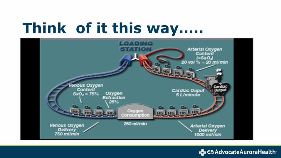
Perfusion, Perfusion, Perfusion• The Cardiovascular system serves to circulate oxygenated blood to
the organs and deoxygenated blood back to the lungs to be reoxygenated.
• In order to function properly, the heart needs:
• the right amount volume to circulate
• low resistance (pressure) from the lungs and proper resistance from the PV system
• good contractility
Think of it this way…..
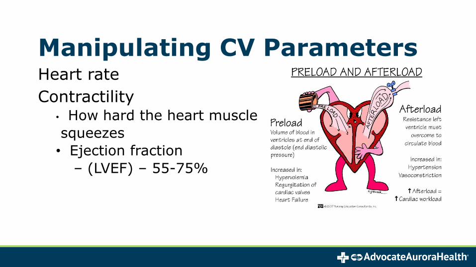
Manipulating CV ParametersHeart rate
Contractility• How hard the heart muscle
squeezes
• Ejection fraction
– (LVEF) – 55-75%

CO = HR x SV (stroke volume)
Determinants of SV • Preload
• Contractility
• Afterload
Preload is Impacted by Starling’s LawStarling’s Law
a.) Increased stretch = increased volume = better ejection
b.) Stretch is within physiological limits
Impacted by volume and pressure
The most important component of SV
Measured by CVP
• (Normal 2-6)
• Increased peep can increase CVP
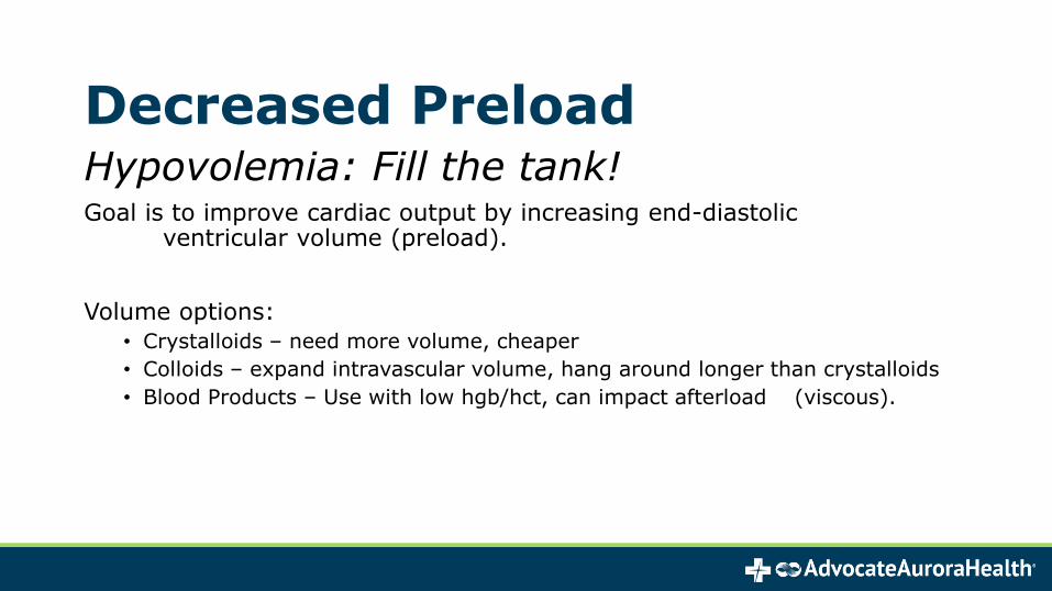
Decreased PreloadHypovolemia: Fill the tank!Goal is to improve cardiac output by increasing end-diastolic
ventricular volume (preload).
Volume options:
• Crystalloids – need more volume, cheaper
• Colloids – expand intravascular volume, hang around longer than crystalloids
• Blood Products – Use with low hgb/hct, can impact afterload (viscous).
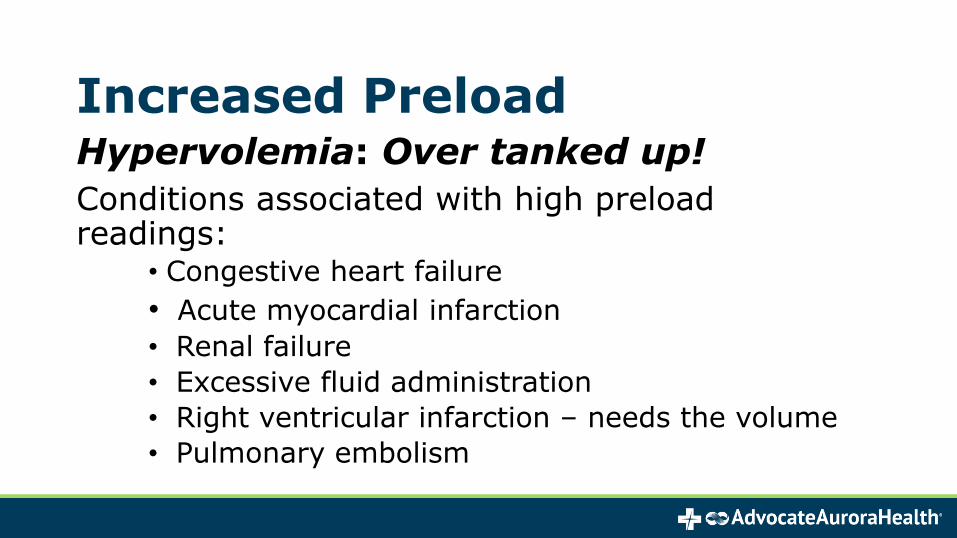
Increased PreloadHypervolemia: Over tanked up!
Conditions associated with high preload readings:
• Congestive heart failure
• Acute myocardial infarction
• Renal failure
• Excessive fluid administration
• Right ventricular infarction – needs the volume
• Pulmonary embolism
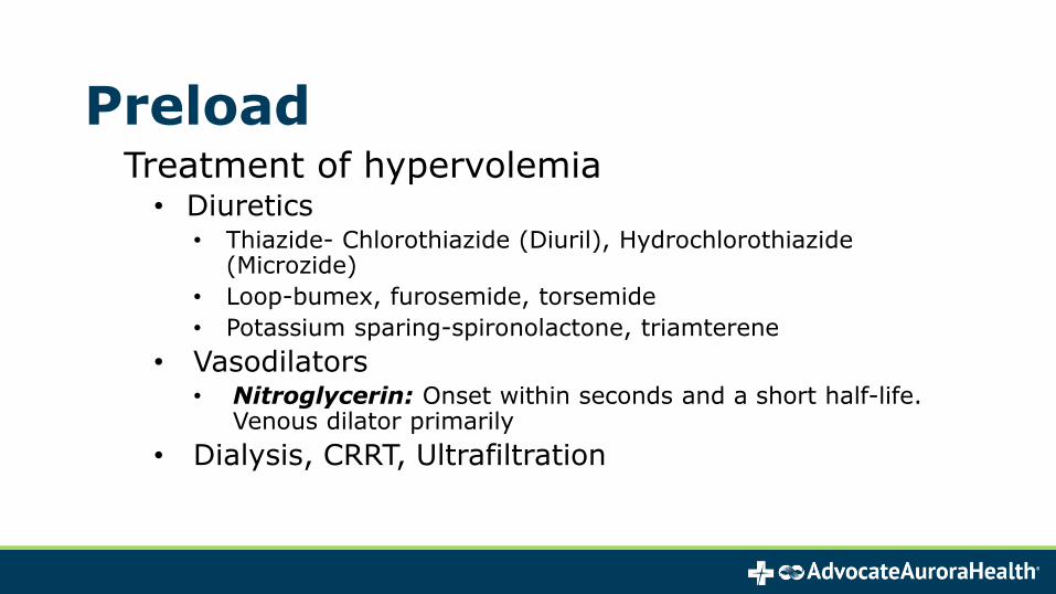
PreloadTreatment of hypervolemia
• Diuretics• Thiazide- Chlorothiazide (Diuril), Hydrochlorothiazide
(Microzide)
• Loop-bumex, furosemide, torsemide
• Potassium sparing-spironolactone, triamterene
• Vasodilators• Nitroglycerin: Onset within seconds and a short half-life.
Venous dilator primarily
• Dialysis, CRRT, Ultrafiltration

Afterload Causes:Inflow
• Increased CVP and PAWP (wedge pressure)
• Reduced cardiac output -volume
Outflow• Valves & vessels
• Right Heart: pulmonic valve & pulmonary artery
• Left Heart: aortic valve & aorta
High systemic vascular resistance (SVR)
Increased BP (usually the cause)
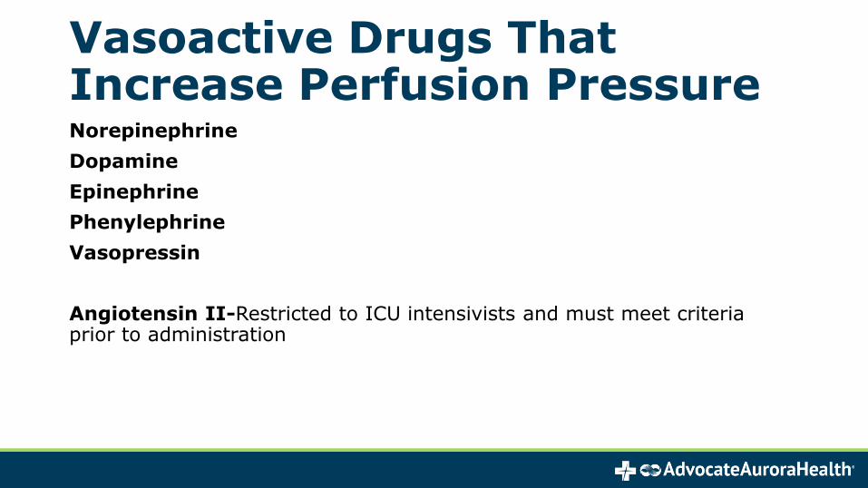
Vasoactive Drugs That Increase Perfusion PressureNorepinephrine
Dopamine
Epinephrine
Phenylephrine
Vasopressin
Angiotensin II-Restricted to ICU intensivists and must meet criteria prior to administration
Increased AfterloadTreatment of increased afterload:
• Vasodilators (Nitroprusside and Nitroglycerin)
• Ace Inhibitors (ACE’s and ARB’s)
• Calcium Channel Blocking agents (Antihypertensive, antiarrhythmic)
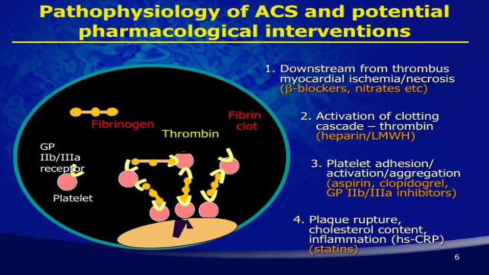

ST Depression vs. ST Elevation

Immediate InterventionsTreatment• Oxygen
• Nitro (0-4 mg SL x 2)
• Prevent platelet activation -Aspirin (325 mg chewable)
• Morphine
• Beta blockers-Lopressor (5 mg IV x3 – If patient BP can tolerate)
• Anticoagulants-Plavix
Work-Up
• 12 lead ECG with in 10 minutes
• CXR
• Labs
Cardiogenic Shock

Pathophysiology of Cardiogenic Shock• Sympathetic NS Stimulation increases HR & contractility
- If protracted, increases myocardial O2 demand
- Can worsen myocardial ischemia
• Kidneys retain fluid due to shock process
• This increases pre-load
• Can lead to acute heart failure & pulmonary edema, AMI

Etiologies of Cardiogenic Shock• Acute MI
• Cardiomyopathy
• Arrythmias
• Post cardiopulmonary bypass
• Structural or mechanical heart defects
• Cardiac compression or obstruction
• Other (acidosis, hypoxemia, electrolyte abnormalities,
negative inotropic drugs)
Signs & Symptoms of Decreased CO • Decreased mental status
• Hypotension
• Pallor
• Cool extremities
• Decreased peripheral pulses
• Oliguria

Optimizing Hemodynamics• May require vasoactive meds to increase myocardial
contractility & decrease afterload
• Adjust with information from Swan
• Often require balloon pump in cardiogenic shock

Signs and Symptoms of Heart Failure
• Fatigue• Weakness• Shortness of Breath
Decreased Forward Flow
Left Heart Failure
Increased Backup of Flow
Pulmonary Congestion
Increased work of the right side of Heart
Right Heart Failure
Back up of Flow
• Increased JVP• Edema• Ascites
• Shortness of Breath
• Orthopnea• Paroxysmal
Pulmonary Edema
* Life-threatening complication of CHF
- Results from LV failure
* Pressure in pulmonary vessels > 18-25 mmHg
* Fluid leaks from pulmonary capillaries into the
interstitial tissue & intra-alveolar spaces
Signs & Symptoms Pulmonary Edema
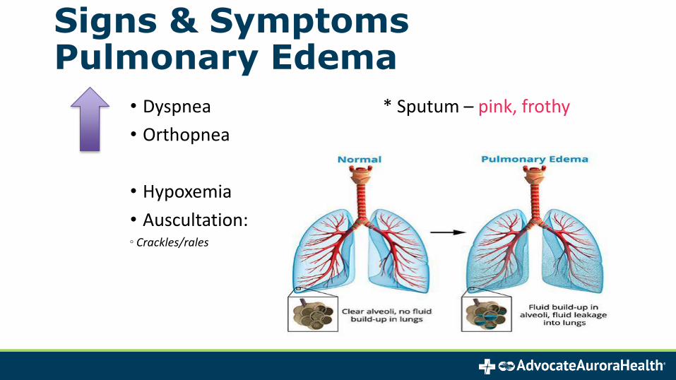
• Dyspnea * Sputum – pink, frothy
• Orthopnea
• Hypoxemia
• Auscultation:◦ Crackles/rales

Treatment of Pulmonary Edema*Treatment is the same as the treatment for left-sided heart failure• Oxygen
- may need Bipap or mechanical ventilation• Decrease preload with loop diuretic (furosemide)
• Morphine or nitroglycerin vasodilate, decreasing afterload and preload, also decreases anxiety
* Dobutamine or milrinone to improve cardiac output
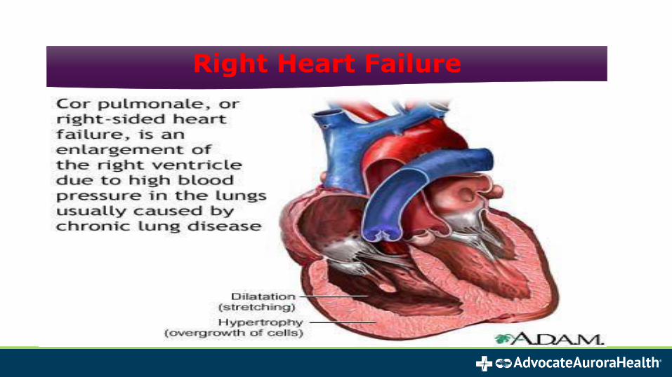
Right Heart Failure

What Causes Right Heart Failure?
• Pulmonary Disease
- COPD, Chronic Bronchitis
• Essential Pulmonary HTN
• Pulmonary Emboli
• Isolated Right Coronary Ischemia
• Left side heart failure can lead to right side failure
Nursing Considerations:• Assess patient for:
- worsening symptoms- responsiveness to medications
• Check lab results – Hypo/Hyperkalemia, Magnesium levels• Daily weights• Accurate intake and output• Positioning – high fowler’s position, legs elevated when sitting• Safety• Educate patient on:
- detection of early signs and symptoms – notify MD for 2-3 lbs/day or 5 lbs/week
- Cardiac diet – 2-3 g Na daily- Fluid Restriction - 2L/day - smoking cessation- compliance with medication regimen - light to moderate --exercise- vaccination
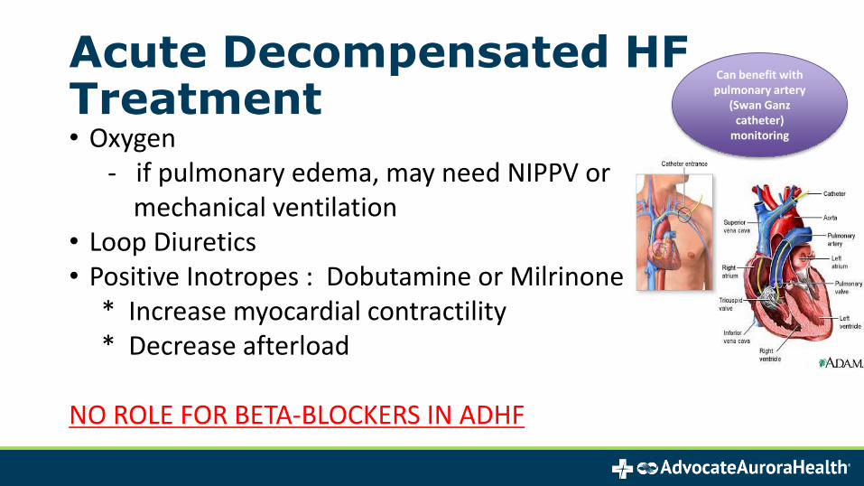
Acute Decompensated HFTreatment• Oxygen
- if pulmonary edema, may need NIPPV ormechanical ventilation
• Loop Diuretics• Positive Inotropes : Dobutamine or Milrinone
* Increase myocardial contractility* Decrease afterload
NO ROLE FOR BETA-BLOCKERS IN ADHF
Can benefit with pulmonary artery
(Swan Ganz catheter)
monitoring

Thank you very much!!!
AcknowledgementsWe would like to thank the following contributors for the development of this course content:
• Sandra Cebrij MSN, RN, CCRN, TNCC
– Critical Care Educator, Advocate Good Samaritan Hospital
• Grace Hamoay BSN, RN, CCRN
– Clinical Practice Specialist-CVTU & CSDU, Advocate Christ Medical Center
• Anne Siwinski BFA, BSN, RN
– Clinical Practice Specialist-ASHU, Advocate Christ Medical Center
• Jennifer Kennedy BSN, RN, CCRN
– Nurse Clinician-CVICU, Aurora St Luke’s Medical Center

References• Burns, S.M. (2014). AACN essentials of critical care nursing
(3rd ed.). New York, NY:McGraw-Hill Companies.
• McCance, K. L., & Huether, S. E. (2010). Pathophysiology: The biologic basis for disease in adults and children. St. Louis, MO: Elsevier.
• Zarbano, C., & Professional Education Systems, Inc. (2016). CCRN prep class. Lecture, Eau Clair, Wisconsin.
Critical Care AcademyMultisystem-- Sepsis
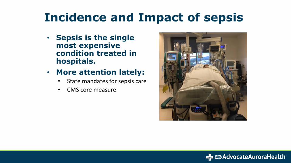
Incidence and Impact of sepsis
• Sepsis is the single most expensive condition treated in hospitals.
• More attention lately:• State mandates for sepsis care
• CMS core measure
SEPSIS 100
What is Sepsis?

SEPSIS 100
Dysregulated host response to an infection, causing life threatening organ dysfunction.
Begins with bacteria somewhere in the body. Hyperinflammatory state occurs.
Organ failure often develops from a variety of processes.
SEPSIS 200
What causes perfusion to the body and capillary beds to become compromised
when a Systemic Inflammatory Response is occurring?

SEPSIS 200
Micro-emboli and vasodilation.

SEPSIS PATHOPHYSIOLOGY

“Except on few occasions, the patient appears to die from the body's response to infection rather than from it.”
Sir William Osler – 1904The Evolution of Modern Medicine

Identify and treat EARLY!No CURE for sepsis! We have to catch and treat the infection early.
Your knowledge of what to watch for (SIRS) (Infection) (Organ dysfunction) is VERY important.
Nurse manual screening for Sepsis is most sensitive
When you see changes in your patient’s condition, perform an assessment including vital signs.

SIRS Criteria
SIRS (2 or more of the following) in the past 6 hours:
• 1. Documented Temp >38.3 or < 36
• 2. Documented HR >90
• 3. Documented RR > 20• 4. WBC >12,000 or WBC < 4,000 or >10% bands
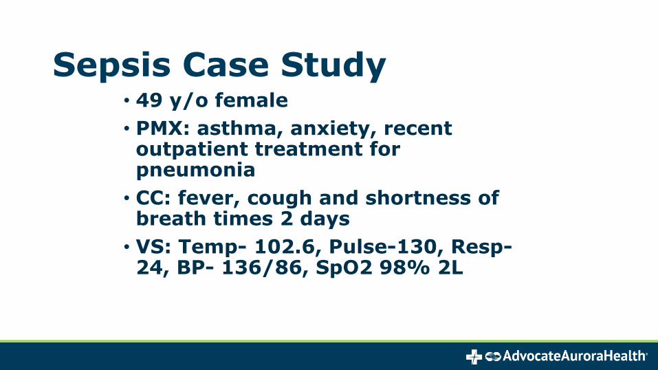
Sepsis Case Study• 49 y/o female
• PMX: asthma, anxiety, recent outpatient treatment for pneumonia
• CC: fever, cough and shortness of breath times 2 days
• VS: Temp- 102.6, Pulse-130, Resp-24, BP- 136/86, SpO2 98% 2L
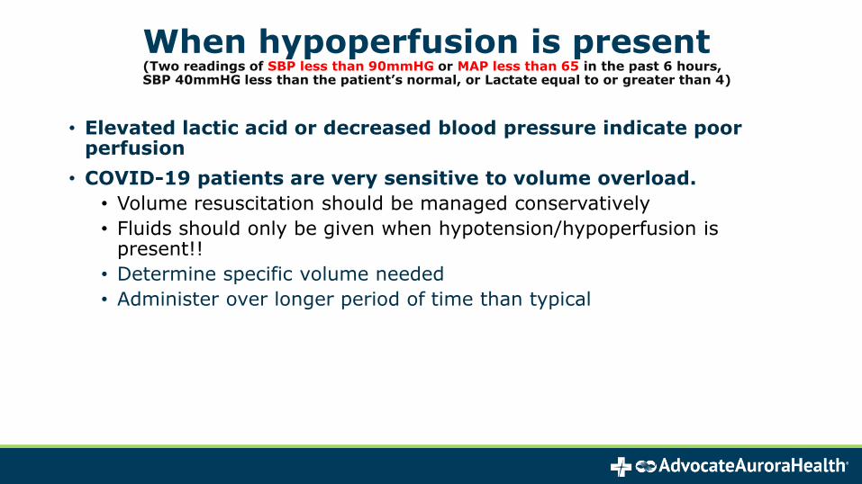
When hypoperfusion is present (Two readings of SBP less than 90mmHG or MAP less than 65 in the past 6 hours, SBP 40mmHG less than the patient’s normal, or Lactate equal to or greater than 4)
• Elevated lactic acid or decreased blood pressure indicate poor perfusion
• COVID-19 patients are very sensitive to volume overload.
• Volume resuscitation should be managed conservatively
• Fluids should only be given when hypotension/hypoperfusion is present!!
• Determine specific volume needed
• Administer over longer period of time than typical
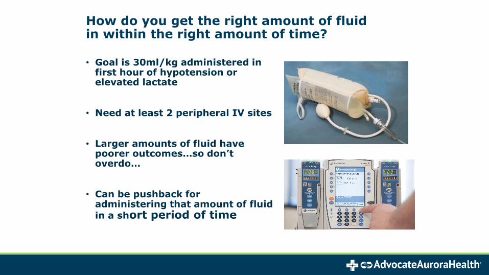
How do you get the right amount of fluid in within the right amount of time?
• Goal is 30ml/kg administered in first hour of hypotension or elevated lactate
• Need at least 2 peripheral IV sites
• Larger amounts of fluid have poorer outcomes…so don’t overdo…
• Can be pushback for administering that amount of fluid
in a short period of time
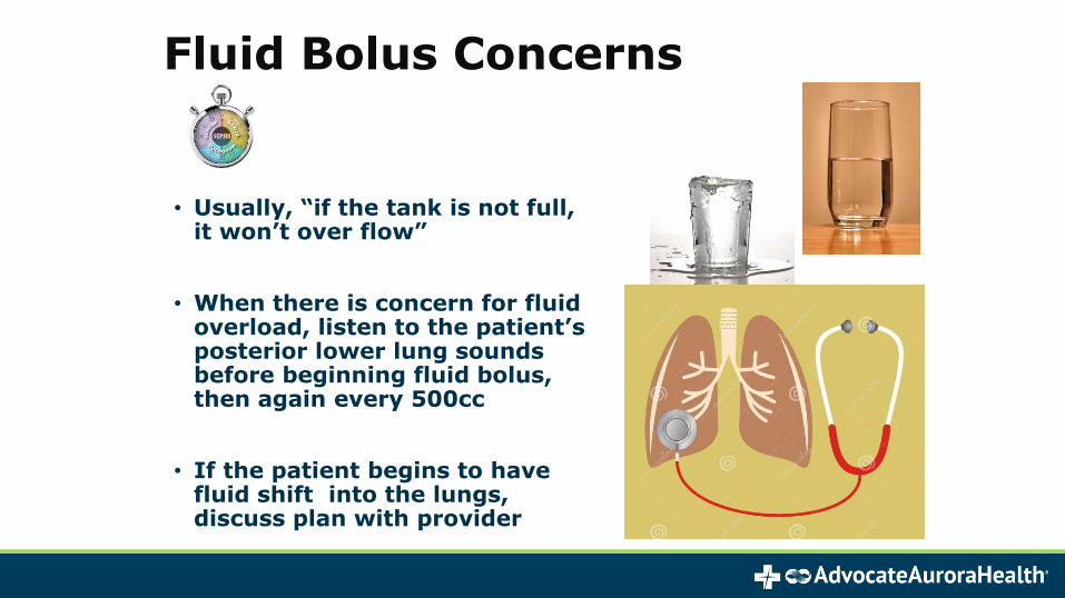
Fluid Bolus Concerns
• Usually, “if the tank is not full, it won’t over flow”
• When there is concern for fluid overload, listen to the patient’s posterior lower lung sounds before beginning fluid bolus, then again every 500cc
• If the patient begins to have fluid shift into the lungs, discuss plan with provider

Septic Shock Treatment Bundle 100
How do we decide this is Septic Shock?
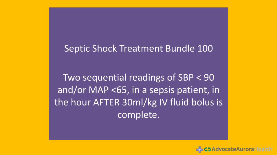
Septic Shock Treatment Bundle 100
Two sequential readings of SBP < 90 and/or MAP <65, in a sepsis patient, in
the hour AFTER 30ml/kg IV fluid bolus is complete.

Septic Shock Treatment Bundle 200
When there is lack of vascular tone, resulting in low perfusion to all organs,
What needs to be started?

Septic Shock Treatment Bundle 200
Vasopressors?

Vasopressors
• NORepinephrine (Levophed) as first line vasopressor
• Additional vasopressor could be EPINEPhrine
• Vasopressin can be added to increase MAP or to help titrate off other pressors
• Dopamine should only be used if there is bradycardia
• Phenylephrine can be used as salvage therapy.
• Angiotension II is a new vasopressor that could be added
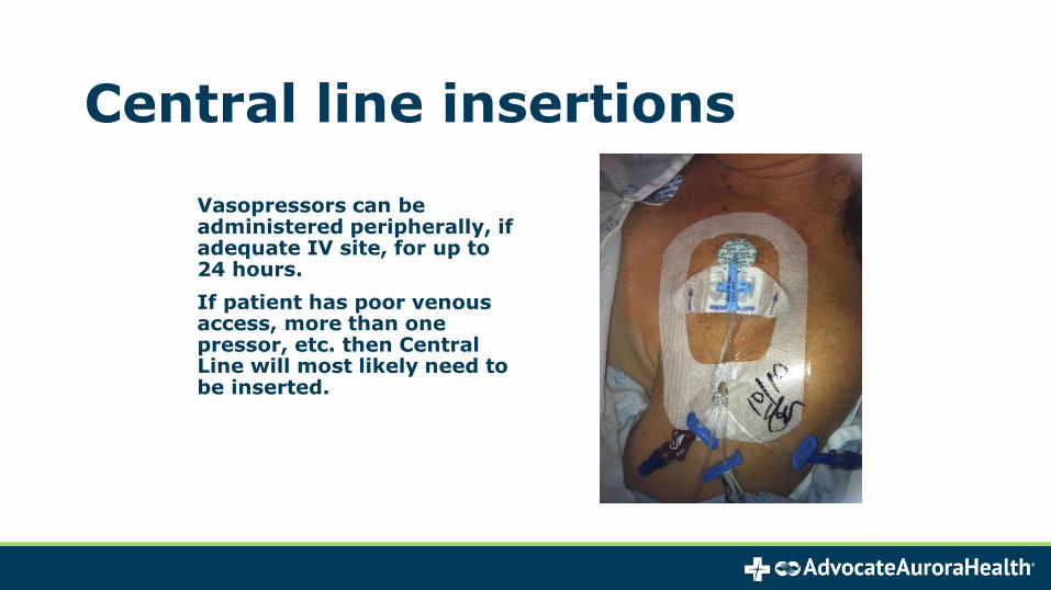
Central line insertions
Vasopressors can be administered peripherally, if adequate IV site, for up to 24 hours.
If patient has poor venous access, more than one pressor, etc. then Central Line will most likely need to be inserted.
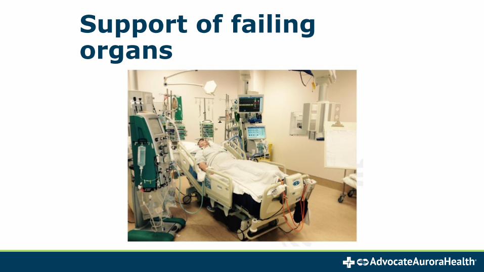
Support of failing organs

Primary Topic of Next Section
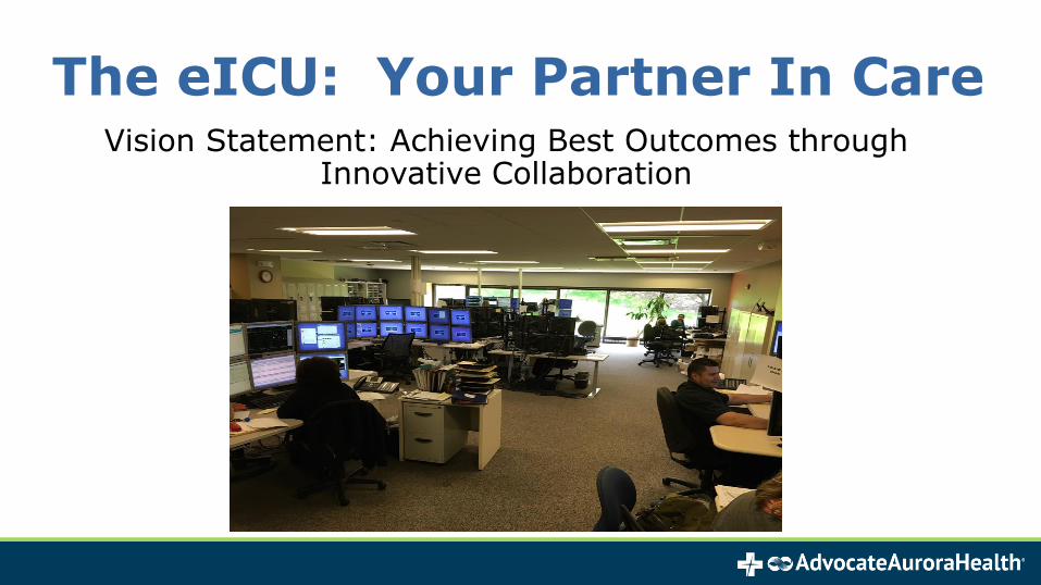
The eICU: Your Partner In CareVision Statement: Achieving Best Outcomes through
Innovative Collaboration
Advocate Aurora eICU
The 2nd largest eICU in the country
• Advocate established in 2003
• Aurora established in 2004
Providing 24/7/365 clinical support to approximately 775 adult critical care beds
Both eICUs:
• Cover beds within the system and outreach sites
• Utilize eMobile carts to monitor patients outside of the ICU
Clinical Coverage:
• All Intensivists are Critical Care Board Certified
Medicine/Pulmonary/Trauma/Surgery/Neuro/Anesthesia/Cardiothoracic
• eRNs are required to have a minimum of 5 years of Critical Care experience (average = 15-20 years)
125-350 patients per eIntensivist
35-45 patients per eRN
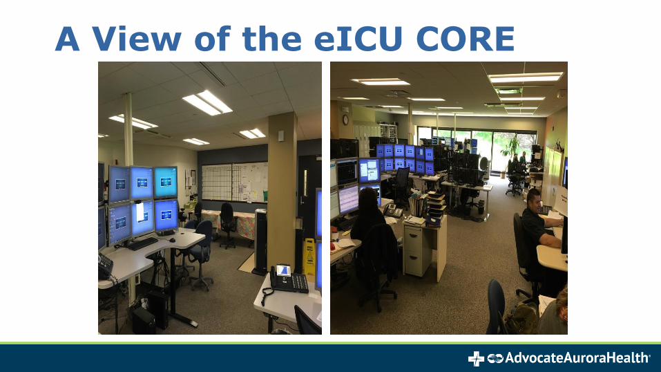
A View of the eICU CORE

eIntensivist Workstation
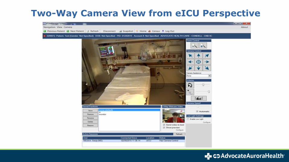
Two-Way Camera View from eICU Perspective
What’s the Benefit of eICU for me at the Bedside?
• Direct observation of patient
• Collaboration and support for staff/pt’s
• Orders/ Assistance
• Monitoring
• Education/ Mentoring
• Quality Improvement

How to Contact eICU
Illinois – 630-575-8340
Wisconsin – 414-747-7730
(Some units have bedside eICU buttons to push for immediate 2-way assistance)

eICU Tools – Used to help support you at the bedside

eICU Report Sheet

eCareManager Alerts

Phone Call Triage Work Flow
Acknowledgements:
We would like to thank the following contributors
for the development of this course content:
• Theresa Brindise, Director eICU
• Cindy Welsh, VP Adult Critical Care and eICU

Advance Directives an Essential Element of Critical Care
Legacy Advocate System Policies/Forms
Policies:
• Advance Directives
• Medical Decision-Making for the Non-decisional Adult Patient
Forms:
• Substitute Decision-Maker Identification form
• Health Care Surrogate Act Physician Certification form for Forgoing Life-Sustaining Treatment
• Close Friend Affidavit, Adult
• Close Friend Affidavit, Minor

Legacy Aurora System Policies
In Wisconsin, Declaration of Incapacity must be signed by two physicians

Why Do We Have to Always Ask?
Patient Autonomy and Safety
▪ Federal Patient Self Determination Act
▪ CMS standards
Compliance with State Laws
▪ Power of Attorney Law and Health Surrogate Law
▪ Turning to the right Substitute Decision-Maker
▪ Documentation requirements
▪ Wisconsin DNR Law

Who is Responsible for Determining Decision-Making Capacity (DMC)?
• Patients are decisional unless assessed otherwise by Attending Physician (or > second year resident)- “Attending” = any physician or consultant involved (may include
eICU physician, on case-by-case basis)
- The law does not allow determination by APN’s/PA’s
Any member of the healthcare team can screen for decisional capacity
• If it is believed the patient may/does lack decisional capacity, it is responsibility of the physician to assess, and provide documentation

Elements of Informed Decision-Making
The Patient:
• Understands the situation requiring decision
• Understands all medically reasonable options to address situation, including option of not selecting an option
• Is aware of risks, burdens and benefits of each option
• Manipulates information logically
• Can communicate a choice
Appelbaum, P., “Assessment of Patients’ Competence to Consent to Treatment”, NEJM, 2007; 357: 1834-60.
Types of Advance Directives in Illinois
• IL Guardianship
• Living Will Act
• Powers of Attorney for Health Care Act
• Health Care Surrogate Act
• Five Wishes
Types of Advance Directives in WisconsinLiving Will: A legal document that states your wishes in the event of a life-altering emergency. Does not name a surrogate decision-maker. This was the first right-to-die legal document.
Health Care Power of Attorney: In addition to information found in a living will, does name a surrogate decision-maker.
Five Wishes: In addition to Health Care Power of Attorney, addresses your beliefs about what is acceptable and unacceptable in terms of quality of life, and your wishes under these circumstances.

Guardianship• Guardianship supersedes all other documents
• Court appointed surrogate/agent
• Able to obtain within short time

Healthcare Power of Attorney –Illinois
Provides the person designated broad powers to make health care decisions for the patient including power to consent, withdraw care, admit and discharge
Healthcare Surrogate/Next-of-Kin
In the absence of a Power of Attorney for Health Care
• The Physician appoints a surrogate decision maker
• The nurse, Social Worker, Case Manager may assist in identification of appropriate individual
Allows decision making powers for medical care
• Not accountable for healthcare finances
• One-time use
Surrogate Decision Maker Qualities
An adult:• Who has decisional capacity, • Available upon reasonable inquiry,• Willing to make decisions on behalf of a patient who
lacks decisional capacity, and• Identified by the attending physician in accordance
with the provisions of the Healthcare Surrogate Act/Next-of-Kin
Illinois and Wisconsin law define the progression of appropriate surrogates
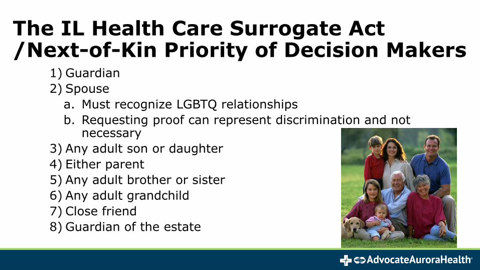
The IL Health Care Surrogate Act/Next-of-Kin Priority of Decision Makers
1) Guardian
2) Spouse
a. Must recognize LGBTQ relationships
b. Requesting proof can represent discrimination and not necessary
3) Any adult son or daughter
4) Either parent
5) Any adult brother or sister
6) Any adult grandchild
7) Close friend
8) Guardian of the estate

This standardized statement contains all legal requirements for physician to
document patient lacks decisional capacity.
Documentation of Lack of Decisional Capacity - Illinois

IL Health Care Surrogate Act:When Treatment Is Withdrawn
Complete “Substitute Decision-Maker Identification”
Removing/Foregoing Life-Sustaining Treatment decisions:
• Complete “Health Care Surrogate Act Physician Certification form for Forgoing Life-Sustaining Treatment”
• Two physicians required:
• An attending and
• Concurring physician (can be a > second-year resident)
•Follow form steps and documentation.
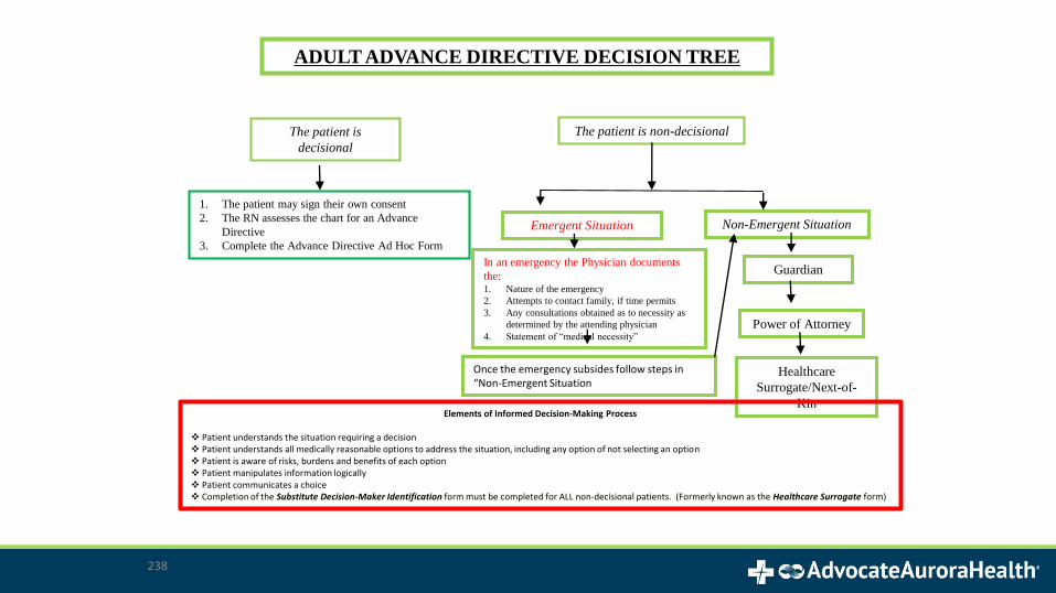
ADULT ADVANCE DIRECTIVE DECISION TREE
The patient is
decisional
1. The patient may sign their own consent
2. The RN assesses the chart for an Advance
Directive
3. Complete the Advance Directive Ad Hoc Form
In an emergency the Physician documents
the:1. Nature of the emergency
2. Attempts to contact family, if time permits
3. Any consultations obtained as to necessity as
determined by the attending physician
4. Statement of “medical necessity”
Emergent Situation Non-Emergent Situation
Guardian
Power of Attorney
Healthcare
Surrogate/Next-of-
Kin
The patient is non-decisional
Once the emergency subsides follow steps in “Non-Emergent Situation
238
Elements of Informed Decision-Making Process
❖ Patient understands the situation requiring a decision❖ Patient understands all medically reasonable options to address the situation, including any option of not selecting an option❖ Patient is aware of risks, burdens and benefits of each option❖ Patient manipulates information logically❖ Patient communicates a choice❖ Completion of the Substitute Decision-Maker Identification form must be completed for ALL non-decisional patients. (Formerly known as the Healthcare Surrogate form)

Emergent Response

Emergency ResponseCOVID-19/Pt Under Investigation (PUI)
SAFETY FIRST
• Limit number of people in emergency area
• Team huddle before to refine process
• PPE monitor at door for donning & doffing
• Limit supplies brought into room
• Keep door cracked open (just enough to hear and pass equip)
• Clean supplies and area after event
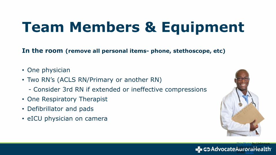
Team Members & Equipment
In the room (remove all personal items- phone, stethoscope, etc)
• One physician
• Two RN’s (ACLS RN/Primary or another RN)
- Consider 3rd RN if extended or ineffective compressions
• One Respiratory Therapist
• Defibrillator and pads
• eICU physician on camera
This Photo by Unknown Author is licensed under CC BY

Team PPE
Impervious gown/ hazmat suit: Intubator, RT
Full face shield: Intubator, RT, RNs,
Protective head cover: Intubator, RT, RN
Shoe covers: Intubator, RT – not felt to be needed
Isolation gown: everyone else in room
N95 mask – EVERYONE
Eye protection (not your glasses) – EVERYONE

Team Members and EquipmentOutside the room:
• Crash cart
• One Pharmacist for medication preparation
• One RN for communications between inside and outside staff
- PPE Monitor and facilitator of resources
- event documentation on paper
• One leader to prevent any additional personnel entry
• One back up RT to obtain resp supplies/ventilator
Social Distance when possible
Airway Management
• Nonrebreather facemask (NRB) at 15 L/min applied over patient’s mouth prior to initiating chest compressions
• NO Bag Valve Mask (ambu bag)
• Place surgical mask over patient's face and nose or over NRB
• Connect directly to ventilator
• Do not disconnect endotracheal/trach tube from ventilator

Chest Compressions
• CHEST COMPRESSION CPR ONLY, apply external compression device
when/where available.
• Bring defibrillator into room
• Stop chest compressions during intubation attempts

Post Emergency Response
• Team huddle for support and refining the process
• Return crash cart
• Return airway equipment (double bagged & labeled)
• Nursing staff with PPE currently on begin cleaning process
This Photoby Unknow
Emergency Response – All Patients
• Limit multiple staff and equipment in the room
• Follow the above guidelines and this will be accomplished
If you reflect back “Shouldn’t we have been doing this for all isolation patients anyway”???
Wouldn't all codes run more smoothly with less
people in the room?
This Photo by Unknown Author is licensed under CC BY
De-escalation of Care
De-escalation of Care
• Limitation of Emergency Treatment (LET)/Do-Not-Resuscitate (DNR)
• Provider Orders for Life Sustaining Treatment (POLST) (Illinois Only)
• Palliative Care/Hospice
• Declaration of Brain Death
• Organ Donation

HospiceHospice care includes the 5 key aspects of quality
▪Expert pain and symptom management,
▪Allowing the patient a natural death,
▪ Providing psychosocial and spiritual support to patients and families,
▪Grief and bereavement support (before and after death), and
▪Support of staff
Hospice Benefits• Family is able to visit (one visitor)
• Expert emotional, spiritual, social and physical care
• Improved symptom management
• Grief and bereavement support
• Appropriate use of medical resources
• Improved patient/family satisfaction
• Prevention of moral distress for ICU staff
Distress occurs when one knows the ethically correct thing to do, but is prevented from acting on that perceived obligation

We Are A Part of a Large System….
What you may not know:
▪ Participation in the organ donation process is mandated by CMS
▪ Uniform Anatomical Gift Act
▪ Department of Health and Human Services (HRSA)
▪ UNOS
▪ Regional and OPO areas of service
Organ Donation and COVID-19
• There is no change in the process; organ donations are still occurring
• Calls to Organ Procurement Centers must still occur
- Calls take time so your assistance is needed

❖ Organ donation
▪ Ventilated patients
▪ Patient referred when physician determines imminent death
• Brain death—total, irreversible loss of function of entire brain
and brain stem
• Cardiac death (DCD)—cessation of both breathing and cardiac
function (after family/physician decision to withdraw life-
sustaining therapies)
❖ Tissue donation
▪ Ventilated and non-ventilated patients
▪ Patient referred before or as soon as possible after death

When is organ donation an opportunity?
❖ For any ventilated patient in a critical care setting▪ If there is a declaration of brain death (DBD)
▪ If there is a decision to withdraw life-sustaining therapies, with the expectation that
death will occur (DCD)
❖ Hospital staff refer these patients to Gift of Hope
▪ When they exhibit any one of the following:
• Fixed and dilated pupils - No response to painful stimuli
• No corneal reflex - No spontaneous respirations
• No gag or cough
▪ Or prior to discussions with the family regarding:
• Change of code status to DNR
• Discontinuation of life-sustaining therapies

The benefits of donation

Take Care
Stress Symptoms
These symptoms or reactions are NORMAL reactions by
NORMAL people to ABNORMAL events!
Physical
Cognitive
Emotional
Care of the Nurse• Be alert to the team behaviors and interactions
• Exercise within 24 hours
• Talk to each other
• Don’t shut off the people that care about you
• Get some extra rest
• Maintain normal routine as much as possible
• Eat healthy food
• Request help if you think you need it!

Covid Specific Content
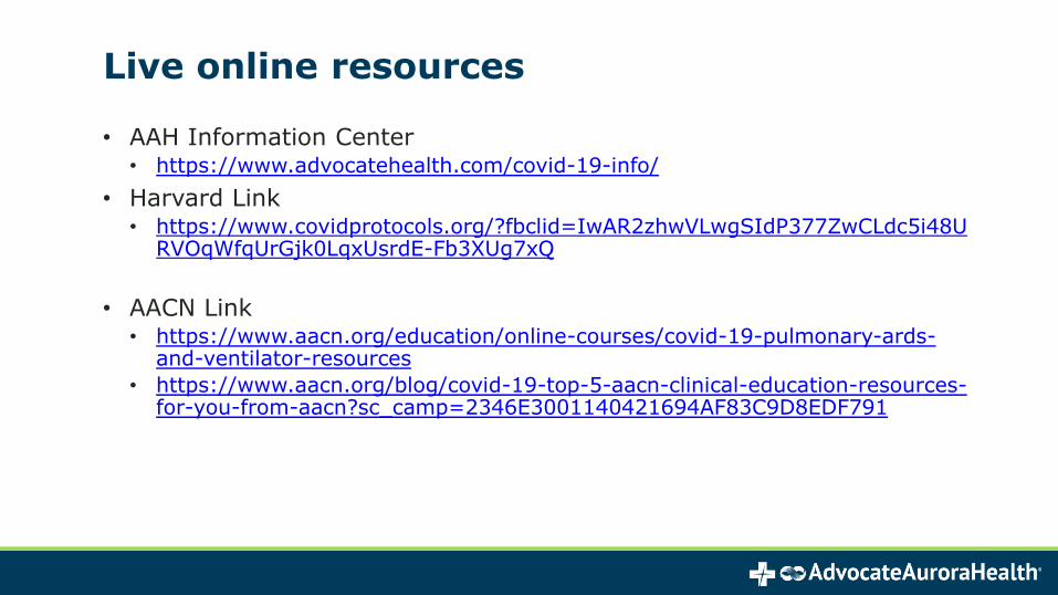
Live online resources
• AAH Information Center• https://www.advocatehealth.com/covid-19-info/
• Harvard Link• https://www.covidprotocols.org/?fbclid=IwAR2zhwVLwgSIdP377ZwCLdc5i48U
RVOqWfqUrGjk0LqxUsrdE-Fb3XUg7xQ
• AACN Link• https://www.aacn.org/education/online-courses/covid-19-pulmonary-ards-
and-ventilator-resources
• https://www.aacn.org/blog/covid-19-top-5-aacn-clinical-education-resources-for-you-from-aacn?sc_camp=2346E3001140421694AF83C9D8EDF791