critical care - medpage today
TRANSCRIPT

This Provisional PDF corresponds to the article as it appeared upon acceptance. Copyedited andfully formatted PDF and full text (HTML) versions will be made available soon.
Discovery and validation of cell cycle arrest biomarkers in human acute kidneyinjury
Critical Care 2013, 17:R25 doi:10.1186/cc12503
Kianoush Kashani ([email protected])Ali Al-Khafaji ([email protected])
Thomas Ardiles ([email protected])Antonio Artigas ([email protected])
Sean M Bagshaw ([email protected])Max Bell ([email protected])Azra Bihorac ([email protected])
Robert Birkhahn ([email protected])Cynthia M Cely ([email protected])
Lakhmir S Chawla ([email protected])Danielle L Davison ([email protected])
Thorsten Feldkamp ([email protected])Lui G Forni ([email protected])
Michelle NG Gong ([email protected])Kyle J Gunnerson ([email protected])
Michael Haase ([email protected])James Hackett ([email protected])
Patrick M Honore ([email protected])Eric AJ Hoste ([email protected])
Olivier Joannes-Boyau ([email protected])Michael Joannidis ([email protected])
Patrick Kim ([email protected])Jay L Koyner ([email protected])
Daniel T Laskowitz ([email protected])Matthew E Lissauer ([email protected])
Gernot Marx ([email protected])Peter A McCullough ([email protected])
Scott Mullaney ([email protected])Marlies Ostermann ([email protected])
Thomas Rimmele ([email protected])Nathan I Shapiro ([email protected])
Andrew D Shaw ([email protected])Jing Shi ([email protected])
Amy M Sprague ([email protected])Jean-Louis Vincent ([email protected])
Christophe Vinsonneau ([email protected])Ludwig Wagner ([email protected])Michael G Walker ([email protected])
R Gentry Wilkerson ([email protected])
Critical Care
© 2013 Kashani et al.This is an open access article distributed under the terms of the Creative Commons Attribution License (http://creativecommons.org/licenses/by/2.0),
which permits unrestricted use, distribution, and reproduction in any medium, provided the original work is properly cited.

Kai Zacharowski ([email protected])John A Kellum ([email protected])
ISSN 1364-8535
Article type Research
Submission date 29 November 2012
Acceptance date 16 January 2013
Publication date 6 February 2013
Article URL http://ccforum.com/content/17/1/R25
This peer-reviewed article can be downloaded, printed and distributed freely for any purposes (seecopyright notice below).
Articles in Critical Care are listed in PubMed and archived at PubMed Central.
For information about publishing your research in Critical Care go to
http://ccforum.com/authors/instructions/
Critical Care
© 2013 Kashani et al.This is an open access article distributed under the terms of the Creative Commons Attribution License (http://creativecommons.org/licenses/by/2.0),
which permits unrestricted use, distribution, and reproduction in any medium, provided the original work is properly cited.

Discovery and validation of cell cycle arrest biomarkers in human acute kidney injury
Kianoush Kashani1, Ali Al-Khafaji 2, Thomas Ardiles3, Antonio Artigas4, Sean M Bagshaw5,
Max Bell6, Azra Bihorac7, Robert Birkhahn8, Cynthia M Cely9, Lakhmir S Chawla10,
Danielle L Davison10, Thorsten Feldkamp11, Lui G Forni12, Michelle Ng Gong13, Kyle J
Gunnerson14, Michael Haase15, James Hackett16, Patrick M Honore17, Eric AJ Hoste18, Olivier
Joannes-Boyau19, Michael Joannidis20, Patrick Kim21, Jay L Koyner22, Daniel T Laskowitz23,
Matthew E Lissauer24, Gernot Marx25, Peter A McCullough26, Scott Mullaney27, Marlies
Ostermann28, Thomas Rimmelé29, Nathan I Shapiro30, Andrew D Shaw31, Jing Shi32, Amy M
Sprague33, Jean-Louis Vincent34, Christophe Vinsonneau35, Ludwig Wagner36, Michael G
Walker32, R Gentry Wilkerson37, Kai Zacharowski38 and John A Kellum39*
1Division of Pulmonary and Critical Care Medicine, Mayo Clinic, 200 First Street SW,
Rochester, MN 55905, USA.
2Department of Critical Care Medicine, University of Pittsburgh School of Medicine, 3550
Terrace Street, Pittsburgh, PA 15213, USA.
3Department of Critical Care, Maricopa Integrated Health System, 2601 E Roosevelt Street,
Phoenix, AZ 85008, USA.
4Critical Care Center, Sabadell Hospital, CIBER Enfermedades Respiratorias, Autonomous
University of Barcelona, Parc Tauli s/n, Sabadell, Barcelona 8208, Spain.
5Division of Critical Care Medicine, Faculty of Medicine and Dentistry, University of
Alberta, 3C1.12 Walter C. Mackenzie Centre, 8440 112 Street NW, Edmonton, Alberta T6G

2B7, Canada.
6Department of Anesthesia and Intensive Care Medicine, Karolinska University Hospital,
Karolinskavagen, Solna, Stockholm SE-171 76, Sweden.
7Department of Anesthesiology, University of Florida, 1660 SW Archer Road, Gainesville,
FL 32611, USA.
8Department of Emergency Medicine, New York Methodist Hospital, 506 6th Street,
Brooklyn, NY 11215, USA.
9Bruce W. Carter Department of Veterans Affairs Medical Center, 1201 NW 16th Street,
Miami, FL 33125, USA.
10Department of Anesthesiology and Critical Care Medicine, George Washington University
Medical Center, 900 23rd Street NW, Washington, DC 20037, USA.
11Department of Nephrology, University Hospital Essen, University Duisburg-Essen,
Hufelandstrasse 55, Essen, 45147, Germany.
12Intensive Care Medicine, Western Sussex Hospitals Trust, Lyndhurst Road, Worthing, West
Sussex, BN11 2DH, UK.
13Department of Medicine, Montefiore Medical Center, Albert Einstein College of Medicine,
111 East 210th Street, Bronx, NY 10467, USA.
14Departments of Anesthesiology and Emergency Medicine, Virginia Commonwealth
University Medical Center, 1200 East Broad Street, Richmond, VA 23298, USA.
15Department of Nephrology, Otto-von-Guericke-Universitat Magdeburg, Leipziger Strasse
44, Magdeburg, 39120, Germany.

16Hackett & Associates, Inc., 14419 Rancho Del Prado Trail, San Diego, CA 92127, USA.
17ICU Department, Universitair Ziekenhuis Brussel (UZB), Vrije Universiteit Brussel (VUB),
Laarbeeklaan 101, Brussels 1090, Belgium.
18Intensive Care Unit, Ghent University Hospital, De Pintelaan 185, Ghent, 9000, Belgium.
19Anaesthesiology and Critical Care Department 2, University Hospital of Bordeaux, 1
Avenue De Magellon, Pessac, 33600, France.
20Department of Internal Medicine, ICU, Medical University Innsbruck, Anichstrasse 35,
Innsbruck, A-6020, Austria.
21Traumatology, Surgical Critical Care and Emergency Surgery, Hospital of the University of
Pennsylvania, 3400 Spruce Street, Philadelphia, PA 19104, USA.
22Department of Medicine, University of Chicago, 6030 South Ellis Avenue, Chicago, IL
60637, USA.
23Department of Medicine, Duke University Medical Center, 2301 Erwin Road, Durham, NC
27710, USA.
24Department of Surgery, University of Maryland School of Medicine, 22 South Greene
Street, Baltimore, MD 21201, USA.
25Department of Intensive Care, Universitätsklinikum der RWTH Aachen, Pauwelsstrasse 30,
Aachen, 52074, Germany.
26Department of Medicine, St John Providence Health System, Providence Hospitals and
Medical Centers, Providence Park Heart Institute, 47601 Grand River Avenue, Novi, MI
48374, USA.

27Department of Medicine, University of California San Diego, 200 West Arbor Drive, San
Diego, CA 92103, USA.
28Department of Critical Care, King’s College London, Guy’s and St Thomas’ Hospital,
Westminster Bridge Road, London, SE1 7EH, UK.
29Service D’Anesthésie Réanimation, Edouard Herriot Hospital, Hospices civils de Lyon, 5
Place d’Arsonval, Lyon, 69003, France.
30Department of Emergency Medicine, Beth Israel Deaconess Medical Center, 1 Deaconess
Road, Boston, MA 2215, USA.
31Department of Anesthesia, Duke University Medical Center/Durham Veterans Affairs
Medical Center, 508 Fulton Street, Durham, NC 27705, USA.
32Walker Biosciences, 6321 Allston Street, Carlsbad, CA 92009, USA.
33Department of Medicine, Joseph M. Still Research Foundation, 3675 J. Dewey Gray Circle,
Augusta, GA 30909, USA.
34Department of Intensive Care, Erasme University Hospital, Route De Lennik 808, Brussels,
1070, Belgium.
35Department of Intensive Care, Hospital Marc Jacquet, 2 Rue Freteau De Peny, Melun,
77011, France.
36Department of Internal Medicine, Medical University of Vienna, Spitalgasse 23, Vienna
1090, Austria.
37Department of Emergency Medicine, Tampa General Hospital, 1 Davis Boulevard, Tampa,
FL 33606, USA.

38Clinic of Anesthesiology, Intensive Care Medicine and Pain Therapy, University Hospital
Frankfurt, Theodor-Stern-Kai 7, Frankfurt am Main, 60590, Germany.
39Department of Critical Care Medicine, University of Pittsburgh, School of Medicine, 3550
Terrace Street, Pittsburgh, PA 15213, USA.
*Corresponding author: John A Kellum, [email protected]
{1st-level heading}Abstract
Introduction: Acute kidney injury (AKI) can evolve quickly and clinical measures of
function often fail to detect AKI at a time when interventions are likely to provide benefit.
Identifying early markers of kidney damage has been difficult due to the complex nature of
human AKI, in which multiple etiologies exist. The objective of this study was to identify and
validate novel biomarkers of AKI.
Methods: We performed two multicenter observational studies in critically ill patients at risk
for AKI - discovery and validation. The top two markers from discovery were validated in a
second study (Sapphire) and compared to a number of previously described biomarkers. In
the discovery phase, we enrolled 522 adults in three distinct cohorts including patients with
sepsis, shock, major surgery, and trauma and examined over 300 markers. In the Sapphire
validation study, we enrolled 744 adult subjects with critical illness and without evidence of
AKI at enrollment; the final analysis cohort was a heterogeneous sample of 728 critically ill
patients. The primary endpoint was moderate to severe AKI (KDIGO stage 2 to 3) within 12
hours of sample collection.
Results: Moderate to severe AKI occurred in 14% of Sapphire subjects. The two top
biomarkers from discovery were validated. Urine insulin-like growth factor-binding protein 7
(IGFBP7) and tissue inhibitor of metalloproteinases-2 (TIMP-2), both inducers of G1 cell
cycle arrest, a key mechanism implicated in AKI, together demonstrated an AUC of 0.80
(0.76 and 0.79 alone). Urine [TIMP-2]•[IGFBP7] was significantly superior to all previously
described markers of AKI (P <0.002), none of which achieved an AUC >0.72. Furthermore,
[TIMP-2]•[IGFBP7] significantly improved risk stratification when added to a nine-variable
clinical model when analyzed using Cox proportional hazards model, generalized estimating
equation, integrated discrimination improvement or net reclassification improvement. Finally,

in sensitivity analyses [TIMP-2]•[IGFBP7] remained significant and superior to all other
markers regardless of changes in reference creatinine method.
Conclusions: Two novel markers for AKI have been identified and validated in independent
multicenter cohorts. Both markers are superior to existing markers, provide additional
information over clinical variables and add mechanistic insight into AKI.
Trial registration: ClinicalTrials.gov number NCT01209169.
Journal: Critical Care
Article Type: Research
Received: 29 November 2012
Revised: 12 January 2013
Accepted: 16 January 2013
Published: 6 February 2013
© 2013 Kashani et al.; licensee BioMed Central Ltd. This is an open access article distributed
under the terms of the Creative Commons Attribution License
(http://creativecommons.org/licenses/by/2.0), which permits unrestricted use, distribution,
and reproduction in any medium, provided the original work is properly cited.
{1st-level heading}Introduction
Acute kidney injury (AKI) is a vexing clinical problem, in part, because it is difficult to
identify before there is loss of organ function, which may then become irreversible [1].
Patients developing AKI have a markedly increased risk of death prior to hospital discharge
[2,3] and survivors also appear to be at significant short- and long-term risk for complications
[4,5]. Available therapies are mainly predicated on supportive measures and the removal of
nephrotoxic agents [6]. Thus, risk assessment for AKI is recommended by clinical practice
guidelines [6]. However, risk stratification remains very difficult, mainly due to limited
sensitivity and specificity of the available diagnostic tests for AKI [7]. Prior efforts at
identifying biomarkers for AKI have been hampered by the heterogeneous nature of the
condition. Many different etiologies for AKI have been reported (for example sepsis,
nephrotoxins, ischemia), and in any given patient the cause is typically thought to be
multifactorial [8]. Here we report the results of a prospective, multicenter investigation in
which two novel biomarkers for AKI were identified in a discovery cohort of critically ill
adult patients and subsequently validated using a clinical assay and compared to existing
markers of AKI in an independent validation cohort of heterogeneous critically ill patients.
{1st-level heading}Materials and methods
{2nd-level heading}Subjects
We conducted a two-stage program in which we first collected blood and urine samples from
three distinct cohorts (Discovery study) to identify novel protein biomarkers for AKI. These
single-center studies were used to identify the best biomarkers among 340 proteins, including
novel candidates and previously described biomarkers such as kidney injury marker-1 (KIM-
1), neutrophil gelatinase-associated lipocalin (NGAL), cystatin-C, interleukin-18 (IL-18), pi-
glutathione S-transferase (pi-GST), and liver fatty acid-binding protein (L-FABP). Data from

all three cohorts were pooled for analysis. A fourth cohort (Sapphire study) was assembled
from 35 clinical sites in North America and Europe and used to validate the performance of
the best biomarkers (urine tissue inhibitor of metalloproteinases-2 (TIMP-2) and insulin-like
growth factor-binding protein 7 (IGFBP7)) from the Discovery study (Figure 1). The
Sapphire study was approved by the Western Institutional Review Board (Olympia,
Washington, USA). In addition, the study protocols were approved by investigational review
boards/ethics committees as required, by each participating institution. All subjects (or
authorized representatives) provided written informed consent.
The Sapphire study was designed and reported according to the STROBE guidelines
[9]. As shown in Figure 1, the Discovery study enrolled patients who were admitted to an
intensive care unit (any type), were at least 18 years of age and typically had at least one
recognized risk factor for AKI. The Sapphire (validation) study enrolled critically ill patients
who were at least 21 years of age, admitted to the intensive care unit within 24 hours of
enrollment, expected to remain in the ICU with a urinary catheter for at least 48 hours and
were critically ill (respiratory or cardiovascular dysfunction). Patients with known existing
moderate or severe AKI (KDIGO [6] stage 2 or 3) were excluded. Sample size for the
Sapphire study was based on the results of the Discovery study and is explained in detail in
Additional file 1.
{2nd-level heading}Sample and data collection
Paired urine and blood samples were collected at enrollment and up to 18 hours later by
standard methods and centrifuged. Plasma (EDTA), serum and urine supernatants were
frozen, shipped on dry ice, stored at ≤-70°C and thawed immediately prior to analysis.
Clinical data including patient demographics, prior health history, serum creatinine, and
hourly urine output as available in the hospital record were collected. Samples were analyzed

at Astute Medical by technicians blinded to clinical data. Password-protected, anonymized
clinical data collected with electronic case-report forms resided on servers at independent
sites (Acumen Healthcare Solutions, Plymouth, MN, USA and Medidata Solutions, New
York, NY, USA for Discovery and Sapphire studies, respectively).
{2nd-level heading}Clinical endpoints
AKI status was classified using the RIFLE [10] or AKIN criteria [11] together as described in
the recent KDIGO international guideline [6] based on the serum creatinine (sCR) and urine
output (UO) available in the hospital record. The primary endpoint for the Sapphire study
was the development of moderate or severe AKI (KDIGO stage 2 or 3) within 12 hours of
sample collection. The reference values for serum creatinine were obtained as follows: if at
least five values were available the median of all values available from six months to six days
prior to enrollment was used. Otherwise, the lowest value in the five days prior to enrollment
was used. If no pre-enrollment creatinine was available, the creatinine value at the time of
enrollment was used (see Additional file 1 for full details). We performed sensitivity analyses
by repeating the primary analysis using several different methods of reference creatinine
assignment. Details including the sensitivity analyses are given in Additional file 1.
Secondary endpoints for the purpose of characterizing the patient population included renal
replacement therapy at any time during hospitalization, survival and major adverse kidney
events. We defined major adverse kidney events (MAKE30) as the composite of death, use of
renal replacement therapy, or persistence of renal dysfunction (defined by serum creatinine
≥200% of reference) at hospital discharge truncated at 30 days [12].
{2nd-level heading}Biomarker selection
Candidate biomarkers were identified through hypotheses based on AKI pathophysiology.
Medline was searched from March 1995 to January 2011 for full reports of original research

and review articles with the terms ‘Acute kidney injury’ OR ‘Acute renal failure’ AND/OR
including one or more of the following terms: inflammation, apoptosis, necrosis, endothelial
injury, cell-cell and cell-matrix adhesion, cytoprotection, oxidative processes and cell cycle.
Abstracts were downloaded for all titles of potential relevance. Full papers were downloaded
when the abstract was deemed relevant. A total of 340 candidate biomarkers were identified
for analysis in the Discovery study. Proteins expressed in the kidney and peripherally (for
example, in leukocytes) were included in the analyses. Biomarkers were ranked by ability to
predict development of AKI RIFLE I or F within 12 to 36 hours. All possible combinations of
two to four biomarkers (novel or previously described) were ranked to ensure that any
biomarker that might contribute in top-performing combinations of biomarkers was retained.
{2nd-level heading}Laboratory methods
Biomarkers were measured with single or multiplexed immunoassays using standard ELISA,
Luminex 200 (Luminex, Austin, TX, USA), MSD SECTOR Imager 6000 (Meso Scale
Discovery, Gaithersburg, MD, USA), or Astute140™ Meter (Astute Medical, San Diego, CA,
USA) platforms. Immunoassays were either developed by Astute Medical or obtained from
vendors and used as recommended by the vendor or modified to optimize performance.
Novel biomarkers were measured with research assays (TIMP-2: R&D Systems,
Minneapolis, MN, USA; IGFBP7: Millipore, Billerica, MA, USA) in the Discovery study
and with the NephroCheck™ Test (Astute Medical, San Diego, CA, USA) in the Sapphire
study. The NephroCheck Test was developed to simultaneously measure the two top-
performing biomarkers (urine [TIMP-2]•[IGFBP7]) from the Discovery study using a
platform that can be used clinically. Previously described biomarkers of AKI (including urine
KIM-1, urine and plasma NGAL, plasma cystatin-C, urine IL-18, urine pi-GST, and urine L-
FABP) were measured with commercially available assays (see Additional file 1).

{2nd-level heading}Statistical analysis
The primary analysis was based on area under the receiver-operating characteristics curve
(AUC) comparing [TIMP-2]•[IGFBP7] to previously described biomarkers for the
development of the primary endpoint (KDIGO stage 2 to 3 within 12 hours of sample
collection, for samples collected within 18 hours of enrollment). We also characterized the
distributions of [TIMP-2]•[IGFBP7] values and several existing marker levels for AKI by
severity and for various non-AKI conditions. We characterized risk for KDIGO stage 2 to 3
within 12 hours of sample collection and for MAKE30 by [TIMP-2]•[IGFBP7]. We calculated
relative risk for KDIGO stage 2 to 3 by tertile. We computed the AUCs for novel and existing
biomarkers in several subgroups of patients (see Additional file 1). We constructed a model
based on the clinical variables found to be associated with the primary endpoint (P <0.1) and
examined whether the addition of [TIMP-2]•[IGFBP7] improved risk prediction using time to
event, integrated discrimination improvement (IDI), category-free net reclassification
improvement (cfNRI) and risk assessment plot analyses (see Additional file 1). Statistical
analyses and biomarker selection in the Discovery study were performed by Astute Medical.
The primary statistical analyses for the Sapphire study were performed by a team of
independent statisticians (MW, JS, and JH). Statistical analyses were performed using SAS
9.3 (SAS Institute, Cary, NC, USA) and R 2.12 [13]. For all analyses, two-sided P values less
than 0.05 were considered statistically significant. Categorical variables were analyzed using
the Fisher exact test or logistic regression. AUC was calculated as empirical AUC with
bootstrap confidence intervals to handle subjects with more than one sample collected within
18 hours of enrollment. Differences between AUCs were tested using bootstrap sampling.
Time to event analyses used Cox proportional hazards regression with the log transform of
[TIMP-2]•[IGFBP7] because the distribution was right-skewed. Tests of trend in relative risk
across tertiles used the Jonckheere-Terpstra test [14].

{1st-level heading}Results
{2nd-level heading}Subject characteristics and event rates
We enrolled 744 subjects in the validation cohort, 460 (62%) from North American sites and
284 (38%) from Europe (Figure 1). Sixteen patients (2%) were excluded from the analysis
cohort because of withdrawal of consent, loss to follow-up, or invalid or missing test results
leaving 728 subjects for the analysis. Demographic information for the analysis cohort is
depicted in Table 1. Overall, 101 subjects (14%) in the analysis cohort met the primary
endpoint of moderate or severe AKI (11% stage 2, 2.5% stage 3) within 12 hours. In addition,
218 (30%) developed AKI within seven days (22% stage 2, 8% stage 3) and 49 (6.7%)
underwent renal replacement therapy during the hospital stay truncated at 30 days. A total of
121 (17%) died prior to hospital discharge truncated at 30 days. Finally, 161 subjects (22%)
met the MAKE30 endpoint.
{2nd-level heading}Novel biomarker performance
Urinary insulin-like growth factor binding protein (IGFBP) 7 and tissue inhibitor of
metalloproteinase (TIMP)-2 were the best-performing markers in the discovery study (AUC
= 0.77 and 0.75, respectively, for RIFLE-I/F within 12 to 36 hours; Table S8 in Additional
file 1), and were therefore the markers we sought to validate in the Sapphire study. Because
these markers appeared to have additive predictive value when used together in the discovery
cohort, we made the decision to use the combination (a simple two-marker panel) as the
primary readout for the validation. In order to ensure that this readout could be interpreted
using a commercial assay platform, we used the NephroCheck Test for the Sapphire study.
The test result is a simple multiplication of the two markers ([TIMP-2]•[IGFBP7]) (see

Additional file 1). In the Sapphire study, [TIMP-2]•[IGFBP7] exhibited an AUC of 0.80 for
development of AKI (stage 2 or 3) within 12 hours and alone IGFBP7 and TIMP-2 each
exhibited an AUC of 0.76 and 0.79 respectively (Figure 2 and Table S1 in Additional file 1).
{2nd-level heading}Comparison of biomarker performance to previously described AKI
biomarkers
Figure 2 also shows the AUCs for several previously described AKI biomarkers (urine and
plasma NGAL, plasma cystatin-C, and KIM-1, IL-18, pi-GST, and L-FABP in the urine). The
AUC for urine [TIMP-2]•[IGFBP7] was significantly greater (P <0.002) than any of these
existing biomarkers. We also examined the performance of urine [TIMP-2]•[IGFBP7]
compared to various other markers including urine KIM-1 and urine NGAL in terms of
discrimination between AKI of different severities and various non-AKI conditions including
chronic kidney disease (Figure 3). Unlike existing markers, [TIMP-2]•[IGFBP7] showed
clear separation between AKI and non-AKI conditions.
{2nd-level heading}Risk of AKI and MAKE30 by [TIMP-2]•[IGFBP7] result
Risk of AKI (KDIGO stage 2 to 3 within 12 hours) and MAKE30 elevated sharply for [TIMP-
2]•[IGFBP7] above 0.3 and almost quintupled and doubled, respectively, for [TIMP-
2]•[IGFBP7] above 2.0 (Figure 4). Relative risk for AKI (KDIGO 2 to 3 within 12 hours) was
also examined by tertile (data not shown). Compared to the lowest tertile, subjects with a
result in the middle tertile had a 3-fold relative risk (P <0.001) and those in the highest tertile
had a nearly 10-fold relative risk (P <0.001).
{2nd-level heading}Additional information from biomarkers over clinical variables
We also examined whether [TIMP-2]•[IGFBP7] enhances predictive ability over clinical
variables. [TIMP-2]•[IGFBP7] significantly improved risk prediction when added to a nine-

parameter clinical model (including serum creatinine at matched time points with
biomarkers) for the primary endpoint, using time to event, IDI, cfNRI and risk assessment
plot analyses (Tables S4-S6 in Additional file 1 and Figure S3 in Additional file 1). All
analyses showed significant enhancement by the addition of [TIMP-2]•[IGFBP7] with
[TIMP-2]•[IGFBP7] remaining strongly associated with AKI in all models.
{2nd-level heading}Sensitivity analyses
Finally, we performed a variety of sensitivity analyses (Table S7 in Additional file 1). We
examined several methods of assigning the serum creatinine reference value. We also
examined the effect of including or excluding patients who had (unbeknownst to the
investigators at the time) reached the endpoint prior to sample collection and including only
the enrollment sample (see Additional file 1). For all sensitivity analyses, our conclusions
were unchanged and the [TIMP-2]•[IGFBP7] AUC was not different from the primary
analysis (point estimate for AUC within the 95% confidence interval) and was higher than the
AUC of all previously described biomarkers tested.
{1st-level heading}Discussion
To our knowledge this is the first report of an AKI biomarker study that used a development-
validation approach with separate patient cohorts in the context of a large prospective
multicenter trial framework. Our results are striking not only in terms of identifying new
robust markers that have improved performance characteristics when directly compared with
existing methods for detecting risk for AKI, but also provide significant additional
information over clinical data as evidenced by IDI, cfNRI and Cox models. Furthermore,
these molecules are known to be associated with mechanisms recently implicated in the

pathogenesis of AKI [15-17]. Thus, our results are important on two levels, development of
new diagnostics and bolstering understanding of the mechanism of disease.
AKI poses both unique opportunities and challenges for development of biomarkers
to aid in risk assessment. The ability to sample fluid ‘proximal’ to the site of injury, (that is
urine), is an important advantage. However, AKI is also challenging because traditional
methods of biomarker discovery often rely on model systems where pathogenesis is well
understood or on tissues taken from patients with disease [18] and, since biopsies are rarely
obtained from patients with AKI, these tissues are not easy to obtain. Further challenges exist
because AKI is not a single disease but a complex syndrome with multiple underlying
etiologies [6,19]. Animal models are usually the source of tissue for many biomarker
discovery programs, but these rarely, if ever, exemplify the full complexity of human AKI
[20]. For these reasons, we chose to discover potential biomarkers in critically ill humans
with and without AKI as opposed to relying on animal models. This approach has the distinct
advantage of being immediately relevant because the discovered biomarkers are active within
the same context of disease encountered in clinical practice. Rather than force our current
understanding of the disease mechanisms on the discovery process, we required candidate
markers to discriminate risk class (that is, high or low risk of moderate to severe AKI in 12 to
36 hours). Once the best-performing markers had been identified, we tested their performance
in a second group of adult critical care patients, thus requiring them to show robust utility
across multiple institutions and patient subtypes. Finally, we subjected the new markers to
analyses that tested their ability to enhance discrimination over robust clinical models. For
these reasons we believe that these markers are the most promising early markers of AKI
reported to date.
We chose to assess risk of moderate to severe AKI rather than all AKI because this

severity (corresponding to KDIGO stage 2 and 3) has been shown to be associated with a
significantly increased incidence of clinically important outcomes such as need for renal
replacement therapy, in hospital death, and persistent renal dysfunction [2,3].
Our results also help shed additional light on the pathogenesis of AKI. Our analysis
included more than 300 molecules representing multiple biologic pathways believed to be
important in the pathogenesis of AKI. It is notable therefore that IGFBP7 and TIMP-2 are
both involved with the phenomenon of G1 cell cycle arrest during the very early phases of
cell injury (Figure 5) [21-24]. AKI engages a series of extremely complex cellular and
molecular pathways involving endothelial, epithelial, inflammatory, and interstitial cells.
These mechanisms include cell cycle, immunity, inflammation, and apoptosis pathways.
Recently, it has been shown that, similar to other epithelia, renal tubular cells enter a short
period of G1 cell-cycle arrest following injury from experimental sepsis [25] or ischemia [26].
It is believed that this prevents cells from dividing when the DNA may be damaged and
arrests the process of cell division until the damage can be repaired lest resulting in the cell’s
demise or senescence [22]. Interestingly, these markers perform very well in patients with
sepsis (AUC 0.82) and post-surgery (AUC 0.85) (Figure S1 in Additional file 1). Also of
interest is that IGFBP7 is superior to TIMP-2 in surgical patients while TIMP-2 is best in
sepsis-induced AKI (Figure S2 in Additional file 1). These differences may underlie subtle
but important mechanistic differences between various etiologies of AKI, and the two
biomarkers are involved in slightly different, pathways (Figure 5). These results also support
the use of the two biomarkers, which together provide the most consistent result across
cohorts.
Markers of cell-cycle arrest such as TIMP-2 and IGFBP7 may signal that the renal
epithelium has been stressed and has shut down function but may still be able to recover
without permanent injury to the organ. Importantly, both TIMP-2 and IGFBP7 appear to be
able to signal in autocrine and paracrine fashions [24,27-30] thus spreading the ‘alarm’ from
the site of injury. In terms of timing, this signal could be ideal as it may be early enough that
treatment can still alter the outcome - further study will be required to test this hypothesis.
Finally, TIMP-2 and IGFBP7 are known to be involved in the response to a wide variety of
insults (inflammation, oxidative stress, ultraviolet radiation, drugs, and toxins) [16,23,24].
This may help explain why they correspond to risk for AKI, a syndrome known for its
multiple etiologies even in the same patient.
Our study has important limitations. Although we measured more than 300 candidates
in our discovery study, many taken from unbiased ‘omics’ approaches, our list is by no means
exhaustive. Furthermore, because we felt that the most important unanswered question was
early risk stratification, we chose to study patients without evidence of AKI and sought to
predict its clinical manifestation over the next 12 hours. Thus, we emphasized molecules with
a rapid response to injury. We recognize that progression of disease and recovery are also
important clinical questions and our results do not directly address these areas.
{1st-level heading}Conclusions
Urine TIMP-2, and IGFBP7, two novel biomarkers for risk stratification of AKI, were
discovered and validated in more than 1,000 critically ill patients. These markers performed
better than any other biomarker reported to date, showed significant enhancement over
clinical variables, are mechanistically relevant, and can be easily measured with existing
technology. Indeed, we chose to validate the two-marker panel ([TIMP-2]•[IGFBP7]) using a
clinical rather than a research assay so as to facilitate rapid translation into clinical practice.
The introduction of this new test should significantly improve the ability of physicians caring

for critically ill patients to identify risk of impending AKI; and also facilitate future AKI
research by permitting more accurate identification of high-risk patients for enrollment into
intervention trials.
{1st-level heading}Key messages
• Urine insulin-like growth factor-binding protein 7 (IGFBP7) and tissue inhibitor of
metalloproteinases-2 (TIMP-2) are new biomarkers for AKI and perform better than
existing markers for predicting the development of moderate or severe AKI (KDIGO
stage 2 or 3) within 12 hours of sample collection.
• [TIMP-2]•[IGFBP7] significantly improved risk stratification when added to a nine-
variable clinical model when analyzed using Cox proportional hazards model, generalized
estimating equation, integrated discrimination improvement or net reclassification
improvement.
• Risk for major adverse kidney events (death, dialysis or persistent renal dysfunction)
within 30 days (MAKE30) elevated sharply for [TIMP-2]•[IGFBP7] above 0.3 and
doubled when values were >2.0.
• Both IGFBP7 and TIMP-2 are inducers of G1 cell-cycle arrest, a key mechanism
implicated in AKI.
{1st-level heading}Abbreviations
AKI, Acute kidney injury; AUC, area under the receiver-operating characteristics curve;
cfNRI, category-free net reclassification improvement; IDI, integrated discrimination
improvement; IGFBP7, insulin-like growth factor-binding protein 7; IL-18, interleukin-18;

KIM-1, kidney injury molecule-1; L-FABP, liver fatty acid-binding protein; MAKE, major
adverse kidney event; NGAL, neutrophil gelatinase-associated lipocalin; pi-GST, pi-
Glutathione S-transferase; TIMP-2, tissue inhibitor of metalloproteinases-2.
{1st-level heading}Competing interests
KK, AA-K, TA, SB, MB, AB, CC, DD, LF, MG, JH, PH, EH, OJ, PK, JLK, ML, PM, SM,
MO, TR, JS, AS, JV, LW, MW, GW and KZ report no conflicts of interest. AA is on the
Scientific Advisory Board for Ferrer and for Gambro. RB has received grants for research
from Alere and Abbott Diagnostics. LC has consulting agreements with Abbott Medical,
Affymax, Alere, AM Pharma, Astute Medical, Covidien, Gambro, Nxstage Medical, Sanofi,
and Bonner Kiernan Law Offices. LC has applied for research support from Eli Lilly and
owns stock in MAKO Corporation for an orthopedic surgical robot. TF has received grant
support from B. Braun Meslungen, Fresenius and Roche. TF has received lecture fees from
Alexion, Amgen, B. Braun Meslungen, BMS, Cellpharm, Novartis, Otsuka, Roche, and Teva.
KG has received research grants from Spectral Diagnostics. MH has received lecture fees
from Abbott Diagnostics and Alere, both involved in the development of NGAL. MJ has
received speaker fees from Astute Medical, Baxter, Fresenius, and Gambro. MJ has received
consulting fees from Gambro, Baxter, Fresenius, and AM Pharma. DL is a consultant for
Astute Medical for which he has received compensation in the form of stock options. GM has
received research grants and payment for lectures from B. Braun Meslungen and from
CytoSorbents. NS has received research funding from Alere, Cheetah Medical, and Thermo
Fisher. AS has received fees for expert testimony from Abbott Laboratories and is a Medical
Advisory Board member for FAST diagnostics for optical GFR measurement and a Scientific
Advisory Board member for NxStage Medical for CRRT in the ICU. CV has received

research funds from Gambro. JK has received consulting fees from Astute Medical, Alere,
Opsona, Aethlon, AM Pharma, Cytosorbents, venBio, Gambro, Baxter, Abbott Diagnostics,
Roche, Spectral Diagnostics, Sangart, and Siemens. JK has also received research grants from
Astute Medical, Alere, Cytosorbents, Gambro, Baxter, Kaneka, and Spectral Diagnostics, and
has licensed unrelated technologies through the University of Pittsburgh to Astute Medical,
Cytosorbents and Spectral Diagnostics.
{1st-level heading}Authors’ contributions
MW and JS had full access to all of the data in the study and take responsibility for the
integrity of the data and the accuracy of the data analysis. Design and conduct of the study:
JK, LC and KK designed the study in conjunction with the sponsor. KK was the Principal
Investigator for the Discovery study and JK and LC were the Principal Investigator and Co-
Principal Investigator, respectively, for the Sapphire study. Data collection: KK, DL, and LW
enrolled subjects, gathered and interpreted the data for the discovery study. AA-K, TA, AA,
SB, MB, AB, RB, CC, DD, TF, LF, MG, KG, MH, PH, EH, OJ, MJ, PK, JLK, KK, ML, GM,
PM, SM, MO, TR, AS, NS, AS, JV, CV, GW, and KZ enrolled subjects, gathered and
interpreted the data for the Sapphire study. Management: Sapphire study clinical data were
managed by a contract research organization (Synteract, Carlsbad, CA, USA) and could be
entered or modified only from the collection site with the site investigator’s permission.
Discovery study clinical data were managed by Astute Medical (San Diego, CA, USA).
Statistical analysis: MW, JS, and JH performed the statistical analysis for the Sapphire study.
Astute Medical performed the statistical analysis for the Discovery study. Interpretation of
the data: All authors reviewed the data and participated in discussions related to
interpretation. Preparation, review or approval of the manuscript: JK, KK, LC, AS, and MW
wrote the paper. All authors reviewed and edited the paper and have seen and approved the
final draft.
{1st-level heading}Acknowledgements
The authors would like to acknowledge the many staff, coordinators, and investigators whose
work was essential to completion of this study. A complete list of Sapphire investigators and
support personnel is provided in the online appendix and is available online [31].
This study was funded by Astute Medical. For the Discovery study, the Mayo cohort
was paid based on patients enrolled while the Vienna and Duke cohorts were paid a research
grant per annum. Sapphire study sites were paid based on patient enrollment and data entry.
The study sponsor (Astute Medical) was primarily responsible for trial design, data
collection, data analysis, and data interpretation, with crucial assistance from the trial lead
investigators (JK, LC, KK) and the independent biostatisticians (MW, JS, JH).
{1st-level heading}References
1. Kellum JA, Bellomo R, Ronco C: Kidney attack. JAMA 2012, 307:2265-2266.
2. Hoste EA, Clermont G, Kersten A, Venkataraman R, Angus DC, De Bacquer D,
Kellum JA: RIFLE criteria for acute kidney injury are associated with hospital
mortality in critically ill patients: a cohort analysis. Crit Care 2006, 10:R73.
3. Murugan R, Karajala-Subramanyam V, Lee M, Yende S, Kong L, Carter M, Angus
DC, Jellum JA: Acute kidney injury in non-severe pneumonia is associated with an

increased immune response and lower survival. Kidney Int 2010, 77:527-535.
4. Hobson CE, Yavas S, Segal MS, Schold JD, Tribble CG, Layon AJ, Bihorac A: Acute
kidney injury is associated with increased long-term mortality after
cardiothoracic surgery. Circulation 2009, 119:2444-2453.
5. Bihorac A, Yavas S, Subbiah S, Hobson CE, Schold JD, Gabrielli A, Layon AJ, Segal
MS: Long-term risk of mortality and acute kidney injury during hospitaliz ation
after major surgery. Ann Surg 2009, 249:851-858.
6. Kellum JA, Lameire N, Aspelin P, Barsoum RS, Burdmann EA, Goldstein SL, Herzog
CA, Joannidis M, Kribben A, MacLeod AM, Mehta RL, Murray PT, Naicker S, Opal
SM, Schaefer F, Schetz M, Uchino S: KDIGO Clinical Practice Guideline for Acute
Kidney Injury 2012. Kidney International Supplements 2012, 2:1-138.
7. Siew ED, Ware LB, Ikizler TA: Biological markers of acute kidney injury. J Am Soc
Nephrol 2011, 22:810-820.
8. Uchino S, Kellum JA, Bellomo R, Doig GS, Morimatsu H, Morgera S, Schetz M, Tan
I, Bouman C, Macedo E, Gibney N, Tolwani A, Ronco C: Acute renal failure in
critically ill patients: a multinational, multicenter study. JAMA 2005, 294:813-818.
9. von Elm E, Altman DG, Egger M, Pocock SJ, Gøtzsche PC, Vandenbroucke JP:
STROBE Initiative. The Strengthening the Reporting of Observational Studies in
Epidemiology (STROBE) statement: guidelines for reporting observational
studies. Ann Intern Med. 2007, 147:573-577.
10. Bellomo R, Ronco C, Kellum JA, Mehta RL, Palevsky P: Acute renal failure -
definition, outcome measures, animal models, fluid therapy and information

technology needs: The Second International Consensus Conference of the Acute
Dialysis Quality Initiative (ADQI) Group. Crit Care 2004, 8:R204-R212.
11. Mehta RL, Kellum JA, Shah SV, Molitoris BA, Ronco C, Warnock DG, Levin A:
Acute Kidney Injury Network: report of an initiative to improve outcome s in
acute kidney injury. Crit Care 2007, 11:R31.
12. Palevsky PM, Molitoris BA, Okusa MD, Levin A, Waikar SS, Wald R, Chertow GM,
Murray PT, Parikh CR, Shaw AD, Go AS, Faubel SG, Kellum JA, Chinchilli VM, Liu
KD, Cheung AK, Weisbord SD, Chawla LS, Kaufman JS, Devarajan P, Toto RM, Hsu
CY, Greene T, Mehta RL, Stokes JB, Thompson AM, Thompson BT, Westenfelder CS,
Tumlin JA, Warnock DG, et al.: Design of clinical trials in acute kidney injury:
report from an NIDDK workshop on trial methodology. Clin J Am Soc Nephrol.
2012, 7:844-850.
13. The R Project for Statistical Computing [www.R-project.org]
14. Hollander M, Wolfe DA: Nonparametric Statistical Methods, Second Edition. New
York: John Wiley & Sons; 1999.
15. Bonventre JV, Yang L: Cellular pathophysiology of ischemic acute kidney injury. J
Clin Invest 2011, 121:4210-4221.
16. Price PM, Safirstein RL, Megyesi J: The cell cycle and acute kidney injury. Kidney
Int 2009, 76:604-613.
17. Sharfuddin AA, Molitoris BA: Pathophysiology of ischemic acute kidney injury.
Nat Rev Nephrol 2011, 7:189-200.
18. Rifai N, Gillette MA, Carr SA: Protein biomarker discovery and validation: the

long and uncertain path to clinical utility. Nat Biotechnol 2006, 24:971-983.
19. Chawla LS, Amdur RL, Amodeo S, Kimmel PL, Palant CE: The severity of acute
kidney injury predicts progression to chronic kidney disease. Kidney Int 2011,
79:1361-1369.
20. Doi K, Leelahavanichkul A, Yuen PS, Star RA: Animal models of sepsis and sepsis-
induced kidney injury. J Clin Invest 2009, 119:2868-2878.
21. Devarajan P: Update on mechanisms of ischemic acute kidney injury. J Am Soc
Nephrol 2006, 17:1503-1520.
22. Rodier F, Campisi J, Bhaumik D: Two faces of p53: aging and tumor suppression.
Nucleic Acids Res 2007, 35:7475-7484.
23. Boonstra J, Post JA: Molecular events associated with reactive oxygen species and
cell cycle progression in mammalian cells. Gene 2004, 337:1-13.
24. Seo DW, Li H, Qu CK, Oh J, Kim YS, Diaz T, Wei B, Han JW, Stetler-Stevenson WG:
Shp-1 mediates the antiproliferative activity of tissue inhibitor of
metalloproteinase-2 in human microvascular endothelial cells. J Biol Chem 2006,
281:3711-3721.
25. Yang QH, Liu DW, Long Y, Liu HZ, Chai WZ, Wang XT: Acute renal failure during
sepsis: potential role of cell cycle regulation. J Infect 2009, 58:459-464.
26. Witzgall R, Brown D, Schwarz C, Bonventre JV: Localization of proliferating cell
nuclear antigen, vimentin, c-Fos, and clusterin in the postischemic kidney.
Evidence for a heterogenous genetic response among nephron segments, and a
large pool of mitotically active and dedifferentiated cells. J Clin Invest 1994,

93:2175-2188.
27. Seo DW, Li H, Guedez L, Wingfield PT, Diaz T, Salloum R, Wei BY, Stetler-Stevenson
WG: TIMP-2 mediated inhibition of angiogenesis: an MMP-independent
mechanism. Cell 2003, 114:171-180.
28. Stetler-Stevenson WG: Tissue inhibitors of metalloproteinases in cell signaling:
metalloproteinase-independent biological activities. Sci Signal 2008, 1:re6.
29. Wajapeyee N, Serra RW, Zhu X, Mahalingam M, Green MR: Oncogenic BRAF
induces senescence and apoptosis through pathways mediated by the secreted
protein IGFBP7. Cell 2008, 132:363-374.
30. Zuo S, Liu C, Wang J, Wang F, Xu W, Cui S, Yuan L, Chen X, Fan W, Cui M, Song G:
IGFBP-rP1 induces p21 expression through a p53-independent pathway, leading
to cellular senescence of MCF-7 breast cancer cells. J Cancer Res Clin Oncol 2012,
138:1045-1055.
31. Sapphire investigators [http://www.ccm.pitt.edu/sapphire-investigators]
32. Vincent JL, Morena R, Takala J, Willatts S, De Mendonca A, Bruining H, Reinhart
CK, Suter PM, Thijs LG: The SOFA (Sepsis-related Organ Failure Assessment)
score to describe organ dysfunction/failure. Intensive Care Med 1996, 22:707-710.
Figure 1. Study design and number of patients in cohorts. 1Risk factors included sepsis,
hypotension, major trauma, hemorrhage, radiocontrast exposure, or major surgery or
requirement for ICU admission. All enrolled patients were in the ICU. 2Risk factors included
hypotension, sepsis, IV antibiotics, radiocontrast exposure, increased intra-abdominal
pressure with acute decompensated heart failure, or severe trauma as the primary reason for
ICU admission and likely to be in the ICU for 48 hours. 3Critical illness was defined as
admission to an ICU and sepsis-related organ failure assessment (SOFA) score [32] ≥2 for
respiratory or ≥1 for cardiovascular. 4Initially patients with acute kidney injury (AKI) stage 1
were also excluded but this was changed at the first protocol amendment. 5A total of 728
patients had test results for urinary biomarkers. A total of 726 patients had test results for
plasma biomarkers.
Figure 2. Area under the receiver-operating characteristics curve (AUC) for novel
urinary biomarkers and existing biomarkers of acute kidney injury for the primary
Sapphire study endpoint (KDIGO stage 2 or 3 within 12 hours of sample collection).
Samples were collected within 18 hours of enrollment. The AUC for urinary [TIMP-
2]•[IGFBP7] is larger than for the existing biomarkers (P value <0.002). IGFBP7, insulin-like
growth factor-binding protein 7; IL-18, interleukin-18; KIM-1, kidney injury marker-1; L-
FABP, liver fatty acid-binding protein; NGAL, neutrophil gelatinase-associated lipocalin; pi-
GST, pi-Glutathione S-transferase; TIMP-2, tissue inhibitor of metalloproteinases-2.
Figure 3. Discrimination between non-AKI conditions and AKI of different severities
for (A) urine [TIMP-2]•[IGFBP7], (B) urine NGAL, and (C) urine KIM -1. Open boxes
represent Sapphire subjects who did not have AKI (of any stage) within seven days. Shaded
boxes represent Sapphire subjects stratified by maximum AKI stage within 12 hours of
sample collection. Boxes and whiskers show interquartile ranges and total observed ranges
(censored by 1.5 times the box range), respectively. Samples were collected within 18 hours
of enrollment. AKI, acute kidney injury; IGFBP7, insulin-like growth factor-binding protein
7; KIM-1, kidney injury marker-1; NGAL, neutrophil gelatinase-associated lipocalin; TIMP-
2, tissue inhibitor of metalloproteinases-2.
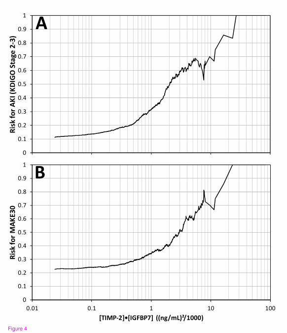
Figure 4. Risk for KDIGO stage 2 to 3 AKI (A) and MAKE30 (B) as a function of urine
[TIMP-2]•[IGFBP7]. Risk at each [TIMP-2]•[IGFBP7] value along the abscissa was
calculated as follows: the number of samples positive for the endpoint that had [TIMP-
2]•[IGFBP7] above the abscissa value divided by the total number of samples that had
[TIMP-2]•[IGFBP7] above the abscissa value. Slightly more than 50% of the samples had a
[TIMP-2]•[IGFBP7] value above 0.3 where risk began to elevate sharply and about 10% of
the samples had a [TIMP-2]•[IGFBP7] value above 2.0 where risk almost doubled and
quintupled for MAKE30 and AKI, respectively. AKI, acute kidney injury; IGFBP7, insulin-
like growth factor-binding protein 7; TIMP-2, tissue inhibitor of metalloproteinases-2.
Figure 5. Proposed mechanistic involvement of the novel biomarkers in AKI: initial
tubular cells sustain injury by various insults. In response to DNA and possibly other
forms of damage, IGFBP7 and TIMP-2 are expressed in the tubular cells. IGFBP7 directly
increases the expression of p53 and p21 and TIMP-2 stimulates p27 expression. These effects
are conducted in an autocrine and paracrine manner via IGFBP7 and TIMP-2 receptors. The p
proteins in turn, block the effect of the cyclin-dependent protein kinase complexes (CyclD-
CDK4 and CyclE-CDK2) on the cell cycle promotion, thereby resulting in G1 cell cycle
arrest for short periods of time presumably to avoid cells with possible damage from
dividing. AKI, acute kidney injury; IGFBP7, insulin-like growth factor-binding protein 7;

TIMP-2, tissue inhibitor of metalloproteinases-2.

Table 1. Baseline characteristics for Sapphire study patients.
Endpoint positive
Endpoint negative All patients
P values
All patients 101 627 728 Male 65 (64%) 384 (61%) 449 (62%) 0.58 Age1 65 (57-77) 64 (52-73) 64 (53-73) 0.048 Race 0.98 White 81 (80%) 492 (78%) 573 (79%) Black 11 (11%) 76 (12%) 87 (12%) Other/Unknown 9 (9%) 59 (9%) 68 (9%) Chronic comorbidities Chronic kidney disease 14 (14%) 51 (8%) 65 (9%) 0.14 Diabetes mellitus 39 (39%) 171 (27%) 210 (29%) 0.064 Congestive heart failure 23 (23%) 99 (16%) 122 (17%) 0.17 Coronary artery disease 33 (33%) 187 (30%) 220 (30%) 0.48 Hypertension 76 (75%) 357 (57%) 433 (59%) 0.001 Chronic obstructive pulmonary disease 21 (21%) 141 (22%) 162 (22%) 0.80 Cancer 25 (25%) 163 (26%) 188 (26%) 0.53 ICU type 0.47 Medical 40 (40%) 185 (30%) 225 (31%) Surgical 24 (24%) 155 (25%) 179 (25%) Combined ICU 14 (14%) 133 (21%) 147 (20%) Cardiac surgery 6 (6%) 55 (9%) 61 (8%) Neurologic 5 (5%) 34 (5%) 39 (5%) Coronary care unit 5 (5%) 25 (4%) 30 (4%) Trauma 4 (4%) 20 (3%) 24 (3%) Other/Unknown 3 (3%) 20 (3%) 23 (3%) Reason for ICU admission2 Respiratory 47 (47%) 263 (42%) 310 (43%) 0.39 Surgery 32 (32%) 215 (34%) 247 (34%) 0.65 Cardiovascular 41 (41%) 202 (32%) 243 (33%) 0.11 Sepsis 26 (26%) 110 (18%) 136 (19%) 0.055 Neurological 8 (8%) 62 (10%) 70 (10%) 0.72 Trauma 4 (4%) 51 (8%) 55 (8%) 0.16 Other 21 (21%) 105 (17%) 126 (17%) 0.32 Enrollment serum creatinine1,3 1.4 (0.9-1.8) 0.9 (0.7-1.2) 0.9 (0.7-1.2) <0.001 APACHE III 1,4 85 (59-106) 67 (51-88) 69 (51-91) <0.001
Baseline characteristics are shown for all patients in the study and patients that are either
negative or positive for the primary study endpoint (KDIGO stage 2 or 3 within 12 hours).
1Median (interquartile range); 2percentages for reason for ICU admission do not sum to 100%
because more than one reason can be given; 3value in hospital record closest to enrollment
time; 4calculated from source data by the study sponsor. APACHE III, Acute Physiology and

Chronic Health Evaluation III.

Additional files
Additional file 1
Title: Supplemental data
Description: Supplemental and supporting data unrelated to the primary analyses are
provided in Additional File 1. The additional data file also contains lists of study personnel,
detailed laboratory methods and sensitivity analyses.


[TIMP-2]•[IGFBP7]
Urine TIMP-2
Urine IGFBP7
Urine NGAL
Plasma Cystatin C
Urine KIM-1
Plasma NGAL
Urine IL-18
Urine pi-GST
Urine L-FABP
0.5 0.6 0.7 0.8 0.9
AUC (with 95% CI)
Fig
ure
2

�
���
���
���
���
���
���
��
��
���
������������� ��������������
�
���
���
���
���
���
���
��
��
���
�
���� ��� � �� ���
�������������
�������� ���!"�#�� ��$�%&'�(%)����
"
�Figure 4

Figure 5

Additional files provided with this submission:
Additional file 1: Sapphire_Additional File 1_Crit Care_R3_FINAL.doc, 492Khttp://ccforum.com/imedia/1577311466900551/supp1.doc