creport - downloads.hindawi.comdownloads.hindawi.com/journals/crie/2018/7261264.pdf · goiter. e...
TRANSCRIPT

Case ReportRemarkable Presentation: Anaplastic Thyroid CarcinomaArising from Chronic Hyperthyroidism
Habib G. Zalzal ,1 Jeffson Chung ,1 and Jessica A. Perini2
1Department of Otolaryngology-Head and Neck Surgery, West Virginia University School of Medicine, Morgantown, WV, USA2Section of Endocrinology, Department of Internal Medicine, West Virginia University School of Medicine, Morgantown, WV, USA
Correspondence should be addressed to Jeffson Chung; [email protected]
Received 27 January 2018; Accepted 25 February 2018; Published 1 April 2018
Academic Editor: Carlo Capella
Copyright © 2018 Habib G. Zalzal et al.This is an open access article distributed under the Creative Commons Attribution License,which permits unrestricted use, distribution, and reproduction in any medium, provided the original work is properly cited.
Background. Undifferentiated anaplastic carcinoma rarely develops from chronic hyperthyroidism. Although acute hyperthy-roidism can develop prior to anaplastic transformation, chronic hyperthyroidism was thought to be a protective measureagainst thyroid malignancy. Methods. A 79-year-old female presented acutely to the hospital with dyspnea. She had been takingmethimazole for chronic hyperthyroidism due to toxic thyroid nodules, previously biopsied as benign. Upon admission, imagingshowed tracheal compression, requiring a total thyroidectomy with tracheostomy for airway management. Results. Pathologydemonstrated undifferentiated anaplastic thyroid carcinoma. The patient passed away shortly after hospital discharge. Despitetreatment with methimazole for many years, abrupt enlargement of her toxic multinodular goiter was consistent with malignanttransformation. Chronic hyperthyroidism and toxic nodules are rarely associated with thyroid malignancy, with only one previousreport documenting association with anaplastic thyroid carcinoma. Conclusion. Progressive thyroid enlargement and acuteworsening of previously controlled hyperthyroidism should promote concern for disease regardless of baseline thyroid function.
1. Introduction
Historically, chronic hyperthyroidism had been consideredprotective against thyroid carcinoma. Some data suggesta lower incidence of papillary thyroid cancer in thosewith lower TSH levels [1]. This presumably arises from adecreased stimulatory effect on thyroid tissue presented bythe low serum thyroid stimulatory hormone (TSH) found inhyperthyroidism. However, with the increasing incidence ofthyroid cancer seen over the past years, there appears to bean improved understanding that thyroid carcinomas can alsoarise in glands that are thyrotoxic due toGraves’ disease, toxicmultinodular goiter, and autonomously functioning thyroidadenomas [2]. Recent review of the literature has shown therisk of malignancy associated with toxic hot nodules rangesfrom 1 to 10.3% [2] or even up to 15% in those with Graves’disease [3]. Most of these documented malignancies aredifferentiated thyroid carcinomas and very rarely medullarythyroid cancer [4].
Anaplastic thyroid carcinoma (ATC) represents oneof the most aggressive endocrine tumors and constitutes
approximately between 1.6 and 5% of all thyroidmalignancies[5]. ATC comes with a dismal prognosis limited to a 10–20%mean survival at 12months [6]. Patients with ATC experiencesignificant local compressive symptoms due to a rapidlyevolving central neck mass (77%) along with dysphagia(40%), hoarseness (40%), and stridor (24%) [7]. Metastasesare noted in 50% of patients at the time of diagnosis, mostcommonly in the lungs (80%), bone (6–16%), and brain(5–13%) [7]. Not uncommonly patients also develop thyro-toxicosis. Although thyrotoxicosis can develop in a glandwith ATC, it is rare to find ATC develop from underlyinglongstanding hyperthyroidism. We found only one othercase in the literature of ATC arising from a patient withchronic hyperthyroidism [8]. In this report, we present ourexperience with a patient who developed ATC after manyyears of hyperthyroidism and toxic multinodular goiter.
2. Patient
A 79-year-old Caucasian female presented to our institutionwith chest pain, dyspnea, and a rapidly enlarging thyroid
HindawiCase Reports in EndocrinologyVolume 2018, Article ID 7261264, 4 pageshttps://doi.org/10.1155/2018/7261264
2 Case Reports in Endocrinology
(a) (b)
(c)
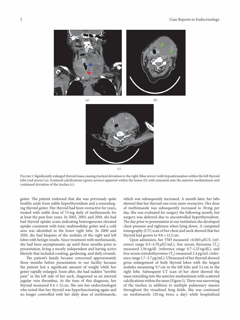
Figure 1: Significantly enlarged thyroidmass causing tracheal deviation to the right (blue arrow) with hypoattenuation within the left thyroidlobe (red arrow) (a). Scattered calcifications (green arrows) apparent within the lesion (b) with extension into the anterior mediastinum andcontinued deviation of the trachea (c).
goiter. The patient endorsed that she was previously quitehealthy aside from stable hyperthyroidism and a nonenlarg-ing thyroid goiter. Her thyroid had been overactive for years,treated with stable dose of 7.5mg daily of methimazole forat least the past four years. In 2002, 2003, and 2010, she hadhad thyroid uptake scans indicating heterogeneous elevateduptake consistent with toxic multinodular goiter and a coldarea was identified in the lower right lobe. In 2000 and2010, she had biopsies of the nodules of the right and leftlobes with benign results. Since treatment with methimazole,she had been asymptomatic up until three months prior topresentation, living a mostly independent and having activelifestyle that included cooking, gardening, and daily errands.
The patient’s family became concerned approximatelythree months before presentation to our facility becausethe patient lost a significant amount of weight while hergoiter rapidly enlarged. Soon after, she had sudden “terriblepain” in the left side of her neck, diagnosed as an internaljugular vein thrombus. At the time of this diagnosis, herthyroid measured 8.4 × 12 cm. She saw her endocrinologistwho noted that her thyroid was hyperfunctioning again andno longer controlled with her daily dose of methimazole,
which was subsequently increased. A month later, her labsshowed that her thyroid was even more overactive. Her doseof methimazole was subsequently increased to 30mg perday. She was evaluated for surgery the following month, butsurgery was deferred due to uncontrolled hyperthyroidism.The day prior to presentation at our institution she developedchest pressure and tightness when lying down. A computedtomography (CT) scan of her chest and neck showed that herthyroid had grown to 9.8 × 13.2 cm.
Upon admission, her TSH measured <0.003 𝜇IU/L (ref-erence range 0.5–4.70 𝜇IU/mL), free serum thyroxine (T
4)
measured 1.56 ng/dL (reference range 0.7–1.25 ng/dL), andfree serum triiodothyronine (T
3) measured 2.4 pg/mL (refer-
ence range 1.7–3.7 pg/mL). Ultrasound of her thyroid showedgross enlargement of both thyroid lobes with the largestnodules measuring 9.7 cm in the left lobe and 5.1 cm in theright lobe. Subsequent CT scan of her chest showed themass extending into the anterior mediastinum with scatteredcalcificationswithin themass (Figure 1).Therewas narrowingof the trachea in addition to multiple pulmonary massesthroughout the visualized lung fields. She was continuedon methimazole (20mg twice a day) while hospitalized
Case Reports in Endocrinology 3
(a) (b)
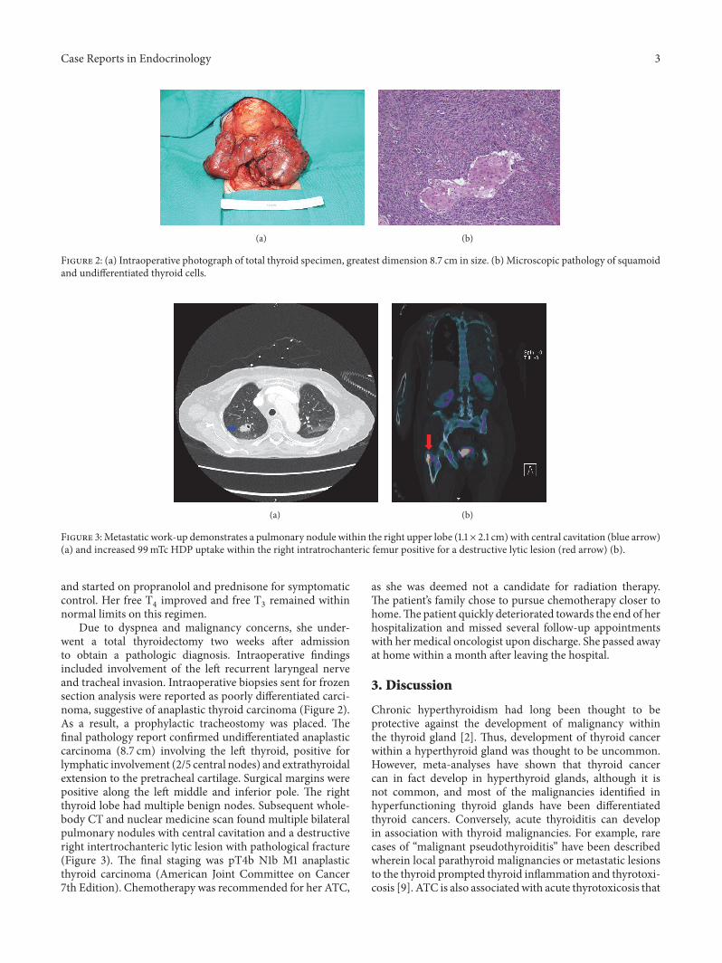
Figure 2: (a) Intraoperative photograph of total thyroid specimen, greatest dimension 8.7 cm in size. (b) Microscopic pathology of squamoidand undifferentiated thyroid cells.
(a) (b)
Figure 3:Metastatic work-up demonstrates a pulmonary nodule within the right upper lobe (1.1 × 2.1 cm) with central cavitation (blue arrow)(a) and increased 99mTc HDP uptake within the right intratrochanteric femur positive for a destructive lytic lesion (red arrow) (b).
and started on propranolol and prednisone for symptomaticcontrol. Her free T
4improved and free T
3remained within
normal limits on this regimen.Due to dyspnea and malignancy concerns, she under-
went a total thyroidectomy two weeks after admissionto obtain a pathologic diagnosis. Intraoperative findingsincluded involvement of the left recurrent laryngeal nerveand tracheal invasion. Intraoperative biopsies sent for frozensection analysis were reported as poorly differentiated carci-noma, suggestive of anaplastic thyroid carcinoma (Figure 2).As a result, a prophylactic tracheostomy was placed. Thefinal pathology report confirmed undifferentiated anaplasticcarcinoma (8.7 cm) involving the left thyroid, positive forlymphatic involvement (2/5 central nodes) and extrathyroidalextension to the pretracheal cartilage. Surgical margins werepositive along the left middle and inferior pole. The rightthyroid lobe had multiple benign nodes. Subsequent whole-body CT and nuclear medicine scan found multiple bilateralpulmonary nodules with central cavitation and a destructiveright intertrochanteric lytic lesion with pathological fracture(Figure 3). The final staging was pT4b N1b M1 anaplasticthyroid carcinoma (American Joint Committee on Cancer7th Edition). Chemotherapy was recommended for her ATC,
as she was deemed not a candidate for radiation therapy.The patient’s family chose to pursue chemotherapy closer tohome.The patient quickly deteriorated towards the end of herhospitalization and missed several follow-up appointmentswith her medical oncologist upon discharge. She passed awayat home within a month after leaving the hospital.
3. Discussion
Chronic hyperthyroidism had long been thought to beprotective against the development of malignancy withinthe thyroid gland [2]. Thus, development of thyroid cancerwithin a hyperthyroid gland was thought to be uncommon.However, meta-analyses have shown that thyroid cancercan in fact develop in hyperthyroid glands, although it isnot common, and most of the malignancies identified inhyperfunctioning thyroid glands have been differentiatedthyroid cancers. Conversely, acute thyroiditis can developin association with thyroid malignancies. For example, rarecases of “malignant pseudothyroiditis” have been describedwherein local parathyroid malignancies or metastatic lesionsto the thyroid prompted thyroid inflammation and thyrotoxi-cosis [9]. ATC is also associatedwith acute thyrotoxicosis that

4 Case Reports in Endocrinology
develops around the time of acute malignant transformation[2, 10]. A 2007 review of the association between ATC andthyrotoxicosis outlines eight cases in the literature [10]. Theauthors discuss the development of acute thyrotoxicosis withanaplastic thyroid cancer as a consequence of rapid leakageof thyroid hormone into the bloodstream from destroyedthyrocytes. Phillips et al. argue that the acute hyperthyroidstate in ATC is caused by a hyperfunctioning metastatictumor [10]. Thyrotoxicosis associated with ATC has beentermed “anaplastic pseudothyroiditis” [11].
This situation, however, differs from our patient in thatthyrotoxicosis did not originate from the anaplastic tumorbut predated the malignant transformation by several years.Our case shows the unique condition in which anaplasticthyroid cancer developed from a hyperthyroid patient despiteseveral years of methimazole treatment. ATC is associatedwith a previous history of thyroid goiter and is known tobe more common in geographic regions of endemic iodinedeficiency [7] but not associated with glands that havepreviously been hyperfunctioning.
To our knowledge, a 2014 report by Marcelino et al. is theonly published case that associates chronic hyperthyroidismwith subsequent development of ATC [8].Their study reportsa similar presentation in a 70-year-old male who had beendiagnosedwith a toxic nodule three years prior to diagnosis ofATC. He was conservatively managed with methimazole and𝛽-adrenergic blockers.Three years later, he re-presented withdyspnea, hoarseness, and dysphagia similar to our patient. Asubsequent neck CT scan showed a suspiciousmass involvingthe left thyroid lobe and isthmus (7 × 6 × 5 cm), with invasionof the surrounding soft tissue, trachea, and recurrent nerve.Their patient was not a surgical candidate and underwentradiotherapy without much improvement in thyroid sizebefore passing away several weeks later of airway compres-sion [8]. Similar to Marcelino et al., previous work-up ofour patient’s thyroid nodules, including biopsies of bilateralnodules, was negative for malignancy. While her thyroiduptake scans previously identified a cold nodule in the lowerright thyroid lobe, her surgical pathologic specimen of the leftthyroid lobe identified the anaplastic carcinomic tissue whileher right thyroid nodule was negative for anaplastic disease.In addition, previous thyroid scans had elevated and patchyuptake in the left lobe despite the pathologic finding. Thedevelopment of ATC from differentiated thyroid carcinomais well established [7], although our patient did not have thisdiagnosis, based on the negative FNA years prior. Whetherour patient developed “de novo” ATC similar to what wastheorized by Marcelino et al. is debatable, but plausibleconsidering the fact that the previously toxic left thyroidnodule was the onewhich developedATC in our patient.Thiswas despite the presence of a cold nodule in the right thyroidlobe which remained negative for malignancy after removal.
Undifferentiated anaplastic thyroid carcinoma is a rareand underreported condition in the setting of chronic hyper-thyroidism. There exists only one other reported case of thisassociation in the literature, also involving a septuagenarianwho developed rapid thyroid enlargement with trachealcompression years after diagnosis and control of hyperthy-roidism with methimazole. While current recommendations
for treatment of hyperthyroidism involve medical therapy,the presence of any nontoxic nodule in the thyroid, andany toxic nodule with suspicious features, should warrantwork-up with ultrasound, fine needle aspirate, and ongoingsurveillance. Our case demonstrates that even those withnegative biopsy and longstanding hyperthyroidism are at riskof development of anaplastic thyroid carcinoma.
Conflicts of Interest
The authors deny any conflicts of interest or funding sourcesassociated with this research study. No competing financialinterests exist.
References
[1] E. Fiore, T. Rago, M. A. Provenzale et al., “Lower levels of TSHare associated with a lower risk of papillary thyroid cancer inpatients with thyroid nodular disease: thyroid autonomy mayplay a protective role,” Endocrine-Related Cancer, vol. 16, no. 4,pp. 1251–1260, 2009.
[2] K. Pazaitou-Panayiotou, K. Michalakis, and R. Paschke, “Thy-roid cancer in patients with hyperthyroidism,” Hormone andMetabolic Research, vol. 44, no. 4, pp. 255–262, 2012.
[3] J. L. Kraimps,M.H. Bouin-Pineau,M.Mathonnet et al., “Multi-centre study of thyroid nodules in patients withGraves’ disease,”British Journal of Surgery, vol. 87, no. 8, pp. 1111–1113, 2000.
[4] K. Pazaitou-Panayiotou, P. Perros, M. Boudina et al., “Mortalityfrom thyroid cancer in patients with hyperthyroidism: TheTheagenion Cancer Hospital experience,” European Journal ofEndocrinology, vol. 159, no. 6, pp. 799–803, 2008.
[5] V. Kumar, B. Blanchon, X. Gu et al., “Anaplastic thyroid cancerand hyperthyroidism,” Endocrine Pathology, vol. 16, no. 3, pp.245–250, 2005.
[6] J. L. Pasieka, “Anaplastic thyroid cancer,” Current Opinion inOncology, vol. 15, no. 1, pp. 78–83, 2003.
[7] G. Nagaiah, A. Hossain, C. J. Mooney, J. Parmentier, and S. C.Remick, “Anaplastic thyroid cancer: a review of epidemiology,pathogenesis, and treatment,” Journal of Oncology, vol. 2011,Article ID 542358, pp. 1–13, 2011.
[8] M. Marcelino, P. Marques, L. Lopes, V. Leite, and J. J. deCastro, “Anaplastic carcinoma and toxicmultinodular goiter: anunusual presentation,” European Thyroid Journal, 2014.
[9] I. B. Rosen,H. G. Strawbridge, P. G.Walfish, and J. Bain, “Malig-nant pseudothyroiditis: A new clinical entity,” The AmericanJournal of Surgery, vol. 136, no. 4, pp. 445–449, 1978.
[10] J. S. Phillips,D. R. Pledger, andA.W.Hilger, “Rapid thyrotoxico-sis in anaplastic thyroid carcinoma,”The Journal of Laryngology& Otology, vol. 121, no. 7, pp. 695–697, 2007.
[11] S. Basaria, R. Udelsman, J. Tejedor-Sojo,W.H.Westra, and A. S.Krasner, “Anaplastic pseudothyroiditis,” Clinical Endocrinology,vol. 56, no. 4, pp. 553–555, 2002.

Stem Cells International
Hindawiwww.hindawi.com Volume 2018
Hindawiwww.hindawi.com Volume 2018
MEDIATORSINFLAMMATION
of
EndocrinologyInternational Journal of
Hindawiwww.hindawi.com Volume 2018
Hindawiwww.hindawi.com Volume 2018
Disease Markers
Hindawiwww.hindawi.com Volume 2018
BioMed Research International
OncologyJournal of
Hindawiwww.hindawi.com Volume 2013
Hindawiwww.hindawi.com Volume 2018
Oxidative Medicine and Cellular Longevity
Hindawiwww.hindawi.com Volume 2018
PPAR Research
Hindawi Publishing Corporation http://www.hindawi.com Volume 2013Hindawiwww.hindawi.com
The Scientific World Journal
Volume 2018
Immunology ResearchHindawiwww.hindawi.com Volume 2018
Journal of
ObesityJournal of
Hindawiwww.hindawi.com Volume 2018
Hindawiwww.hindawi.com Volume 2018
Computational and Mathematical Methods in Medicine
Hindawiwww.hindawi.com Volume 2018
Behavioural Neurology
OphthalmologyJournal of
Hindawiwww.hindawi.com Volume 2018
Diabetes ResearchJournal of
Hindawiwww.hindawi.com Volume 2018
Hindawiwww.hindawi.com Volume 2018
Research and TreatmentAIDS
Hindawiwww.hindawi.com Volume 2018
Gastroenterology Research and Practice
Hindawiwww.hindawi.com Volume 2018
Parkinson’s Disease
Evidence-Based Complementary andAlternative Medicine
Volume 2018Hindawiwww.hindawi.com
Submit your manuscripts atwww.hindawi.com