craniomaxillofac trauma reconstr bone graft in cranifacial surgery / orthodontic courses by indian...
TRANSCRIPT

Craniomaxillofac Trauma Reconstr. 2009 October; 2(3): 125–134.
Prepublished online 2009 April 14. doi: 10.1055/s-0029-1215875
PMCID: PMC3052656
Bone Grafts in Craniofacial Surgery
Mohammed E. Elsalanty, M.D., Ph.D.1 and David G. Genecov, M.D.2
Author information ► Copyright and License information ►
Abstract
Reconstruction of cranial and maxillofacial defects is a challenging task. The standard reconstruction method has been bone grafting. In this review, we shall describe the biological principles of bone graft healing, as pertinent to craniofacial reconstruction. Different types and sources of bone grafts will be discussed, as well as new methods of bone defect reconstruction.
Keywords: Bone grafts, craniofacial, maxillofacial, reconstruction, bone, vascularized, bone substitutes, bone augmentation, regenerationBone defects in the craniomaxillofacial skeleton vary from the small (few millimeters) periodontal defects to the large segmental defects resulting from trauma, surgical excision, or cranioplasty. Such defects typically have complex three-dimensional structural needs, which are difficult to restore. In cranial vault defects, the underlying brain needs permanent

protection. Segmental jaw defects require restoration of mechanical integrity, temporomandibular joint function, and intermaxillary dental occlusion. Maintaining acceptable facial esthetics is another unique consideration in the treatment of facial defects, which cannot be underestimated. Bone grafts remain the gold standard for reconstructing segmental bone defects. We will overview the status of bone grafting techniques for craniofacial reconstruction, their biological foundation, as well as future directions.
The earliest report of a bone grafting procedure came in an 1682 book by Job Janszoo van Meekeren, a surgeon in Amsterdam. In this account, the author reported a case in Russia, where the surgeon restored a cranial defect using a cranial bone graft from a dead dog.
In 1881, Sir William MacEwen of Rothesay, Scotland, published the first case report of successful inter human transfer of bone grafts. He used tibial bone wedges excised from three donors, during surgical correction of skeletal deformity, to reconstruct a humeral defect in a 3-year-old child. Subsequent clinical reports helped to establish the efficacy of autogenous bone grafts in defect reconstruction.
MECHANISM OF ACTION OF BONE GRAFTS
A bone graft is defined as any implanted material that promotes bone healing, whether alone or in combination with other material. Augmentation of bone healing at the recipient

site occurs through one or more of the following mechanisms: osteoconduction, osteoinduction, and osteogenesis.
An osteoconductive material simply allows, or directs, new bone formation along its surfaces. Examples include bone graft matrix and synthetic osteoconductive polymers.
An osteoinductive graft supplies recruitment and/or differentiation factors for bone-forming cells at the recipient site.
An osteogenic graft supplies induced or inducible, bone-forming cells to the recipient site. Accordingly, an ideal bo ne graft is the one that functions through all three mechanisms by providing a template that directs three-dimensional bone growth (osteoconduction), recruits and induces differentiation of resident bone-forming cells, and supplies more bone-forming cells to the recipient site. Such grafts include cancellous and vascularized bone grafts.
Bone grafts can be employed for functions other than to stimulate bone formation within a defect. An onlay graft laid over facial bone surfaces could augment the cheek prominence or restore facial contour. In this case, more emphasis is directed toward the rate of graft resorption. Those grafts that are known for their slow resorption, such as calvarial and cortical bone, or nonresorption, such as synthetic materials, are preferred. Such grafts might also be used for their mechanical properties wherever mechanical support or

immediate protection of vital structures is required, as in reconstructing orbital floor or calvarial defects.
Slow resorption is a disadvantage if the graft is used to augment bone formation at the recipient site. Graft incorporation is inversely proportional to how solid the graft is and how slow it resorbs. Therefore, osteoconductive graft materials with interconnected internal spaces that reach the outer surface are better scaffolds for directing three-dimensional bone invasion of the graft. This architecture provides more surface area along which native osteoclasts can attach themselves and start dissolving the graft, which is the first stage in graft incorporation.
TYPES OF BONE GRAFTS
Bone grafts can be divided into the following subtypes: autografts, allografts, xenografts, synthetic materials, and any combination thereof. Autografting is the transfer of graft material obtained from one anatomic site to another within the same subject. It includes transferring cancellous, cortical, corticocancellous, or vascularized bone or aspirated bone marrow. Autografts have the advantage of retaining at least some osteogenic cells and do not trigger an immune response. However, the total amount of bone that can be transferred is limited, and there can be high morbidity at the donor site.Grafts that are transferred between two genetically matched subjects, identical twins in humans, are called isografts. They
would be expected to have the same advantages and disadvantages as autografts.Grafts that are transferred between two genetically unmatched subjects are called allografts. Bone allografts are unique in that the cellular component is typically removed to minimize their rejection. In addition, they are thoroughly treated to eliminate any possibility of disease transmission. Therefore, allografts can be subdivided according to their source, processing method, or available form.With advancement in biomaterials technology, the use of animal-derived tissues for human tissue reconstruction is on the rise. These types of grafts are called xenografts. Several bone xenografts have been developed and are commercially available. They are typically in the form of bovine or porcine collagen and can be used either alone or in combination with a synthetic carrier.Synthetic bone substitutes and bone-augmenting preparations have been the focus of extensive research and have recently spawned a huge industry. Synthetic skeletal materials include osteoconductive polymers in the form of blocks, granules, or cements and osteoinductive proteins.8 Synthetic osteoinductive proteins that have been extensively studied in bone reconstruction include differentiation factors, such as bone morphogenic protein (BMP)-2 and -7,11,12,13,14,15 and angiogenic factors, such as vascular endothelial growth factor (VEGF).16,17,18Go to:
Go to:

INCORPORATION OF BONE GRAFTS INTO THE RECIPIENT SITE
It is true a bone graft may be used for its bulk or mechanical properties or to stimulate bone formation at the recipient site without necessarily being integrated into the newly formed bone. However, when bone grafts are used to bridge a critical-size bone defect, they are expected to become incorporated into the bed. Incorporation of the bone graft in the recipient site involves two essential steps: first is the bony union between the edges of the graft to the edges of native bone segments, and second is graft remodeling, or gradual resorption of the graft material itself, concomitant with its replacement by new bone.19,20,21 Graft remodeling can be of secondary importance in case of vascularized grafts, where the bone should be viable from the time of implantation. In this case, the remodeling process is expected be similar to that of normal bone.
Ideally, the whole bone graft should be incorporated into the recipient site. In other words, the space that the graft originally occupies should ultimately become viable bone permanently accessible to the physiological remodeling mechanisms. That process is typically very slow, and perfect outcome cannot always be achieved. Many factors determine how far the incorporation process will proceed. These factors may be pertinent to the graft itself, graft bed (recipient site), or the interface in between.
Factors related to the graft include the graft type, porosity, and mechanism of action. Autogenous cancellous and corticocancellous grafts are better incorporated due to their

porous architecture, allowing easy cellular and vascular invasion. The graft trabeculae have a large surface area that is covered by osteoblasts, making it osteogenic as well as osteoconductive for three-dimensional bone growth. Additionally, due to the extensive vascular invasion, the bone matrix can readily be demineralized and its proteins exposed through the actions of osteoclasts. This leads to the release of osteoinductive matrix proteins.
By contrast, autogenous cortical bone grafts are more solid. The only available access for cellular and vascular invasion of such grafts is the junction with the adjacent bone segments, making the integration process slow and rarely complete.19 This deficiency can be eliminated by using vascularized bone, which provides excellent long-term viability at the recipient site, even in large defects.8,19 In fact, the only factor to worry about regarding integration of a viable vascularized graft is its mechanical stability.8
As in fracture repair, rigid fixation of the graft to its bed is essential. Bone formation requires very low tissue strain levels. In addition, the ratio between the graft size and the contact area with circulation is a major determinant of how fast the graft can be incorporated, if at all. Large bone grafts with only minimal contact to bleeding, viable bone edges at the recipient site are expected to take a long time to become incorporated. One way to expose more of the graft core to the circulation is to mince the graft in a bone mill and pack it into the raw bed, given that it can be shielded from undue tissue strains.

Another important factor in determining graft incorporation is vascularity and viability of the graft bed. The bone graft typically needs to be attached to viable, bleeding bone edges. Too much reaming or excessive heat generation during saw cutting can cause necrosis of the bone edges and delay union to the graft.9 Radiotherapy can jeopardize tissue vascularity, eliminating the option of reconstruction using a nonvascularized bone graft. In such cases, a vascularized bone graft should be used, given that reasonably viable bone edges can be found to connect to the graft, dependable vessels can be used for microvascular anastomosis, and absence of infection. Some reports suggest the use of hyperbaric oxygen therapy to promote tissue perfusion before reconstruction.22,23,24 Finally, graft incorporation depends also on the overall physiological healing capacity of the body.
The biological process leading to graft incorporation is very similar to that of fracture repair. In brief, the cascade starts with the surgical hematoma, which involves the recruitment of platelets and white blood cells and the subsequent release of essential growth factors and cytokines. The recruited monocytes differentiate into osteoclasts and start removing the necrotic bone edges, with the demineralization of the matrix and release of bone augmenting factors. This leads to differentiation of osteoblasts and triggering the union between the graft and native bone edges. In the meantime, new blood vessels form within the granulation tissue and begin tunneling their way into the graft.

Since the early studies in bone transplantation immunity, it has been widely believed that at least some autograft-carried osteoblasts survive the transplantation process.25,26,27,28,29 Cell survival is also believed to occur more often in vascularized autografts than in nonvascularized autografts and in cancellous more than in cortical autografts.8,30 These cells can play an essential role early during the incorporation process.8,31 Graft incorporation has been summarized by Bauer and Muschler into five major steps8:
1. Hematoma formation, release of bone inducing factors and cellular recruitment
2. Inflammation and development of fibrovascular tissue, connecting the graft to the adjacent bone
3. Vascular invasion of the graft4. Focal resorption of the graft by recruited
osteoclasts5. New bone formation, union between the graft and
the surrounding bone, and graft remodelingGo to:
Go to:
SOURCES OF AUTOGENOUS BONE GRAFTS FOR CRANIOFACIAL RECONSTRUCTION
Free Nonvascularized Bone Grafts
ILIAC CRESTThe iliac crest is one of the most common donor site for bone grafts, both vascularized and nonvascularized. Large segments
of cortical, corticocancellous, or cancellous bone can be quickly obtained for different-sized defects. Furthermore, the location of the ilium allows harvesting by a separate surgical team to save operation time. A full-thickness iliac crest graft would have two thick cortices with ample amount of trabecular bone in between and can very closely resemble the thickness and height of mandibular bone. The graft shows reasonable long-term survival, and rehabilitation with osseointegrated dental implants is possible.32,33,34 Mandibular defects could be filled using nonvascularized iliac bone with a 70% success rate.35 The graft could be implanted as corticocancellous blocks or particulate cancellous bone carried within either a titanium mesh tray or a crib of alloplastic rib bone. However, the rate of successful union drops sharply when the defect is longer than 6 cm.35,36Posterior iliac crest graft can also be used for craniofacial reconstruction. However, the patient has to be tilted to the prone position, which eliminates the advantage of a simultaneous two-team approach. Donor site morbidity rate for anterior iliac crest grafts is around 23%, and much less for posterior iliac crest.37 Complications include postoperative pain, iliac or acetabular fractures or instability, persistent hematoma, herniation of abdominal contents, vascular injury, lateral femoral cutaneous nerve injury, and unsightly contour defects along the iliac crest.38,39,40,41CALVARIAL GRAFTThis is one of the most popular cortical bone grafts in craniofacial reconstruction, mainly for its mechanical

properties and very slow resorption rate.8 This makes it ideal for facial augmentation, orbital roof and floor reconstruction, and covering cranial defects. Typically, only the outer cortex is used, although a full-thickness graft could be taken and split into two grafts (Fig. 1). Typically, the skull continues to grow until the age of 8, continues to thicken until the age of 20, and
is thickest at the parietal region. This area can provide ∼8 × 10 cm of bone and is considered the safest to harvest.42
Figure 1Technique of splitting cranial bone using a reciprocating saw.
However, there are several key anatomic facts to consider before harvesting a calvarial bone graft:
1. Thickness of the calvarium is highly variable to the point of being unpredictable, even within the parietal region.43 Preoperative radiographic measurement of the bone thickness should give an idea of the area of bone that can safely be harvested.
2. The dura is tightly adherent to the inner cortex and can easily be injured if the inner cortex is to be harvested with the graft.
3. Various important vascular structures exist immediately beneath the bone at various sites, including the superior sagittal sinus in the midline.
4. The two cortices fuse together and the bone can become quite thin laterally and inferiorly to the temporal line, the attachment of the temporalis muscle, and at suture sites.
5. Other anatomic variables, including transcortical emissary veins, subcortical vessels, and aberrant arachnoid plexuses (within the cortical calvarium), should also be considered.42
The temporoparietal region provides more curved bone, which would be more suitable for orbital or malar reconstruction.44 However, straight grafts can be harvested more posteriorly (i.e., from the occipitoparietal region). In any case, the bone is typically harvested as narrow strips (5 to 6 cm long × 1.5 to 2 cm wide) to avoid graft fracture during harvest. Then, several strips can be fixed together and used as one graft.Calvarial bone can be harvested at three levels: partial-thickness outer cortex, full-thickness outer cortex, and bicortical.42 Partial-thickness outer cortex can be harvested using a very sharp osteotome to curl off a sheet of cortical bone from the outer cortical plate. This technique can be used in children between the age of 4 and 8 years and can yield enough bone to fill a small defect.
In adults, full-thickness outer cortex can safely be harvested and is therefore the most commonly used calvarial graft. If a craniotomy has already been performed, the inner cortex can be harvested from the bone flap and used in the reconstruction, leaving the outer cortex to be placed back in
its original position. This technique maintains the contour of the calvarium. If large quantities of bone are needed, bicortical grafts may be harvested, followed by splitting of the two cortices to double the surface of the graft. It is obvious that harvesting a bicortical calvarial graft would have the most complications hazard.
Complications of calvarial grafts include surface deformity at the donor and/or recipient site and graft fracture during harvest. Less commonly, dural exposure or tear can occur. If the dura is injured, the tear should be totally exposed, by expanding the bone defect with a rongeur, and patched with a temporalis fascia or, more recently, a synthetic graft. Intracranial hemorrhage after calvarial bone harvesting has been reported but is extremely rare.42CHIN GRAFT
Up to 3 cm of cortical and corticocancellous bone can be shaved off the chin bone through an intraoral approach. This can be sufficient for small defects, such as cleft palate and orthognathic osteotomy defects. Because of its slow resorption, it can be used as an onlay graft for facial augmentation.
RETROMOLAR GRAFTA small block of cortical or corticocancellous bone can be chiseled off the area behind the third molar.45 This graft has the same indications as chin grafts; however, the amount of available bone is much smaller.TIBIAL GRAFT

The anterior surface of the tibial plateau can be a good source of cortical or corticocancellous bone grafts. Mechanical stiffness of the tibial cortex can be useful in augmentation of atrophic alveolar ridge for implant placement, facial bone augmentation, or bridging an osteotomy defect.
RIB GRAFTNonvascularized rib was the first autogenous bone graft used for reconstruction of mandibular segmental defects.45 Osseous or osseochondral segments can be harvested from ribs 5 to 7 and can either be used in full or split thickness (Fig. 2). Costochondral grafts remain very popular in the treatment of ascending mandibular ramus and condylar defects.46,47,48,49 Side effects and complications include postoperative chest wall pain, pleural injury leading to pleuritis or pneumothorax, and facial asymmetry due to overgrowth of the graft.47,50,51
Figure 2Mandibular reconstruction with an osseocartilaginous rib graft.
Although they were frequently used for facial bone augmentation, bridging osteotomy defects, and orbital floor reconstruction, osseous rib grafts are now rarely used in craniofacial reconstruction.45 In addition to the problems mentioned previously, the amount and quality of bone obtained are inadequate for most reconstruction procedures. The

availability of other sources of bone graft with better quality and quantity, as well as with safer approaches and synthetic bone substitute materials, has rendered rib grafting less popular.REIMPLANTATION OF RESECTED BONE SEGMENTSLimited studies have tested the possibility of “recycling” native bone segments that were removed as a part of tumor excision.52,53,54,55 Intuitively, if tumor cells were successfully eradicated from the excised segments, they would be ideal for reconstructing the remaining defects. Resected mandibular segments could be reimplanted intact or hollowed out to remove trabecular bone, with use of the cortical bone shell as a tray for autogenous cancellous grafts. Larger long-term studies are needed to validate the safety and efficacy of this technique.
Regional Pedicled Bone Grafts
PEDICLED RIBIn 1980, Cuono and Ariyan reported their successful use of the pectoralis major–attached rib as an osteomyocutaneous flap for oromandibular reconstruction.56 However, subsequent reports showed flap necrosis rates ranging from 21 to 75%.57,58 The rib graft can also be carried along the latissimus dorsi or serratus anterior flaps.45 In all the above-mentioned flaps, the pedicle only allows to rib graft to reach the lower third of the face, limiting its use to mandibular defects. As mentioned earlier, rib grafts are not suitable for

such defects. Thus, these flaps are used only for soft tissue, and not bone, reconstruction.45PEDICLED CLAVICLESternocleidomastoid muscle (SCM) flaps have been extensively studied but not widely used. Several reports suggested the possibility of transferring clavicular periosteum59 and bone segments of the clavicle itself.60 The bone segment can either be partial or full thickness and can be utilized in reconstruction of small mandibular bone defects.The technique preserves the neurovascular supply of the SCM muscle, thus allowing for its use in dynamic facial reconstruction.61 This is particularly advantageous in cases where restoration of facial muscles, lower lip competence, mastication, or tongue movements is attempted. However, preserving the SCM muscle raises some concern in oncology cases due to the possibility of cervical lymph node involvement. In addition, the unsightly contour defect at the donor site and in the lower neck is another disadvantage of this flap.61PEDICLED TEMPORAL BONEThe temporalis flap is one of the earliest described muscle flaps.62 Over the years, it became one of the main techniques for reconstructing paralyzed facial muscles and midfacial full-thickness defects.63,64 More relevant to our review, partial or full-thickness temporal bone can be raised with the muscle flap. It can be used to reconstruct maxillary, palatal, orbital rim, orbital floor, or ascending mandibular ramus defects. It can also be used as an onlay graft for facial augmentation.62

However, significant donor site morbidity has been reported when calvarial bone is carried with the flap. These include limitation of mouth opening, which can be permanent, in addition to the mentioned complications of calvarial grafts.45
Vascularized Bone Grafts
Although not widely used for midface, upper face, or cranial reconstruction, vascularized bone grafting is considered the gold standard for large mandibular defect reconstruction. Because the graft's blood supply is coming through the anastomosis, it is independent of the condition of the recipient site. That makes it the most resistant to conditions like poor vascularity, extensive scarring, and previous radiotherapy of the bed.45 Moreover, they show less resorption than nonvascularized grafts and can immediately take endosteal implants for permanent dental restoration.65 They are ideal for primary reconstruction, unlike free grafts that have very high failure rate in primary reconstruction. Another advantage is the possibility of simultaneous soft tissue and bone reconstruction with the same composite flap. Success rates of vascularized grafts is more than 90%.36,66,67However, vascularized bone grafts are much more demanding and are technique sensitive as compared with nonvascularized grafts. Harvesting and an anastomosis require special surgical training and equipment. They add significantly to the operation time in cases of primary reconstruction, which can increase postoperative morbidity and mortality.66,68 Microvascular reconstruction is mostly limited

to mandibular defects, with the most commonly used vascularized grafts being the fibula, iliac crest, scapula, and radius. Detailed description of these techniques is beyond the scope of this review.Go to:
Go to:
ALLOPLASTIC BONE GRAFTS IN CRANIOFACIAL RECONSTRUCTION
Demineralized bone matrix (DBM) allografts have been frequently used in craniofacial reconstruction.69,70,71,72,73 Various DBM preparations are commercially available, varying from particles to blocks to sheets of different sizes. Generally, the smaller particles incorporate into the recipient bed faster than larger blocks or cortical sheets.8 A recent study has shown that the bone augmenting properties of DBM vary from one commercial preparation to another.74In addition to its osteoconductive and osteoinductive properties, DBM has some degree of mechanical stiffness, rendering it useful in reconstructing large cranial vault defects after cranioplasty procedures.73Sheets of cortical DBM could be molded into various shapes to match the three-dimensional configuration of the defect, providing a semirigid shield for the underlying brain during the regeneration process.Despite the overwhelming experimental evidence supporting the role of DBM as a bone augmenting material, incorporation of such allografts into recipient sites in human patients could be extremely slow. Replacement of DBM with new calcified

bone has been inconsistent, typically takes several months, especially in large defects.75,76 During that period, mechanical stiffness of the DBM implants is not high enough to protect the underlying brain, necessitating the use of protective helmets. The process of graft incorporation and new bone formation can be markedly accelerated with the addition of bone augmenting factors, such as BMP-2.75Go to:
Go to:
SYNTHETIC BONE SUBSTITUTES AND BONE AUGMENTING FACTORS
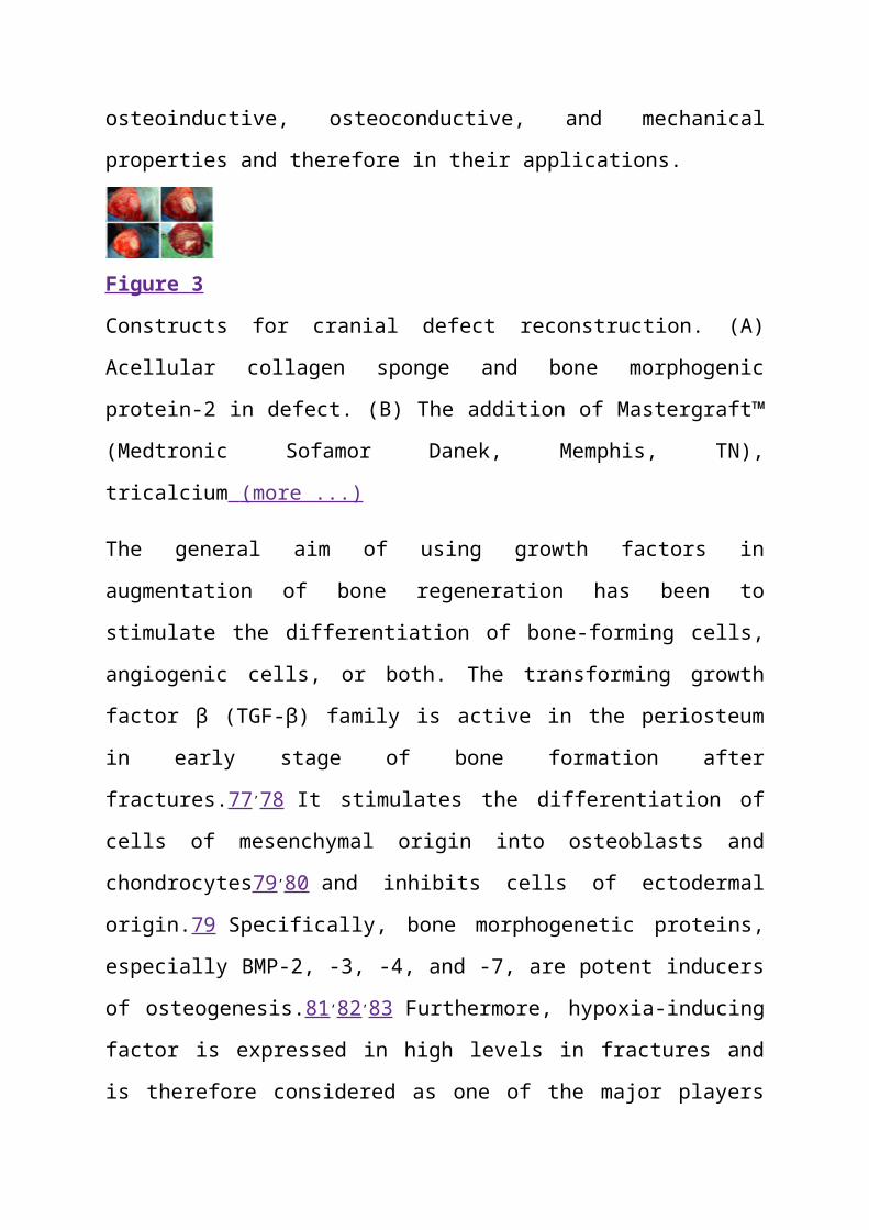
Advances in tissue engineering have provided a myriad of new tools for bone grafting. Growth factors, whether extracted or synthetic, adhesion molecules, and osteoconductive materials are becoming more available for bone reconstruction (Fig. 3). These factors and materials vary widely in their osteoinductive, osteoconductive, and mechanical properties and therefore in their applications.
Figure 3Constructs for cranial defect reconstruction. (A) Acellular collagen sponge and bone morphogenic protein-2 in defect. (B) The addition of Mastergraft™ (Medtronic Sofamor Danek, Memphis, TN), tricalcium (more ...)
The general aim of using growth factors in augmentation of bone regeneration has been to stimulate the differentiation of

bone-forming cells, angiogenic cells, or both. The transforming growth factor β (TGF-β) family is active in the periosteum in early stage of bone formation after fractures.77,78 It stimulates the differentiation of cells of mesenchymal origin into osteoblasts and chondrocytes79,80 and inhibits cells of ectodermal origin.79 Specifically, bone morphogenetic proteins, especially BMP-2, -3, -4, and -7, are potent inducers of osteogenesis.81,82,83 Furthermore, hypoxia-inducing factor is expressed in high levels in fractures and is therefore considered as one of the major players in stimulation of angiogenic factors expression.84 In fracture sites, hypoxia regulates osteoblast production of vascular modulators, such as VEGF and members of the TGF-β, insulin-like growth factor, and fibroblast growth factor families.85 Recruited osteoclasts have been reported to produce heparinase, which releases VEGF from heparin in an active form, stimulating local angiogenesis and further osteoclast activity.86On the other hand, the vascular response during bone regeneration is extremely sensitive to the mechanical environment.87 Endothelial cells subjected to mechanical forces, hypoxia, or VEGF stimulation could start producing BMP-2.88,89 Other products of endothelial cells, including endothelin-1 and endothelial-derived angiotensin II, can also stimulate osteoblasts during bone healing.90 Of these factors, BMP-2, BMP-7, and VEGF have shown the most potential for successful clinical use.75,91,92 It has been reported that platelet-rich plasma (PRP) promotes angiogenesis and osteogenesis via the presence of growth factors, which include
platelet-derived growth factor, platelet-derived endothelial cell growth factor, and TGF-β.93,94,95Kim and coworkers reported that demineralized bone and PRP produced a significantly higher percentage of bone regeneration as compared with the use of demineralized bone alone.96 However, Marden and coworkers found that platelet-derived growth factor inhibited bone regeneration induced by osteogenin, a bone morphogenetic protein, in rat craniotomy defects.97 In our experience, we found no evidence that PRP either promotes or interferes with osteogenesis occurring in the presence of exogenous recombinant human bone morphogenetic protein-2 (rhBMP2).75Two types of bone substitute materials have been used in craniofacial reconstruction: calcium phosphate cements and calcium sulfate (plaster of paris).98 Several preparations of calcium phosphates are commercially available for bone defect reconstruction. They have been successfully used to block cerebrospinal fluid leaks,99 obliterate the frontal sinus,100 and reconstruct contour defects in the cranium.101 Calcium sulfate hemihydrate, in combination with porous ceramic hydroxyapatite granules, has also been successfully used for cranial defect reconstruction.102
One major problem with cranial reconstruction has been how to maintain mechanical stability and protection for the underlying brain until sufficient bone regenerates to give permanent protection. Temporary stability can be provided with either resorbable or nonresorbable fixation materials. During growth, nonresorbable metal fixation should be
removed after reconstruction so as not to interfere with subsequent cranial remodeling. Nonresorbable fixation materials include titanium and cobalt chrome, the latter being easier to remove due to lack of osseointegration.
Several forms of resorbable fixation materials are available, which are mostly different forms of polylactate and polyglycolate polymers.103,104 When using these materials, however, it should be noted that the time needed to lose mechanical stiffness is much shorter than the resorption time. Additionally, there might be some interaction between certain bone graft materials, such as DBM or hydroxyapatite cements and some resorbable materials, such as Lactosorb® (Walter Lorenz Surgical, Inc., Jacksonville, FL).75,105Go to:
Go to:
FUTURE DIRECTIONS
Autogenous bone grafts remain the gold standard for surgical reconstruction of bone defects. However, advances in tissue engineering and biomaterials technology will provide more tools for these procedures. Several problems remain that limit the wide utilization of such options, including regulatory requirements, high costs, lack of randomized controlled human studies, uncertain long-term results, as well as method-specific limitations.
Go to:
Go to:

References
1. Meekeren J J. [Observationes Medico-Chirugicae]. Amsterdam: Ex Officina Henrici & Vidnae Theodori Boom; 1682.
2. Sanan A, Haines S J. Repairing holes in the head: a history of cranioplasty. Neurosurgery.1997;40:588–603. [PubMed]
3. Macewen W. Observations concerning transplantation of bone illustrated by a case of inter-human osseous transplantation, whereby over two-thirds of the shaft of a humerus was restored.Proc Roy Soc Lond. 1881;32:232–247.
4. Meikle M C. On the transplantation, regeneration and induction of bone: the path to bone morphogenetic proteins and other skeletal growth factors. Surgeon. 2007;5:232–243. [PubMed]
5. Albee F H. Fundamentals in bone transplantation: experiences in three thousand bone graft operations. JAMA. 1923;81:1429–1432.
6. Phemister D B. The fate of transplanted bone and regenerative power of its various constituents.Surg Gynecol Obstet. 1914;19:303–333.
7. Phemester D. Treatment of ununited fractures by onlay bone grafts without screw or tie fixation and without breaking down of fibrous union. J Bone Joint Surg Am. 1947;29:946–960.[PubMed]

8. Bauer T W, Muschler G F. Bone graft materials. An overview of the basic science. Clin Orthop Relat Res. 2000;(371):10–27. [PubMed]
9. Khan S N, Cammisa F P, Jr, Sandhu H S, Diwan A D, Girardi F P, Lane J M. The biology of bone grafting. J Am Acad Orthop Surg. 2005;13:77–86. [PubMed]
10. Laurencin C T, El-Amin S F. Xenotransplantation in orthopaedic surgery. J Am Acad Orthop Surg. 2008;16:4–8. [PubMed]
11. Boden S D. The ABCs of BMPs. Orthop Nurs. 2005;24:49–52. quiz 53–54. [PubMed]
12. Boyne P J, Salina S, Nakamura A, Audia F, Shabahang S. Bone regeneration using rhBMP-2 induction in hemimandibulectomy type defects of elderly sub-human primates. Cell Tissue Bank.2006;7:1–10. [PubMed]
13. Seto I, Marukawa E, Asahina I. Mandibular reconstruction using a combination graft of rhBMP-2 with bone marrow cells expanded in vitro. Plast Reconstr Surg. 2006;117:902–908. [PubMed]
14. Urist M R, Sato K, Brownell A G, et al. Human bone morphogenetic protein (hBMP) Proc Soc Exp Biol Med. 1983;173:194–199. [PubMed]
15. Wikesjö U M, Qahash M, Thomson R C, et al. rhBMP-2 significantly enhances guided bone regeneration. Clin Oral Implants Res. 2004;15:194–204. [PubMed]
16. Geiger F, Lorenz H, Xu W, et al. VEGF producing bone marrow stromal cells (BMSC) enhance vascularization and resorption of a natural coral bone substitute. Bone. 2007;41:516–522.[PubMed]
17. Ito H, Koefoed M, Tiyapatanaputi P, et al. Remodeling of cortical bone allografts mediated by adherent rAAV-RANKL and VEGF gene therapy. Nat Med. 2005;11:291–297. [PMC free article][PubMed]
18. Peng H, Usas A, Olshanski A, et al. VEGF improves, whereas sFlt1 inhibits, BMP2-induced bone formation and bone healing through modulation of angiogenesis. J Bone Miner Res.2005;20:2017–2027. [PubMed]
19. Dell P C, Burchardt H, Glowczewskie F P., Jr A roentgenographic, biomechanical, and histological evaluation of vascularized and non-vascularized segmental fibular canine autografts.J Bone Joint Surg Am. 1985;67:105–112. [PubMed]
20. Goldberg V M, Stevenson S. Natural history of autografts and allografts. Clin Orthop Relat Res.1987;(225):7–16. [PubMed]
21. Stevenson S, Li X Q, Davy D T, Klein L, Goldberg V M. Critical biological determinants of incorporation of non-vascularized cortical bone grafts. Quantification of a complex process and
structure. J Bone Joint Surg Am. 1997;79:1–16. [PubMed]
22. Myers R A, Marx R E. Use of hyperbaric oxygen in postradiation head and neck surgery. NCI Monogr. 1990;(9):151–157. [PubMed]
23. Vudiniabola S, Pirone C, Williamson J, Goss A N. Hyperbaric oxygen in the therapeutic management of osteoradionecrosis of the facial bones. Int J Oral Maxillofac Surg. 2000;29:435–438. [PubMed]
24. Yildiz S, Cimsit M, Ilgezdi S, et al. Hyperbaric oxygen therapy used to treat radiation injury: two case reports. Ostomy Wound Manage. 2006;52:14–16, 18, 20. [PubMed]
25. Burwell R G. Studies in the transplantation of bone. V. The capacity of fresh and treated homografts of bone to evoke transplantation immunity. J Bone Joint Surg Br. 1963;45-B:386–401. [PubMed]
26. Chalmers J. Transplantation immunity in bone homografting. J Bone Joint Surg Br. 1959;41-B:160–179. [PubMed]
27. De Bruyn P P, Kabisch W T. Bone formation by fresh and frozen, autogenous and homogenous transplants of bone, bone marrow and periosteum. Am J Anat. 1955;96:375–417. [PubMed]

28. Heiple K G, Chase S W, Herndon C H. A comparative study of the healing process following different types of bone transplantation. J Bone Joint Surg Am. 1963;45:1593–1616. [PubMed]
29. Kruyt M C, Dhert W J, Oner C, Blitterswijk C A van, Verbout A J, de Bruijn J D. Osteogenicity of autologous bone transplants in the goat. Transplantation. 2004;77:504–509. [PubMed]
30. Doi K, Tominaga S, Shibata T. Bone grafts with microvascular anastomoses of vascular pedicles: an experimental study in dogs. J Bone Joint Surg Am. 1977;59:809–815. [PubMed]
31. Zhang X, Xie C, Lin A S, et al. Periosteal progenitor cell fate in segmental cortical bone graft transplantations: implications for functional tissue engineering. J Bone Miner Res.2005;20:2124–2137. [PubMed]
32. Güven O. Rehabilitation of severely atrophied mandible using free iliac crest bone grafts and dental implants: report of two cases. J Oral Implantol. 2007;33:122–126. [PubMed]
33. Laine J, Vähätalo K, Peltola J, Tammisalo T, Happonen R P. Rehabilitation of patients with congenital unrepaired cleft palate defects using free iliac crest bone grafts and dental implants. Int J Oral Maxillofac Implants. 2002;17:573–580. [PubMed]

34. Sekine J, Sano K, Ikeda H, Inokuchi T. Rehabilitation by means of osseointegrated implants in oral cancer patients with about four to six years follow-up. J Oral Rehabil. 2006;33:170–174.[PubMed]
35. Pogrel M A, Podlesh S, Anthony J P, Alexander J. A comparison of vascularized and nonvascularized bone grafts for reconstruction of mandibular continuity defects. J Oral Maxillofac Surg. 1997;55:1200–1206. [PubMed]
36. Foster R D, Anthony J P, Sharma A, Pogrel M A. Vascularized bone flaps versus nonvascularized bone grafts for mandibular reconstruction: an outcome analysis of primary bony union and endosseous implant success. Head Neck. 1999;21:66–71. [PubMed]
37. Ahlmann E, Patzakis M, Roidis N, Shepherd L, Holtom P. Comparison of anterior and posterior iliac crest bone grafts in terms of harvest-site morbidity and functional outcomes. J Bone Joint Surg Am. 2002;84-A:716–720. [PubMed]
38. Boone D W. Complications of iliac crest graft and bone grafting alternatives in foot and ankle surgery. Foot Ankle Clin. 2003;8:1–14. [PubMed]
39. Nocini P F, Bedogni A, Valsecchi S, et al. Fractures of the iliac crest following anterior and posterior bone graft harvesting. Review of the
literature and case presentation. Minerva Stomatol.2003;52:441–448, 448–452. [PubMed]
40. Velchuru V R, Satish S G, Petri G J, Sturzaker H G. Hernia through an iliac crest bone graft site: report of a case and review of the literature. Bull Hosp Jt Dis. 2006;63:166–168. [PubMed]
41. Zijderveld S A, ten Bruggenkate C M, Den Bergh J P van, Schulten E A. Fractures of the iliac crest after split-thickness bone grafting for preprosthetic surgery: report of 3 cases and review of the literature. J Oral Maxillofac Surg. 2004;62:781–786. [PubMed]
42. Frodel J L. Calvarial bone graft harvesting techniques: considerations for their use with rigid fixation techniques in the craniomaxillofacial region. In: In: Greenberg A, Prein J, editor.Craniomaxillofacial Reconstructive and Corrective Bone Surgery. New York: Springer-Verlag; 2002. pp. 700–712.
43. Pensler J, McCarthy J G. The calvarial donor site: an anatomic study in cadavers. Plast Reconstr Surg. 1985;75:648–651. [PubMed]
44. Powell N B, Riley R W. Cranial bone grafting in facial aesthetic and reconstructive contouring.Arch Otolaryngol Head Neck Surg. 1987;113:713–719. [PubMed]
45. Ehrenfeld M, Hagenmaier C. Autogenous bone grafts in maxillofacial reconstruction. In: In:
Greenberg A, Prein J, editor. Craniomaxillofacial Reconstructive and Corrective Bone Surgery.New York: Springer-Verlag; 2002. pp. 295–309.
46. Güzel M Z, Arslan H, Saraç M. Mandibular condyle reconstruction with inlay application of autogenous costochondral graft after condylectomy: Cerrahpaşa's technique. J Oral Maxillofac Surg. 2007;65:615–620. [PubMed]
47. Medra A M. Follow up of mandibular costochondral grafts after release of ankylosis of the temporomandibular joints. Br J Oral Maxillofac Surg. 2005;43:118–122. [PubMed]
48. Poswillo D E. Biological reconstruction of the mandibular condyle. Br J Oral Maxillofac Surg.1987;25:100–104. [PubMed]
49. Troulis M J, Tayebaty F T, Papadaki M, Williams W B, Kaban L B. Condylectomy and costochondral graft reconstruction for treatment of active idiopathic condylar resorption. J Oral Maxillofac Surg. 2008;66:65–72. [PubMed]
50. Peltomäki T, Isotupa K. The costochondral graft: a solution or a source of facial asymmetry in growing children. A case report. Proc Finn Dent Soc. 1991;87:167–176. [PubMed]
51. Siavosh S, Ali M. Overgrowth of a costochondral graft in a case of temporomandibular joint ankylosis. J Craniofac Surg. 2007;18:1488–1491. [PubMed]

52. Bradley P F. A two-stage procedure for reimplantation of autogenous freeze-treated mandibular bone. J Oral Maxillofac Surg. 1982;40:278–284. [PubMed]
53. Marciani R D, Giansanti J S, Massey G B. Reimplantation of freeze-treated and saline-treated mandibular bone. J Oral Surg. 1976;34:314–319. [PubMed]
54. Plezia R A, Weaver A W, Pietruk T, Gilbert H D. Evaluation of osteogenesis following immediate and delayed reimplantation of frozen autogenous mandibular bone. Oral Surg Oral Med Oral Pathol. 1983;56:341–350. [PubMed]
55. Rossi G, Arrigoni G. Reimplantation of the mandibular condyle in cases of intraoral resection and reconstruction of the mandible. J Maxillofac Surg. 1979;7:1–5. [PubMed]
56. Cuono C B, Ariyan S. Immediate reconstruction of a composite mandibular defect with a regional osteomusculocutaneous flap. Plast Reconstr Surg. 1980;65:477–484. [PubMed]
57. Biller H F, Krespi Y P, Lawson W, Baek S M. A one-stage flap reconstruction following resection for stomal recurrence. Otolaryngol Head Neck Surg. 1980;88:357–360. [PubMed]
58. Lam K H, Wei W I, Siu K F. The pectoralis major costomyocutaneous flap for mandibular

reconstruction. Plast Reconstr Surg. 1984;73:904–910. [PubMed]
59. Tovi F, Gittot A. Sternocleidomastoid myoperiosteal flap for the repair of laryngeal and tracheal wall defects. Head Neck Surg. 1983;5:447–451. [PubMed]
60. Siemssen S O, Kirkby B, O'Connor T P. Immediate reconstruction of a resected segment of the lower jaw, using a compound flap of clavicle and sternomastoid muscle. Plast Reconstr Surg.1978;61:724–735. [PubMed]
61. Urken M L, Biller H F. Muscle and musculocutaneous flaps: sternocleidomastoid. In: In: Urken ML, Cheney ML, Sullivan MJ, editor. Atlas of Regional and Free Flaps for Head and Neck Reconstruction. New York: Raven Press; 1995. pp. 49–64.
62. Cheney M L. Muscle and musculocutaneous flaps: temporalis. In: In: Urken ML, Cheney ML, Sullivan MJ, editor. Atlas of Regional and Free Flaps for Head and Neck Reconstruction. New York: Raven Press; 1995. pp. 65–76.
63. Cheney M L, McKenna M J, Megerian C A, Ojemann R G. Early temporalis muscle transposition for the management of facial paralysis. Laryngoscope. 1995;105(9 Pt 1):993–1000. [PubMed]

64. Rubin L R, Mishriki Y, Lee G. Anatomy of the nasolabial fold: the keystone of the smiling mechanism. Plast Reconstr Surg. 1989;83:1–10. [PubMed]
65. Chana J S, Chang Y M, Wei F C, et al. Segmental mandibulectomy and immediate free fibula osteoseptocutaneous flap reconstruction with endosteal implants: an ideal treatment method for mandibular ameloblastoma. Plast Reconstr Surg. 2004;113:80–87. [PubMed]
66. Haughey B H, Wilson E, Kluwe L, et al. Free flap reconstruction of the head and neck: analysis of 241 cases. Otolaryngol Head Neck Surg. 2001;125:10–17. [PubMed]
67. Keller E E, Tolman D E, Eckert S. Endosseous implant and autogenous bone graft reconstruction of mandibular discontinuity: a 12-year longitudinal study of 31 patients. Int J Oral Maxillofac Implants. 1998;13:767–780. [PubMed]
68. Komisar A. The functional result of mandibular reconstruction. Laryngoscope. 1990;100:364–374. [PubMed]
69. Chen T M, Wang H J. Cranioplasty using allogeneic perforated demineralized bone matrix with autogenous bone paste. Ann Plast Surg. 2002;49:272–277. discussion 277–279. [PubMed]

70. Moss S D, Joganic E, Manwaring K H, Beals S P. Transplanted demineralized bone graft in cranial reconstructive surgery. Pediatr Neurosurg. 1995;23:199–204. discussion 204–205.[PubMed]
71. Salyer K E, Bardach J, Squier C A, Gendler E, Kelly K M. Cranioplasty in the growing canine skull using demineralized perforated bone. Plast Reconstr Surg. 1995;96:770–779. [PubMed]
72. Salyer K E, Gendler E, Menendez J L, Simon T R, Kelly K M, Bardach J. Demineralized perforated bone implants in craniofacial surgery. J Craniofac Surg. 1992;3:55–62. [PubMed]
73. Salyer K E, Gendler E, Squier C A. Long-term outcome of extensive skull reconstruction using demineralized perforated bone in Siamese twins joined at the skull vertex. Plast Reconstr Surg.1997;99:1721–1726. [PubMed]
74. Bae H W, Zhao L, Kanim L E, Wong P, Delamarter R B, Dawson E G. Intervariability and intravariability of bone morphogenetic proteins in commercially available demineralized bone matrix products. Spine. 2006;31:1299–1306. discussion 1307–1308. [PubMed]
75. Elsalanty M E, Por Y C, Genecov D G, et al. Recombinant human BMP-2 enhances the effects of materials used for reconstruction of large cranial
defects. J Oral Maxillofac Surg. 2008;66:277–285. [PubMed]
76. Por Y C, Barceló C R, Salyer K E, et al. Bone generation in the reconstruction of a critical size calvarial defect in an experimental model. Ann Acad Med Singapore. 2007;36:911–919. [PubMed]
77. Andrew J G, Hoyland J, Andrew S M, Freemont A J, Marsh D. Demonstration of TGF-beta 1 mRNA by in situ hybridization in normal human fracture healing. Calcif Tissue Int. 1993;52:74–78. [PubMed]
78. Bourque W T, Gross M, Hall B K. Expression of four growth factors during fracture repair. Int J Dev Biol. 1993;37:573–579. [PubMed]
79. Lind M. Growth factors: possible new clinical tools. A review. Acta Orthop Scand. 1996;67:407–417. [PubMed]
80. Massagué J. The transforming growth factor-beta family. Annu Rev Cell Biol. 1990;6:597–641.[PubMed]
81. Barnes G L, Kostenuik P J, Gerstenfeld L C, Einhorn T A. Growth factor regulation of fracture repair. J Bone Miner Res. 1999;14:1805–1815. [PubMed]
82. Eingartner C, Coerper S, Fritz J, Gaissmaier C, Koveker G, Weise K. Growth factors in distraction osteogenesis. Immuno-histological pattern of TGF-beta1 and IGF-I in human callus induced by

distraction osteogenesis. Int Orthop. 1999;23:253–259. [PubMed]
83. Ishidou Y, Kitajima I, Obama H, et al. Enhanced expression of type I receptors for bone morphogenetic proteins during bone formation. J Bone Miner Res. 1995;10:1651–1659.[PubMed]
84. Pacicca D M, Patel N, Lee C, et al. Expression of angiogenic factors during distraction osteogenesis. Bone. 2003;33:889–898. [PubMed]
85. Steinbrech D S, Mehrara B J, Saadeh P B, et al. Hypoxia increases insulinlike growth factor gene expression in rat osteoblasts. Ann Plast Surg. 2000;44:529–534. discussion 534–535. [PubMed]
86. Saijo M, Kitazawa R, Nakajima M, Kurosaka M, Maeda S, Kitazawa S. Heparanase mRNA expression during fracture repair in mice. Histochem Cell Biol. 2003;120:493–503. [PubMed]
87. Wallace A L, Draper E R, Strachan R K, McCarthy I D, Hughes S P. The vascular response to fracture micromovement. Clin Orthop Relat Res. 1994;(301):281–290. [PubMed]
88. Bouletreau P J, Warren S M, Spector J A, et al. Hypoxia and VEGF up-regulate BMP-2 mRNA and protein expression in microvascular endothelial cells: implications for fracture healing. Plast Reconstr Surg. 2002;109:2384–2397. [PubMed]

89. Sorescu G P, Sykes M, Weiss D, et al. Bone morphogenic protein 4 produced in endothelial cells by oscillatory shear stress stimulates an inflammatory response. J Biol Chem. 2003;278:31128–31135. [PubMed]
90. von Schroeder H P, Veillette C J, Payandeh J, Qureshi A, Heersche J N. Endothelin-1 promotes osteoprogenitor proliferation and differentiation in fetal rat calvarial cell cultures. Bone.2003;33:673–684. [PubMed]
91. Kaigler D, Wang Z, Horger K, Mooney D J, Krebsbach P H. VEGF scaffolds enhance angiogenesis and bone regeneration in irradiated osseous defects. J Bone Miner Res. 2006;21:735–744.[PubMed]
92. Ripamonti U, Ma S S, Cunningham N S, Yeates L, Reddi A H. Reconstruction of the bone—bone marrow organ by osteogenin, a bone morphogenetic protein, and demineralized bone matrix in calvarial defects of adult primates. Plast Reconstr Surg. 1993;91:27–36. [PubMed]
93. Kawase T, Okuda K, Saito Y, Amizuka N, Suzuki H, Yoshie H. Platelet-rich plasma provides nucleus for mineralization in cultures of partially differentiated periodontal ligament cells. In Vitro Cell Dev Biol Anim. 2005;41:171–176. [PubMed]
94. Kawase T, Okuda K, Wolff L F, Yoshie H. Platelet-rich plasma-derived fibrin clot formation

stimulates collagen synthesis in periodontal ligament and osteoblastic cells in vitro. J Periodontol.2003;74:858–864. [PubMed]
95. Okuda K, Kawase T, Momose M, et al. Platelet-rich plasma contains high levels of platelet-derived growth factor and transforming growth factor-beta and modulates the proliferation of periodontally related cells in vitro. J Periodontol. 2003;74:849–857. [PubMed]
96. Kim S G, Kim W K, Park J C, Kim H J. A comparative study of osseointegration of Avana implants in a demineralized freeze-dried bone alone or with platelet-rich plasma. J Oral Maxillofac Surg. 2002;60:1018–1025. [PubMed]
97. Marden L J, Fan R S, Pierce G F, Reddi A H, Hollinger J O. Platelet-derived growth factor inhibits bone regeneration induced by osteogenin, a bone morphogenetic protein, in rat craniotomy defects. J Clin Invest. 1993;92:2897–2905. [PMC free article] [PubMed]
98. Pou A M. Update on new biomaterials and their use in reconstructive surgery. Curr Opin Otolaryngol Head Neck Surg. 2003;11:240–244. [PubMed]
99. Costantino P D, Hiltzik D H, Sen C, et al. Sphenoethmoid cerebrospinal fluid leak repair with hydroxyapatite cement. Arch Otolaryngol Head Neck Surg. 2001;127:588–593. [PubMed]
100. Petruzzelli G J, Stankiewicz J A. Frontal sinus obliteration with hydroxyapatite cement.Laryngoscope. 2002;112:32–36. [PubMed]
101. Baker S B, Weinzweig J, Kirschner R E, Bartlett S P. Applications of a new carbonated calcium phosphate bone cement: early experience in pediatric and adult craniofacial reconstruction. Plast Reconstr Surg. 2002;109:1789–1796. [PubMed]
102. Costantino P D, Hiltzik D, Govindaraj S, Moche J. Bone healing and bone substitutes. Facial Plast Surg. 2002;18:13–26. [PubMed]
103. Tiainen J, Leinonen S, Ilomäki J, et al. Comparison of the pull-out forces of bioabsorbable polylactide/glycolide screws (Biosorb and Lactosorb) and tacks: a study on the stability of fixation in human cadaver parietal bones. J Craniofac Surg. 2002;13:538–543. [PubMed]
104. Wiltfang J, Merten H A, Schultze-Mosgau S, Schrell U, Wénzel D, Kessler P. Biodegradable miniplates (LactoSorb): long-term results in infant minipigs and clinical results. J Craniofac Surg.2000;11:239–243. discussion 244–245. [PubMed]
105. Genecov D G, Kremer M, Agarwal R, et al. Norian craniofacial repair system: compatibility with resorbable and nonresorbable plating

materials. Plast Reconstr Surg. 2007;120:1487–1495.[PubMed]
Articles from Craniomaxillofacial Trauma & Reconstruction are provided here courtesy of Thieme Medical Pike, Bethesda MD, 20894 USA