cranial anatomy and phylogenetic affinities … · cranial anatomy and phylogenetic affinities of...
TRANSCRIPT
BioOne sees sustainable scholarly publishing as an inherently collaborative enterprise connecting authors, nonprofit publishers, academic institutions, researchlibraries, and research funders in the common goal of maximizing access to critical research.
CRANIAL ANATOMY AND PHYLOGENETIC AFFINITIES OF THEPERMIAN PARAREPTILE MACROLETER POEZICUSAuthor(s): LINDA A. TSUJISource: Journal of Vertebrate Paleontology, 26(4):849-865. 2006.Published By: The Society of Vertebrate PaleontologyDOI: http://dx.doi.org/10.1671/0272-4634(2006)26[849:CAAPAO]2.0.CO;2URL: http://www.bioone.org/doi/full/10.1671/0272-4634%282006%2926%5B849%3ACAAPAO%5D2.0.CO%3B2
BioOne (www.bioone.org) is a nonprofit, online aggregation of core research in the biological, ecological, andenvironmental sciences. BioOne provides a sustainable online platform for over 170 journals and books publishedby nonprofit societies, associations, museums, institutions, and presses.
Your use of this PDF, the BioOne Web site, and all posted and associated content indicates your acceptance ofBioOne’s Terms of Use, available at www.bioone.org/page/terms_of_use.
Usage of BioOne content is strictly limited to personal, educational, and non-commercial use. Commercial inquiriesor rights and permissions requests should be directed to the individual publisher as copyright holder.

CRANIAL ANATOMY AND PHYLOGENETIC AFFINITIES OF THE PERMIAN PARAREPTILEMACROLETER POEZICUS
LINDA A. TSUJI*Department of Biology, University of Toronto at Mississauga, 3359 Mississauga Rd. N., Mississauga, Ontario, Canada,
ABSTRACT—The Parareptilia, the sister group to the Eureptilia, is poorly known, with many taxa in need of adequatedescription. One such taxon, Macroleter poezicus is found in Middle Permian strata of the Mezen River Basin in theArkhangel’sk Province of Russia. The cranial anatomy of Macroleter is described from four new well-preserved speci-mens, and 89 cranial characters are incorporated into a phylogenetic analysis of parareptiles. A single most parsimonioustopology is found, consisting of 205 steps, with the novel result that Macroleter is the taxon most closely related topareiasaurs. This result has important implications for the phylogeny of the Parareptilia as well as for the identity of adisputed element (the tabular) in the skull of pareiasaurs.
INTRODUCTION
Continued disagreement concerning turtle origins has resultedin an increased focus on the Parareptilia, a previously enigmaticfossil group. Not only have there been arguments identifyingturtles as diapsid (deBraga and Rieppel, 1997; Rieppel and Reisz,1999; Rieppel, 2000) or parareptilian (Reisz and Laurin, 1991;Lee, 1993, 1995, 1997a, b; Laurin and Reisz, 1995) reptiles, buteven those who conclude that turtles are parareptiles argue overtheir exact position within the clade. Attempts to sort out theorigin of turtles have been frustrated by the fact that many of thetaxa within Parareptilia are insufficiently known, inhibiting theability to discern evolutionary trends within the group, howeverrecent studies have described some of the lesser-known Permianparareptilian taxa, incorporating them into a phylogeny of earlyamniotes (deBraga and Reisz, 1996; Lee, 1997a; Berman et al.,2000; Modesto, 2000; Reisz and Scott, 2002). The majority of thewell-known parareptiles come from the Late Permian of theKaroo Basin, South Africa, but fossils from this clade are alsoknown from sites on practically every continent, including Northand South America, Antarctica, Asia, and Europe, with thegreatest numbers coming from the expanse of the Russian Plat-form. Temporally, the range of the non-testudinine parareptilesextends from the Early Permian to the Late Triassic. Minimumdivergence times based on the latest phylogenies imply that theproganosaurs (mesosaurs + parareptiles, sensu Anderson andModesto, 2005) diverged from the Eureptilia at the latest in theLate Carboniferous (Modesto, 2000), indicating the presence ofextensive ghost lineages for many clades within Parareptilia.
The distinctive assemblage of parareptiles from Russia plays akey role in our understanding of the diversity of Permian amni-otes. Most of these taxa, however, have been only briefly de-scribed, and have been only sporadically included in phyloge-netic analyses of the clade. One particularly interesting amniote-bearing site, the Mezen River Basin in the Arkhangel’skProvince of Russia, is dated to the Late Kazanian to Early Ta-tarian in the Middle Permian, and is believed to be contempo-raneous with the Tapinocephalus Zone of the Beaufort Seriesin South Africa (Rubidge, 1995). This area is host to an impor-tant faunal assemblage that is dominated by parareptilian taxa(Ivakhnenko et al., 1997). Discovered in 1935 by N. I. Novozhi-
lov in localities along the Mezen, Pyoza, and Kimzha rivers of theMezen River watershed (Tverdokhlebova and Ivakhnenko,1984), this fauna was first described by Efremov (1938, 1940).The assemblage is notable for the apparent preferential preser-vation of small terrestrial amniotes, and contains an intriguingmix of synapsids, a single small diapsid, and a number of para-reptiles. Oddly, no anamniotes, very common to other contem-poraneous faunas, have been found in any of the many localitiesscattered throughout the basin. The synapsids of the Mezen as-semblage represent both basal synapsid ‘pelycosaurs’, thevaranopids Mesenosaurus and Pyozia and the caseid Ennatosau-rus, along with the more derived therapsids Biarmosuchus andNiaftasuchus. Also present in the assemblage is a small diapsidLanthanolania ivakhnenkoi (Modesto and Reisz, 2003) and theparareptiles Lanthaniscus efremovi, Bashkyroleter mesensis,Nyctiphruretus acudens, Nycteroleter ineptus, and Macroleterpoezicus (Ivakhnenko et al., 1997). Most of the taxa are very lowin abundance, with the small Nyctiphruretus dominating the as-semblage. Nyctiphruretus comprises over 67% of the specimensfound in the basin, with Macroleter comprising over 8% of theknown specimens.
Initially these two common taxa, belonging to the Nyctiphru-retidae (Efremov, 1938) and the Nycteroleteridae (Romer, 1956)respectively, were difficult to classify. The nycteroleterids in par-ticular superficially resemble anamniotes, possessing a definitetemporal emargination and conspicuous dermal sculpturing, andthey have indeed been allied with diadectids and other anamni-ote taxa (Olson, 1947; Heaton, 1980). Subsequent analyses, how-ever, have shown that nycteroleterids are clearly amniotes, andhave placed these animals well within the Parareptilia, usually asor within the sister taxon to the group including the pareiasaursand procolophonids (Lee, 1995, 1997a; deBraga and Reisz, 1996;deBraga and Rieppel, 1997; Hill, 2005;). There remains, how-ever, some debate about their exact position within Parareptilia,and even their monophyly as a clade, due in large part to a lackof detailed knowledge about the anatomy of the group.
The largest of the nycteroleterids from the Mezen River basin,Macroleter poezicus was named in 1984 (Tverdokhlebova andIvakhnenko, 1984). At last count, Macroleter had been found in13 different sites throughout the basin, the third most commontaxon in the assemblage after Nyctiphruretus and the varanopidMesenosaurus. Until a misidentified specimen from the ChickashaFormation in Oklahoma was recognized as a species of Macro-leter (Reisz and Laurin, 2001), the genus was monospecific, and
* Current address: Humboldt-Universität zu Berlin, Museum fürNaturkunde, D-10099 Berlin, Germany.
Journal of Vertebrate Paleontology 26(4):849–865, December 2006© 2006 by the Society of Vertebrate Paleontology
849
was restricted to the localities within the Mezen Basin. Macro-leter poezicus can now be studied in more detail due to collectingexpeditions to this latter area in the 1990s, which have producedadditional specimens. These new specimens consist of well pre-served and relatively undistorted cranial, and for the first time,post-cranial material of the genus. This paper details a redescrip-tion of the cranial anatomy of Macroleter poezicus from the newspecimens, and incorporates this morphological information intoa phylogenetic analysis of the Parareptilia with the aim of re-solving Macroleter’s relationship to pareiasaurs and procolo-phonids.
Institutional Abbreviations—PIN, Paleontological Instituteof the Russian Academy of Sciences, Moscow, Russia; UTM,University of Toronto at Mississauga, Mississauga, Canada.
Anatomical Abbreviations—a. max. for., anterior maxillaryforamen; a, angular; ar, articular; art. sp., articulation area of thesplenial; atc, atlas centrum; ati, atlas intercentrum; atr, atlas rib;axc, axis centrum; axi, axis intercentrum; bo, basioccipital; br,basisphenoid rostrum; cbl, ceratobranchial; cl, clavicle; clin, cli-noid process; co, coronoid; d, dentary; dors. sel., dorsum sellae;ect, ectopterygoid; eo, exoccipital; ept, epipterygoid; f, frontal;for. orb., foramen orbitonasale; h1, hyoid; hyp, insertion of hyp-axial musculature; j, jugal; la, lacrimal; m, maxilla; n, nasal; op,opisthotic; p, parietal; pal, palatine; pbs, parabasisphenoid; pf,postfrontal; pm, premaxilla; po, postorbital; pp, postparietal; prf,prefrontal; pro, prootic; pt, pterygoid; q, quadrate; qj, quadra-tojugal; sa, surangular; s, stapes; scl, scleral ossicle; sel, sellaturcica; so, supraoccipital; sp, splenial; sq, squamosal; sub. for.,suborbital foramen; st, supratemporal; t. f., temporal fenestra; t,tabular; v, vomer; vic, vidian canal; I-XII, foramen for cranialnerves I-XII.
SYSTEMATIC PALEONTOLOGY
REPTILIA Laurenti, 1768PARAREPTILIA Olson, 1947
ANKYRAMORPHA deBraga and Reisz, 1996MACROLETER POEZICUS Tverdokhlebova and
Ivakhnenko, 1984
Revised Diagnosis
Macroleter poezicus is a medium-sized parareptile distin-guished by the following cranial autapomorphies: maxilla vomeranterior contact, basicranial articulation and basipterygoid pro-cess facing anteriorly, pterygoids meet anterior to basicranialarticulation; sculptureless round indentation just anterior tofronto-parietal suture; posterior portion of suture between pari-etals highly depressed, skull roof v-shaped in occipital view.
Holotype—PIN 3586/1, consisting of an entire skeletonLocality and Horizon—Mezen River Basin, Arkhangel’sk
Province, Russia. Uppermost Kazanian or lowermost Tatarian,Middle Permian (Ivakhnenko et al., 1997).
Referred Specimens—PIN 4543/3, mostly complete skull andpostcranial material; PIN uncataloged immature specimen, com-plete skull and postcranial material; UTM/Mezen/2001/1, largeskull, some postcranial material; UTM/Mezen/2001/2, skull andpostcranial material anterior to pelvic girdle.
OSTEOLOGICAL DESCRIPTION
Reconstruction
The skull of Macroleter was reconstructed in dorsal, ventral,lateral, and occipital views (Fig. 1), based primarily on two speci-mens, PIN 4543/3 and PIN (uncataloged), which are virtuallycomplete and show little crushing or warping. For the ventralview PIN 4543/3 was the basis for the reconstruction. The dorsalreconstruction was composed based on a combination of PIN
4543/3 and PIN (uncataloged). The lateral reconstruction wasbased primarily on PIN (uncataloged), and secondarily on UTM/Mezen/2001/2. The occipital view was largely reconstructed fromthe information available from the two best preserved skulls, butthere also exists a slide of a skull now missing (R. Reisz, pers.comm., 2005), in occipital view, which was also used as a refer-ence.
Skull
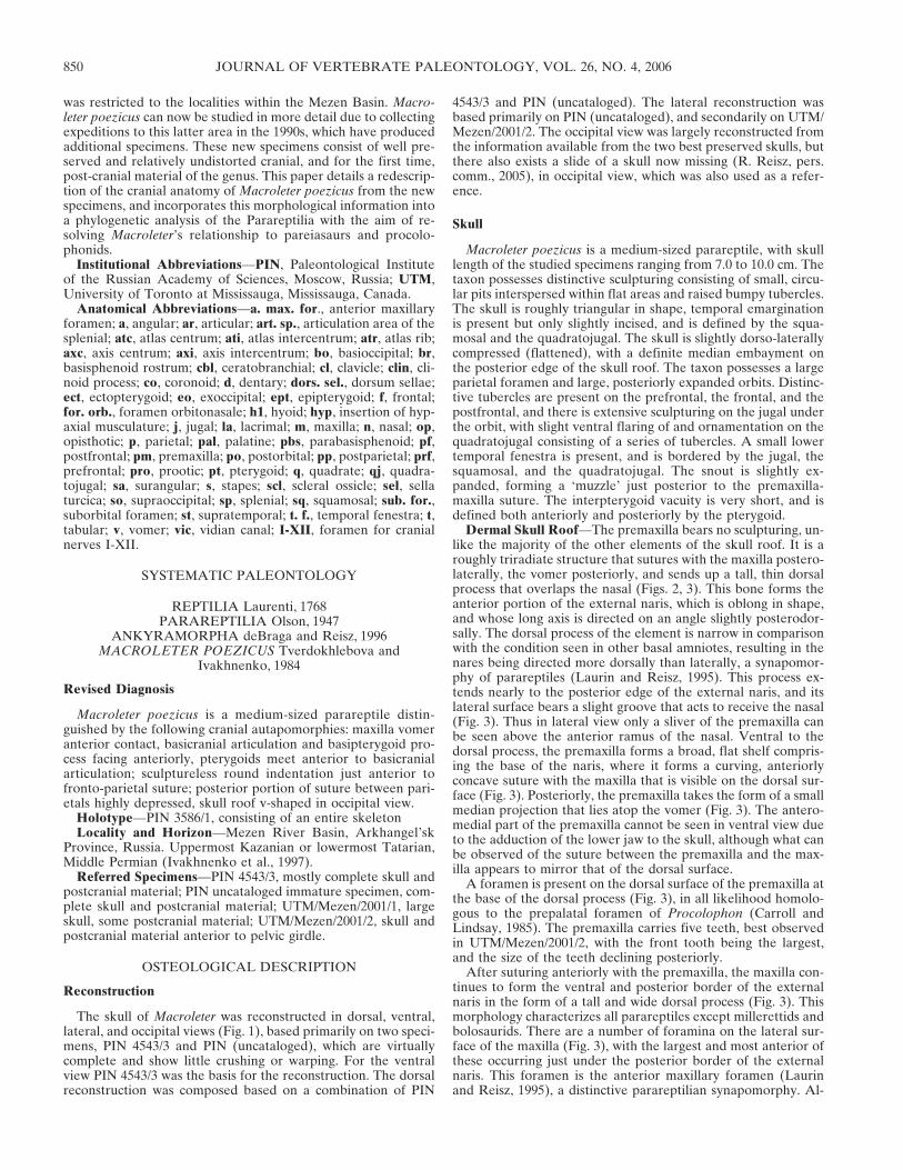
Macroleter poezicus is a medium-sized parareptile, with skulllength of the studied specimens ranging from 7.0 to 10.0 cm. Thetaxon possesses distinctive sculpturing consisting of small, circu-lar pits interspersed within flat areas and raised bumpy tubercles.The skull is roughly triangular in shape, temporal emarginationis present but only slightly incised, and is defined by the squa-mosal and the quadratojugal. The skull is slightly dorso-laterallycompressed (flattened), with a definite median embayment onthe posterior edge of the skull roof. The taxon possesses a largeparietal foramen and large, posteriorly expanded orbits. Distinc-tive tubercles are present on the prefrontal, the frontal, and thepostfrontal, and there is extensive sculpturing on the jugal underthe orbit, with slight ventral flaring of and ornamentation on thequadratojugal consisting of a series of tubercles. A small lowertemporal fenestra is present, and is bordered by the jugal, thesquamosal, and the quadratojugal. The snout is slightly ex-panded, forming a ‘muzzle’ just posterior to the premaxilla-maxilla suture. The interpterygoid vacuity is very short, and isdefined both anteriorly and posteriorly by the pterygoid.
Dermal Skull Roof—The premaxilla bears no sculpturing, un-like the majority of the other elements of the skull roof. It is aroughly triradiate structure that sutures with the maxilla postero-laterally, the vomer posteriorly, and sends up a tall, thin dorsalprocess that overlaps the nasal (Figs. 2, 3). This bone forms theanterior portion of the external naris, which is oblong in shape,and whose long axis is directed on an angle slightly posterodor-sally. The dorsal process of the element is narrow in comparisonwith the condition seen in other basal amniotes, resulting in thenares being directed more dorsally than laterally, a synapomor-phy of parareptiles (Laurin and Reisz, 1995). This process ex-tends nearly to the posterior edge of the external naris, and itslateral surface bears a slight groove that acts to receive the nasal(Fig. 3). Thus in lateral view only a sliver of the premaxilla canbe seen above the anterior ramus of the nasal. Ventral to thedorsal process, the premaxilla forms a broad, flat shelf compris-ing the base of the naris, where it forms a curving, anteriorlyconcave suture with the maxilla that is visible on the dorsal sur-face (Fig. 3). Posteriorly, the premaxilla takes the form of a smallmedian projection that lies atop the vomer (Fig. 3). The antero-medial part of the premaxilla cannot be seen in ventral view dueto the adduction of the lower jaw to the skull, although what canbe observed of the suture between the premaxilla and the max-illa appears to mirror that of the dorsal surface.
A foramen is present on the dorsal surface of the premaxilla atthe base of the dorsal process (Fig. 3), in all likelihood homolo-gous to the prepalatal foramen of Procolophon (Carroll andLindsay, 1985). The premaxilla carries five teeth, best observedin UTM/Mezen/2001/2, with the front tooth being the largest,and the size of the teeth declining posteriorly.
After suturing anteriorly with the premaxilla, the maxilla con-tinues to form the ventral and posterior border of the externalnaris in the form of a tall and wide dorsal process (Fig. 3). Thismorphology characterizes all parareptiles except millerettids andbolosaurids. There are a number of foramina on the lateral sur-face of the maxilla (Fig. 3), with the largest and most anterior ofthese occurring just under the posterior border of the externalnaris. This foramen is the anterior maxillary foramen (Laurinand Reisz, 1995), a distinctive parareptilian synapomorphy. Al-
JOURNAL OF VERTEBRATE PALEONTOLOGY, VOL. 26, NO. 4, 2006850
though the lateral surface of the maxilla is relatively smoothventrally, dorsally it begins to show slight sculpturing, becomingextensively so at a level halfway up the external naris (Fig. 3).The maxilla is just excluded from the ventral rim of the orbit bycontact between the lacrimal anteriorly, and the jugal posteri-orly. The posterior ramus of the maxilla thins to a sliver as itapproaches the level of the posterior border of the orbit, whereit sutures with the thin anterior ramus of the quadratojugal. Thiscondition contrasts with that of the majority of pareiasaurs andprocolophonids, in which the quadratojugal is excluded fromcontact with the maxilla, though it appears that the two bones domake contact in some pareiasaurs.
Removal of the skull roof in PIN (uncataloged) allows a clearview of the dorsal surface of the maxilla (Fig. 3). Anteriorly, themedial edge of the bone lies in very close proximity to the vomer,and at one point slightly overlaps the lateral flange of this ele-ment. As a result, the anterior extent of the choana is largelyclosed off or reduced to a small channel if it is present at all.There is a large pocket on the dorsal surface of the maxilla, thebeginning of the supramaxillary canal (Heaton, 1979), whichcontinues posteriorly over the tooth row. Posteromedially, ananterior process of the vomer forms an anteromedially directedsuture with the maxilla, deflecting the posterior end of thechoana towards the midline, a character that Macroleter andNyctiphruretus share with pareiasaurs. The posterodorsal edge ofthe maxilla is formed by a suture with the lacrimal as the latterelement rises dorsally from the palate.
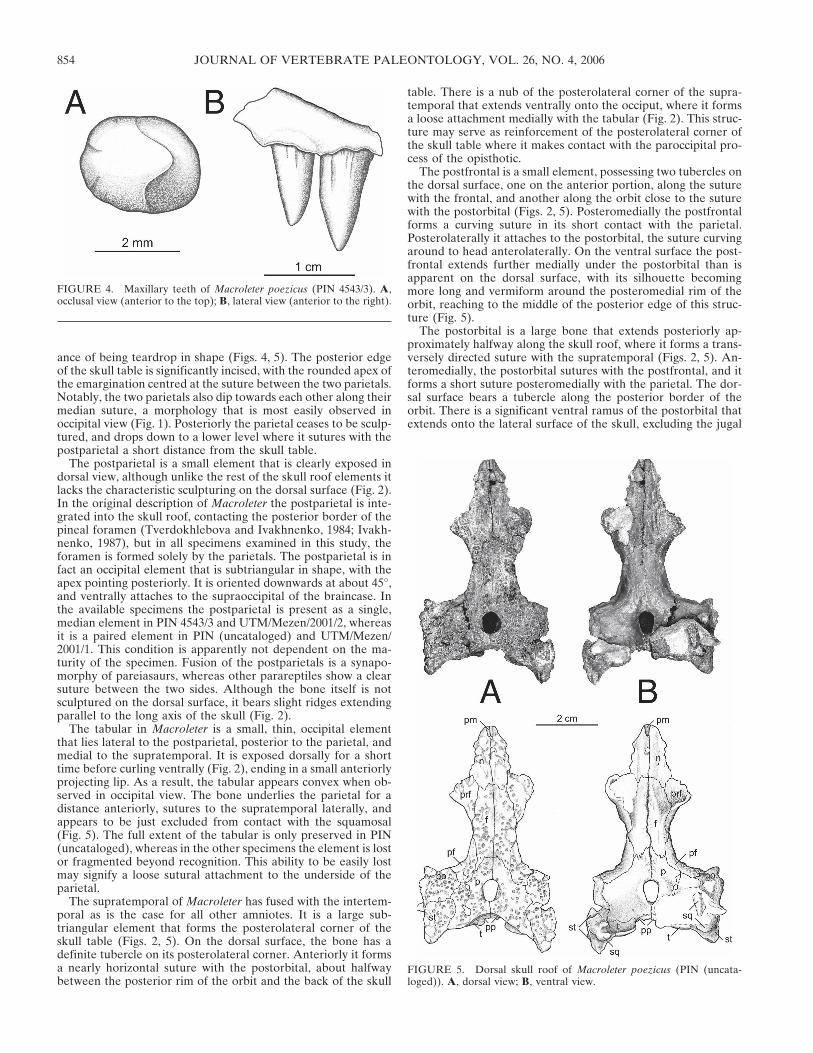
In the larger individuals, the maxilla bears 18–20 teeth. There
is a caniniform area in which the teeth are larger than the othersin the row, but there is no single caniniform tooth. In this areathe maxilla swells slightly laterally, indicating that these largerteeth were implanted deeply. Because of this implantation, thesnout region of the skull is slightly blunt anteriorly, a morphol-ogy that is similar to that of pareiasaurs. The teeth themselvesare fairly broad at the base and subcircular in cross section. Theyare slightly recurved, such that the tip just reaches the level of theposterior side of the tooth in occlusal view. The teeth also showa shovel-shaped morphology, being slightly mediolaterally com-pressed, with two faint cutting edges more closely placed relativeto each other on the lingual side of the tooth (Fig. 4). The baseof the majority of the teeth shows fluting consisting of longitu-dinal grooves of varying depths and heights, most visible on thelabial side. These grooves are very similar in morphology tothose seen in Colobomycter (Modesto, 1999), and millerettids(Gow, 1972). This fluting is also present on some of the largerpremaxillary teeth.
The lacrimal, in contrast with the condition seen in most earlytetrapods, is a shortened bone in most parareptiles, and is ex-cluded from contact with the naris in most parareptiles savemillerettids and pareiasaurs, though only barely so in the case ofMacroleter. It is quite a long bone nonetheless, extending fartheranteriorly than the prefrontal (Fig. 2), in contrast to the condi-tion depicted in other nycteroleterids (Ivakhnenko et al., 1997).About midway down the lateral surface of the lacrimal, theheavy sculpturing that characterizes the dermal skull roof ceases,and the element becomes very smooth, a texture which continues
FIGURE 1. Cranial reconstruction of Macroleter poezicus. A, dorsal view; B, ventral view; C, left lateral view; D, occipital view. Small dotted linesdelineate within element, non-sutural features, dashed lines represent inferred sutural contacts or element shape.
TSUJI—CRANIAL ANATOMY OF MACROLETER 851
on the lateral surface as it forms a short suture with the jugalalong the ventral margin of the orbit (Fig. 3). This suture extendsdown into the orbit to meet the palate. No lacrimal foramen canbe seen on the exposed posterior surface of the ventral process ofthe lacrimal, though this feature may lie further laterally and hasbeen obscured from view in these specimens. The foramen or-bitonasale is represented by a gap in the area around the con-fluence of the lacrimal, the prefrontal medially, and the under-lying palatine (Fig. 3).
As a pair, the nasals are roughly wing-shaped, their anteriorprocesses excluded from contact with each other for the firstthird by the dorsal process of the premaxilla (Fig. 2). From theirslightly pointed origin at the anterior end, the nasals widen lat-erally to form the medial and dorsal border of the naris. Poste-rior to this structure, the nasal forms a short suture with thedorsal process of the maxilla. Continuing posteriorly, the antero-
lateral edge of the nasal is formed primarily by a short undulat-ing suture with the lacrimal, and the lateral border is orientedslightly posteromedially as a suture with the prefrontal. The pos-terior border of the nasal is formed by contact with the frontalvia a horizontally directed undulating suture, with the two nasalsmeeting in a relatively straight median suture. The dorsal surfaceof the bone shows the distinctive sculpturing pattern, with anotable tubercle just posterior to the naris, whereas the ventralsurface is smooth (Fig. 5).
The prefrontal is a stout bone that forms the majority of theanteromedial border of the orbit (Figs. 2, 3, 5). It has a broad,robust ventral extension, which is sutured firmly to the palatine,very clearly demonstrated by PIN (uncataloged) (Fig. 3). Thisthickened state is common for parareptiles, including the bolo-saurids and procolophonids (Laurin and Reisz, 1995), and a firmsuture is also found in pareiasaurs (Lee, 1995). As the bone
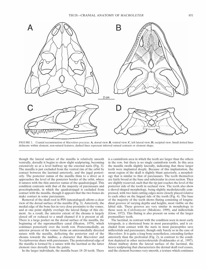
FIGURE 2. Skull of Macroleter poezicus (PIN 4543/3). A, dorsal view; B, ventral view.
JOURNAL OF VERTEBRATE PALEONTOLOGY, VOL. 26, NO. 4, 2006852
extends anteriorly along the skull roof, it is incised by the lacri-mal laterally, and appears slimmer on the dorsal surface. Ante-riorly, the prefrontal extends only two-thirds of the extent of thelacrimal onto the snout region. On each of the specimens exam-ined, there is a prominent tubercle on the prefrontal a shortdistance away from the orbital rim. In lateral view, the frontaldisplays a noticeable hump comprising the majority of the levelchange between the relatively flat snout area of the skull and theskull table (Fig. 3).
More than twice as long as it is wide, the frontal is subrectan-gular in shape, and noticeably longer than the nasal (Figs. 2, 5).Posterior to the nasal suture, the frontal widens very slightly untilit forms a small portion of the dorsal border of the orbit, al-though it is nearly excluded from this structure by the posteriorand anterior process of the prefrontal and the postfrontal, re-spectively. On its posterior half, the frontal meets the postfrontalin a primarily anteroposteriorly directed suture that extendsslightly medially, tapering the bone posteriorly. The suture withthe parietal initially extends laterally from the midline, butshortly curves posterolaterally, meeting the postfrontal-frontal
suture at an acute angle, the posterolateral edges of the frontalsthus wrapping back around the anterior extension of the pariet-als (Figs. 2, 5). There is a raised tubercle close to the lateral edgewhere it contributes to the orbit. A distinctive sub-circular dentis also present, centered just anterior to the fronto-parietal su-ture, in each of the observed specimens of Macroleter, includingthose specimens that show little distortion (Figs. 2, 5). This de-pression is bereft of sculpturing.
In dorsal view, the paired parietal is a roughly triangular ele-ment, forming an undulating, posterolaterally-oriented sutureanteriorly with the frontal (Figs. 2, 5). Ventrally, the parietalunderlaps the postorbital and the supratemporal so that it ap-pears larger on the ventral surface than it does dorsally, therebyhelping to reinforce the skull roof. A large pineal foramen lies onthe suture between the two parietals, displaced slightly anteriorlyin relation to the full size of the bone, though it appears to beposteriorly placed due to the median embayment of the skulltable. The foramen itself is laterally constricted by a small flangeon the ventral surface of the bone, directed slightly anterolater-ally from the posterior edge of the foramen, giving it the appear-
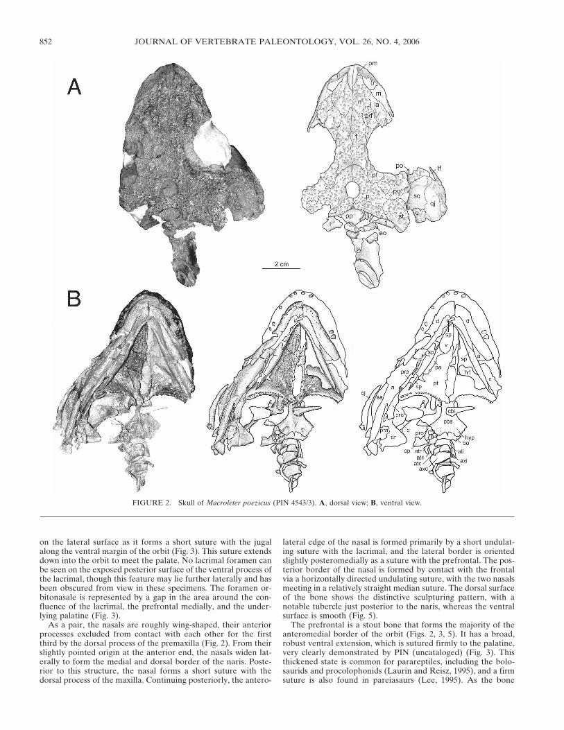
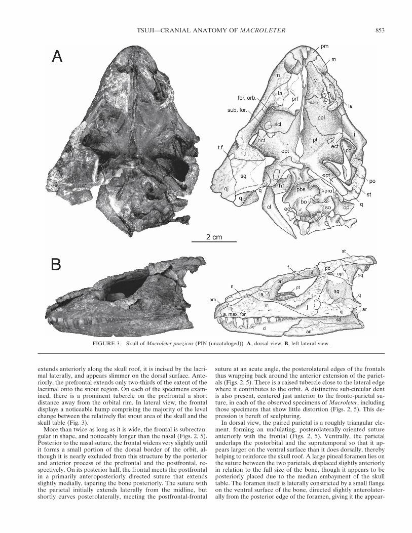
FIGURE 3. Skull of Macroleter poezicus (PIN (uncataloged)). A, dorsal view; B, left lateral view.
TSUJI—CRANIAL ANATOMY OF MACROLETER 853
ance of being teardrop in shape (Figs. 4, 5). The posterior edgeof the skull table is significantly incised, with the rounded apex ofthe emargination centred at the suture between the two parietals.Notably, the two parietals also dip towards each other along theirmedian suture, a morphology that is most easily observed inoccipital view (Fig. 1). Posteriorly the parietal ceases to be sculp-tured, and drops down to a lower level where it sutures with thepostparietal a short distance from the skull table.
The postparietal is a small element that is clearly exposed indorsal view, although unlike the rest of the skull roof elements itlacks the characteristic sculpturing on the dorsal surface (Fig. 2).In the original description of Macroleter the postparietal is inte-grated into the skull roof, contacting the posterior border of thepineal foramen (Tverdokhlebova and Ivakhnenko, 1984; Ivakh-nenko, 1987), but in all specimens examined in this study, theforamen is formed solely by the parietals. The postparietal is infact an occipital element that is subtriangular in shape, with theapex pointing posteriorly. It is oriented downwards at about 45°,and ventrally attaches to the supraoccipital of the braincase. Inthe available specimens the postparietal is present as a single,median element in PIN 4543/3 and UTM/Mezen/2001/2, whereasit is a paired element in PIN (uncataloged) and UTM/Mezen/2001/1. This condition is apparently not dependent on the ma-turity of the specimen. Fusion of the postparietals is a synapo-morphy of pareiasaurs, whereas other parareptiles show a clearsuture between the two sides. Although the bone itself is notsculptured on the dorsal surface, it bears slight ridges extendingparallel to the long axis of the skull (Fig. 2).
The tabular in Macroleter is a small, thin, occipital elementthat lies lateral to the postparietal, posterior to the parietal, andmedial to the supratemporal. It is exposed dorsally for a shorttime before curling ventrally (Fig. 2), ending in a small anteriorlyprojecting lip. As a result, the tabular appears convex when ob-served in occipital view. The bone underlies the parietal for adistance anteriorly, sutures to the supratemporal laterally, andappears to be just excluded from contact with the squamosal(Fig. 5). The full extent of the tabular is only preserved in PIN(uncataloged), whereas in the other specimens the element is lostor fragmented beyond recognition. This ability to be easily lostmay signify a loose sutural attachment to the underside of theparietal.
The supratemporal of Macroleter has fused with the intertem-poral as is the case for all other amniotes. It is a large sub-triangular element that forms the posterolateral corner of theskull table (Figs. 2, 5). On the dorsal surface, the bone has adefinite tubercle on its posterolateral corner. Anteriorly it formsa nearly horizontal suture with the postorbital, about halfwaybetween the posterior rim of the orbit and the back of the skull
table. There is a nub of the posterolateral corner of the supra-temporal that extends ventrally onto the occiput, where it formsa loose attachment medially with the tabular (Fig. 2). This struc-ture may serve as reinforcement of the posterolateral corner ofthe skull table where it makes contact with the paroccipital pro-cess of the opisthotic.
The postfrontal is a small element, possessing two tubercles onthe dorsal surface, one on the anterior portion, along the suturewith the frontal, and another along the orbit close to the suturewith the postorbital (Figs. 2, 5). Posteromedially the postfrontalforms a curving suture in its short contact with the parietal.Posterolaterally it attaches to the postorbital, the suture curvingaround to head anterolaterally. On the ventral surface the post-frontal extends further medially under the postorbital than isapparent on the dorsal surface, with its silhouette becomingmore long and vermiform around the posteromedial rim of theorbit, reaching to the middle of the posterior edge of this struc-ture (Fig. 5).
The postorbital is a large bone that extends posteriorly ap-proximately halfway along the skull roof, where it forms a trans-versely directed suture with the supratemporal (Figs. 2, 5). An-teromedially, the postorbital sutures with the postfrontal, and itforms a short suture posteromedially with the parietal. The dor-sal surface bears a tubercle along the posterior border of theorbit. There is a significant ventral ramus of the postorbital thatextends onto the lateral surface of the skull, excluding the jugal
FIGURE 4. Maxillary teeth of Macroleter poezicus (PIN 4543/3). A,occlusal view (anterior to the top); B, lateral view (anterior to the right).
FIGURE 5. Dorsal skull roof of Macroleter poezicus (PIN (uncata-loged)). A, dorsal view; B, ventral view.
JOURNAL OF VERTEBRATE PALEONTOLOGY, VOL. 26, NO. 4, 2006854
from forming most of the posterior margin of the orbit (Fig. 3).This ventral process is enforced by a thickening of the bone thatcan be seen extending into the orbit on its internal surface (Fig.3). The orbits are expanded similarly to procolophonids, more soon the posterodorsal edge, though not to the extent seen inNyctiphruretus.
The jugal forms most of the ventral rim of the orbit. It beginslong and thin anteriorly in a short suture with the lacrimal, andthen flares posteriorly, where it forms the anterior border of thesmall temporal fenestra of Macroleter (Figs. 2, 3). The bone in-creases in height posteriorly, forming a gently wavering, pos-terodorsally directed suture with the squamosal. The tall dorsalexpansion of the jugal forms the ventral part of the posterioredge of the orbit, but the bone does not reach the skull table, asthe anteroventrally directed suture with the postorbital termi-nates its dorsal extent. Posteriorly, the jugal is just excluded fromthe temporal emargination. On the internal (medial) surface, thejugal sutures anteriorly and medially with the palatine, and fur-ther posteriorly with the small ectopterygoid (Fig. 3).
A prominent bumpy ridge of sculpturing projects laterally aconsiderable distance at the posteroventral border of the orbit,and consists of a series of tubercles and pits. Although the jugalis sculptured on its entire lateral surface, this sculpturing doesnot extend very far ventrally, and only very sparsely under theaforementioned ornamentation (Fig. 3). This morphology maybe homologous to the ornamentation in the form of bosses seenin pareiasaurs.
The squamosal is a broad plate of bone that forms a largesection of the temporal emargination, and has a restricted dorsalcontribution to the temporal fenestra (Figs. 2, 3). It is firmlysutured to the underside of the skull roof, and the detached skulltable of PIN (uncataloged) allows for the examination of thisarea of the squamosal in ventral view. The articulation of thedermal skull roof and palate with the paroccipital process of thebraincase is very complicated, with the confluence of the ptery-goid, the quadrate, the squamosal, the tabular, and the supra-temporal within a small area. The squamosal is broadly suturedto the ventral surface of the supratemporal, a short distance fromthe posterolateral corner. There is a structure that has doublecondyle-like processes, a larger posterior one and a smaller an-terior one, separated by a groove into which the paroccipitalprocess of the opisthotic fits. The notch itself is directed at anangle, twisted slightly from the short axis of the skull (Fig. 5).
Laterally, the squamosal defines the majority of the temporalemargination, and sutures posteriorly with the quadrate, which itoverlaps to a large extent as the latter bone extends anteriorly tosuture with the pterygoid. The ventral suture with the quadra-tojugal gently undulates, and is directed slightly posterodorsally.A large portion of the squamosal in the area of the temporalemargination is unsculptured. There is a thick ridge borderingthe posterior part of the temporal emargination, possibly ho-mologous to the tympanic ridge. A posterolaterally directedflange of the squamosal wraps on to the occiput, but this areacannot be observed directly because none of the specimens areexposed in occipital view. The quadrate foramen has been ex-posed in PIN (uncataloged), however, and is contained betweenthe occipital flange of the squamosal, the quadrate, and thequadratojugal (Fig. 1).
The quadratojugal is thin anteriorly where it sutures to theposterior process of the jugal, but it expands posteriorly to aheight that is about half of the total length of the bone, a mor-phology that is typical of parareptiles (Laurin and Reisz, 1995)(Figs. 2, 3). Its anterior edge forms the posterior border of thesmall temporal fenestra, with a process extending ventral andanterior to this structure to contact the maxilla. The quadrato-jugal forms the ventral portion of the temporal emargination,with its posterior edge overlapping the quadrate. The free pos-teroventral edge of the quadratojugal extends slightly below the
ventral edge of the maxilla, and on this portion of the bone thesculpturing becomes more ornate, with prominent tubercles vis-ible on the ventrolateral flange, especially towards the posterioredge (Figs. 2, 3).
Palate—The vomer, the most anterior element of the palate,carries a shagreen of denticles on the ventral surface, althoughthe anterior portion is obscured in PIN 4543/3 by the firm ad-duction of the mandible to the skull (Fig. 2). The vomer suturesanteriorly to the premaxilla and forms the anterolateral edge ofthe choana. Posterior to its suture with the premaxilla the vomerexpands laterally, a structure termed the alar flange by Damianiand Modesto (2001). This flange results in the appearance of aninwardly curving, medially incised choana, a morphology that isa distinctive feature of pareiasaurs and some testudines. Thevomer is very wide in Macroleter such that the alar flange closesoff most of the anterior extent of the choana (Fig. 3). The ventralsurface of the palate displays a prominent ridge of denticles oneach side extending posteriorly along the suture between the pairof vomers. This ridge divides into two parallel ridges about half-way along the bone, and continues onto the pterygoid. The dor-sal surface of the bone is smooth but bears, along with the pala-tine, part of the curving orbitonasal ridge for the articulation ofthe orbitonasal membrane, forming a depression for the para-septal cartilage of the nasal capsule, a morphology very similar tothat seen in captorhinids (Heaton, 1979). The vomer also dis-plays a median dorsal flange that contributes to the nasal septum(Fig. 3). This distinctive feature was recently proposed as anautapomorphy of the pareiasaurian vomer (Damiani andModesto, 2001), and its presence in Macroleter, along with theextensive alar flange also thought to be a pareiasaurian autapo-morphy, serves to strengthen the plausibility of a close relation-ship between these taxa.
The palatine is a partially denticulate element articulating an-teromedially with the vomer (Figs. 2, 3). Posteromedially thepalatine forms a suture with the pterygoid, and its posterolateralborder is formed on the dorsal surface by a suture with theectopterygoid. However, the exact position of this latter suturecannot be seen in ventral view because the ectopterygoid is miss-ing from PIN 4543/3. The ventral surface of the palatine bearsdenticles, most notably on the continuation of the anterolaterallydirected ridges from the pterygoid in addition to a field of smalldenticles scattered over the surface (Fig. 2). Dorsally the palatinebears the posterior portion of the orbitonasal ridge that contin-ues from the vomer. An anterior process of the palatine extendsalong the lateral edge of the choana to suture with the maxilla,approximately halfway to the front of the dorsal exposure of thechoana. This morphology results in the palatine forming the en-tire posterior, and part of the lateral border of the choana, afeature also found in pareiasaurs and some testudines (Gaffney,1979; Lee 1995, 1997a). Because of the wedge-shaped anteriorextent of this process the internal naris does not run parallel tothe maxilla for its entire extent, but rather is inflected mediallyalong its posterior edge.
The pterygoid is the dominant element of the palate in Macro-leter, and is broadly triangular in shape, contacting the vomeranteriorly, the palatine anterolaterally, the quadrate posterolat-erally, and articulating posteriorly with the parabasisphenoid(Figs. 2, 3). On the ventral surface there is a high ridge bearingdenticles that extends on a diagonal anterolaterally from close tothe midline, dividing into two parallel ridges a short distancefrom its origin, which continue onto the palatine. The ventralsurface of the pterygoid also bears a denticulated double ridgethat lines the edge of the interpterygoid vacuity, continuing an-teriorly along the median edge of each pterygoid and rostrallyonto the vomer. There are denticles scattered over the anteriorramus of the pterygoid, but the remainder of the element islargely devoid of denticles. The suture between the pterygoidand the palatine on the ventral surface of the palate is angled
TSUJI—CRANIAL ANATOMY OF MACROLETER 855
anteromedially, with the anterior process of the pterygoid ex-tending just past the posterior end of the choana. On the dorsalsurface seen in PIN (uncataloged), the pterygoid presents a tallthin flange of bone dorsally, which continues anteriorly past themain portion of the bone, medial to the vomer. This flange in-terdigitates with a smaller, but similar, flange of the vomer thatprojects posteriorly (Fig. 3). The combination of these twoflanges serves to divide the interorbital region posteriorly, andanteriorly forms the ventral osseous portion of the internarialseptum.
The two pterygoids meet anterior and posterior to the veryshort interpterygoid vacuity (Fig. 3), which is just less than 15%of the total length of the skull in PIN (uncataloged). A shortinterpterygoid vacuity is a character that defines the group in-cluding the bolosaurids, procolophonids and pareiasaurs. Thedorsal surface of the pterygoid is smooth, and shows an oddsuture pattern with the ectopterygoid. The pterygoid, in concertwith the palatine, appears to incise the dorsal exposure of theectopterygoid, dividing the latter bone into two sections.
The transverse flange of the pterygoid is recurved, and bearslarge and well-formed teeth. The largest of these teeth are gentlyfluted at the base, bearing a resemblance to those in the maxil-lary tooth row. These teeth also possess slight cutting edges, andappear to be implanted in a subthecodont fashion, with definitesockets visible where teeth have been lost (Fig. 2). Unlike mostprocolophonids, the transverse flange is extensively recurved,such that it is pointing almost directly laterally, and is ratherlarge. This character has often been used to place the nycterol-eterids in a more basal position within parareptiles, the morederived of which show an anteriorly or anterolaterally directedtransverse flange (Laurin and Reisz, 1995; Lee, 1995).
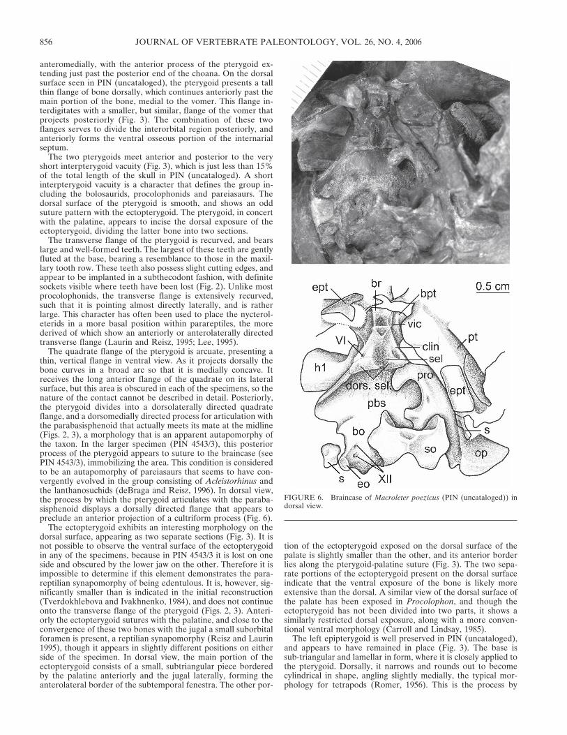
The quadrate flange of the pterygoid is arcuate, presenting athin, vertical flange in ventral view. As it projects dorsally thebone curves in a broad arc so that it is medially concave. Itreceives the long anterior flange of the quadrate on its lateralsurface, but this area is obscured in each of the specimens, so thenature of the contact cannot be described in detail. Posteriorly,the pterygoid divides into a dorsolaterally directed quadrateflange, and a dorsomedially directed process for articulation withthe parabasisphenoid that actually meets its mate at the midline(Figs. 2, 3), a morphology that is an apparent autapomorphy ofthe taxon. In the larger specimen (PIN 4543/3), this posteriorprocess of the pterygoid appears to suture to the braincase (seePIN 4543/3), immobilizing the area. This condition is consideredto be an autapomorphy of pareiasaurs that seems to have con-vergently evolved in the group consisting of Acleistorhinus andthe lanthanosuchids (deBraga and Reisz, 1996). In dorsal view,the process by which the pterygoid articulates with the paraba-sisphenoid displays a dorsally directed flange that appears topreclude an anterior projection of a cultriform process (Fig. 6).
The ectopterygoid exhibits an interesting morphology on thedorsal surface, appearing as two separate sections (Fig. 3). It isnot possible to observe the ventral surface of the ectopterygoidin any of the specimens, because in PIN 4543/3 it is lost on oneside and obscured by the lower jaw on the other. Therefore it isimpossible to determine if this element demonstrates the para-reptilian synapomorphy of being edentulous. It is, however, sig-nificantly smaller than is indicated in the initial reconstruction(Tverdokhlebova and Ivakhnenko, 1984), and does not continueonto the transverse flange of the pterygoid (Figs. 2, 3). Anteri-orly the ectopterygoid sutures with the palatine, and close to theconvergence of these two bones with the jugal a small suborbitalforamen is present, a reptilian synapomorphy (Reisz and Laurin1995), though it appears in slightly different positions on eitherside of the specimen. In dorsal view, the main portion of theectopterygoid consists of a small, subtriangular piece borderedby the palatine anteriorly and the jugal laterally, forming theanterolateral border of the subtemporal fenestra. The other por-
tion of the ectopterygoid exposed on the dorsal surface of thepalate is slightly smaller than the other, and its anterior borderlies along the pterygoid-palatine suture (Fig. 3). The two sepa-rate portions of the ectopterygoid present on the dorsal surfaceindicate that the ventral exposure of the bone is likely moreextensive than the dorsal. A similar view of the dorsal surface ofthe palate has been exposed in Procolophon, and though theectopterygoid has not been divided into two parts, it shows asimilarly restricted dorsal exposure, along with a more conven-tional ventral morphology (Carroll and Lindsay, 1985).
The left epipterygoid is well preserved in PIN (uncataloged),and appears to have remained in place (Fig. 3). The base issub-triangular and lamellar in form, where it is closely applied tothe pterygoid. Dorsally, it narrows and rounds out to becomecylindrical in shape, angling slightly medially, the typical mor-phology for tetrapods (Romer, 1956). This is the process by
FIGURE 6. Braincase of Macroleter poezicus (PIN (uncataloged)) indorsal view.
JOURNAL OF VERTEBRATE PALEONTOLOGY, VOL. 26, NO. 4, 2006856
which the palate would have braced against the skull roof, withthe unfinished dorsal tip in Macroleter indicative that this contactwas most likely via a cartilaginous connection in this animal. Inlateral view the epipterygoid can be seen just poking out abovethe broken portion of the skull (Fig. 3), and falls short of makingcontact with the skull roof. There is also no indication on theunderside of the skull roof where this attachment would haveoccurred, supporting the inference that the articulation was car-tilaginous. In Macroleter, the epipterygoid does not take part inthe basipterygoid articulation.
The quadrate is largely obscured from ventral view in thespecimens available for examination at this time due to the at-tachment of the mandible to the skull. A sliver of the quadratecan be seen in lateral view (Fig. 3), but the posterior flanges ofthe quadratojugal and the squamosal cover the majority of thelateral surface. The occipital flange of the squamosal obscuresthe quadrate as it extends dorsally, with the quadrate interposedbetween the squamosal medially and the pterygoid laterally as itapproaches the skull roof. The anterior ramus of the quadrate bywhich it contacts the pterygoid is long, extending approximatelytwo-thirds of the length of the pterygoid, and it broadly overlapsthis bone medially.
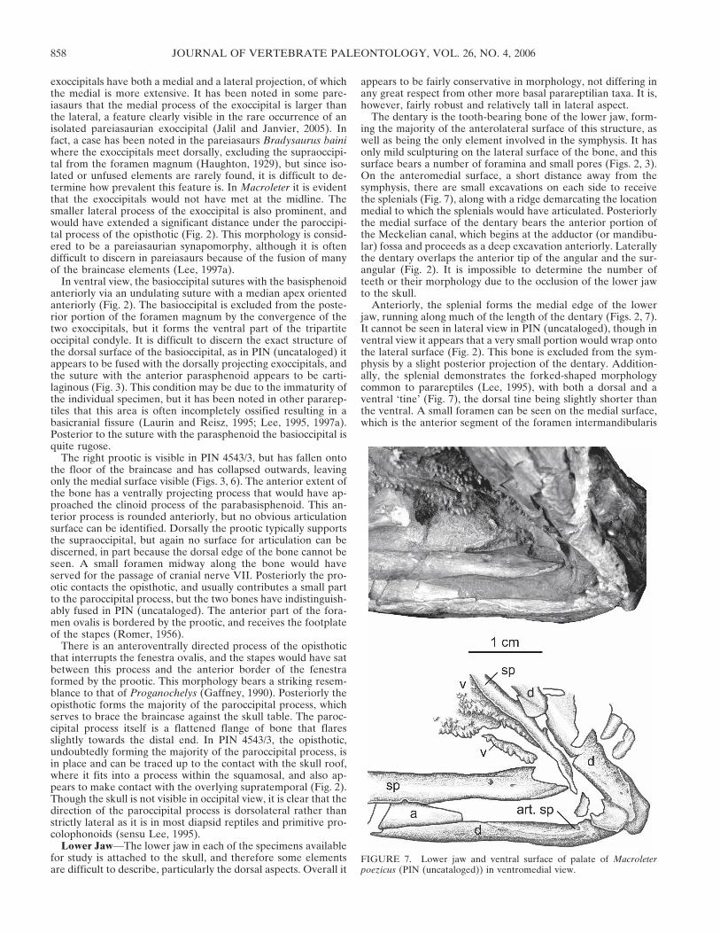
Braincase—There is a rare opportunity to examine the brain-case of Macroleter whereby the skull roof can be removed (andreplaced) in PIN (uncataloged). The braincase can thus be ob-served in dorsal view, and this feature, combined with the well-preserved ventral surface of PIN 4543/3, allows for a thoroughdescription. Detailed descriptions of parareptilian braincases arerare (Ivakhnenko, 1973; Kemp, 1974; Spencer, 2000), but most ofthe general features and much of the anatomy is comparable tomodern reptiles (Carroll and Lindsay, 1985). Despite the factthat PIN (uncataloged) is immature and lacks some fully ossifiedbones, many of the elements of the braincase have fused, sug-gesting that those of a full-sized individual would have beenquite massive and fully fused.
The parasphenoid consists of the sheath of dermal bone un-derlying most of the braincase and forming the cultriform pro-cess when this structure is present. This bone is partially suturedto the basisphenoid, and in many parts of the braincase these twoelements are indistinguishable. Unless specifically referring toone or the other of the fused elements, the structure will hence-forth be referred to as the parabasisphenoid.
The single, median parabasisphenoid is a complicated struc-ture. Anteriorly, the basipterygoid processes of Macroleter showa different morphology from those of known procolophonids(Ivakhnenko, 1973; Carroll and Lindsay, 1985; Spencer, 2000), asthose of the latter project anterolaterally from their origin on theanteromedial surface of the parabasisphenoid, and are separatedmedially by the basisphenoid rostrum (sensu Spencer, 2000),which normally bears the cultriform process. The basipterygoidprocesses of Macroleter, on the other hand, are small and projectdirectly anteriorly, with only a small space between them. Ven-trally, the suture between the parabasisphenoid and the ptery-goids is tightly convoluted, and the processes themselves are veryshort (Fig. 2). A great deal of infolding can be seen on the ventralsurface in PIN 4543/3, with the basipterygoid processes suturedfirmly to the pterygoid, and there is no doubt that very littlekinesis was possible in this area. This area does not appear to beso closely attached in the dorsal view of PIN (uncataloged), al-though the basipterygoid processes are still short and anteriorlydirected with little separation between them (Fig. 6). This dif-ference in joint morphology may be because the latter specimenis immature, and the area has yet to fully ossify. A fused basi-pterygoid joint is a synapomorphy of pareiasaurs, and also occursconvergently in Lanthanosuchus. Its presence in Macroleter isrecognized here for the first time.
At the anteromedial end of the parabasisphenoid is the dorsalbody, or basisphenoid rostrum (Fig. 6). It is from this structure
that the cultriform process of the parasphenoid extends whenpresent, but similar to the morphology of Leptopleuron (Spen-cer, 2000), there is no discernable trace of this structure in Mac-roleter. Rather, the dorsal body extends anterodorsally in highrelief, and does not continue anteriorly. The cultriform process isa shortened structure in procolophonids and pareiasaurs, butrarely to the extent seen here. Caudal to the dorsal body lies asignificant depression, the sella turcica (termed the retractor pitby Heaton, 1979), in which would have lain the pituitary body(Fig. 6). Rising posterodorsally on either side of this depressionare the clinoid processes, which are smooth and rounded on themedial and lateral sides, and flare both dorsally and mediolater-ally from their origin at the base of the dorsal body. There is aforamen piercing the posterodorsal end of the clinoid processesfor the passage of cranial nerve VI, almost identical to the mor-phology seen in millerettids (Gow, 1972). The nerve would havepassed from the floor of the braincase posteriorly through theforamina to the pituitary fossa then extended laterally into theorbit (Romer, 1956). The two clinoid processes are generallyconnected transversely by a vertical sheath of bone; the dorsumsellae. In PIN (uncataloged), however, the clinoid processes endin unfinished bone, and no transverse sheet is present. It is pos-sible that the dorsum sellae was lost or broken, or it is also likelythat it had yet to fully ossify in this specimen.
The parasphenoid sheath of this bone complex is visible inventral view, and consists of a constricted waist that flares pos-teriorly (Figs. 2, 6). A notable feature of the ventral surface ofthis element is the presence of paired excavations for the inser-tion of the hypaxial cervical musculature, almost identical tothose found in mesosaurs (Modesto, 2006). Most other para-reptiles lack this feature, with the ventral surface of the para-sphenoid being smooth, or bearing a paired or a single mediantubercle. Posteriorly the parasphenoid sutures with the basi-occipital.
The supraoccipital is a single, median element that is dorso-ventrally flattened. It is present in PIN (uncataloged), but is notin place because it has fallen anteriorly, leaving only the dorsalsurface readily available for observation (Figs. 3, 6). The elementis slightly ventrally concave, and contains a low median sagittalridge that is typical for parareptiles, as is the notable reduction inthe lateral extent of the bone. There is an anterior semi-circularcrest whose apex is centred on the median sagittal ridge (Fig. 6),which serves as the surface for articulation dorsally with thepostparietal. The supraoccipital, when in place, would haveangled posteroventrally, with the posterior edge forming the dor-sal border of the foramen magnum. A central notch in the pos-terior edge of the bone results in a foramen magnum that hasbeen reconstructed as keyhole shaped (Fig. 1).
The left exoccipital is exposed in dorsal view in PIN (uncata-loged) (Fig. 6), and in PIN 4543/3 careful preparation has re-vealed much of the dorsal and part of the lateral surface. Nosuture can be discerned between the exoccipital and the basi-occipital on the lateral surface, and perhaps because PIN (un-cataloged) is a juvenile, most of the dorsal process for articula-tion with the supraoccipital has been broken, or was not fullyossified and subsequently drifted away. In PIN 4543/3, however,nearly the entirety of the exoccipitals can be seen, revealing thedorsal, and a good deal of the lateral surface of the bones. At thebase of the exoccipital where it meets the basioccipital, two fo-ramina are present for the passage of cranial nerve XII. Two ofthese foramina can be seen on the medial surface in PIN (un-cataloged), and presumably the same two can be seen continuingthrough to the lateral surface in PIN 4543/3.
The two exoccipitals meet below the foramen magnum, ex-cluding the basioccipital from the posterior part of this struc-ture (Fig. 2). From their ventral origin in chunky, boot shapedbases, the exoccipitals rise as small columns that form the lateralmargins of the foramen magnum. The dorsal processes of the
TSUJI—CRANIAL ANATOMY OF MACROLETER 857
exoccipitals have both a medial and a lateral projection, of whichthe medial is more extensive. It has been noted in some pare-iasaurs that the medial process of the exoccipital is larger thanthe lateral, a feature clearly visible in the rare occurrence of anisolated pareiasaurian exoccipital (Jalil and Janvier, 2005). Infact, a case has been noted in the pareiasaurs Bradysaurus bainiwhere the exoccipitals meet dorsally, excluding the supraoccipi-tal from the foramen magnum (Haughton, 1929), but since iso-lated or unfused elements are rarely found, it is difficult to de-termine how prevalent this feature is. In Macroleter it is evidentthat the exoccipitals would not have met at the midline. Thesmaller lateral process of the exoccipital is also prominent, andwould have extended a significant distance under the paroccipi-tal process of the opisthotic (Fig. 2). This morphology is consid-ered to be a pareiasaurian synapomorphy, although it is oftendifficult to discern in pareiasaurs because of the fusion of manyof the braincase elements (Lee, 1997a).
In ventral view, the basioccipital sutures with the basisphenoidanteriorly via an undulating suture with a median apex orientedanteriorly (Fig. 2). The basioccipital is excluded from the poste-rior portion of the foramen magnum by the convergence of thetwo exoccipitals, but it forms the ventral part of the tripartiteoccipital condyle. It is difficult to discern the exact structure ofthe dorsal surface of the basioccipital, as in PIN (uncataloged) itappears to be fused with the dorsally projecting exoccipitals, andthe suture with the anterior parasphenoid appears to be carti-laginous (Fig. 3). This condition may be due to the immaturity ofthe individual specimen, but it has been noted in other pararep-tiles that this area is often incompletely ossified resulting in abasicranial fissure (Laurin and Reisz, 1995; Lee, 1995, 1997a).Posterior to the suture with the parasphenoid the basioccipital isquite rugose.
The right prootic is visible in PIN 4543/3, but has fallen ontothe floor of the braincase and has collapsed outwards, leavingonly the medial surface visible (Figs. 3, 6). The anterior extent ofthe bone has a ventrally projecting process that would have ap-proached the clinoid process of the parabasisphenoid. This an-terior process is rounded anteriorly, but no obvious articulationsurface can be identified. Dorsally the prootic typically supportsthe supraoccipital, but again no surface for articulation can bediscerned, in part because the dorsal edge of the bone cannot beseen. A small foramen midway along the bone would haveserved for the passage of cranial nerve VII. Posteriorly the pro-otic contacts the opisthotic, and usually contributes a small partto the paroccipital process, but the two bones have indistinguish-ably fused in PIN (uncataloged). The anterior part of the fora-men ovalis is bordered by the prootic, and receives the footplateof the stapes (Romer, 1956).
There is an anteroventrally directed process of the opisthoticthat interrupts the fenestra ovalis, and the stapes would have satbetween this process and the anterior border of the fenestraformed by the prootic. This morphology bears a striking resem-blance to that of Proganochelys (Gaffney, 1990). Posteriorly theopisthotic forms the majority of the paroccipital process, whichserves to brace the braincase against the skull table. The paroc-cipital process itself is a flattened flange of bone that flaresslightly towards the distal end. In PIN 4543/3, the opisthotic,undoubtedly forming the majority of the paroccipital process, isin place and can be traced up to the contact with the skull roof,where it fits into a process within the squamosal, and also ap-pears to make contact with the overlying supratemporal (Fig. 2).Though the skull is not visible in occipital view, it is clear that thedirection of the paroccipital process is dorsolateral rather thanstrictly lateral as it is in most diapsid reptiles and primitive pro-colophonoids (sensu Lee, 1995).
Lower Jaw—The lower jaw in each of the specimens availablefor study is attached to the skull, and therefore some elementsare difficult to describe, particularly the dorsal aspects. Overall it
appears to be fairly conservative in morphology, not differing inany great respect from other more basal parareptilian taxa. It is,however, fairly robust and relatively tall in lateral aspect.
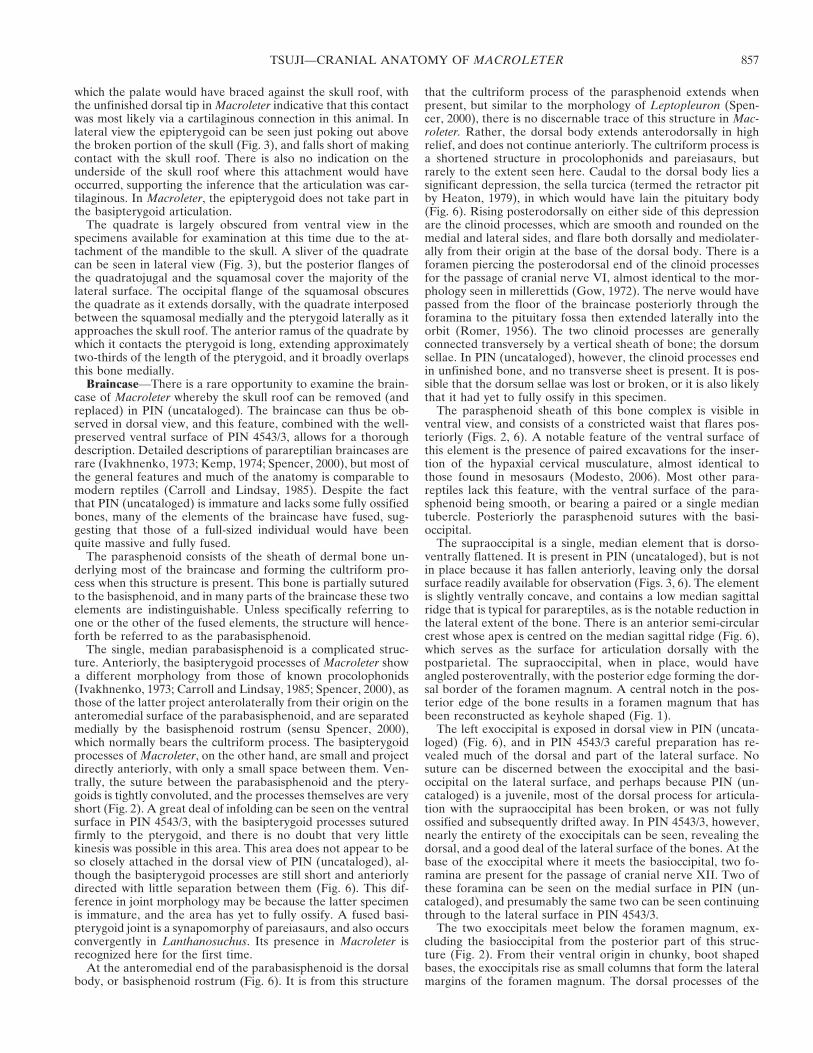
The dentary is the tooth-bearing bone of the lower jaw, form-ing the majority of the anterolateral surface of this structure, aswell as being the only element involved in the symphysis. It hasonly mild sculpturing on the lateral surface of the bone, and thissurface bears a number of foramina and small pores (Figs. 2, 3).On the anteromedial surface, a short distance away from thesymphysis, there are small excavations on each side to receivethe splenials (Fig. 7), along with a ridge demarcating the locationmedial to which the splenials would have articulated. Posteriorlythe medial surface of the dentary bears the anterior portion ofthe Meckelian canal, which begins at the adductor (or mandibu-lar) fossa and proceeds as a deep excavation anteriorly. Laterallythe dentary overlaps the anterior tip of the angular and the sur-angular (Fig. 2). It is impossible to determine the number ofteeth or their morphology due to the occlusion of the lower jawto the skull.
Anteriorly, the splenial forms the medial edge of the lowerjaw, running along much of the length of the dentary (Figs. 2, 7).It cannot be seen in lateral view in PIN (uncataloged), though inventral view it appears that a very small portion would wrap ontothe lateral surface (Fig. 2). This bone is excluded from the sym-physis by a slight posterior projection of the dentary. Addition-ally, the splenial demonstrates the forked-shaped morphologycommon to parareptiles (Lee, 1995), with both a dorsal and aventral ‘tine’ (Fig. 7), the dorsal tine being slightly shorter thanthe ventral. A small foramen can be seen on the medial surface,which is the anterior segment of the foramen intermandibularis
FIGURE 7. Lower jaw and ventral surface of palate of Macroleterpoezicus (PIN (uncataloged)) in ventromedial view.
JOURNAL OF VERTEBRATE PALEONTOLOGY, VOL. 26, NO. 4, 2006858
(the foramen intermandibularis oralis of Heaton, 1979). The pos-terior part of the intermandibular foramen (foramen interman-dibularis caudalis of Heaton, 1979) can be seen enclosed be-tween the splenial, prearticular, and angular. Posteriorly thesplenial overlaps the anterior end of the prearticular.
Very little of the coronoid is visible in the available specimensof Macroleter, although the top of the coronoid process can beseen in the dorsal view of PIN (uncataloged) (Fig. 3). This pro-cess appears to be well defined, and composed primarily of thesingle coronoid.
The anterior part of the prearticular lies between the splenialmedially and the angular laterally. The posterior end of theprearticular of PIN 4543/3 has been fragmented, but can be seento have covered the medial surface of the articular. The posteriorend closely approaches but does not extend beyond the base ofthe retroarticular process.
The surangular forms the dorsal part of the lateral surface ofthe mandible, and is slightly sculptured on its lateral surface(Figs. 2, 3). It sutures ventrally with the angular, and its posteriorborder overlies the lateral exposure of the articular. Also on thelateral surface, the dorsal portion of the surangular is deflectedoutwards, forming a shelf. The size of this feature, however, mayhave been exaggerated in PIN 4543/3 and PIN (uncataloged) dueto the crushing associated with the skull being forced down ontothe lower jaw.
The large angular forms the ventral extent of the lateral sur-face of the lower jaw, constituting about half its total length(Figs. 2, 3). Its lateral surface is characterized by faint sculpturingconsisting of a series of shallow intricate ridges. Dorsally, theangular forms a roughly horizontal, though slightly wavering su-ture with the surangular. The angular sutures anteriorly with thedentary and underlies it as the dentary wraps around the ventralsurface of the jaw. Ventrally, the angular can be seen extendingmedial to the dentary, its anterior extent covered by an overlap-ping portion of the splenial (Fig. 2).
A small sliver of the articular is visible on the lateral surface ofthe lower jaw, and is just visible projecting posterior to the an-gular (Fig. 3). The retroarticular process can be partially recon-structed, because both the lateral and a portion of the medialsurfaces can be seen in two of the specimens. It appears to retainthe procolophonian morphology of being transversely expandedand dorsally concave (Laurin and Reisz, 1995). In PIN 4543/3, adorsal projection of the articular has been crushed inwards, withthe mesial side of this process becoming visible in ventral view(Fig. 2). It is clear from what can be seen in this view that theretroarticular process is concave dorsally from the area of articu-lation, ending in a tall pointed process. This process is overlainmedially by a thin sheath of the prearticular, but it is quite largeand robust considering the size of the animal.
Hyoid Apparatus—Macroleter possesses a large ossified hyoidapparatus, a feature shared with a number of other parareptiles,including Procolophon, Owenetta, and various pareiasaurs(Lee, 1995; Reisz and Scott, 2002). The larger, median element,which is referred to by some as the copula or the corpus hyoi-deum (Reisz and Scott, 2002), is approximately 16 mm in width,and is shaped like a bow tie, with a central constriction, and twoflaring ends (Fig. 2). The ossification of this element may be acommon feature of the Parareptilia, as first noted by Gow(1977), but as it is not sutured to any other element, it is easilylost. This structure exists in modern reptiles but is primarilycartilaginous. A potential ossified basibranchial has recentlybeen found in the early eureptile Protorothyris (J. Müller, pers.comm., November 2005), suggesting its ossification may in factbe primitive for early amniotes. The other element of the hyoidapparatus is the ceratohyal, which is roughly an elongated coneshape (Fig. 2). This paired element would have attached to eitherend of the copula.
PHYLOGENETIC ANALYSIS
A phylogenetic analysis of 16 taxa was completed to determinethe position of Macroleter poezicus within the parareptiles. Thir-teen Paleozoic and Early Mesozoic ingroup taxa were consideredin the present analysis: Eunotosaurus, Millerettidae, Acleistorhi-nus, Lanthanosuchus, Eudibamus, Belebey, Procolophon, Owen-etta, Barasaurus, Nyctiphruretus, Macroleter poezicus, Bradysau-rus, and Scutosaurus. Three outgroup taxa, Synapsida, Capto-rhinidae, and Mesosauridae, were included in the analysis.Synapsida is a large, diverse group that was scored following themethodology of Laurin and Reisz (1995), whereby coding wasbased on a composite of four basal synapsid groups to try todetermine the ancestral state for the node.
The analysis consisted of 89 characters (see Appendix 1 forcharacter list, Appendix 2 for character matrix). The majority ofthe characters were taken from Reisz and colleagues (in press),which was in turn based on Laurin and Reisz (1995), with twonew characters added (40, 45). Since this investigation involvedthe study of the cranial anatomy of Macroleter, the focus was onthe addition and redefinition of cranial characters. Charactersfor taxa other than Macroleter were scored either based on in-formation from previous analyses or descriptions of specific taxain the literature (including Laurin and Reisz, 1995; Lee, 1995,1997a, b; Meckert, 1995; deBraga and Reisz, 1996; deBraga andRieppel, 1997; Modesto, 1999a, 2006; Reisz et al., in press). Thedata set was analysed using the branch-and-bound parsimonyalgorithm in PAUP* 4.0b10 (Swofford, 2002). Character stateswere optimized using the DELTRAN algorithm, were allweighted equally, and left unordered. To assess the robustness ofthe topology, a bootstrap analysis was run using a heuristicsearch with random step-wise addition, with 100 repetitions for1000 replicates. A decay analysis was also performed.
Results
A single most parsimonious tree was found, with a length of205 steps (Fig. 8). The consistency index (CI) was 0.5561, and0.5473 when uninformative characters were excluded. The ho-moplasy index (HI) was 0.4439. The retention index (RI) was0.6750, and the rescaled consistency index (RC) was 0.3754. Theresults of the bootstrap analysis, along with the decay indices aredisplayed in Figure 8. Nodes were assigned letters for discussion.
DISCUSSION
Phylogenetic Implications
The phylogeny produced by this analysis differs substantiallyfrom that of other recent studies (Lee, 1997a; Jalil and Janvier,2005), which suggested that a strongly supported, monophyleticNyctiphruretia (including the nyctiphruretids and the nycterol-eterids) formed the sister group to Clade H (Lee, 1997a). Thepresent study does not replicate these results, although this maybe in part because ‘nyctiphruretians’ are poorly known in theliterature. The results of this analysis preclude a monophyleticNyctiphruretia; forcing Macroleter and Nyctiphruretus to bemonophyletic resulted in a tree three steps longer than the 205 ofthe most parsimonious reconstruction. However, 19.1% of thecharacters could not be scored for Nyctiphruretus, including alllower jaw characters, because no description or illustration ofthese elements exists in the literature. This missing data may bethe cause of the low bootstrap support (45%) for Node K. Al-though it has been argued that the presence of missing data haslittle influence on the topology of a tree, assuming the charactershave been scored correctly (Kearney, 2002; Kearney and Clark,2003; Müller, 2004), the inclusion of more data would likely ei-ther improve support for the current grouping, or support amonophyletic Nyctiphruretia. Indeed, if Nyctiphruretus is re-
TSUJI—CRANIAL ANATOMY OF MACROLETER 859
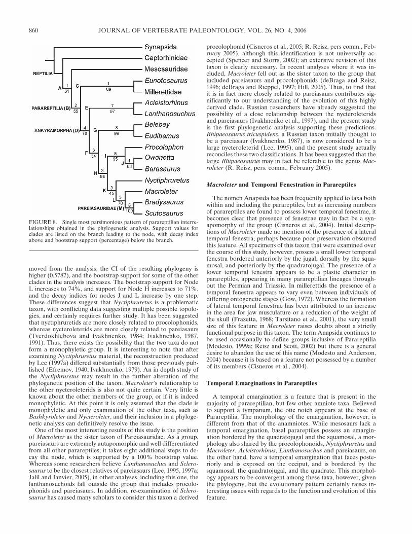
moved from the analysis, the CI of the resulting phylogeny ishigher (0.5787), and the bootstrap support for some of the otherclades in the analysis increases. The bootstrap support for NodeL increases to 74%, and support for Node H increases to 71%,and the decay indices for nodes J and L increase by one step.These differences suggest that Nyctiphruretus is a problematictaxon, with conflicting data suggesting multiple possible topolo-gies, and certainly requires further study. It has been suggestedthat nyctiphruretids are more closely related to procolophonids,whereas nycteroleterids are more closely related to pareiasaurs(Tverdokhlebova and Ivakhnenko, 1984; Ivakhnenko, 1987,1991). Thus, there exists the possibility that the two taxa do notform a monophyletic group. It is interesting to note that afterexamining Nyctiphruretus material, the reconstruction producedby Lee (1997a) differed substantially from those previously pub-lished (Efremov, 1940; Ivakhnenko, 1979). An in depth study ofthe Nyctiphruretus may result in the further alteration of thephylogenetic position of the taxon. Macroleter’s relationship tothe other nycteroleterids is also not quite certain. Very little isknown about the other members of the group, or if it is indeedmonophyletic. At this point it is only assumed that the clade ismonophyletic and only examination of the other taxa, such asBashkyroleter and Nycteroleter, and their inclusion in a phyloge-netic analysis can definitively resolve the issue.
One of the most interesting results of this study is the positionof Macroleter as the sister taxon of Pareiasauridae. As a group,pareiasaurs are extremely autapomorphic and well differentiatedfrom all other parareptiles; it takes eight additional steps to de-cay the node, which is supported by a 100% bootstrap value.Whereas some researchers believe Lanthanosuchus and Sclero-saurus to be the closest relatives of pareiasaurs (Lee, 1995, 1997a;Jalil and Janvier, 2005), in other analyses, including this one, thelanthanosuchoids fall outside the group that includes procolo-phonids and pareiasaurs. In addition, re-examination of Sclero-saurus has caused many scholars to consider this taxon a derived
procolophonid (Cisneros et al., 2005; R. Reisz, pers comm., Feb-ruary 2005), although this identification is not universally ac-cepted (Spencer and Storrs, 2002); an extensive revision of thistaxon is clearly necessary. In recent analyses where it was in-cluded, Macroleter fell out as the sister taxon to the group thatincluded pareiasaurs and procolophonids (deBraga and Reisz,1996; deBraga and Rieppel, 1997; Hill, 2005). Thus, to find thatit is in fact more closely related to pareiasaurs contributes sig-nificantly to our understanding of the evolution of this highlyderived clade. Russian researchers have already suggested thepossibility of a close relationship between the nycteroleteridsand pareiasaurs (Ivakhnenko et al., 1997), and the present studyis the first phylogenetic analysis supporting these predictions.Rhipaeosaurus tricuspidens, a Russian taxon initially thought tobe a pareiasaur (Ivakhnenko, 1987), is now considered to be alarge nycteroleterid (Lee, 1995), and the present study actuallyreconciles these two classifications. It has been suggested that thelarge Rhipaeosaurus may in fact be referable to the genus Mac-roleter (R. Reisz, pers. comm., February 2005).
Macroleter and Temporal Fenestration in Parareptiles
The nomen Anapsida has been frequently applied to taxa bothwithin and including the parareptiles, but as increasing numbersof parareptiles are found to possess lower temporal fenestrae, itbecomes clear that presence of fenestrae may in fact be a syn-apomorphy of the group (Cisneros et al., 2004). Initial descrip-tions of Macroleter made no mention of the presence of a lateraltemporal fenestra, perhaps because poor preservation obscuredthis feature. All specimens of this taxon that were examined overthe course of this study, however, possess a small lower temporalfenestra bordered anteriorly by the jugal, dorsally by the squa-mosal, and posteriorly by the quadratojugal. The presence of alower temporal fenestra appears to be a plastic character inparareptiles, appearing in many parareptilian lineages through-out the Permian and Triassic. In millerettids the presence of atemporal fenestra appears to vary even between individuals ofdiffering ontogenetic stages (Gow, 1972). Whereas the formationof lateral temporal fenestrae has been attributed to an increasein the area for jaw musculature or a reduction of the weight ofthe skull (Frazetta, 1968; Tarsitano et al., 2001), the very smallsize of this feature in Macroleter raises doubts about a strictlyfunctional purpose in this taxon. The term Anapsida continues tobe used occasionally to define groups inclusive of Parareptilia(Modesto, 1999a; Reisz and Scott, 2002) but there is a generaldesire to abandon the use of this name (Modesto and Anderson,2004) because it is based on a feature not possessed by a numberof its members (Cisneros et al., 2004).
Temporal Emarginations in Parareptiles
A temporal emargination is a feature that is present in themajority of parareptilian, but few other amniote taxa. Believedto support a tympanum, the otic notch appears at the base ofParareptilia. The morphology of the emargination, however, isdifferent from that of the anamniotes. While mesosaurs lack atemporal emargination, basal parareptiles possess an emargin-ation bordered by the quadratojugal and the squamosal, a mor-phology also shared by the procolophonoids, Nyctiphruretus andMacroleter. Acleistorhinus, Lanthanosuchus and pareiasaurs, onthe other hand, have a temporal emargination that faces poste-riorly and is exposed on the occiput, and is bordered by thesquamosal, the quadratojugal, and the quadrate. This morphol-ogy appears to be convergent among these taxa, however, giventhe phylogeny, but the evolutionary pattern certainly raises in-teresting issues with regards to the function and evolution of thisfeature.
FIGURE 8. Single most parsimonious pattern of parareptilian interre-lationships obtained in the phylogenetic analysis. Support values forclades are listed on the branch leading to the node, with decay indexabove and bootstrap support (percentage) below the branch.
JOURNAL OF VERTEBRATE PALEONTOLOGY, VOL. 26, NO. 4, 2006860
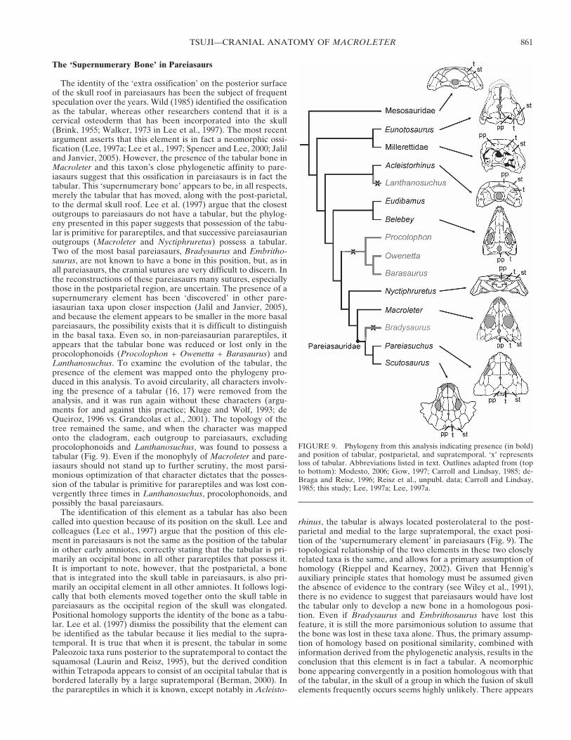
The ‘Supernumerary Bone’ in Pareiasaurs
The identity of the ‘extra ossification’ on the posterior surfaceof the skull roof in pareiasaurs has been the subject of frequentspeculation over the years. Wild (1985) identified the ossificationas the tabular, whereas other researchers contend that it is acervical osteoderm that has been incorporated into the skull(Brink, 1955; Walker, 1973 in Lee et al., 1997). The most recentargument asserts that this element is in fact a neomorphic ossi-fication (Lee, 1997a; Lee et al., 1997; Spencer and Lee, 2000; Jaliland Janvier, 2005). However, the presence of the tabular bone inMacroleter and this taxon’s close phylogenetic affinity to pare-iasaurs suggest that this ossification in pareiasaurs is in fact thetabular. This ‘supernumerary bone’ appears to be, in all respects,merely the tabular that has moved, along with the post-parietal,to the dermal skull roof. Lee et al. (1997) argue that the closestoutgroups to pareiasaurs do not have a tabular, but the phylog-eny presented in this paper suggests that possession of the tabu-lar is primitive for parareptiles, and that successive pareiasaurianoutgroups (Macroleter and Nyctiphruretus) possess a tabular.Two of the most basal pareiasaurs, Bradysaurus and Embritho-saurus, are not known to have a bone in this position, but, as inall pareiasaurs, the cranial sutures are very difficult to discern. Inthe reconstructions of these pareiasaurs many sutures, especiallythose in the postparietal region, are uncertain. The presence of asupernumerary element has been ‘discovered’ in other pare-iasaurian taxa upon closer inspection (Jalil and Janvier, 2005),and because the element appears to be smaller in the more basalpareiasaurs, the possibility exists that it is difficult to distinguishin the basal taxa. Even so, in non-pareiasaurian parareptiles, itappears that the tabular bone was reduced or lost only in theprocolophonoids (Procolophon + Owenetta + Barasaurus) andLanthanosuchus. To examine the evolution of the tabular, thepresence of the element was mapped onto the phylogeny pro-duced in this analysis. To avoid circularity, all characters involv-ing the presence of a tabular (16, 17) were removed from theanalysis, and it was run again without these characters (argu-ments for and against this practice; Kluge and Wolf, 1993; deQueiroz, 1996 vs. Grandcolas et al., 2001). The topology of thetree remained the same, and when the character was mappedonto the cladogram, each outgroup to pareiasaurs, excludingprocolophonoids and Lanthanosuchus, was found to possess atabular (Fig. 9). Even if the monophyly of Macroleter and pare-iasaurs should not stand up to further scrutiny, the most parsi-monious optimization of that character dictates that the posses-sion of the tabular is primitive for parareptiles and was lost con-vergently three times in Lanthanosuchus, procolophonoids, andpossibly the basal pareiasaurs.
The identification of this element as a tabular has also beencalled into question because of its position on the skull. Lee andcolleagues (Lee et al., 1997) argue that the position of this ele-ment in pareiasaurs is not the same as the position of the tabularin other early amniotes, correctly stating that the tabular is pri-marily an occipital bone in all other parareptiles that possess it.It is important to note, however, that the postparietal, a bonethat is integrated into the skull table in pareiasaurs, is also pri-marily an occipital element in all other amniotes. It follows logi-cally that both elements moved together onto the skull table inpareiasaurs as the occipital region of the skull was elongated.Positional homology supports the identity of the bone as a tabu-lar. Lee et al. (1997) dismiss the possibility that the element canbe identified as the tabular because it lies medial to the supra-temporal. It is true that when it is present, the tabular in somePaleozoic taxa runs posterior to the supratemporal to contact thesquamosal (Laurin and Reisz, 1995), but the derived conditionwithin Tetrapoda appears to consist of an occipital tabular that isbordered laterally by a large supratemporal (Berman, 2000). Inthe parareptiles in which it is known, except notably in Acleisto-
rhinus, the tabular is always located posterolateral to the post-parietal and medial to the large supratemporal, the exact posi-tion of the ‘supernumerary element’ in pareiasaurs (Fig. 9). Thetopological relationship of the two elements in these two closelyrelated taxa is the same, and allows for a primary assumption ofhomology (Rieppel and Kearney, 2002). Given that Hennig’sauxiliary principle states that homology must be assumed giventhe absence of evidence to the contrary (see Wiley et al., 1991),there is no evidence to suggest that pareiasaurs would have lostthe tabular only to develop a new bone in a homologous posi-tion. Even if Bradysaurus and Embrithosaurus have lost thisfeature, it is still the more parsimonious solution to assume thatthe bone was lost in these taxa alone. Thus, the primary assump-tion of homology based on positional similarity, combined withinformation derived from the phylogenetic analysis, results in theconclusion that this element is in fact a tabular. A neomorphicbone appearing convergently in a position homologous with thatof the tabular, in the skull of a group in which the fusion of skullelements frequently occurs seems highly unlikely. There appears
FIGURE 9. Phylogeny from this analysis indicating presence (in bold)and position of tabular, postparietal, and supratemporal. ‘x’ representsloss of tabular. Abbreviations listed in text. Outlines adapted from (topto bottom): Modesto, 2006; Gow, 1997; Carroll and Lindsay, 1985; de-Braga and Reisz, 1996; Reisz et al., unpubl. data; Carroll and Lindsay,1985; this study; Lee, 1997a; Lee, 1997a.
TSUJI—CRANIAL ANATOMY OF MACROLETER 861

to be a trend towards cranial simplification in tetrapods in gen-eral, and the acquisition of neomorphic ossifications in amniotesis extremely rare (Sidor, 2001).
CONCLUSION
An in-depth phylogenetic study of the cranial anatomy ofMacroleter poezicus results in a tree showing a close relationshipbetween the pareiasaurs and Macroleter, a relationship oftenpostulated but only now supported by a phylogenetic analysis. Itappears to be increasingly important to determine the relation-ships between the Russian nycteroleterids and nyctiphruretidsand the relationship of these taxa to other parareptiles, espe-cially to pareiasaurs. The phylogeny of pareiasaurs in particularis an interesting issue, with very little description of known taxa,and few existing phylogenetic analyses of this significant, yetenigmatic clade. Further studies of poorly known parareptiliantaxa will not only potentially help to shed light on the origins andrelationships of turtles, but will also foster a greater understand-ing of amniote evolution.
ACKNOWLEDGMENTS
I thank my M.Sc. supervisor R. Reisz for support, advice, andfor suggesting the project. D. Scott was of invaluable help withthe figures and general advice during the course of my time atUTM. Comments on earlier versions of the manuscript by J.Müller and K. Folinsbee significantly improved its quality. I alsothank the other members of the RPL, D. Evans, J. Fröbisch, andH. Maddin for both academic support and non-academic distrac-tion. J. Ting also kindly helped with the figures. A. Shinya and K.Dupuis executed some of the skilled preparation of the speci-mens. This manuscript was greatly improved by the reviews ofM. Lee and S. Modesto.
Many thanks to M. Ivakhnenko and V. Golubev for the loan ofspecimens. Funding was provided by the University of Torontoand NSERC Discovery and National Geographic Society grantsto R. Reisz.
LITERATURE CITED
Berman, D. S. 2000. Origin and early evolution of the amniote occiput.Journal of Paleontology 74:938–956.
Berman, D. S., R. R. Reisz, D. Scott, A. C. Henrici, S. S. Sumida, and T.Martens. 2000. Early Permian bipedal reptile. Science 290:969–972.
Brink, A. S. 1955. On Nanoparia. Palaeontologia Africana 3:57–63.Carroll, R. L., and W. Lindsay. 1985. Cranial anatomy of the primitive
reptile Procolophon. Canadian Journal of Earth Sciences 22:1571–1587.
Cisneros, J. C., R. Damiani, C. Schultz, A. da Rosa, C. Schwanke, L. W.Neto, and P. L. P. Aurelio. 2004. A procolophonid reptile withtemporal fenestration from the Middle Triassic of Brazil. Proceed-ings of the Royal Society of London B 271:1541–1546.
Cisneros, J. C., F. Abdala, and M. C. Malabarba. 2005. Pareiasauridsfrom the Rio do Rasto Formation, Southern Brazil: Biostratigraphicimplications for Permian faunas of the Parana Basin. Revista Bra-sileira de Paleontologia 8:13–24.
de Queiroz, K. 1996. Including characters of interest during tree recon-struction and the problems of circularity and bias in studies of char-acter evolution. American Naturalist 148:700–708.
Damiani, R. J. and S. P. Modesto. 2001. The morphology of the pare-iasaurian vomer. Neues Jahrbuch für Geologie und Paläontologie,Monatshefte 2001:423–434.
deBraga, M., and R. R. Reisz. 1996. The Early Permian reptile Acleis-torhinus pteroticus and its phylogenetic position. Journal of Verte-brate Paleontology 16:384–395.
deBraga, M., and O. Rieppel. 1997. Reptile phylogeny and the interre-lationships of turtles. Zoological Journal of the Linnean Society120:281–354.
Efremov, J. A. 1938. Some new Permian reptiles of the U.S.S.R.Comptes Rendus (Doklady) 19:771–776.
Efremov, J. A. 1940. Die Mesen-Fauna der permischen Reptilien. Neues
Jarbuch für Minerologie, Geologie und Paläontologie Abhandlun-gen, Abteilungen B 84:379–466.
Frazetta, T. H. 1968. Adaptive problems and possibilities in the temporalfenestration of tetrapod skulls. Journal of Morphology 125:145–158.
Gaffney, E. S. 1990. The comparative osteology of the Triassic turtleProganochelys. Bulletin of American Museum of Natural History194:1–263.
Gow, C. E. 1972. The osteology and relationships of the Millerettidae(Reptilia: Cotylosauria). Journal of Zoology, London 167:219–264.
Gow, C. E. 1977. Owenetta in perspective. Palaeontologia Africana 20:115–118.
Gow, C. E. 1997. A reassessment of Eunotosaurus africanus Seeley (Am-niota: Parareptilia). Palaeontologia Africana 33:33–42.
Grandcolas, P., P. Deleporte, L. Desutter-Grandcolas, and C. Daugeron.2001. Phylogenetics and ecology: as many characters as possibleshould be included in the cladistic analysis. Cladistics 17:104–110.
Haughton, S. H. 1929. Pareiasaurian studies. Part II. Notes on somepareiasaurian brain-cases. Annals of the South African Museum28:88–96.
Heaton, M. J. 1979. Cranial anatomy of primitive captorhinid reptilesfrom the Late Pennsylvanian and early Permian, Oklahoma andTexas. Oklahoma Geological Survey Bulletin 127:1–84.
Heaton, M. J. 1980. The Cotylosauria: a reconsideration of a group ofarchaic tetrapods; pp. 467–551 in A. L. Panchen (ed.), The Terres-trial Environment and the Origin of Land Vertebrates. AcademicPress, London.
Hill, R. V. 2005. Integration of morphological data sets for phylogeneticanalysis of Amniota: The importance of integumentary charactersand increased taxon sampling. Systematic Biology 54:530–547.
Ivakhnenko, M. F. 1973. Skull structure in the early procolophonianTichvinskia vjatkensis. Paleontological Journal 1973:74–83.
Ivakhnenko, M. F. 1979. [Permian and Triassic Procolophonia of theRussian Platform]. Trudy Paleontologicheskogo Instituta, Aka-demiia Nauk SSSR 164:1–79. [Russian]
Ivakhnenko, M. F., V. K. Golubev, Y. M. Gubin, N. N. Kalandadze, I. V.Novikov, A. G. Sennikov, and A. S. Rautian. 1997. [Permian and Tri-assic tetrapods of Eastern Europe]. GEOS, Moscow, 216 pp. [Russian]
Jalil, N.-E., and P. Janvier. 2005. Les pareiasaures (Amniota, Pararep-tilia) du Permien superieur du Bassin d’Argana, Maroc. Geodiver-sitas 27:35–132.
Kearney, M. 2002. Fragmentary taxa, missing data, and ambiguity: mis-taken assumptions and conclusions. Systematic Biology 51:369–381.
Kearney, M., and J. M. Clark. 2003. Problems due to missing data inphylogenetic analyses including fossils: a critical review. Journal ofVertebrate Paleontology 23:263–274.
Kemp, T. S. 1974. The braincase and associated structures of the coty-losaur reptile Procolophon trigoniceps Owen. Annals of the SouthAfrican Museum 64:11–26.
Kluge, A. G., and A. J. Wolf. 1993. Cladistics: what’s in a word? Clad-istics 9:183–199.
Laurenti, J. N. 1768. Classis Reptilium. Specimen medicum, exhibens syn-opsis Reptilium emendatum, cum experimentis circa venena et anti-dote Reptilium Austriacorum. J. Thom., Nob. et Trattnern, Vienna.
Laurin, M., and R. R. Reisz. 1995. A reevaluation of early amniotephylogeny. Zoological Journal of the Linnean Society 113:165–223.
Lee, M. S. Y. 1993. The origin of the turtle body plan: bridging a famousmorphological gap. Science 261:1716–1720.
Lee, M. S. Y. 1995. Historical burden in systematics and the interrela-tionships of ’parareptiles’. Biological Reviews 70:459–547.
Lee, M. S. Y. 1997a. Pareiasaur phylogeny and the origin of turtles.Zoological Journal of the Linnean Society 120:197–280.
Lee, M. S. Y. 1997b. A taxonomic revision of pareiasaurian reptiles:implications for Permian terrestrial palaeoecology. Modern Geol-ogy 21:231–298.
Lee, M. S. Y., C. E. Gow, and J. W. Kitching. 1997. Anatomy and rela-tionships of the pareiasaur Pareiasuchus nasicornis from the UpperPermian of Zambia. Palaeontology 40:307–355.
Meckert, D. 1995. The procolophonid Barasaurus and the phylogeny ofearly amniotes. Ph.D. dissertation, McGill University, Montreal, 149pp.
Modesto, S. P. 1999a. Observations on the structure of the Early Permianreptile Stereosternum tumidum Cope. Palaeontologia africana 35:7–19.
Modesto, S. P. 1999b. Colobomycter pholeter from the Lower Permian ofOklahoma: A parareptile, not a protorothyridid. Journal of Verte-brate Paleontology 19:466–472.
JOURNAL OF VERTEBRATE PALEONTOLOGY, VOL. 26, NO. 4, 2006862
Modesto, S. P. 2000. Eunotosaurus africanus and the Gondwanan ances-try of anapsid reptiles. Palaeontologia Africana 36:15–20.
Modesto, S. P. 2006. The cranial skeleton of the Early Permian aquaticreptile Mesosaurus tenuidens: implications for relationships and pa-laeobiology. Zoological Journal of the Linnean Society 146:345–368.
Modesto, S. P., and R. R. Reisz. 2003. An enigmatic new diapsid reptilefrom the Upper Permian of Eastern Europe. Journal of VertebratePaleontology 22:851–855.
Modesto, S. P., and J. S. Anderson. 2004. The phylogenetic definition ofReptilia. Systematic Biology 53:815–821.
Müller, J. 2004. The relationships among diapsid reptiles and the influ-ence of taxon selection; pp. 379–408 in G. Arratia, M. V. H. Wilson,and R. Cloutier (eds.), Recent Advances in the Origin and EarlyRadiation of Vertebrates. Verlag Dr. Friedrich Pfeil, Munich.
Olson, E. C. 1947. The family Diadectidae and its bearing on the classi-fication of reptiles. Fieldiana Geology 11:1–53.
Reisz, R. R., and M. Laurin. 1991. Owenetta and the origin of turtles.Nature 349:324–326.
Reisz, R. R., and M. Laurin. 2001. The reptile Macroleter: first evidence forcorrelation of Upper Permian continental strata of North America andRussia. Geological Society of America Bulletin 113:1229–1233.
Reisz, R. R., and D. Scott. 2002. Owenetta kitchingorum, sp. nov., a smallparareptile (Procolophonia: Owenettidae) from the Lower Triassicof South Africa. Journal of Vertebrate Paleontology 22:244–256.
Reisz, R. R., J. Muller, L. A. Tsuji, and D. Scott. In press. The cranialosteology of Belebey vegrandis (Parareptilia: Bolosauridae) fromthe Middle Permian of Russia, and its bearing on reptilian revolu-tion. Zoological Journal of the Linnean Society.
Rieppel, O., and R. R. Reisz. 1999. The origin and early evolution ofturtles. Annual Review of Ecology and Systematics 30:1–22.
Rieppel, O., and M. Kearney. 2002. Similarity. Biological Journal of theLinnean Society 75:59–82.
Romer, A. S. 1956. Osteology of the Reptiles. The University of ChicagoPress, Chicago, 772 pp.
Rubidge, B. S. (ed.). 1995. Biostratigraphy of the Beaufort Group (Ka-roo Supergroup). Biostratigraphic Series, South African Committeefor Stratigraphy 1:1–45.
Sidor, C. A. 2001. Simplification as a trend in synapsid cranial evolution.Evolution 55:1419–1442.
Spencer, P. S. 2000. The braincase structure of Leptopleuron lacertinumOwen (Parareptilia: Procolophonidae). Journal of Vertebrate Pale-ontology 20:21–30.
Spencer, P. S., and M. S. Y. Lee. 2000. A juvenile Elginia and earlygrowth in pareiasaurs. Journal of Paleontology 74:1191–1195.
Spencer, P. S., and G. W. Storrs. 2002. A re-evaluation of small tetrapodsfrom the Middle Triassic Otter Sandstone Formation of Devon,England. Palaeontology 45:447–467.
Swofford, D. L. 2002. PAUP*: Phylogenetic Analysis Using Parsimony*.4.0b10. Sinauer, Sunderland, Massachusetts.
Tarsitano, S. F., B. Oelofsen, E. Frey, and J. Riess. 2001. The origin oftemporal fenestrae. South African Journal of Science 97:334–336.
Tverdokhlebova, G. I., and M. F. Ivakhnenko. 1984. [Nykteroleteridsfrom the Upper Permian of Eastern Europe]. PaleontologicheskiiZhurnal 1984:98–111. [Russian]
Wild, R. 1985. Ein Schadelrest von Parasaurus geinitzi H. v. Meyer (Rep-tilia; Cotylosauria) aus dem Kupferschiefer (Permian) von Richels-dorf (Hessen). Geologische Blatter fur Nordost-Bayern (Gedenk-schrift Freyberg) 34/35:897–920.
Wiley, E. O., D. Siegel-Causey, D. R. Brooks, and V. A. Funk. 1991. TheCompleat Cladist: A Primer of Phylogenetic Procedures. University ofKansas Museum of Natural History, Special Publication No. 19, 158 pp.
Submitted 15 March, 2006; accepted 3 July 2006.
APPENDIX 1. Description of characters used in the phylogeneticanalysis.
1. Narial shelf: absent (0); present (1). (Laurin and Reisz, 1995 no. 1)2. Frontal orbital contact: present (0); absent (1). (Modified from Lau-
rin and Reisz, 1995 no. 2)3. Frontal lateral lappet: absent (0); large, occupies at least one-third of
the dorsal margin of the orbit (1). (deBraga and Reisz, 1996 no. 7)4. Pineal foramen position: in the middle of the body of the parietal (0);
displaced posteriorly (1); displaced anteriorly (2); absent (3). (de-Braga and Rieppel, 1997 no. 49)
5. Postparietal: paired (0); median (1); greatly reduced or absent (2).(Modified from Laurin and Reisz, 1995 no. 4)
6. Postparietal position: occipital (0); dorsally exposed, integrated intoskull table (1). (Modified from Laurin and Reisz, 1995 no. 5)
7. Prefrontal-palatal contact: absent (0); weak (1); strong (2). (Laurinand Reisz, 1995 no. 6)
8. Prefrontal medial flange: narrow (0); wide (1). (Laurin and Reisz,1995 no. 7)
9. Bulbous medial process of prefrontal: absent (0); present (1). (Laurinand Reisz, 1995 no. 8)
10. Lacrimal narial contact: present (0); absent (1). (Laurin and Reisz,1995 no. 9)
11. Foramen orbitonasale: absent (0); represented by a medial indenta-tion on the lacrimal and a dorsal indentation on the palatine (1);enclosed between prefrontal, lacrimal and palatine (2). (Laurin andReisz, 1995 no. 10)
12. Jugal anterior process: does not extend to anterior orbital rim (0);extends at least to level of anterior orbital rim (1). (Laurin and Reisz,1995 no. 11)
13. Postorbital posterior process shape: increased width, parallelogramoutline in lateral aspect (0); slender, half as wide as it is long (1).(Modified from deBraga and Reisz, 1996 no. 14)
14. Postorbital: close to occiput, posterior extent reaching at least 50%the length of the parietal (0); far from occiput, posterior extent notreaching the level of 50% the length of the parietal (1). (Modifiedfrom Laurin and Reisz, 1995 no. 13)
15. Posterolateral corner of skull roof: formed mostly by supratemporal(0); formed by parietal and small supratemporal or parietal alone (1).(Modified from Laurin and Reisz, 1995 no. 15)
16. Tabular and opisthotic: in contact (0); separated (1). (Laurin andReisz, 1995 no. 16)
17. Tabular size: large and part of skull table (0); small and largelyoccipital (1); absent (2). (Laurin and Reisz, 1995 no. 17)
18. Supratemporal size: large (0); small (1); absent (2). (Laurin andReisz, 1995 no. 18)
19. Premaxillary dorsal process: broad, narial opening faces predomi-nantly laterally (0); narrow, narial opening faces anteriorly (1). (de-Braga and Reisz, 1996 no. 1)
20. Anterodorsal process of the maxilla: absent (0); present (1). (Modi-fied from Laurin and Reisz, 1995 no. 19)
21. Anterior lateral maxillary foramen: equal in size to other maxillaryforamina (0); larger than other foramina (1); the lateral surface of themaxilla lacks large foramina (2). (Laurin and Reisz, 1995 no. 20)
22. Maxilla and quadratojugal: separated (0); in contact (1). (Modifiedfrom Laurin and Reisz, 1995 no. 22)
23. Quadratojugal anterior extent: reaches posterior border of orbit (0);does not reach level of posterior border of orbit (1). (Modified fromLaurin and Reisz, 1995 no. 23)
24. Caniniform region: present (0); absent (1). (Laurin and Reisz, 1995no. 24)
25. Single caniniform maxillary tooth: present (0); absent (1). (Modifiedfrom Laurin and Reisz, 1995 no. 25)
26. Squamosal and post-temporal fenestra: separated (0); in contact (1).(Laurin and Reisz, 1995 no. 26)
27. Occipital flange of squamosal; gently convex all along the posterioredge of the skull (0); convex above quadrate emargination and con-cave medial to tympanic ridge (1); medial to tympanic ridge, facingposterodorsally (2); medial to tympanic ridge, concave, facing pos-terolaterally or ventrolaterally (3). (Modified from Laurin and Reisz,1995 no. 27)
28. Quadratojugal shape: narrow, horizontal dimension exceeds verticaldimension by a factor of at least 3 (0); tall, greatest vertical dimen-sion is at least 1/3 of or greater than horizontal dimension (1). (Modi-fied from Laurin and Reisz, 1995 no. 28)
29. Quadratojugal ornamentation: confluent with the cheek and not or-nate in any manner (0); ornamented, dermal protuberances projectfrom its surface (1). (deBraga and Rieppel, 1997 no. 43)
30. Lower temporal fenestra: absent (0); present (1); distinct morphol-ogy—thin slit (2). (Modified from Laurin and Reisz, 1995 no. 30)
31. Postorbital contribution to lateral temporal fenestra: bordered byjugal, quadratojugal, squamosal, postorbital (0); no contribution bypostorbital (1). (deBraga and Reisz, 1996 no. 20)
32. Quadratojugal-lateral temporal fenestra contribution: quadratojugalexcluded from posterior border (0); quadratojugal contributes to lat-eral temporal fenestra (1). (deBraga and Reisz, 1996 no. 16)
TSUJI—CRANIAL ANATOMY OF MACROLETER 863
33. Temporal emargination: absent (0); with squamosal and supratem-poral (1); with quadratojugal and squamosal (2); facing posteriorlyand exposed on occiput, bordered by squamosal, quadratojugal, andquadrate (3). (Laurin and Reisz, 1995 no. 31)
34. Postorbital region of skull: long (more than 15% of skull length) (0);short (15% of skull length or less) (1). (Laurin and Reisz, 1995 no.32)
35. Ventral margin of postorbital skull region: expanded below ventralextent of maxilla (0); rectilinear (1); emarginated (2). (Modified fromLaurin and Reisz, 1995 no. 33)
36. Quadrate lateral exposure: absent (0); present (1). (Laurin andReisz, 1995 no. 34)
37. Jaw articulation position: even with occiput (0); posterior to occiput(1); anterior to occiput (2). (Modified from Laurin and Reisz, 1995no. 36)
38. Posterior extension of orbit: absent (0); present (1). (Laurin andReisz, 1995 no. 37)
39. Dermal sculpturing: absent (0); tuberosities (1); tuberosities and pits(2); honeycomb pattern of ridges and pits (3). (Laurin and Reisz,1995 no. 38)
40. Sculpturing involving circumorbital bumps: no distinctive ornamen-tation (0); circumorbital tubercles (1).
41. Posterior margin of skull roof: embayed bilaterally (0), with a single,median embayment (1); roughly straight (2). (Modified fromModesto, 1999 no. 125)
42. Interpterygoid vacuity: short, less than 15% of skull length (0); ab-sent (1); long, at least 15% of skull length (2). (Laurin and Reisz,1995 no. 39)
43. Interpterygoid vacuity anterior extent: reaches beyond posterior bor-der of palatine (0); reaches level of palatine or less (1). (Modifiedfrom Reisz et al. unpublished data no. 127)
44. Choana: parallel to maxilla; palatine forms its posterior edge only (0);curved posteromedially; palatine forms its posterior and part of its lat-eral edge (1). (Laurin and Reisz, 1995 no. 40)
45. Alar flange of the vomer: absent (0); present (1).46. Palatine contribution to palate: narrow, restricted to lateral margins
of palate (0); broad, exceeds 50% of the width of the pterygoid (1).(deBraga and Reisz, 1996 no. 31)
47. Arcuate flange of pterygoid: present (0); absent (1). (Laurin andReisz, 1995 no. 42)
48. Cranio-quadrate space: small, quadrate ramus of pterygoid and para-occipital process of opisthotic converge posterolaterally (0); large,quadrate ramus of pterygoid and paraoccipital process of opisthoticare parallel to each other (1). (Laurin and Reisz, 1995 no. 43)
49. Pterygoid anterior extent: reaches level of posterior end of choana(0); posterior to choana (1). (Laurin and Reisz, 1995 no. 44)
50. Transverse flange of the pterygoid: large, approaches cheek, a no-ticeable lateral projection (0); small, does not approach cheek (1).(Modified from Lee, 1997 no. 19)
51. Transverse flange of pterygoid orientation: directed posterolaterallyor transversely (0); directed anterolaterally (1); directed anteriorly(2). (Modified from Laurin and Reisz, 1995 no. 45)
52. Transverse flange of pterygoid dentition: shagreen of denticles, noventral ridge (0); single row of large teeth, no ventral ridge (1);edentulous with ventral ridge (2). (Laurin and Reisz, 1995 no. 46)
53. Quadrate ramus of pterygoid: merges smoothly into transverseflange without distinctive excavation (0); deep excavation on pos-terolateral surface (1). (deBraga and Reisz, 1996 no. 29)
54. Ectopterygoid: large, anteromedial length at least 40% of anterome-dially directed suture with pterygoid (0); small, anteromedial lengthless than 40% of anteromedially-directed suture with pterygoid (1);absent (2). (Modified from Laurin and Reisz, 1995 no. 47)
55. Ectopterygoid dentition: present (0); absent (1). (Laurin and Reisz,1995 no. 48)
56. Ectopterygoid relationship to transverse flange: ectopterygoid distalto transverse flange, does not contribute to flange (0); ectopterygoidmakes contact with tooth-bearing region of transverse flange (1).(deBraga and Reisz, 1996 no. 33)
57. Suborbital foramen: absent (0); present (1); fenestra present (2).(Laurin and Reisz, 1995 no. 49)
58. Basicranial articulation: kinetic/synovial (0); sutured and/or immo-bile (1). (Modified from Lee, 1997 no. 2)
59. Length of basicranial articulation: restricted to anterolateral marginof the parasphenoid (0); extends over much of length of main bodyof parasphenoid (1). (deBraga and Reisz, 1996 no. 36)
60. Parasphenoid pocket for cervical musculature: present (0); absent(1). (Laurin and Reisz, 1995 no. 50)
61. Parasphenoid wings; present, parasphenoid broader posteriorly thanlong (0); absent, parasphenoid narrower posteriorly than long (1).(Modified Laurin and Reisz, 1995 no. 51)
62. Cultriform process: longer than the body of the parasphenoid (0);shorter than the body of the parasphenoid (1). (Laurin and Reisz,1995 no. 52)
63. Parasphenoid teeth: absent (0); present (1). (Laurin and Reisz, 1995no. 53)
64. Supraoccipital: plate-like, no sagittal crest (0); body constricted atmidline, forming sagittal crest (1). (Modified from deBraga andRieppel, 1997 no. 56)
65. Paraoccipital process: vertically broad (0); anteroposteriorly ex-panded (1); narrow (2); tubular, composed of opisthotic (3). (Laurinand Reisz, 1995 no. 56)
66. Paraoccipital process orientation: directed primarily laterally (0); ori-ented obliquely, at an angle of at least 45 degrees from the horizontalplane of the skull (1). (deBraga and Reisz, 1996 no. 44)
67. Contact between paraoccipital process and dermatocranium: to tabu-lar and squamosal (0); to squamosal and supratemporal (1); to su-pratemporal (2) to squamosal and quadrate (3); ends freely (4).(Modified from Laurin and Reisz, 1995 no. 57)
68. Medial wall of inner ear (made of prootic): unossified (0); ossifiedwith acoustic nerve foramina (1). (Laurin and Reisz, 1995 no. 59)
69. Post-temporal fenestra: small, diameter less than half the diameter offoramen magnum (0); large, diameter at least equal to foramen mag-num (1). (Modified from deBraga and Rieppel, 1997 no. 59)
70. Osseous contact between basioccipital and basisphenoid: present (0);absent (1). (Lee, 1993 #A3, scored as per Laurin and Reisz, 1995 no.61)
71. Occipital condyle shape: reniform to circular (0); transversely broad(1). (Laurin and Reisz, 1995 no. 62)
72. Ventral exposure of basioccipital: contributes extensively to ventralsurface of the braincase (0); restricted to condylar region (1). (de-Braga and Reisz, 1996 no. 37)
73. Ventral braincase tubera: absent (0); present and restricted to basi-occipital (1); present, very large and restricted to basisphenoid (2);median (3). (Modified from Laurin and Reisz, 1995 no. 63, and de-Braga and Rieppel, 1996 no. 65)
74. Lateral flange of exoccipital: absent (0); present (1). (Laurin andReisz, 1995 no. 64)
75. Quadrate condyle articular surfaces: strongly convex, antero-posteriorly longer than they are wide (0); nearly flat, antero-posteriorly shorter than they are wide (1). (Modified from Laurinand Reisz, 1995 no. 65)
76. Stapedial dorsal process: ossified (0); unossified (1). (Laurin andReisz, 1995 no. 67)
77. Morphology of marginal dentition: single cusp (0); two or more cusps(1). (Modified from Lee, 1997 no. 59)
78. Foramen intermandibularis: anterior symphysial foramen (0); twoforamina, a symphysial and a posterior foramen located anterior tocoronoid process (1); two foramina, a symphysial and a posteriorforamen located posterior to or at level of coronoid process (2).(Laurin and Reisz, 1995 no. 69)
79. Meckelian fossa orientation: faces mediodorsally, prearticular nar-row (0) faces dorsally, prearticular broad (1). (Laurin and Reisz,1995 no. 70)
80. Fossa meckelii: long, occupies at least 20% of lower jaw length (0);short, occupies less than 20% of lower jaw length (1). (Laurin andReisz, 1995 no. 71)
81. Surangular length: extends beyond coronoid eminence (0); does notextend beyond coronoid eminence (1). (Laurin and Reisz, 1995 no.72)
82. Accessory lateral shelf on surangular anterior to articular region: absent(0); present (1). (Laurin and Reisz, 1995 no. 73)
83. Coronoid number: two or three (0); one (1). (Laurin and Reisz, 1995no. 74)
84. Prearticular extends: beyond the coronoid eminence (0); does notextend beyond coronoid eminence (1); prearticular very long (2).(Laurin and Reisz, 1995 no. 75)
85. Retroarticular process: absent or small and narrow (0); transverselybroad, dorsally concave (1). (Laurin and Reisz, 1995 no. 76)
86. Retroarticular process composition: articular body (0); three or more
JOURNAL OF VERTEBRATE PALEONTOLOGY, VOL. 26, NO. 4, 2006864
elements (articular, prearticular, angular and surangular) (1). (Lau-rin and Reisz, 1995 no. 77)
87. Lateral shelf on articular region: absent (0); on articular (1); onsurangular (2). (Laurin and Reisz, 1995 no. 78)
88. Coronoid process: small or absent, composed of several elements (0);high process composed of coronoid only (1); high, composed primar-ily of dentary (2). (Modified from Laurin and Reisz, 1995 no. 79)
89. Splenial: contributes to symphysis (0); excluded from symphysis (1).(Laurin and Reisz, 1995 no. 80)
Notes: Characters 40 and 45 are new to this study. The majority ofcharacters were adapted from Laurin and Reisz, 1995, but many werealso independently used in subsequent or contemporaneous studies(including Lee, 1995; 1997a; deBraga and Rieppel, 1997).
APPENDIX 2. Data matrix used in this study.
Taxon1
12345678902
12345678903
12345678904
12345678905
1234567890
Synapsida A00B000000 0000000000 00A0000001 0000A00000 0C00000000Mesosauridae 0000000000 ?0?0001000 0101110000 ??01100000 1000001?00Eunotosaurus ?00000???? ?0100110?1 ?011111100 ??20212010 120??????0Millerettidae 0000001000 2010001000 111A10110A 0020112010 1200000000Eudibamus 000???2100 ?0011??210 0?01110?02 1101?01?00 201?????11Belebey 0001202100 20011?0210 0101110002 1101000100 2010010?11Acleistorhinus 001210???1 ?001011011 1110010101 1130102010 1200000100Lanthanosuchus 001C201001 ?0010?1011 ?011112111 1130101020 1200010100Macroleter 0002A02101 2101001011 1100103111 1120002121 1011110100Nyctiphruretus 0001102101 ?0010?2011 1101103100 ??20002120 1211100110Bradysaurus 0102111000 ?1010?2011 1001102110 ??30002021 11111101?0Scutosaurus 0102111000 ?101011011 1101102110 ??30002021 10111101?0Procolophon 10022?2111 21010?1011 1001103100 ??20202100 2010010110Owenetta 1002012111 ?10?0?2011 1000103100 ??20202100 ?210010110Barasaurus 100201?110 ?0010?2011 1001103100 ??20202100 ?210010110Captorhinidae 000C001000 11011?2100 0110010000 ??00100030 0200001000
Taxon6
12345678907
12345678908
12345678909
1234567890
Synapsida 010000000A 00A0000000 00C0000D00 AA0000000Mesosauridae 0102???000 000000??1? 000?000??0 001?1000?Eunotosaurus 01?????001 000010???? ?0???10??? 0???0??0?Millerettidae 0101101001 1010001011 0000010?00 001000000Eudibamus 22111??000 1?0??0???? 11????02?? ???200?20Belebey 2211110000 100??0???0 ???00?0201 10120?021Acleistorhinus 011011111? 1010012??0 1100100??? 1011010?1Lanthanosuchus 0110110111 1010012?0? 11?11?0??? ?????????Macroleter 0111?01100 1101101?1? 0001??02?1 11111??11Nyctiphruretus 01?0011001 110?101011 00001?0??? ??1?????1Bradysaurus 1111101101 1101101110 0001101?11 10111?110Scutosaurus 1111111101 1101101110 0001111211 10111?110Procolophon 1211111001 0101102011 001011021? 111111011Owenetta 1211111001 1101103?1? 00100?0211 101111011Barasaurus 12?1111001 1101103?1? 00100?0??? ?????????Captorhinidae 0002??1001 10A0204?10 0000000100 001000000
Polymorphism is indicated by letters, such that A!0&1, B!1&2, C!0&2, D!0&1&2.
TSUJI—CRANIAL ANATOMY OF MACROLETER 865