cox-2 inhibitors: a novel strategy in the management of...
TRANSCRIPT

Review
s�K
EYNOTEREVIEW
REVIEWS Drug Discovery Today �Volume 21, Number 4 �April 2016
COX-2 inhibitors: a novel strategy inthe management of breast cancerMiłosz Regulski1, Katarzyna Regulska2, Wiesław Prukała3,Hanna Piotrowska1, Beata Stanisz4 and Marek Murias1
1 Poznan University of Medical Sciences, Chair and Department of Toxicology, 30th Dojazd Street,
60-631 Poznan, Poland2Greater Poland Oncology Center, 15th Garbary Street, 61-866 Poznan, Poland3Adam Mickiewicz University in Poznan, Faculty of Chemistry, Nucleosides and Nucleotides Chemistry,6th Grunwaldzka Street, 60-780 Poznan, Poland4 Poznan University of Medical Sciences, Chair and Department of Pharmaceutical Chemistry,
6th Grunwaldzka Street, 60-780 Poznan, Poland
Cyclooxygenase-2 (COX-2) inhibitors are common anti-inflammatory
drugs with pleiotropic, endogenous actions that could be useful in the
management of breast cancer. Here, we provide a complete understanding
of the biochemistry of COX-2 and discuss the various molecular
mechanisms behind its increased expression in breast cancer. We also
analyze the possible mechanisms responsible for the anticancer effect of
COX-2 inhibitors and provide an overview of the available preclinical and
clinical data on the use of COX-2 inhibitors in breast cancer. Finally, we
describe a mathematical model of the relation between the structure and
biological potency of promising new COX-2 inhibitors (trans-stilbenes)
using a 2D quantitative structure–activity relationship (QSAR) technique.
IntroductionBreast cancer is the most common malignancy and the most common cause of cancer mortality
for females in industrialized, Western-lifestyle countries. In the USA, for example, there are
approximately 182,000 new cases diagnosed annually, while each year around 40,000 women die
from this disease [1]. Unfortunately, the incidence of breast cancer is still on the increase,
although the mortality index has decreased over past few years. This decrease is likely to be the
result of the widespread implementation of improved screening techniques, early detection and
novel therapies [2]. In fact, intense scientific research in the field of breast cancer pharmacother-
apy has provided several advanced strategies, such as monoclonal antibodies and small-molecule
kinase inhibitors, which have considerably extended the opportunities for treatment. However,
their costs are relatively high. Furthermore, recent studies on the pathologic nature of this disease
have revealed the mechanisms by which breast cancer develops. For instance, it has been found
that the concentration of prostaglandins (PGs) in breast cancer cells is greater than that in the
corresponding normal tissue, indicating that it is aberrant COX expression, especially COX-2,
Miłosz Regulski is a PhD
candidate in toxicology at
Poznan University of Medical
Sciences. He earned his
Master’s degree in pharmacy
in 2008 from the same
university. His research
currently focuses on the
biological activity of trans-
stilbene derivatives as anti-
inflammatory and anticancer
molecules. He is also interested in QSAR techniques,
drug designing, and biological properties of new
anticancer drugs and COXinhibitors.
Katarzyna Regulska is
studying to be a clinical
pharmacy specialist at Poznan
University of Medical
Sciences. She has a PhD in
pharmaceutical chemistry and
earned her Master’s degree in
pharmacy in 2008. Her
research currently focuses on
the stability and genotoxicity
of angiotensin-converting enzyme inhibitors as well as
the biological activity of COX inhibitors.
Marek Murias is an
associate professor in the
Department of Toxicology,
Poznan University of Medical
Sciences. He obtained his
MSc and PhD from the same
university. He performed his
postdoctoral project, which
was financed by a Marie Curie
Fellowship, at the University of Vienna (Institute of
Pharmaceutical Chemistry) and Medical University of
Vienna (Institute of Pathophysiology). His main
research interests are resveratrol and its analogs,
oxidative stress induced by antioxidants, drug-
metabolizing enzymes, and xenoestrogens and
photodynamic therapy.
Corresponding author: Murias, M. ([email protected])
598 www.drugdiscoverytoday.com1359-6446/� 2015 Elsevier Ltd. All rights reserved.
http://dx.doi.org/10.1016/j.drudis.2015.12.003

Drug Discovery Today � Volume 21, Number 4 �April 2016 REVIEWS
Reviews�KEYNOTEREVIEW
that could be associated with the development and progression of
the malignancy. Thus, various procarcinogenic actions have been
attributed to COX-2, including inhibition of apoptosis, increase in
proliferation, and stimulation of angiogenesis, making its abolish-
ment an encouraging strategy for the management and preven-
tion of breast cancer [3–8]. This approach is additionally favored by
the fact that appropriate inhibitory agents are already widely
available, inexpensive, and relatively well tolerated [9]. In this
article, we review the most important issues concerning the use of
COX-2 inhibitors in breast cancer and provide a full understand-
ing of the impact of COX-2 on tumorigenesis.
COX-2: structure and functionHuman COX family (also named prostaglandin-endoperoxide
synthase or prostaglandin-G/H synthase; EC 1.14.99.1) comprises
three isoenzymes: constitutive (COX-1), inducible (COX-2), and
the recently discovered COX-3. They are involved in the first two
steps of prostanoid biosynthesis (i.e., PGs, thromboxanes, and
prostacyclins). They catalyze the metabolic transformations of
unsaturated fatty acids, such as arachidonic acid (AA), dihomo-
g-linolenic acid, and eicosapentaenoic acid, which are released by
phospholipases A2, C, or D from cell membrane phospholipids
[10,11]. COX enzymes exhibit a dualistic nature by displaying
both COX activity, converting AA into short-lived 15-hydroper-
oxide–PGG2, and PG hydroperoxidase activity, which is crucial in
the process of reduction of PGG2 into PGH2. PGH2 is then spon-
taneously rearranged or enzymatically converted into the biologi-
cally active PGs D, E, and F, prostacyclin PGI2, or thromboxane
(TXA2) [11]. The process of prostanoid biosynthesis by COX is
detailed in Fig. 1.
In its constitutive form, COX-1 is expressed as a housekeeping
enzyme in most human organs and tissues. It is responsible for the
maintenance of internal homeostasis by participating in general
body processes, such as cytoprotection of the gastric mucosa,
platelet aggregation, vascular smooth muscle functioning, and
regulation of glomerular filtration and renal blood flow. By con-
trast, COX-2 usually remains undetected in healthy tissues and
organs. In adults, it is found only in the central nervous system,
kidneys, vesicles, and placenta, whereas in the fetus, it occurs in
heart, kidneys, lungs, and skin [10,11]. COX-2 is a highly inducible
isoform and can be rapidly upregulated in response to various
proinflammatory agents, including cytokines, tumor promoters,
and mitogens, especially in cells involved in inflammation, pain,
fever, tumor, Alzheimer’s disease, or osteoarthritis [12,13]. The
recently discovered COX-3 isoform is still not fully understood.
However, in humans, it exists as a splice variant of COX-1, pri-
marily in cerebral cortex and heart [13].
COX-1 is encoded by ubiquitously expressed housekeeping
gene located on chromosome 9. Consistent with its structure,
COX1 is almost devoid of transcription factor-binding sites and,
hence, is open to only slight regulation. The gene encoding COX-2
is located on chromosome 1 and comprises the following regula-
tory elements: TATA box, NF-IL-6 motif, two AP-2 regions, three
SP1 regions, CRE motif, and E-box. Their presence makes the
transcription of COX2 susceptible to stimulation by bacterial
lipopolysaccharides, the cytokines, such as interleukin (IL)-1b,
IL-2 and tumor necrosis factor (TNF)-a, and the growth factors,
such as epidermal growth factor (EGF), platelet-derived growth
factor (PDGF) and transforming growth factor (TGF)-b. By con-
trast, anti-inflammatory agents, such as corticosteroids, IL-13, IL-
10 or IL-4, can inhibit the expression of COX-2 in vivo [10,11,14].
Both COX-1 and COX-2 exist as integral, membrane-bound
proteins, located primarily on the lumenal side of the endoplas-
matic reticulum (ER) and the nuclear envelope. Importantly, COX-
2 is more concentrated in the latter position. Both enzymes share
common mechanistic features as well as product-substrate profiles.
They catalyze AA transformations with similar kinetics, although
dihomo-g-linolenic acid and eicosapentaenoic acid show a higher
affinity to COX-2 [13]. Structurally, human COX-1 and COX-2
occur as homodimers stabilized by hydrophobic interactions, and
hydrogen and electrolytic bridges. They also exhibit as much as
63% amino acid identity. They vary in terms of their chain length
and glycosylation patterns. Human COX-1 comprises 599 amino
acid residues (molecular weight 70 kDa) with three N-glycosyla-
tion sites at Asn67, Asn103, and Asn147. In addition, there are five
disulfide bonds between Cys35-Cys46, Cys36-Cys158, Cys40-
Cys56, Cys58-Cys68, and Cys568-Cys574 that stabilize its tertiary
structure. By contrast, COX-2 comprises 604 amino acid residues
(molecular weight 72 kDa) with four N-glycosylation sites at
Asn53, Asn130, Asn395, and Asn580, and five disulfide bonds
between Cys21-Cys32, Cys22-Cys145, Cys26-Cys42, Cys44-
Cys54 and Cys555-Cys561 [10,11,14,15] (http://www.uniprot.
org/uniprot/P23219; http://www.uniprot.org/uniprot/P35354)
(Fig. 2).
The primary structure of COX-2 is functionally divided into four
regions, starting from the N terminus: (i) signal peptide (between
Met1 and Thr17) usually removed by post-translational modifica-
tions; (ii) EGF-like domain (between Ala18 and Ser55), which
contains three conserved disulfide bonds; this area is implicated
in the process of homodimerization; (iii) a membrane domain,
which takes the form of four amphipathic alpha-helices and serves
as an anchor sequence; and (iv) acatalytic domain containing two
independent peroxidase and cyclooxygenase active sites, and a
heme-binding sequence [10,14,15] (http://www.uniprot.org/
uniprot/P23219; http://www.uniprot.org/uniprot/P35354).
The COX active site of COX-2 is described as a narrow, hydro-
phobic channel, approximately 8 A wide and 25 A long. It is
restricted by the membrane domain and its outlet is located close
to the cell membrane. Such construction favors the penetration of
lipophilic AA deep inside the active site tunnel to the specific
cyclization spot. The peroxidase catalytic site is on the surface of
the enzyme and is easily accessible to solvent molecules. It con-
tains a heme cofactor bound with His374 [14].
The catalytic mechanism of COX-2-dependent conversion of
AA into PGH2 can be defined as a series of radical reactions
occurring in the following sequence: (i) two-electron reduction
of a peroxide substrate and oxidation of the ferric heme of peroxi-
dase active site with the production of ferryl-oxo-porphyrin radi-
cal; (ii) formation of a tyrosyl radical at Tyr385 of the COX active
site, resulting in the enzyme activation; (iii) abstraction of the pro-
S hydrogen atom from C13 of AA by the tyrosyl radical; the AA in
the COX active site adopts an extended L-shape conformation and
its carboxyl is bound with Arg120 and Tyr341 by hydrogen bonds;
as a result, C13 remains perfectly positioned to interact with
the tyrosyl radical; (iv) formation of the arachidonyl radical local-
ized at C11 and C9, and the interaction with oxygen to produce
www.drugdiscoverytoday.com 599

REVIEWS Drug Discovery Today �Volume 21, Number 4 �April 2016
COOH
COOH
COOH
HO
HO
O
OH
COOH
COOHO
HOOH
COOH
COOH
COOH
COOHCOOH
O
O
OO
OHOH
OH
COOHCOOH
COOH
OH
OH
OH
OH
OH
OH
OH
O
O
OHO
HO O
HO
O
O
O
O
OOH
Arachidonic acid (AA)
(cyclooxygenase active site)Prostaglandin G/H synthase –1/–2
Prostaglandin G/H synthase –1/–2(peroxidase active site)
2O2
AH2
H2O
H2O
H2O
PGI2 synthase
A + H2O
Prostaglandin G2 (PGG2)
Prostaglandin F2α(PGF2α)
Prostaglandin E2(PGE2)
Prostaglandin D2(PGD2)
Prostaglandin H2(PGH2)
Thromboxane A2(TXA2)
Thromboxane B2(TXB2)
Prostaglandin I2(PGI2)= Prostacyclin
Prostaglandin A2(PGA2)
Prostaglandin B2(PGB2) Prostaglandin C2(PGC2)
Prostaglandin J2(PGJ2)
dehydratation
dehydratation
hydratation
isomerisation
PGF2α 9-ketoreductase(PGF2α synthase) (PGF2 synthase)
PGD2 synthase
TXA2 synthase
Drug Discovery Today
FIGURE 1
The biosynthesis of prostanoids by cyclooxygenase (COX) [10,11]. COX converts arachidonic acid (AA) into short-lived 15-hydroperoxide-PGG2 and then reduces itto PGH2. PGH2 is subsequently spontaneously rearranged or enzymatically converted into the biologically active prostaglandins D, E, and F, prostacyclin (PGI2), or
thromboxane (TXA2).
Review
s�K
EYNOTEREVIEW
endoperoxide; (v) interaction of the radical localized at C15 with
the second molecule of oxygen, facilitated by Ser530 and Val349;
(vi) reabstraction of the proton from Tyr385 and formation of
hydroperoxyl–PGG2 with the concomitant regeneration of the
tyrosyl radical; (vii) and conversion of PGG2 into PGH2 at the
peroxidase active site [11,14].
There are three fundamental elements of the COX active site in
COX-2 that distinguish it from COX-1: Val523, Arg513, and
Val343. In COX-1, these are substituted by the long side-chain
amino acids Ile523, His513, and Ile434. These subtle differences
600 www.drugdiscoverytoday.com
contribute to the 17% enlargement of the substrate-binding pock-
et in COX-2 and the increase in its hydrophilicity, enabling COX-2
to recognize bulkier substrates and increasing its substrate spec-
trum. This relation can also be used for the construction of selec-
tive COX-2 inhibitors with large hydrophilic functional groups,
which fit easily into the wider catalytic site of COX-2.
Structural differences between COX-1 and COX-2 also deter-
mine their behavior in the presence of the irreversible inhibitor
acetylsalicylic acid. COX-1 loses its capability for cyclooxygena-
tion almost entirely when acetylated at Ser530, whereas COX-2

Drug Discovery Today � Volume 21, Number 4 �April 2016 REVIEWS
Drug Discovery Today
FIGURE 2
The secondary structure of the cyclooxygenase 2 (COX-2) homodimer with
the inhibitor (celecoxib) bound to the active site (white-dashed oval). The
COX active site is a narrow, hydrophobic channel restricted by the membranedomain with the outlet located close to the cell membrane. PDB ID: 3LN1,
reproduced from [15].
Reviews�KEYNOTEREVIEW
preserves its catalytic function, although its product profile
changes and, instead of PGG2, 15-hydroxyeicosatetraenoic acid
(HETE) is produced [11,14,16].
Prostaglandins and thromboxanesPGs and thromboxanes (eicosanoids), the primary products of
COX, constitute a large family of endogenous, regulatory agents
derived from AA. Chemically, they are part of the prostanoid
TABLE 1
Main biological effects of PGsa
Location Regulatory factor Biological resp
Female reproductive system PGE2, PGF2 Uterine contrac
Male reproductive system PGE1, PGE2, PGE3, PGF2a Stimulation of sejaculation
Cardiovascular system TXA2, PGI2 Regulation of h
TXA2 Vascular perme
PGE2, PGI2 VasodilationPGE2 Angiogenesis
TXA2, PGF2a Vasoconstriction
PGE2, PGI2 Fetal ductus art
Respiratory system PGE2, PGI2 BronchodilationPGF2a, TXA2, PGD2 Bronchospasm
PGF2a, TXA2 Constriction of
Excretory system PGE2, PGI2, Glomerular filtra
Gastrointestinal tract PGE2, PGI2 Cytoprotection,
viscous mucus
Immune system PGE2, PGI2, TXA2, PGD2 Inhibition of lym
lymphocytes
Central nervous system PGE2 Pyrexia, wakefu
PGD2 Sleep regulation
PGE2, PGI2 Hyperalgesiaa Based on [9,17,25–27].
subclass of eicosanoids, and contain two side chains attached to
the adjacent carbons of the cyclopentane ring (PGs) or the tetra-
hydropyran ring (thromboxanes). Their biosynthesis occurs in
almost every human tissue, although each tissue can produce
various eicosanoids depending on its physiological state. In gen-
eral, prostaglandin D is synthesized primarily in mast cells and
brain, prostaglandin F in uterus, prostaglandin I in endothelial
cells, prostaglandin E in the whole body, and thromboxanes in
thrombocytes and macrophages. Given that PGs and thrombox-
anes have a very short half-life, they act locally as autocrine or
paracrine factors via the interaction with specific PG and throm-
boxane receptors [13,17–19]. Their main biological effects are
listed in Table 1.
PGE2 is the most physiologically abundant product of COX-2
because it exists at some level in nearly all cell types. Apart from its
above-mentioned biological effects, such as induction of pain and
inflammation, PGE2 also participates in the mechanisms of cell
proliferation, apoptosis, and metastasis, thereby contributing to
the progression of several human cancers, including colon cancer,
breast cancer, and lung cancer [18,20]. This hormone-like lipid
compound acts on specific G-protein-coupled membrane recep-
tors, termed EP1, EP2, EP3, and EP4 [21]. Each of these induces
different signaling pathways. EP1 is coupled with Gq and its second
messengers are inositol trisphosphate (IP3) and diacylglycerol
(DAG), which are responsible for calcium mobilization. EP2 and
EP4 are coupled with Gs, which stimulates adenyl cyclase to
increase the production of cAMP [an activator of protein kinase
A (PKA), phosphoinositide 3-kinase (PI3K) and glycogen synthase
kinase 3 (GSK3)]. Importantly, GSK3 is implicated in cellular
proliferation by promoting the phosphorylation of b-catenin
and its subsequent degradation by the 26S proteasome. In addition
to PGE2-dependent activation, EP4 is also associated with the
induction of early growth response protein 1 (EGR-1). Finally,
onse
tion; rupture of the follicle, ovulation; pregnancy accommodation
permatozoa, ejaculation, stimulation of female reproductive tract after
emostasis
ability, thrombosis
of veins
eriosus patency
pulmonary vessels
tion rate and renal flow regulation, renin release
reduction of gastric acid secretion, vasodilation in gastric mucosa, release of
phocyte T and B proliferation, stimulation of immature thymocytes and B
lness
, hypothermia
www.drugdiscoverytoday.com 601
REVIEWS Drug Discovery Today �Volume 21, Number 4 �April 2016
OOS
S
F
Br
Drug Discovery Today
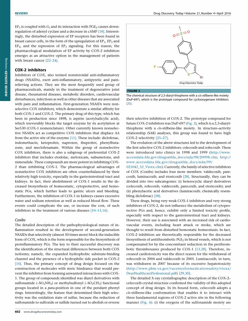
FIGURE 3
The chemical structure of 2,3-diaryl-thiophene with a cis-stilbene-like moiety(DuP-697), which is the prototype compound for cyclooxygenase inhibitors
[25].
Review
s�K
EYNOTEREVIEW
EP3 is coupled with Gi and its interaction with PGE2 causes down-
regulation of adenyl cyclase and a decrease in cAMP [18]. Interest-
ingly, the disturbed expression of EP receptors has been found in
breast cancer cells, in the form of the upregulation of EP1, EP2 and
EP4, and the repression of EP3 signaling. For this reason, the
pharmacological modulation of EP activity by COX-2 inhibitors
emerges as an attractive option in the management of patients
with breast cancer [22–24].
COX-2 inhibitorsInhibitors of COX, also termed nonsteroidal anti-inflammatory
drugs (NSAIDs), exert anti-inflammatory, antipyretic and pain-
relieving actions. They are the most frequently used group of
pharmaceuticals, mainly in the treatment of degenerative joint
disease, rheumatoid diseases, metabolic disorders, cardiovascular
disturbances, infections as well as other diseases that are associated
with pain and inflammation. First-generation NSAIDs were non-
selective COX inhibitors, which demonstrate a similar affinity for
both COX-1 and COX-2. The primary drug of this type, which has
been in production since 1898, is aspirin (acetylsalicylic acid),
which irreversibly blocks the target enzyme by its acetylation at
Ser530 (COX-1 nomenclature). Other currently known nonselec-
tive NSAIDs act as competitive COX inhibitors that displace AA
from the active site of the enzyme [11]. These include: diclofenac,
indomethacin, ketoprofen, naproxen, ibuprofen, phenylbuta-
zone, and meclofenamate. Within the group of nonselective
COX inhibitors, there is also a subgroup of preferential COX-2
inhibitors that includes etodolac, meloxicam, nabumetone, and
nimesulide. These compounds are more potent in inhibiting COX-
2 than inhibiting COX-1. The pharmacological advantages of
nonselective COX inhibitors are often counterbalanced by their
relatively high toxicity, especially in the gastrointestinal tract and
kidneys. In fact, their abolishment of COX-1 results in the de-
creased biosynthesis of homeostatic, cytoprotective, and hemo-
static PGs, which further leads to gastric ulcers and bleeding.
Furthermore, the inhibition of COX-1 in kidneys causes excessive
water and sodium retention as well as reduced blood flow. These
events could complicate the use, or increase the cost, of such
inhibitors in the treatment of various diseases [10–13,16].
CoxibsThe detailed description of the pathophysiological nature of in-
flammation resulted in the development of second-generation
NSAIDs that selectively (almost 50 times more) block the inducible
form of COX, which is the form responsible for the biosynthesis of
proinflammatory PGs. The key to their successful discovery was
the identification of the structural differences between both COX
isoforms; namely, the expanded hydrophobic substrate-binding
channel and the presence of a hydrophilic side pocket in COX-2
[16]. Thus, the primary concept of drug design focused on the
construction of molecules with steric hindrance that would pre-
vent the inhibitor from forming unwanted interactions with COX-
1. The group of compounds identified was diaryl derivatives with
sulfonamide (–SO2NH2) or methylsulfonyl (–SO2CH3) functional
groups located in a para-position in one of the pendant phenyl
rings. Interestingly, the feature that determined their high selec-
tivity was the oxidation state of sulfur, because the reduction of
sulfonamide to sulfoxide or sulfide turned out to abolish or reverse
602 www.drugdiscoverytoday.com
their selective inhibition of COX-2. The prototype compound for
future COX-2 inhibitors was DuP-697 (Fig. 3), which is a 2,3-diaryl-
thiophene with a cis-stilbene-like moiety. In structure–activity
relationship (SAR) analyses, this group was found to have high
COX-2 selectivity [25–27].
The evolution of the above structures led to the development of
the first selective COX-2 inhibitors: celecoxib and rofecoxib. These
were introduced into clinics in 1998 and 1999 (http://www.
accessdata.fda.gov/drugsatfda_docs/nda/98/20998.cfm; http://
www.accessdata.fda.gov/drugsatfda_docs/nda/99/
021042_52_Vioxx.cfm). Currently, the family of selective inhibitors
of COX (Coxibs) includes four more members: valdecoxib, pare-
coxib, lumiracoxib, and etoricoxib [28]. Structurally, they can be
divided into two subcategories: (i) heterocyclic diaryl derivatives
(celecoxib, rofecoxib, valdecoxib, parecoxib, and etoricoxib); and
(ii) phenylacetic acid derivatives (lumiracoxib; chemically resem-
bling diclofenac) [28].
These drugs, being very weak COX-1 inhibitors and very strong
inhibitors of COX-2, do not influence the metabolism of cytopro-
tective PGs and, hence, exhibit only a limited toxicity profile,
especially with respect to the gastrointestinal tract and kidneys.
However, their use is associated with an increased risk of cardio-
vascular events, including heart attack or stroke, which are
thought to result from disturbed hemostatic homeostasis. In fact,
COX-2 inhibitors are theoretically responsible for the decreased
biosynthesis of antithrombotic PGI2 in blood vessels, which is not
compensated for by the concomitant reduction in the prothrom-
botic thromboxanes produced by COX-1 [13,28]. Therefore, in-
creased cardiotoxicity was the direct reason for the withdrawal of
rofecoxib in 2004 and valdecoxib in 2005. Lumiracoxib, in turn,
was withdrawn in 2007 because of its excessive hepatotoxicity
(http://www.pbm.va.gov/vacenterformedicationsafety/vioxx/
DearHealthcareProfessional.pdf) [29,30].
The detailed X-ray crystallographic description of the COX-2–
celecoxib crystal structure confirmed the validity of this adopted
concept of drug design. In its bound form, celecoxib adopts a
cis-stilbene-like conformation that enables it to interact with
three fundamental regions of COX-2 active site in the following
manner (Fig. 4): (i) the oxygens of the sulfonamide moiety are

Drug Discovery Today � Volume 21, Number 4 �April 2016 REVIEWS
HN
H
H
NH
NN
F
FF
Ser339
Gly512
Leu338Val509
AIa513
N
O
O
O OR
RSer339
S
R
R
Leu338
Drug Discovery Today
FIGURE 4
The selective cyclooxygenase 2 (COX-2) inhibitor celecoxib bound to the
active site of murine COX-2 (PDB ID: 3LN1). The hydrogens of its sulfonamide
moiety are H-bound with Leu338, and Ser339. The pyrazole ring and the
trifluoromethyl functional group interact through Van der Waals bonds withSer339, Gly512, Leu338, Val509, and Ala513. Black-dashed lines show
hydrogen bonds, whereas green lines, show hydrophobic interactions. Image
generated using the PoseView software.
HO
OH
OH
3
4
5
6
2
11’
2’
3’
4’
5’
6’
α
β
Drug Discovery Today
FIGURE 5
The structure of trans-resveratrol (3,40 ,5-trihydroxy-trans-stilbene), which is anaturally occurring nonselective cyclooxygenase inhibitor that exhibits anti-
inflammatory, fungicidal, cytotoxic, chemopreventive, and cardioprotective
actions [34].
Reviews�KEYNOTEREVIEW
H-bound with the amino acids located in the hydrophilic side
pocket (His75, Arg499 and Gln178); the pyrazole ring and the
trifluoromethyl functional group interact through Van der Waals
bonds with Ser339, Gly512, Leu338, Val509, Tyr341, and Arg106,
which line the side hydrophobic channel of the enzyme; and (iii)
the aryl ring substituted with the methyl functional group in a
para position forms Van der Waals interactions with the amino
acids from the main hydrophobic channel (Tyr371 and Ser516)
[15,31–33].
trans-Stilbene derivativesAlthough coxibs are the only clinically available selective COX-2
inhibitors, there are also other agents that display a promising
COX-2 selectivity confirmed in in vitro and ex vivo tests. In fact,
numerous compounds without cis-stilbene moiety have been dis-
cussed in the literature and one such group deserving special
attention are the trans-stilbene derivatives. The main stimulus
for their research was the discovery of the biological properties
of resveratrol. This natural compound shows nonselective affinity
to COX-1 (IC50 � 0.5 mmol/l) and COX-2 (IC50 � 1 mmol/l) in vitro
[34,35] and also demonstrates anti-inflammatory, fungicidal, cy-
totoxic, chemopreventive, and cardioprotective actions [34]. Im-
portantly, resveratrol is a stilbene analog that occurs as a cis or trans
isomer, yet only trans-resveratrol (3,40,5-trihydroxy-trans-stilbene)
is biologically active (Fig. 5) [34,35].
Based on these findings, various trans-resveratrol and trans-
stilbene derivatives have become appealing targets for drug design
as potential selective COX-2 inhibitors. The most interesting
structures are presented in Table 2.
One of the most promising groups of selective COX-2 blockers
are hydroxy trans-resveratrol analogs, with 3,30,4,40,5,50-hexahy-
droxy-trans-stilbene (3,30,4,40,5,50-HHS) and piceatannol (3,30,40,5-
tetrahydroxy-trans-stilbene) being the most potent members. In
an immunoenzymatic in vitro assay, the former compound had
700 times higher affinity to COX-2 than to COX-1, whereas the
latter had almost 400 times higher affinity. Their significant
increase in COX-2 selectivity relative to trans-resveratrol was at-
tributed to the presence of numerous hydroxyl groups (–OH) in
the aromatic rings, especially in the 30 position. However, appro-
priate docking experiments showed that both compounds, unlike
coxibs, do not accommodate the hydrophilic side pocket of COX-
2, indicating that their hydroxylation pattern enables them to
create additional hydrogen bonds with the enzyme [35].
Further investigations with trans-stilbenes provided evidence
that the substitution of hydroxyls in resveratrol with methoxy
functionals substantially potentiated their cytotoxic activity, con-
firmed in SAR studies. However, in vitro assays revealed only low
selectivity and weak affinity to COX-2 in this group, with the
exception of 3,30,4,40,5,50-hexamethoxy-trans-stilbene, which
exhibited moderate potency and selectivity (approximately 45
times higher than to COX-1). These results clearly suggest that
the cytotoxic effects of the studied compounds cannot be attrib-
uted to their COX-2-related actions (Table 2) [35].
Another interesting population of trans-stilbene derivatives
comprises the analogs substituted with the following groups in
meta or para positions: –OH, –OCH3, –N(CH3)2, –F, –CF3, and –
NO2, with one phenyl group substituted with naphthyl, or with an
ethylene bridge modified with –CH3 or –C6H5 (Table 2). Interest-
ingly, in in vitro assays, the compounds with –OH and –CF3 at
position 4 displayed a high affinity to COX-2 (IC50 < 2 mmol/l). Of
these, almost all were substituted with –OH or –OCH3 at position 3
www.drugdiscoverytoday.com 603

REVIEWS Drug Discovery Today �Volume 21, Number 4 �April 2016
TABLE 2
trans-Stilbene derivatives that selectively (>50�) and/or potently (IC50 < 2 mmol/l) inhibit COX-2
Chemical formula and name of trans-stilbene derivative
(name of compound in the reference)
COX-1 IC50
(mmol/l)
COX-2 IC50
(mmol/l)
Selectivity index
(COX-1 IC50/COX-2 IC50)
Refs
4-methylsulfonyl-40-fluor-a-phenyl-trans-stilbene (10c)
>100 0.0316 >3164 [37]
4-methylsulfonyl-40-chlor-a-phenyl-trans-stilbene (10e)
>100 0.1138 >878 [37]
4-methylsulfonyl-40-methyl-a-phenyl-trans-stilbene (10b)
>100 0.12 >833 [37]
3,30 ,4,40 ,5,50-heksahydroxy-trans-stilbene (3,30 ,4,40 ,5,50-HHS) (12)
0.748 0.00104 719.23 [35]
3,30 ,40 ,5-tetrahydroxy-trans-stilbene(30-hydroxy-trans-resveratrol,piceatannol) (10)
4.713 0.0113 417.08 [35]
OH3C
OS
F
F
4-methylsulfonyl-30 ,40-difluor-a-phenyl-trans-stilbene (10d)
>100 0.97 >103 [37]
4-(N,N-dimethyl)amine-40-hydroxy-b-methyl-trans-stilbene (1M13)
36.3 0.47 77.23 [36]
4-hydroxy-trans-resveratrol (8) 2.072 0.04537 45.67 [35]
604 www.drugdiscoverytoday.com
Review
s�K
EYNOTEREVIEW

Drug Discovery Today � Volume 21, Number 4 �April 2016 REVIEWS
TABLE 2 (Continued )
Chemical formula and name of trans-stilbene derivative
(name of compound in the reference)
COX-1 IC50
(mmol/l)
COX-2 IC50
(mmol/l)
Selectivity index
(COX-1 IC50/COX-2 IC50)
Refs
3,4-dihydroxy-40-trifluormethyl-a-methyl-trans-stilbene (2M16)
10.7 1.74 6.15 [36]
b-methyl-trans-resveratrol (1M2) 1.9 1.57 1.21 [36]
a-methyl-trans-resveratrol (2M1) 1.9 1.78 1.07 [36]
40-hydroxy-3,5-dimethoxy-trans-
stilbene = pterostilbene (DL2; 1H6)
4.84 [35]; 0.70 [36] 1.19 [35]; 0.82 [36] 4.07 [35]; 0.85 [36] [35,36]
Reviews�KEYNOTEREVIEW
and 5 as well as by –CH3 or –C2H5 in the ethylene bridge. Further-
more, the compound 1M13 (Table 2) with no substituents at
positions 3 and 5, and –N(CH3)2 at position 4 also demonstrated
strong inhibitory activity and moderate selectivity to COX-2
(almost 80 times higher than to COX-1). These findings, in turn,
provided several structural clues determining the selectivity and
potency of trans-stilbene derivatives: (i) the presence of func-
tionals with high electron density at positions 3, 4, and 5 in the
aromatic ring; and (ii) the presence of alkyl functionals in the
ethylene bridge to provide steric hindrance for the flat conforma-
tion of trans-stilbene and to change the special orientation of the
rings relative to the flat structure of the bridge, thereby conferring
a better docking opportunity in the COX-2 active site [36].
A final group of trans-stilbenes includes the derivatives of trans-
1,1,2-triarylethene, four of which exhibit high potency
(IC50 < 1 mmol/l) and high selectivity (>100 times) to COX-2.
Their representative [4-methylsulfonyl-40-fluor-a-phenyl-trans-
stilbene (10c)] is over 3000 times more selective to COX-2 and
over six times more potent than celecoxib. Interestingly, the
general structure of these compounds resembles coxibs, with three
aromatic rings and the methyl sulfonyl functional group in a para
position. The discussed agents also interact similarly to coxibs with
the COX-2 active site: (i) the ring substituted with –SO2CH3
accommodates the hydrophilic side pocket (His90, Arg513, and
Gln192); (ii) the benzene ring binds with the side hydrophobic
channel (Arg120, Leu359, and Tyr 355); and (iii) the benzene ring
substituted with fluorine, chlorine, or methyl in the para position
enters the main hydrophobic channel (Ser530, Ala527, and
Val349). Importantly, the lack of the above substituents complete-
ly abolishes the selectivity to COX-2. This interaction pattern also
indicates that the derivatives of trans-1,1,2-triarylethene prefera-
bly adopt a cis conformation when bound to COX-2 [37].
Unfortunately, the published data on trans-stilbene derivatives
and their ability to inhibit COX-2 are scarce and, hence, more
studies are required (only 100 compounds have been analyzed to
date). The need to extend the scope of this research is primarily
substantiated by the fact that several analogs exhibit pronounced
anticancer activity that could be both COX-2 dependent and
independent. Therefore, we report here some in silico analyses
to support the process of trans-stilbene development.
QSAR 2DA crucial technique in modern drug discovery is computer-aided
drug design (CADD), which involves the optimization of a drug
candidate structure using computer analysis before in vitro and in
vivo experiments. This scientific approach can result in a decrease
in the time and costs necessary for drug elaboration [38,39]. A
common method of CADD is QSAR, which determines the relation
between biological activity and physiochemical properties of a
compound. Such analyses provide essential data on which features
of a compound are most responsible for its biological actions and
then depict these relationships as a mathematical model in the
form of an equation with one, two or more variables [40].
Experimental analysisWe qualified 40 trans-stilbene derivatives in our study, 37 of which
were obtained from the literature [35–37]. The remaining three
new compounds (Table 3) were synthesized and identified accord-
ing to [41]. Out of this group, a training set of compounds was
selected to determine the best 2D QSAR equations. The remaining
www.drugdiscoverytoday.com 605

REVIEWS Drug Discovery Today �Volume 21, Number 4 �April 2016
TABLE 3
New trans-stilbene derivatives as potential COX-1 and COX-2 inhibitorsa
The chemical formula and name of trans-stilbene derivative COX-1 IC50
(mmol/l)bCOX-2 IC50
(mmol/l)bSelectivity index
(COX-1 IC50/COX-2 IC50)
Cl
O
O
OCH3 40-(2-methoxy-2-oxo)-ethoxy-
4-chlor-trans-stilbene (Z6)
40.980 7.522 5.448
N
O
O
O
CH3 40-ethoxy-4-nitro-trans-stilbene (Z9) 84.980 4.017 21.155
N
O
O
O
O
OCH3
40-(2-methoxy-2-oxo)-ethoxy-4-
nitro-trans-stilbene (Z10)
30.830 7.461 4.132
a Based on [41].b The biological activity (IC50) was determined using a commercial ELISA test: COX (ovine/human) Inhibitor Screening Assay Kit – item no. 560131 (Cayman Chemical, USA).
Review
s�K
EYNOTEREVIEW
compounds were used as a test set for the verification of the
parameters of the training set.
For calculation purposes, the biological activity of the com-
pounds (IC50) was expressed as COX-1 pIC50 (= �log COX-1 IC50)
and COX-2 pIC50 (= �log COX-2 IC50). The appropriate equations
were then established basing on three different mathematical
approaches: single linear regression (SLR); multiple linear regres-
sion (MLR); and principal component analysis (PCA). SLR and
MLR were performed in Microsoft Excel 2007, whereas PCA was
calculated using PQStat Software v. 1.6.0 (http://pqstat.pl/pl).
Validation of each training set was performed by using Eqns 1
and 2 [42]:
R2training ¼
SSE
SST¼ 1� SSR
SST¼ 1�
Pni¼1ðy
expi;training�y
predi;trainingÞ
2
Pni¼1ðy
expi;training�g˙
exptrainingÞ
2(1)
Q2training ¼ 1�
Pni¼1ðy
expi;training�y
pred;LOO cvi;training Þ
2
Pni¼1ðy
expi;training�g˙
exptrainingÞ
2(2)
Q2training ¼ R2 obtained from leave-one-out cross-validation (LOO
cv), where SSE was the sum of squared errors of prediction; SSR was
606 www.drugdiscoverytoday.com
the sum of squared residuals; SST was the total sum of squares
(SSE + SSR); yexpi;training was the experimental value of the biological
activity of inhibitor i in the training set; g˙exptraining was the mean
experimental value of the biological activity of the training set;
ypredi;training was the predicted value of the biological activity of inhibitor
i in the training set; and ypred;LOO cvi;training was the predicted value of the
biological activity of inhibitor i left out from the training set.
Each test set was verified with by the calculation of Eqns 3 and 4
[42]:
R2test ¼
SSE
SST¼ 1� SSR
SST¼ 1�
Pni¼1ðy
expi;test�y
predi;testÞ
2
Pni¼1ðy
expi;test�g˙
exptest Þ
2(3)
Q2test ¼ 1�
Pni¼1ðy
expi;test�y
predi;testÞ
2
Pni¼1ðy
expi;test�g˙
exptrainingÞ
2; (4)
where yexpi;test was the experimental value of the biological activity of
inhibitor i in the test set; ypredi;test was the predicted value of the
biological activity of inhibitor i in the test set; g˙exptest was the mean
experimental value of the biological activity of the test set; and
Drug Discovery Today � Volume 21, Number 4 �April 2016 REVIEWS
Reviews�KEYNOTEREVIEW
g˙exptraining was the mean experimental value of the biological activity
of the training set.
A model was qualified as good and acceptable only if all the
calculated parameters were �0.5 and the differences between
corresponding R2 or Q2 were �0.3 [42]. The results of the best
2D QSAR models are presented in Tables S1 and S2 in the supple-
mentary material online [43].
Our observations led to the following conclusions: (i) the mo-
lecular weight, number of H-bond acceptors, dipole moment,
number of rotatable bonds, and sum of atomic polarizabilities
are weak 2D descriptors because of their lack of correlation with
the biological activity of the tested COX-1/-2 inhibitors; (ii) pIC50
against COX-1 correlated with the greater number of descriptors
compared with the pIC50 obtained for COX-2 (e.g., in the PCA, the
prediction of COX-1 affinity was based on six descriptors, whereas
the prediction of COX-2 affinity was based on only 4); (iii) more H-
bond donors (e.g., –OH, –SH, and –NH2 moieties) and a greater
polar surface (TPSA) in the molecule increased the potency of
inhibitors against both COX isoforms in the same manner (similar
coefficients in the corresponding equations), but only the TPSA
model for COX-2 inhibitors had good parameters; (iv) lipophilic
compounds (higher ClogP values) with high mass, surface and
volume (high MR and VdWSA) were weaker COX-1 inhibitors
compared with small hydrophilic molecules with polar groups
and a low-branching or linear structure; (v) hydrophobic com-
pounds with a linear structure and smaller polar surface (i.e., a
reduced number of polar atoms, such as O, S, N, Cl, and F) are
weaker COX-2 inhibitors compared with hydrophilic, branched
compounds that are rich in polar moieties (i.e., lower ClogP,
higher TPSA, and/or VdWSA), such as trans-stilbenes with sulfon-
amide, nitro, trifluoromethyl, hydroxyl, or hydroxyl-alkoxy
groups in one or both aromatic rings.
Similar relations have been suggested by Kang et al. [36], who
observed a higher inhibitory activity against COX-2 for com-
pounds with one aromatic ring substituted with the resveratrol-
like, resorcinol group (H-bond donor and lower ClogP), and with
small hydrophilic or polar moieties (e.g., –OH or –CF3) in the
second ring. However, in terms of COX-1 inhibitors, and contrary
to our findings, the potency was suggested to increase for com-
pounds with the resorcinol group in one ring (H-bond donor and
lower ClogP) and hydrophobic substituents (higher ClogP and
VdWSA, but lower TPSA), such as the naphthalene group, or
electron-rich hydrophobic moieties (lower TPSA and higher
MR), such as –N(Me)2 or –OMe in the other ring.
Our results are consistent with the observations of Murias et al.
[35], who tested 12 resveratrol analogs (methoxy- and hydroxyl-
derivatives) and determined the following relations: (i) a moder-
ately negative correlation between COX-1 pIC50 and MR; and (ii)
strongly positive dependence between COX-2 pIC50 and TPSA
(more –OH than –OMe substituents favor COX-2 selectivity).
Interestingly, no correlation was estimated for logP, suggesting
that lipophilicity makes no significant contribution to the biolog-
ical activity of the tested molecules. However, unlike this obser-
vation, we found a moderately negative relation between ClogP
and pIC50 values for COX-1/-2.
Finally, the study performed by Uddin et al. [37] within
the group of 1,1,2-triarylethenes leads to some ambiguous
conclusions on the relation between their biological activity
(inhibitory potency and selectivity against COX) and physico-
chemical properties. In fact, the researchers have been unable
to establish any clear correlation between pIC50 for COX-1 or
COX-2 and 2D descriptors (mainly ClogP, MR, and TPSA). How-
ever, 1,1,2-triarylethenes have an additional third aromatic ring at
the ethylene bridge, which changes their geometric structure,
unlike classic trans-stilbenes, which comprise only two aromatic
rings. This feature might influence their activity inconsistently
with the classic trans-stilbenes. Therefore, the discussed com-
pounds should not be used for in silico analysis (e.g., QSAR 3D)
with trans-stilbenes because they might cause difficulty in obtain-
ing a reliable model for the prediction of activity against COX.
Based on the above-discussed experiments, the clear differenti-
ation of the descriptors responsible only for COX-1 or COX-2
selectivity is difficult. Nevertheless, we can hypothesize that small,
but more branched and more hydrophilic compounds (richer in
polar groups at both aromatic rings) might be stronger COX-2 than
COX-1 inhibitors. To confirm this, further QSAR studies, such as
3D analysis, are required. Such data would facilitate the develop-
ment of selective trans-stilbenes that might serve as drug candi-
dates for use in preclinical and clinical trials.
Inflammation and breast cancerInflammation is a complex biochemical and immune process that
occurs in vascular tissue in response to various harmful stimuli,
including pathogens, aberrant cells, or irritants. Physiologically,
its main aim is to stimulate host defense mechanisms against
infections or induce tissue repair after damage. One of the primary
mediators of inflammation is PGs biosynthesized by COX-2. They
rapidly accumulate at the damaged site and induce a vascular
response that is responsible for the initiation and maintenance of
the inflammatory state [44]. PGs also participate in the mechanism
of chronic inflammation, which, based on available epidemiologic
studies, has been associated with the development of more than
15% of all human malignancies, including [45–47]: Helicobacter
pylori infection and gastric cancer; chronic viral hepatitis and liver
cancer; Human papilloma virus (HPV) infection and cervical car-
cinoma; parasitic infections, such as Schistosoma haematobium, and
bladder cancer; chronic obstructive pulmonary disease associated
with smoking and lung cancer; chronic inflammation of the
intestines (e.g., ulcerative colitis and Crohn’s disease) and gastro-
intestinal cancer; and ductal carcinoma in situ (a premalignant
form of breast cancer), characterized by high levels of COX-2, and
malignant breast cancer [48].
How does inflammation contribute to cancer? The current
belief is that local inflammatory foci, which undergo a gradual
generalization, are already formed at the stage of carcinogenesis
initiation. They are thought to produce appropriate conditions
for various genetic changes. For instance, one inflammatory
mechanism that facilitates tumor promotion and progression is
the local excessive generation of reactive oxygen species and
reactive nitrogen intermediates. These factors cause oxidative
damage and nitration of DNA leading to the accumulation of
genetic mutations and genome destabilization. As a consequence,
tumor cells can acquire two fundamental features of immortality:
uncontrolled proliferation and resistance to apoptosis. Apart
from DNA damage, inflammation also facilitates the release of
growth factors, such as EGF and fibroblast growth factor (FGF),
www.drugdiscoverytoday.com 607

REVIEWS Drug Discovery Today �Volume 21, Number 4 �April 2016
Review
s�K
EYNOTEREVIEW
into the tumor microenvironment. This additionally accelerates
neoplastic proliferation and, hence, has a crucial role in cancer
promotion. Furthermore, there are many hypoxic and acidic areas
in tumor tissue in which massive cell death occurs, similarly to
that in the areas of tissue injury or infection. This phenomenon
constitutes a misleading signal to the host immune system which
immediately activates signaling pathways responsible for tissue
repair and regeneration, thereby enabling the tumor cells to
survive. Immune cells that exist within the tumor microenviron-
ment, including dendritic cells, lymphocytes, and macrophages,
release various proinflammatory cytokines, such as TNFa, IL1b,
IL6, and IL8, inducing secondary inflammation that further facil-
itates cancer development [10,18,49].
Breast cancer: epidemiology, etiology, and treatmentBreast cancer is the most common invasive cancer in women
worldwide, with an incidence equaling 22.9% of all invasive
cancers in the population as a whole (second place) and 16% of
all female cancers (first place). Furthermore, current prognosis
suggests that the worldwide incidence of breast cancer and related
mortality are both on the rise. According to the WHO, in 2008,
there were over 1,380,000 new cases diagnosed and over 458,000
deaths. However, in 2012, almost 1,700,000 women (25% of all
female cases and 12% of the total) were diagnosed with breast
cancer and 522,000 died, indicating an 18% increase on the 2008
figures. It has been also predicted that the worldwide incidence of
female breast cancer will reach approximately 3.2 million new
cases per year by 2050 [50,51].
The current international incidence of breast cancer varies
significantly, being the lowest in less-developed regions, such as
Eastern Africa and South Asia, and the highest in Northern Europe,
Australia, and the USA (the American Cancer Society estimates
that one in eight women in the USA will develop breast cancer in
her lifetime). Furthermore, female breast cancer is strongly related
to age, with almost 80% of cases diagnosed in the over 50s (Fig. 6)
[50–52].
In most cases, the etiology of breast cancer remains unknown.
However, there are several risk factors predisposing to the devel-
opment of this disease, including: personal or family history of
breast cancer, genetic predisposition (BRCA1 and BRCA2 muta-
tion), female gender, advanced age, hormonal activity exceeding
30 years (early first menstruation and late menopause), long-term
use of hormone replacement therapy, late first delivery or nulli-
parity, exposure to ionizing radiation therapy, alcohol consump-
tion, and some noncancerous breast diseases [50].
As in other cancers, the primary determinant of successful
therapy is early diagnosis. However, treatment is a multistep
process involving surgery, radiotherapy, and systemic therapy
(i.e., hormone therapy, chemotherapy, and targeted treatments).
The drugs of choice include: antiestrogens (tamoxifen, idoxifene,
toremifene, and droloxifene); aromatase inhibitors (letrozole,
anastrozole, and exemestane); gonadotropin-releasing hormone
agonists (goserelin and buserelin); progestogens (megestrol
acetate and medroxyprogesterone acetate); pyrimidine analogs
(5-fluorouracyl and capecitabine); folic acid analogs (methotrex-
ate); alkylating antineoplastic agents (cyclophosphamide);
topoisomerase inhibitors (anthracyclines); microtubule-affecting
drugs (taxanes, such as paclitaxel and docetaxel); monoclonal
608 www.drugdiscoverytoday.com
antibodies (trastuzumab and bevacizumab); and small-molecule ty-
rosine kinase inhibitors (lapatinib) (http://www.cancer.gov/types/
breast/hp/breast-treatment-pdq).
The selection of appropriate chemotherapy regimen for treat-
ment of patients with breast cancer is determined by the individ-
ual phenotype of neoplastic cells and their corresponding
receptor status, described by the level of expression of estrogen
(ER), progesterone (P4), and HER2 receptors. In fact, the increased
response ratio to hormone therapy is associated with the in-
creased expression of ER and P4. By contrast, Her2-positive breast
cancers (20–30% of all breast cancers) are more susceptible
to molecular treatment with monoclonal antibodies and small-
molecule kinase inhibitors (http://www.cancer.gov/types/breast/
hp/breast-treatment-pdq) [53].
Expression of COX-2 in breast cancerThe increased expression of PGE2 and TXA2 in neoplastic cells of
breast cancer, especially in patients with metastatic disease, has
been known since the 1980s, while long-term treatment with
NSAIDs was noted to correlate with a lower risk of breast cancer
development [54–56]. Further analysis of COX2 gene expression
confirmed these observations, showing an elevated concentration
of COX2 mRNA in breast cancer as well as in other human solid
cancers, including colorectal, lung, gastric, bladder, pancreas,
endometrial, and prostate cancer [3]. Furthermore, it has been
shown that, although COX-2 is not physiologically present in
breast epithelium, COX2 expression is increased in 63–85% of
cases of premalignant-stage breast cancers, such as ductal carcino-
ma in situ. Similarly, in approximately 40% of human breast
malignancies (although this varies from 5% to 100% depending
on the approved research protocol), the concentration of COX-2 is
increased [4]. For example, in one immunohistochemical study of
1576 invasive breast carcinomas, there was moderate to strong
COX2 expression in 37% of the samples analyzed [57]. Other
researchers found the increased COX2 expression in all 13 tumor
samples analyzed using RT-PCR [58]. Finally, the overexpression of
COX2 was detected only in two out of 44 tumor samples in a study
using Western blots [59]. Moreover, the excessive expression and
activity of COX2 has been confirmed in experimental rats with
carcinogen-induced breast cancers [using dimethylbenz(a)anthra-
cene, N-nitrosylmethyl urea, or 2-amino-1-methyl-6-phenylimi-
dazol(4,5-b)pyridine], as well as in transgenic mice overexpressing
HER2 53,60,61]. In addition, the most common malignant breast
cancer cell lines (ER+ MCF-7 and ER� MDA-MB-231) have been
shown to have fluctuating levels (from very low to high) of COX2
mRNA and COX-2 protein, which influenced their epithelial–
mesenchymal transition and invasiveness [62–66].
The overexpression of COX2 occurs predominantly in HER2-
positive breast cancers as well as in cells with Ras mutation. This
effect probably stems from the activation of the HER2-coupled Raf/
Mek/ERK pathway, Janus kinase (JNK), and p38 kinases, which
stimulate COX-2 transcription [5,53]. The increased activity of
COX-2, in turn, affects the signaling pathways that involve propro-
liferatory PGs and, hence, the upregulation of this enzyme correlates
with worse patient prognosis and a more aggressive profile of the
tumor. In summary, the features of tumors that overexpress COX-2
are: negative ER but positive HER2 status, increased microvessel
density, and a tendency to form distant metastases [6,67].

Drug Discovery Today � Volume 21, Number 4 �April 2016 REVIEWS
Female rates
Age at diagnosis
(a)
(b)
Ave
rag
e n
um
ber
of
case
s p
er y
ear
7,500
4,500
3,000
1,500
00 to04 05 to 10 to 15 to 20 to 25 to 30 to 35 to 40 to 45 to 50 to 55 to 60 to 65 to 70 to 75 to 80 to
09
> 64.8
45.8 - 64.8
33.9 - 45.824.1 - 33.9< 24.1
No data Not applicable
14 19 24 29 34 39 44 49 54 59 64 69 74 79 84
0
100
200
300
400
500
85+
6,000
Female cases
Rat
e p
er 1
00,0
00
Drug Discovery Today
FIGURE 6
Incidence of breast cancer. (a) Breast cancer prevalence in the UK showing the average number of new cases per year (pink bars) and age-specific incidence rates
(pink line) (Data from http://www.cancerresearchuk.org/health-professional/cancer-statistics/statistics-by-cancer-type/breast-cancer). Female breast cancer isstrongly related to age with most cases diagnosed in the over 50s. (b) Worldwide incidence of breast cancer showing the age-standardized rate. Breast cancer is
less frequent in less-developed regions, such as Eastern Africa and South Asia, whereas the highest incidence occurs in Northern Europe, Australia, and the USA
[52].Reviews�KEYNOTEREVIEW
The association between COX-2 and breast cancerNumerous in vivo experiments involving various mouse models of
breast cancer clearly indicate that the inhibition of COX-2
restrains the development of breast tumors. For example, nonse-
lective inhibitors, such as indomethacin, flurbiprofen, and aspirin,
have been found to reduce the frequency of carcinogen-induced
tumors [68–72]. In addition, preferable or selective COX-2 inhi-
bitors, such as celecoxib and nimesulide, delayed the onset of
tumorigenesis and decreased the disease incidence. Importantly,
celecoxib was more effective than ibuprofen [60,73]. Furthermore,
available genetic studies show that the suppressed expression of
COX2 in transgenic HER2/neu mice decreased the number of
neoplastic lesions in breast when compared with wild-type ani-
mals [53]. Finally, the overexpression of COX2 was sufficient for
the initiation of carcinogenesis in HER2/neu mice after multiple
gestations, which confirms that COX-2 has a crucial role in breast
cancer development [74].
Further research on animal models proved that COX-2 influ-
ences both cancer initiation and/or growth and vascularization.
Indeed, a decreased density of microvessels was observed in ani-
mals with the induced downregulation of COX2, which also
correlated with the decreased expression of genes responsible
for angiogenesis, including VEGF, Angiopoietin 1 and 2 (Ang1
and Ang2), Fetal liver kinase 1 (Flk1), and Fms-related tyrosine
www.drugdiscoverytoday.com 609

REVIEWS Drug Discovery Today �Volume 21, Number 4 �April 2016
Review
s�K
EYNOTEREVIEW
kinase 1 (Flt1). Interestingly, the excessive production of COX-2 in
breast cancer, accompanied by the increased biosynthesis of PGE2,
also impacts the formation of bone metastases, an effect that is
associated with the induction of IL-8, IL-11, RANK ligand
(RANKL), and osteoclasts [3,7].
On the molecular level, several mechanisms of COX-2-depen-
dent neoplastic initiation and progression in breast cancer have
been identified. These include antiapoptotic and proangiogenic
activity, induction of matrix metalloproteinases (MMPs), in-
creased synthesis of antiadhesive proteins, immunosuppression,
stimulation of aromatase P450, and formation of mutagens in vivo.
ApoptosisApoptosis, as one of the most vital elements associated with body
functioning and physiological cell turnover, is recognized as pro-
grammed cell death. In physiology, it is defined as the process of
elimination of redundant, damaged, mutated, or used cells that
might be harmful to the host organism. The principal rule of
apoptosis is based on the degradation of cellular components by
cysteine proteases, named caspases, that can be activated through
either extrinsic or intrinsic pathways. Both mechanisms are so-
phisticated and energy dependent. The extrinsic (or death recep-
tor) pathway is initiated by an external apoptotic signal that
activates death receptors on the plasma membrane (e.g., TNF
receptor 1). This is followed by formation of the death-inducing
signaling complex and stimulation of the caspase cascade, leading
to lysis. Intrinsic (mitochondrial) apoptosis is associated with DNA
damage and the consequent overexpression of cytoplasmic proa-
poptotic proteins, such as B-cell lymphoma-extra large (BCL-xL),
BH3 interacting domain death agonist (BID) and BCL2-associated
X protein (BAX) (normally counterbalanced by BCL-2 in nona-
poptotic cells). Their accumulation disturbs the integrity of the
mitochondrial membrane and causes the release of cytochrome c
into the cytosol. Cytochrome c further interacts with ATP, caspase
9, and Apoptotic peptidase activating factor 1 (Apaf-1) forming an
apoptosome that stimulates the caspase cascade. This event finally
causes protein degradation and cell death [75,76]. The mechanism
of apoptosis is tightly regulated and any abnormalities can lead to
significant pathologies, such as cancer, autoimmune lymphopro-
liferative syndrome, AIDS, ischemia, Parkinson’s disease, Alzhei-
mer’s disease, Huntington’s disease, and amyotrophic lateral
sclerosis [77].
The dysfunction of normal cell turnover in breast cancer stems
from the COX-2-dependent overexpression of antiapoptotic BCL-
2 in malignant cells, accompanied by downregulation of proa-
poptotic BAX and BCL-xL. Breast cancer cells can also become
resistant to apoptosis by the COX-2-related activation of serine–
treonine kinase Akt, which is a vital component of the Akt/
phosphatidylinositol 3,4,5-trisphosphate (PIP3)/mammalian tar-
get of rapamycin (mTOR) signaling pathway, which promotes cell
survival. This effect was confirmed in an in vitro study with ER–
HER2+ cell lines [78]. In addition, other in vitro studies on breast
cancer cells indicate that the selective COX-2 inhibitor celecoxib
activates the intrinsic pathway of apoptosis by stimulation of
caspase 3 and caspase 9, but not caspase 8 [3]. Finally, COX-2
could effect its antiapoptotic activity by the degradation of AA,
which is a known promoter of programmed cell death (it partici-
pates in the conversion of sphingomyelin to ceramide). Therefore,
610 www.drugdiscoverytoday.com
the overexpression of COX2 in breast cancer leads to its increased
metabolism, thereby decreasing the apoptotic potential of malig-
nant cells [53,79,80].
ProliferationCell proliferation occurs as a cyclic process of cell growth and
division. It is associated with the biochemical and biophysical
transformations that are regulated by a highly controlled series of
events, named the cell cycle. Physiologically, this crucial molecular
mechanism is divided into four phases: G1, S, G2 (collectively called
interphase), and phase M (mitosis). During G1, the intense synthesis
of lipids and proteins occurs, leading to an in increase in cell mass
and size. In S, DNA replication and duplication of chromatin in the
cell nucleus occurs. In G2, cells continue protein (mainly tubulin)
synthesis and any damage of DNA is repaired. Cell division occurs
during the M phase and involves karyokinesis (i.e., the division of
the nucleus) and cytokinesis (i.e., the division of the cytosol). After
division, each daughter cell enters the interphase of its own cycle
[81]. The organization of the cell cycle is controlled in an ordered
and directional manner by the regulatory cyclins and cyclin-depen-
dent kinases (CDKs). Their main function is to supervise the coor-
dinated progression of the cell cycle from one into the following
phase by phosphorylation of the target cell cycle proteins [82].
Another key protein regulating the cell cycle is tumor protein
p53, which ensures the integrity of DNA and functions as a tumor
suppressor. Interestingly, COX-2 and its products, such as PGE1,
PGE2 and PGI2, also participate in the cell proliferation because they
influence the increased synthesis of DNA during tissue regeneration,
as confirmed in an in vitro study on rat hepatocytes [80] and in
mammary epithelial cells in the presence of EGF [83].
The fundamental nature of cell proliferation means that any
abnormalities in its progress result in the development of neoplas-
tic tissue. Published data clearly indicate that disturbed COX2
expression in breast cancer is one of the elements that determines
this uncontrolled proliferation. The upregulation of COX-2 con-
tributes to the increased intracellular concentration of cyclin D1,
which regulates the progression from G1 to S phase [84]. Thus, the
inhibition of COX-2 with celecoxib in breast cancer cells results in
cell cycle G1 arrest, which decreases the number of cells in S and
G2/M phase, thereby suppressing cell division [3,85]. This benefi-
cial effect has been confirmed in several animal studies as well as in
one randomized Phase II clinical trial conducted within a popula-
tion of patients with primary breast cancer. Interestingly, this
study showed that the preoperative administration of celecoxib
causes G2/M arrest associated with the activation of p53 and
GADD45A, the protein that inhibits cyclin B1 and B2, both of
which have a role in the G2/M transition [86,87]. In addition, there
is also the hypothesis that the increased proliferation of breast
cancer cells is dependent on the PGE2-related stimulation of EGFR
and the consequent stimulation of MAPKs, which are critical
mediators of mitogenic pathways in cells [18].
AngiogenesisAngiogenesis is an important part of neoplastic development
because it is the formation of new blood vessels on the basis of
the pre-existing ones. This complex multistep process is required
for tumor growth, spread, and metastasis. The function of the
pathologic cancer vasculature is to provide an essential blood
Drug Discovery Today � Volume 21, Number 4 �April 2016 REVIEWS
Reviews�KEYNOTEREVIEW
supply for nutrients, oxygen, and metabolite exchange. It also serves
as a way to invade other tissues. Physiologically, the regulation of
angiogenesis depends on the balance between proangiogenic sub-
stances [e.g., VEGF, PDGF, TGFa, TGFb, basic FGF (bFGF), IL-8 and
hypoxia inducible factor alpha (HIFa)] and antiangiogenic factors,
which include angiostatin, endostatin, and thrombospondin [88].
The increased concentration of the former stimulates the adjacent
endothelial cells to proliferate and migrate towards tumor tissue in a
process facilitated by the digestion of extracellular matrix by MMPs
[88]. Importantly, the overexpression of COX2 in tumor blood
vessels also has a crucial role in tumor angiogenesis, especially in
colon, breast, lung, prostate, pancreas, and head and neck cancers
[8,89]. In fact, it is COX-2 that activates MMPs in a complex
mechanism involving nuclear factor (NF)-kappa B. This enzyme
also influences endothelial migration by TXA2 [8,90,91]. Further-
more, it has been observed that the increased activity of COX-2
correlates with the secretion of proangiogenic substances by epi-
thelial neoplastic cells, endothelial neoplastic cells, fibroblasts, and
macrophages [90–92]. Hence, the mechanism of COX-2-dependent
Stimulus
ER
ROS
BCL-2BAX
AC
ATP cAMP
Aut
ocrin
e
AA
EP
EP
FGFR
COX-2
COX-2
Neoplastic cells
Fibroblastic cells
FGF
FGF
PKA
PKA
VEG
VEGF
HypoxiaATP
AC
cAMP
Cell growth
Cell survival
Par
acrin
e
Par
acrin
e
Membranephospholipids
Ca2+
Aut
ocrin
e
Aut
ocrin
e
Estradiol (E2)
PLA2
PGE2
PGE2
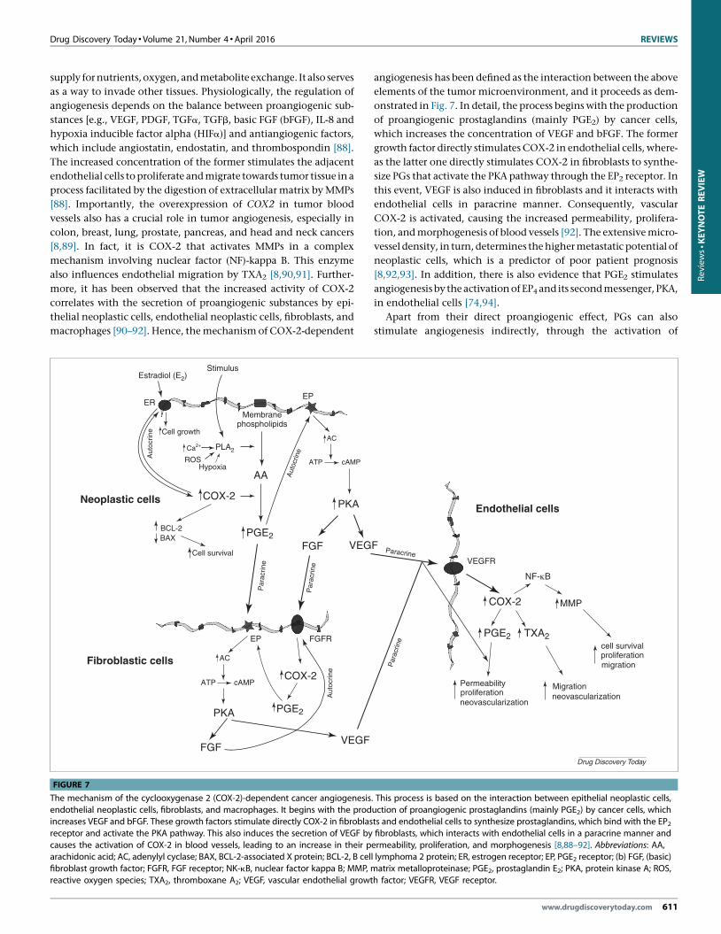
FIGURE 7
The mechanism of the cyclooxygenase 2 (COX-2)-dependent cancer angiogenesisendothelial neoplastic cells, fibroblasts, and macrophages. It begins with the prod
increases VEGF and bFGF. These growth factors stimulate directly COX-2 in fibroblas
receptor and activate the PKA pathway. This also induces the secretion of VEGF by
causes the activation of COX-2 in blood vessels, leading to an increase in their parachidonic acid; AC, adenylyl cyclase; BAX, BCL-2-associated X protein; BCL-2, B cell
fibroblast growth factor; FGFR, FGF receptor; NK-kB, nuclear factor kappa B; MMP, m
reactive oxygen species; TXA2, thromboxane A2; VEGF, vascular endothelial growt
angiogenesis has been defined as the interaction between the above
elements of the tumor microenvironment, and it proceeds as dem-
onstrated in Fig. 7. In detail, the process begins with the production
of proangiogenic prostaglandins (mainly PGE2) by cancer cells,
which increases the concentration of VEGF and bFGF. The former
growth factor directly stimulates COX-2 in endothelial cells, where-
as the latter one directly stimulates COX-2 in fibroblasts to synthe-
size PGs that activate the PKA pathway through the EP2 receptor. In
this event, VEGF is also induced in fibroblasts and it interacts with
endothelial cells in paracrine manner. Consequently, vascular
COX-2 is activated, causing the increased permeability, prolifera-
tion, and morphogenesis of blood vessels [92]. The extensive micro-
vessel density, in turn, determines the higher metastatic potential of
neoplastic cells, which is a predictor of poor patient prognosis
[8,92,93]. In addition, there is also evidence that PGE2 stimulates
angiogenesis by the activation of EP4 and its second messenger, PKA,
in endothelial cells [74,94].
Apart from their direct proangiogenic effect, PGs can also
stimulate angiogenesis indirectly, through the activation of
NF-κB
COX-2 MMP
Endothelial cells
FVEGFR
Permeability Migration
migration
cell survival
proliferation
proliferation
neovascularizationneovascularization
Par
acrin
e
Paracrine
PGE2 TXA2
Drug Discovery Today
. This process is based on the interaction between epithelial neoplastic cells,uction of proangiogenic prostaglandins (mainly PGE2) by cancer cells, which
ts and endothelial cells to synthesize prostaglandins, which bind with the EP2 fibroblasts, which interacts with endothelial cells in a paracrine manner and
ermeability, proliferation, and morphogenesis [8,88–92]. Abbreviations: AA, lymphoma 2 protein; ER, estrogen receptor; EP, PGE2 receptor; (b) FGF, (basic)
atrix metalloproteinase; PGE2, prostaglandin E2; PKA, protein kinase A; ROS,
h factor; VEGFR, VEGF receptor.
www.drugdiscoverytoday.com 611

REVIEWS Drug Discovery Today �Volume 21, Number 4 �April 2016
Review
s�K
EYNOTEREVIEW
monocytes that infiltrate cancer tissue. In fact, the increased
production of proinflammatory factors, such as IL-2, results in
further recruitment of macrophages to the so-formed inflamma-
tory foci, recognized by macrophages as the area of damaged
tissue. Thus, the arriving macrophages induce a healing process,
which involves the connection of the damaged region to blood
vessels, formation of supportive connective tissue, and stimula-
tion of regeneration [84,92].
Finally, in one Phase II clinical trial in patients with breast
cancer, preoperative adjuvant therapy with celecoxib resulted in
improved matrix stability associated with the downregulation of
MMP-2 and MMP-9. Interestingly, this observation was explained
by the celecoxib-dependent activation of TIMP metallopeptidase
inhibitor 1–3 (TIMP1, TIMP2, and TIMP3), and Reversion-induc-
ing-cysteine-rich protein with Kazal motifs (RECK), which are
known antagonists of MMPs [87].
ImmunosuppressionCOX-2 has a significant role in breast tumor immune escape. PGs,
especially PGE2, negatively influence the activity of T and B
lymphocytes, natural killer (NK) cells, and dendritic cells. They
also reduce the synthesis of TNFa and augment the activity of
immunosuppressive IL-10 [53]. These abnormalities, in turn, abol-
ish the efficiency of host defenses in detecting and rejecting
malignant cells, thereby promoting their uncontrolled prolifera-
tion [95]. The hypothesis of COX-2-related immunosuppression is
supported by published studies in which the decreased expression
of COX2 in breast cancer cells resulted in increased tissue infiltra-
tion by cytotoxic T lymphocytes (CD8+). Also, the application of
selective COX-2 inhibitors in human breast cancers induced the
enhanced recruitment of immune cells to the tumor microenvi-
ronment [85,95].
Association between COX-2 and aromatase P450Aromatase P450 belongs to the family of proteins associated with
cytochrome P450. It is responsible for the conversion of andro-
genic steroids into estrone and estradiol, and has a crucial role in
the local production of these hormones in estrogen-dependent
breast cancer (approximately 60–70% of all breast tumors). The
interaction between estrogens and their receptors, in turn, con-
stitutes the critical stimulus for the development of these tumors
because it directly induces the growth and proliferation of ma-
lignant cells. Hence, it is no surprise that the upregulation of
aromatase P450 determines the progression of breast cancer with
an ER+ status. Interestingly, the expression of CYP19 encoding
aromatase P450 is dependent on auto- and paracrine activation
by PGE2, which, by interacting with EP2 and EP4, causes the
accumulation of cAMP, PKC, and PKA [96]. For this reason, in
human breast cancer, there is a positive correlation between
CYP19 expression and the concentration of COX-2 within the
tumor microenvironment [97]. Available experimental data sup-
port these hypotheses. In fact, in model transgenic mice with
silenced COX2, the activity of aromatase appears to be signifi-
cantly lower, whereas in mice overexpressing COX2, the enzyme
is upregulated [47]. In addition, the impact of COX-2 on the local
production of estrogens could explain the chemopreventive
properties of NSAIDs in postmenopausal estrogen-dependent
breast cancer [47].
612 www.drugdiscoverytoday.com
Production of mutagens in vivoA mutagen is any endo- or exogenic substance that damages DNA
directly, thereby contributing to the development of neoplasia.
One endogenic mutagen [malondialdehyde (MDA)] is formed
consequently to the enzymatic or nonenzymatic degradation of
PGH2, which is a known product of COX-2. The detrimental effect
of MDA is, in turn, associated with its tendency to form adducts
with deoxynucleotides, leading to point and frameshift mutations
[53]. In addition, COX-2 acting as a peroxidase could catalyze the
oxidation of aromatic amines, heterocyclic amines, dihydrodiol
derivatives, and multiring aromatic hydrocarbons, producing var-
ious carcinogenic compounds [98]. In fact, it has been experimen-
tally shown that the preferential COX-2 inhibitor nimesulide
reduces the production of a mutagen named 8-oxo-7,8-dihydro-
20-deoxyguanosine in mucosa during early colonic inflammation
in rats [99].
COX-2 inhibitors in breast cancer: current and futuredirectionsThe use of COX-2 inhibitors in breast cancer could be primarily
justified by their chemopreventive properties, which have been
found to be beneficial in both primary and secondary prophylaxis.
Appropriate studies with animal models have shown such strate-
gies to be effective. For instance, the decreased incidence of
carcinogen-induced breast malignancies after the application of
flurbiprofen and indomethacin (nonselective COX inhibitors) has
been shown. Similarly, some chemopreventive actions have been
observed for selective and preferential COX-2 inhibitors, including
celecoxib, rofecoxib, and nimesulide. What is more, these agents
additionally resulted in growth inhibition in cancer cells with ER+,
and ER� HER2+ phenotypes (http://www.cancer.gov/types/
breast/hp/breast-treatment-pdq), whereas celecoxib has also been
found to delay the onset of HER2/neu-induced tumors. Important-
ly, celecoxib also acts more effectively in retarding the develop-
ment of DMBA-induced breast cancer compared with nonselective
ibuprofen [100].
Moreover, the available epidemiological data indicate that the
long-term use of NSAIDs significantly lowers the incidence of
breast cancer. This effect was confirmed in 1980 within a group of
4876 patients treated with indomethacin (compared with age-
matched controls) [101]. In 1996, it was estimated that the
administration of NSAIDs three times a week for at least 1 year
lowered the risk of breast cancer development by 66% (a study
comparing 511 females with newly diagnosed breast cancer using
NSAIDs versus 1534 controls) [102]. Consistent results were
achieved in studies conducted in 1994 (the incidence ratio among
aspirin users was 0.7) [103] and 2000 (retarded growth of breast
cancer among NSAIDs users) [104]. In addition, a more recent
meta-analysis of nine case–control studies and seven cohort
studies from 2003 showed a slight but significant decrease in
breast cancer incidence among users of acetylsalicylic acid and
other nonaspirin NSAIDs []105. Also, in a case–control study from
2006, a reduction of breast cancer risk of 71% was observed in
women using selective COX-2 inhibitors (celecoxib and rofe-
coxib) in standard doses for 2 years [106]. Finally, large meta-
analyses from 2012 confirmed that aspirin is protective against
cancer development and its regular intake significantly reduces
the risk of breast cancer (odds ratio = 0.88) [107]. However, there

Drug Discovery Today � Volume 21, Number 4 �April 2016 REVIEWS
Reviews�KEYNOTEREVIEW
are also epidemiological studies indicating that there is no rela-
tion between NSAIDs use and breast cancer incidence [108–110].
This inconsistence could be explained by the nonuniform expres-
sion of COX-2 in different subtypes of this malignancy.
Apart from their prophylactic effects, COX-2 inhibitors could be
also used in the treatment of breast cancer. This concept is sub-
stantiated by the results of experiments on rats that showed that
100-day therapy with ibuprofen decreased the size of DMBA-
induced breast cancer by 40%, whereas in nontreated controls
the neoplastic lesions increased by 180% [61]. Similarly, celecoxib
used for 6 weeks contributed to the decrease of tumor mass by 32%
[111].
Alternatively, COX-2 inhibitors could be introduced into adju-
vant anticancer therapy. A promising strategy in this respect
involves their co-administration with taxanes, because these cy-
tostatic drugs have the ability to induce the transcription of COX2,
which might result in the decreased efficiency of monotherapy
[112]. Another possibility is the combination therapy of a COX-2
inhibitor and an aromatase inhibitor, such as letrozole or anastro-
zole, in the treatment of hormone-dependent breast cancers.
Finally, a therapeutic regimen including a COX-2 inhibitor and
trastuzumab in malignancies overexpressing HER2 should also be
considered [113]. However, the results of available studies are not
satisfactory. In trastuzumab-refractory metastatic breast cancer,
COX-2 inhibition was shown to be inactive yet well tolerated in a
group of 12 patients (a randomized Phase II clinical trial) [114]. In
postmenopausal metastatic breast cancer, the improved efficacy
and end-point benefits of celecoxib in combination with an aro-
matase inhibitor were reported, but only in tamoxifen-resistant
women [115]. Finally, the results of a randomized Phase II study in
111 postmenopausal women with advanced breast cancer treated
with exemestane and celecoxib indicated a longer time to progres-
sion with no additional adverse effects associated with the use of
combination therapy [116].
There is also a study that analyzed the benefits of simultaneous
use of celecoxib (400 mg) with FEC chemotherapy (5-FU 500 mg/
m2, epirubicin 75 mg/m2, and cyclophosphamide 500 mg/m2) in
neoadjuvant treatment of locally advanced breast tumors. Inter-
estingly, the results showed that the clinical and pathological
responses for the combined treatment arm (FEC + celecoxib)
equaled 81.3 and 87.5%, respectively, versus 62.5 and 62.5% in
the group treated with FEC only [117].
Despite these promising results, more clinical data are required
to decide whether the described strategies provide any substantial
benefits to patients with breast cancer.
Concluding remarksGreat progress has been made since COX-2 was first established as a
potential therapeutic or preventive target in breast cancer, espe-
cially in our understanding of the link between the activity of the
enzyme and the malignancy. The detailed description of the
pathomechanism of COX-2-related cancer progression revealed
that selective COX-2 inhibitors together with conventional thera-
py (radiotherapy or chemotherapy) could represent a modern and
effective approach in the management of breast cancer. Indeed,
this idea is supported by the results of several experimental studies,
yet the clinical benefits of this novel therapeutic mode are cur-
rently too limited [61,86,88,101–120]. Appropriate, randomized
clinical trials are needed for the final assessment of the clinical
value associated with the strategies discussed here. Also, the
identification of patients who could benefit from their implemen-
tation is required. Finally, more attention should be paid to the
discovery and development of new safer and more effective inhi-
bitors, such as trans-stilbenes, to provide patients with more
convenient and less toxic therapeutic options. This can be done
by the use of different analytical methods, including in silico QSAR
techniques, which constitute an important research tool in con-
temporary pharmaceutical research.
Conflict of interestNone declared.
AcknowledgmentThis study was financially supported by The National Science
Centre (NCN) of Poland, grant no UMO-2011/01/N/NZ4/01137.
Appendix A. Supplementary dataSupplementary data associated with this article can be found, in
the online version, at http://dx.doi.org/10.1016/j.drudis.2015.12.
003.
References
1 Jemal, A. et al. (2008) Cancer statistics. CA Cancer J. Clin. 58, 71–96
2 Jemal, A. et al. (2011) Global cancer statistics. CA Cancer J. Clin. 61, 69–90
3 Yoshinaka, R. et al. (2006) COX-2 inhibitor celecoxib suppresses tumor growth and
lung metastasis of a murine mammary cancer. Anticancer Res. 26 (6B), 4245–4254
4 Ranger, G.S. and Mokbel, K. (2003) COX-2 inhibitors and breast cancer. ANZ J.
Surg. 73 (8), 565–566
5 Subbaramaiah, K. et al. (2002) Cyclooxygenase-2 is overexpressed in HER-2/neu-
positive breast cancer: evidence for involvement of AP-1 and PEA3. J. Biol. Chem.
277 (21), 18649–18657
6 Dannenbergd, A.J. and Howe, L.R. (2003) The role of COX-2 in breast and cervical
cancer. COX-2: a new target for cancer prevention and treatment. Prog. Exp. Tumor
Res. 37, 90–106
7 Singh, B. et al. (2007) COX-2 involvement in breast cancer metastasis to bone.
Oncogene 26 (26), 3789–3796
8 Gupta, G.P. et al. (2007) Mediators of vascular remodelling co-opted for sequential
steps in lung metastasis. Nature 446 (7137), 765–770
9 Mazhar, D. et al. (2006) COX inhibitors and breast cancer. Br. J. Cancer 94, 346–350
10 Burdan, F. et al. (2006) Cyklooksygenaza i prostanoidy – znaczenie biologiczne.
(Cyclooxygenase and prostanoids – biological implications).Postepy. Hig. Med.
Dosw. 60, 129–141 (in Polish)
11 Simmons, D.L. et al. (2004) Cyclooxygenase isozymes: the biology of
prostaglandin synthesis and inhibition. Pharmacol. Rev. 56, 387–437
12 Turini, M.E. and DuBois, R.N. (2002) Cyclooxygenase-2: a therapeutic target.
Annu. Rev. Med. 53, 35–57
13 Claria, J. (2003) Cyclooxygenase-2 biology. Curr. Pharm. Des. 9, 2177–2190
14 Chandrasekharan, N.V. and Simmons, D.L. (2004) Protein family review: the
cyclooxygenases. Genome Biol. 5, 241 1–7
15 Wang, J.L. et al. (2010) The novel benzopyran class of selective cyclooxygenase-2
inhibitors. Part 2: the second clinical candidate having a shorter and favorable
human half-life. Bioorg. Med. Chem. Lett. 20, 7159–7163
16 Hinz, B. and Brune, K. (2002) Cyclooxygenase-2 – 10 years later. J. Pharmacol. Exp.
Ther. 300, 367–375
17 Narumiya, S. and FitzGerald, G.A. (2001) Genetic and pharmacological analysis of
prostanoid receptor function. J. Clin. Invest. 108, 25–30
www.drugdiscoverytoday.com 613
REVIEWS Drug Discovery Today �Volume 21, Number 4 �April 2016
Review
s�K
EYNOTEREVIEW
18 Sobolewski, C. et al. (2010) The role of cyclooxygenase-2 in cell proliferation and
cell death in human malignancies. Int. J. Cell Biol. 2010, 215158
19 Kelly, R.W. (1981) Prostaglandin synthesis in the male and female reproductive
tract. J. Reprod. Fertil. 62, 293–304
20 Zarghi, A. and Arfaei, S. (2011) Selective COX-2 inhibitors: a review of their
structure–activity relationships. Iran J. Pharm. Res. 10, 655–683
21 Breyer, R.M. et al. (2001) Prostanoid receptors: subtypes and signaling. Annu. Rev.
Pharmacol. Toxicol. 41, 661–690
22 Kundu, N. et al. (2014) Prostaglandin E receptor EP4 is a therapeutic target in breast
cancer cells with stem-like properties. Breast Cancer Res. Treat. 143, 19–31
23 Reader, J. et al. (2011) Prostaglandin E2 EP receptors as therapeutic targets in breast
cancer. Cancer Metastasis Rev. 30, 449–463
24 Ma, X. et al. (2010) Prostaglandin E receptor EP1 suppresses breast cancer
metastasis and is linked to survival differences and cancer disparities. Mol. Cancer
Res. 8, 1310–1318
25 Gans, K.R. et al. (1990) Anti-inflammatory and safety profile of DuP 697, a novel orally
effective prostaglandin synthesis inhibitor. J. Pharmacol. Exp. Ther. 254, 180–187
26 Kalgutkar, A.S. and Zhao, Z. (2001) Discovery and design of selective
cyclooxygenase-2 inhibitors as non-ulcerogenic, anti-inflammatory drugs with
potential utility as anti-cancer agents. Curr. Drug Targets 2, 79–106
27 Marnett, L.J. and Kalgutkar, A.S. (1999) Cyclooxygenase 2 inhibitors: discovery,
selectivity and the future. Trends Pharmacol. Sci. 20, 465–469
28 Pratico, D. and Dogne, J.M. (2005) Selective cyclooxygenase-2 inhibitors
development incardiovascular medicine. Circulation 112, 1073–1079
29 European Medicines Agency (2005) Public Statement on Valdyn (valdecoxib):
Withdrawal of the Marketing Authorisation in the European Union. EMA
30 European Medicines Agency (2007) European Medicines Agency Recommends
Withdrawal of the Marketing Authorisations for Lumiracoxib-containing Medicines.
EMA
31 Llorens, O. et al. (1999) Structural basis of the dynamic mechanism of ligand
binding to cyclooxygenase. Bioorg. Med. Chem. Lett. 9, 2779–2784
32 Michaux, C. and Charlier, C. (2004) Structural approach for COX-2 inhibition.
Mini Rev. Med. Chem. 4, 603–615
33 Walker, M.C. and Kurumbail, R.G. (2001) A three-step kinetic mechanism for
selective inhibition of cyclooxygenase-2 by diarylheterocyclic inhibitors. Biochem.
J. 357, 709–718
34 Zykova, T.A. et al. (2008) Resveratrol directly targets COX-2 to inhibit
carcinogenesis. Mol. Carcinog. 47, 797–805
35 Murias, M. et al. (2004) Resveratrol analogues as selective cyclooxygenase-2
inhibitors: synthesis and structure–activity relationship. Bioorg. Med. Chem. 12,
5571–5578
36 Kang, S. et al. (2009) Synthesis and biological evaluation of a library of resveratrol
analogues as inhibitors of COX-1, COX-2 and NF-kappaB. Bioorg. Med. Chem. 17,
1044–1054
37 Uddin, M.J. et al. (2005) Design and synthesis of (E)-1,1,2-triarylethenes: novel
inhibitors of the cyclooxygenase-2 (COX-2) isozyme. Bioorg. Med. Chem. Lett. 15,
439–442
38 Kapetanovic, I.M. (2008) Computer-aided drug discovery and development
(CADDD): in silico-chemico-biological approach. Chem. Biol. Interact. 171, 165–
176
39 Selassie, C.D. (2003) History of quantitative structure–activity relationships. In
Burger’s Medicinal Chemistry and Drug Discovery (6th edn) (Abraham, D.J., ed.), pp.
1–48, John Wiley & Sons
40 Puzyn, T. et al. eds (2010) Recent Advances in QSAR Studies. Methods and Applications,
Springer
41 Prukała, W. et al. (1988) New (E)-40-halo substituted stilbenyloxyalkanocarboxylic
acids and their derivatives. Pol. J. Chem. 62, 251–255
42 Veerasamy, R. et al. (2011) Validation of QSAR models-strategies and importance.
Int. J. Drug Des. Discov. 2, 511–519
43 Regulski, M. et al. (2015) 2D QSAR analysis of trans-stilbene derivatives as COX-1/
COX-2 inhibitors. 8th Polish-German Symposium on Pharmaceutical Science
‘Retrospects, Insights And Prospects’ Kiel, Germany, May 29–30, Kiel University
Abstract PH-04
44 Całkosinski, I. et al. (2009) Charakterystyka odczynu zapalnego. (Characterization
of an inflammatory response).Postepy. Hig. Med. Dosw. 63, 395–408 (in Polish)
45 Stasik, Z. et al. (2008) Białko C-reaktywne u chorych na nowotwory złosliwe. (C-
reactive protein in cancer patients).Nowotwory 58, 441–446 (in Polish)
46 Triantafillidis, J.K. et al. (2009) Colorectal cancer and inflammatory bowel disease:
epidemiology, risk factors, mechanisms of carcinogenesis and prevention
strategies. Anticancer Res. 29, 2727–2737
47 Howe, L.R. (2007) Inflammation and breast cancer. Cyclooxygenase/
prostaglandin signaling and breast cancer. Breast Cancer Res. 9, 210
614 www.drugdiscoverytoday.com
48 Regulska, K. et al. (2013) The renin-angiotensin system as a target of novel
anticancer therapy. Curr. Pharm. Des. 19, 7103–7125
49 Rakoff-Nahoum, S. (2006) Why cancer and inflammation? Yale J. Biol. Med. 79,
123–130
50 Tao, Z. et al. (2015) Breast cancer: epidemiology and etiology. Cell Biochem.
Biophys. 72, 333–338
51 Ferlay, J. et al. (2013) Cancer incidence and mortality patterns in Europe: Estimates
for 40 countries in 2012. Eur. J. Cancer 49, 1374–1403
52 WHO (2012) Breast Cancer. Estimated Incidence, Mortality and Prevalence Worldwide
in 2012. WHO
53 Howe, L.R. et al. (2001) Cyclooxygenase-2: a target for the prevention and
treatment of breast cancer. Endocr. Relat. Cancer 8, 97–114
54 Bennett, A. et al. (1977) Prostaglandins and breast cancer. Lancet 310, 624–626
55 Tan, W.C. et al. (1974) Studies of prostaglandins in rat mammary tumors induced
by 7,12-dimethylbenz(a)anthracene. Cancer Res. 34, 3229–3231
56 Rolland, P.H. et al. (1980) Prostaglandin in human breast cancer: evidence
suggesting that an elevated prostaglandin production is a marker of high
metastatic potential for neoplastic cells. J. Natl. Cancer Inst. 64, 1061–1070
57 Ristimaki, A. et al. (2002) Prognostic significance of elevated cyclooxygenase-2
expression in breast cancer. Cancer Res. 62, 632–635
58 Parrett, M.L. et al. (1997) Cyclooxygenase-2 gene expression in human breast
cancer. Int. J. Oncol. 10, 503–508
59 Hwang, D. and Byrne, J. (1998) Expression of cyclooxygenase-1 and
cyclooxygenase-2 in human breast cancer. J. Natl. Cancer Inst. 90, 455–460
60 Nakatsugi, S. et al. (2000) Chemoprevention by nimesulide, a selective
cyclooxygenase-2 inhibitor, of 2-amino-1-methyl-6-phenylimidazo[4,5-
b]pyridine (PhIP)-induced mammary gland carcinogenesis in rats. Jpn. J. Cancer
Res. 91, 886–892
61 Robertson, F.M. et al. (1998) Ibuprofen-induced inhibition of cyclooxygenase
isoform gene expression and regression of rat mammary carcinomas. Cancer Lett.
122, 165–175
62 Liu, X.H. and Rose, D.P. (1996) Differential expression and regulation of
cyclooxygenase-1 and -2 in the human breast cancer cell lines. Cancer Res. 56,
5125–5127
63 Bocca, C. et al. (2011) Antiproliferative effects of COX-2 inhibitor celecoxib on
human breast cancer cell lines. Mol. Cell. Biochem. 350, 59–70
64 Bocca, C. et al. (2014) Expression of Cox-2 in human breast cancer cells as a critical
determinant of epithelial-to-mesenchymal transition and invasiveness. Expert
Opin. Ther. Targets 18, 121–135
65 Thill, M. et al. (2009) Prostaglandin metabolizing enzymes in correlation with
vitamin D receptor in benign and malignant breast cell lines. Anticancer Res. 29,
3619–3625
66 Zhao, X. et al. (2008) Cyclooxygenase-2 expression during immortalization and
breast cancer progression. Cancer Res. 68, 467–475
67 Ranger, G.S. et al. (2004) Elevated cyclooxygenase-2 expression correlates with
distant metastases in breast cancer. Anticancer Res. 24, 2349–2351
68 Carter, C.A. et al. (1983) Effect of the prostaglandin synthetase inhibitor
indomethacin on 7,12-dimethylbenz(a)anthracene-induced mammary
tumorigenesis in rats fed different levels of fat. Cancer Res. 43, 3559–3562
69 McCormick, D.L. et al. (1985) Modulation of rat mammary carcinogenesis by
indomethacin. Cancer Res. 45, 1803–1808
70 Noguchi, M. et al. (1991) Effects of the prostaglandin synthetase inhibitor
indomethacin on tumorigenesis, tumor proliferation, cell kinetics, and receptor
contents of 7,12-dimethylbenz(a)anthracene-induced mammary carcinoma in
Sprague-Dawley rats fed a high- or low-fat diet. Cancer Res. 51, 2683–2689
71 McCormick, D.L. and Moon, R.C. (1983) Inhibition of mammary carcinogenesis
by flurbiprofen, a non-steroidal anti-inflammatory agent. Br. J. Cancer 48, 859–861
72 Suzui, N. et al. (1997) Inhibitory effects of diallyl disulfide or aspirin on 2-amino-1-
methyl-6-phenylimidazo[4,5-bpyridine-induced mammary carcinogenesis in rats.
Jpn. J. Cancer Res. 88, 705–711
73 Harris, R.E. et al. (2000) Chemoprevention of breast cancer in rats by celecoxib, a
cyclooxygenase 2 inhibitor. Cancer Res. 60, 2101–2103
74 Liu, C.H. et al. (2001) Overexpression of cyclooxygenase-2 is sufficient to induce
tumorigenesis in transgenic mice. J. Biol. Chem. 276, 18563–18569
75 Pai, S.I. et al. (2006) Prospects of RNA interference therapy for cancer. Gene Ther.
13, 464–477
76 Dabbagh, A. and Rajaei, S. (2013) The role of anesthetic drugs in liver apoptosis.
Hepat. Mon. 13, e13162
77 Elmore, S. (2007) Apoptosis: a review of programmed cell death. Toxicol. Pathol. 35,
495–516
78 Glynn, S.A. et al. (2010) COX-2 activation is associated with Akt phosphorylation
and poor survival in ER-negative, HER2-positive breast cancer. BMC Cancer 10, 626

Drug Discovery Today � Volume 21, Number 4 �April 2016 REVIEWS
Reviews�KEYNOTEREVIEW
79 Cao, Y. and Prescott, S.M. (2002) Many actions of cyclooxygenase-2 in cellular
dynamics and in cancer. J. Cell Physiol. 190, 279–286
80 Cao, Y. et al. (2000) Intracellular unesterified arachidonic acid signals apoptosis.
Proc. Natl. Acad. Sci. U. S. A. 97, 11280–11285
81 Yamasaki, L. (2003) Role of the RB tumor suppressor in cancer. Cancer Treat. Res.
115, 209–239
82 Lim, S. and Kaldis, P. (2013) Cdks, cyclins and CKIs: roles beyond cell cycle
regulation. Development 140, 3079–3093
83 Bandyopadhyay, G.K. et al. (1987) Linoleate metabolites enhance the in vitro
proliferative response of mouse mammary epithelial cells to epidermal growth
factor. J. Biol. Chem. 262, 2750–2756
84 Lim, S.C. (2013) Role of COX-2, VEGF and cyclin D1 in mammary infiltrating duct
carcinoma. Oncol. Rep. 10, 1241–1249
85 Im, S-A. et al. (2004) Expression of cyclooxygenase-2 in Korean breast cancer
patients and the in vitro growth inhibitory effect of celecoxib in breast cancer cells.
J. Clin. Oncol. 22, 9715
86 Liu, W. et al. (2003) Combination of radiation and Celebrex (celecoxib) reduce
mammary and lung tumor growth. Am. J. Clin. Oncol. 26, S103–S109
87 Brandao, R.D. et al. (2013) A randomised controlled phase II trial of pre-operative
celecoxib treatment reveals anti-tumour transcriptional response in primary
breast cancer. Breast Cancer Res. 15, R29
88 Huang, Z. and Bao, S.D. (2004) Roles of main pro- and anti-angiogenic factors in
tumor angiogenesis. World J. Gastroenterol. 10, 463–470
89 Leahy, K.M. et al. (2000) Role of cyclooxygenases in angiogenesis. Curr. Med. Chem.
7, 1163–1170
90 Zha, S. et al. (2004) Cyclooxygenases in cancer: progress and perspective. Cancer
Lett. 215, 1–20
91 Divvela, A.K.C. et al. (2010) Pathogenic role of cyclooxygenase-2 in cancer. J.
Health Sci. 56, 502–516
92 Fosslien, E. (2001) Review: molecular pathology of cyclooxygenase-2 in cancer-
induced angiogenesis. Ann. Clin. Lab. Sci. 31, 325–348
93 Visscher, D.W. et al. (1993) Prognostic significance of image morphometric
microvessel enumeration in breast carcinoma. Anal. Quant. Cytol. Histol. 15, 88–92
94 Zhang, Y. and Daaka, Y. (2011) PGE2 promotes angiogenesis through EP4 and PKA
Cg pathway. Blood 118, 5355–5564
95 Markosyan, N. et al. (2013) Mammary carcinoma cell derived cyclooxygenase 2
suppresses tumor immune surveillance by enhancing intratumoral immune
checkpoint activity. Breast Cancer Res. 15, R75
96 Mokbel, K. (2002) The evolving role of aromatase inhibitors in breast cancer. Int. J.
Clin. Oncol. 7, 279–283
97 Brueggemeier, R.W. et al. (1999) Correlation of aromatase and cyclooxygenase
gene expression in human breast cancer specimens. Cancer Lett. 140, 27–35
98 Wiese, F.W. et al. (2001) Carcinogen substrate specificity of human COX-1 and
COX-2. Carcinogenesis 22, 5–10
99 Tardieu, D. et al. (2000) The COX-2 inhibitor nimesulide suppresses superoxide
and 8-hydroxy-deoxyguanosine formation, and stimulates apoptosis in mucosa
during early colonic inflammation in rats. Carcinogenesis 21, 973–976
100 Howe, L.R. et al. (2002) Celecoxib, a selective cyclooxygenase inhibitor, protects
against human epidermal growth factor receptor 2 (HER-2)/neu-induced breast
cancer. Cancer Res. 62, 5405–5407
101 Friedman, G.D. and Ury, H.K. (1980) Initial screening for carcinogenicity of
commonly used drugs. J. Natl. Cancer Inst. 65, 723–733
102 Harris, R.E. et al. (1996) Nonsteroidal antiinflammatory drugs and breast cancer.
Epidemiology 7, 203–205
103 Schreinemachers, D.M. and Everson, R.B. (1994) Aspirin use and lung, colon, and
breast cancer incidence in a prospective study. Epidemiology 5, 138–146
104 Sharpe, C.R. et al. (2000) Nested case–control study of the effects of non-steroidal
anti-inflammatory drugs on breast cancer risk and stage. Br. J. Cancer 83, 112–120
105 Gonzalez-Perez, A. et al. (2003) Effects of non-steroidal anti-inflammatory drugs on
cancer sites other than colon and rectum: a meta-analysis. BMC Cancer 3, 28
106 Harris, R.E. et al. (2006) Reduction in the risk of human breast cancer by selective
cyclooxygenase-2 (COX-2) inhibitors. BMC Cancer 6, 27
107 Algra, A.M. and Rothwell, P.M. (2012) Effects of regular aspirin on long-term
cancer incidence and metastasis: a systematic comparison of evidence from
observational studies versus randomised trials. Lancet Oncol. 13, 518–527
108 Paganini-Hill, A. et al. (1989) Aspirin use and chronic diseases: a cohort study of the
elderly. BMJ 299, 1247–1250
109 Egan, K.M. et al. (1996) Prospective study of regular aspirin use and the risk of
breast cancer. J. Natl. Cancer Inst. 88, 988–993
110 Rosenberg, L. (1995) Nonsteroidal anti-inflammatory drugs and cancer. Prev. Med.
24, 107–109
111 Alshafie, G.A. et al. (2000) Chemotherapeutic evaluation of Celecoxib, a
cyclooxygenase-2 inhibitor, in a rat mammary tumor model. Oncol. Rep. 7, 1377–
1381
112 Subbaramaiah, K. et al. (2000) Microtubule-interfering agents stimulate the
transcription of cyclooxygenase-2. Evidence for involvement of ERK1/2 AND p38
mitogen-activated protein kinase pathways. J. Biol. Chem. 275, 14838–14845
113 Falandry, C. et al. (2009) Role of combination therapy with aromatase and
cyclooxygenase-2 inhibitors in patients with metastatic breast cancer. Ann. Oncol.
20, 615–620
114 Dang, C.T. et al. (2004) Phase II study of celecoxib and trastuzumab in metastatic
breast cancer patients who have progressed after prior trastuzumab-based
treatments. Clin. Cancer Res. 10, 4062–4067
115 Falandry, C. et al. (2009) Celecoxib and exemestane versus placebo and
exemestane in postmenopausal metastatic breast cancer patients: a double-blind
phase III GINECO study. Breast Cancer Res. Treat. 116, 501–508
116 Dirix, L.Y. et al. (2003) Final results from an open-label, multicenter, controlled
study of exemestane +/�celecoxib in postmenopausal women with advanced
breast cancer (ABC) progressed on tamoxifen (T). Proc. Am. Soc. Clin. Oncol. 22, 20
117 Arun, B. and Goss, P. (2004) The role of COX-2 inhibition in breast cancer
treatment and prevention. Semin. Oncol. 31 (2 Suppl. 7), 22–29
118 Moore, R.J. et al. (2000) Enhanced antitumor activity by co-administration of
celecoxib and the chemotherapeutic agents cyclophosphamide and 5-FU. Proc.
Am. Assoc. Cancer Res. 41, 409
119 Milas, L. et al. (1999) Enhancement of tumor response to gamma-radiation by an
inhibitor of cyclooxygenase-2 enzyme. J. Natl. Cancer Inst. 91, 1501–1504
120 Dittmann, K.H. et al. (2008) Celecoxib induced tumor cell radiosensitization by
inhibiting radiation induced nuclear EGFR transport and DNA-repair: a COX-2
independent mechanism. Int. J. Radiat. Oncol. Biol. Phys. 70, 203–212
www.drugdiscoverytoday.com 615