covid-19 pandemic: the deadly respiratory disease of 21st
TRANSCRIPT
Current Research in Pharmaceutical Sciences 2021; 11 (01): 01-18
2
ISSN: 2250 – 2688
Received: 15/01/2021
Revised: 31/01/2021
Accepted: 08/02/2021
Published: 08/04/2021
Shweta Jain
Sir Madan Lal Institute of Pharmacy,
Etawah (U.P.) India;
Pankaj Kumar Jain
Community Medicine, Uttar Pradesh
University of Medical Sciences, Saifai,
Etawah (U.P.) India
Ramakant Yadav
Department of Neurology, Uttar
Pradesh University of Medical Sciences,
Saifai, Etawah (U.P.) India
Surendra Kumar Jain
Sagar Institute of Research and
Technology–Pharmacy,Bhopal (M.P.)
India
Ankur Vaidya
Pharmacy College Saifai, Uttar Pradesh
University of Medical Sciences, Saifai,
Etawah (U.P.) India
Correspondence
Ankur Vaidya
Pharmacy College Saifai, Uttar Pradesh
University of Medical Sciences, Saifai,
Etawah (U.P.) India
E mail:
DOI: 10.24092/CRPS.2021.110101
Website: www.crpsonline.com
Quick Response Code:
COVID-19 pandemic: The deadly respiratory disease of 21st
century
Shweta Jain, Pankaj Kumar Jain, Ramakant Yadav, Surendra Kumar Jain,
Ankur Vaidya
ABSTRACT
The sudden outbreak of 2019 novel coronavirus (2019-nCoV) caused by the novel
severe acute respiratory syndrome coronavirus 2 (SARS-CoV-2) originated from Wuhan, China.
SARS-CoV-2 causes severe respiratory illness and becomes a major threat for humanity.
Recently the entire scientist, researchers and physicians all over the countries focused to find the
treatment of this pandemic disease. Numerous drugs and or vaccines have been trialed for
prevention and treatment against 2019-nCoV but no therapy has been shown effective to date.
Currently, numerous vaccines are under clinical investigation and mRNA-1273 vaccine (LNP-
encapsulated mRNA vaccine encoding S protein) from Moderna is ahead. Although chloroquine,
hydroxychloroquine, remdesivir and many other drugs had recommended against SARS-CoV-2,
but still they are not the guarantee treatment of COVID-19. Recently, India, America, Russia and
China introduced vaccines against COVID-19 in the market, however assurance of their 100%
effectiveness are doubtful. The speed of daily new cases threatens the world and urges the
scientist to crack this pandemic condition.
KEYWORDS 2019-nCoV; Chloroquine; COVID-19; Moderna; Respiratory disease;
Remdesivir
1. INTRODUCTION
Humans have faced lots of virus epidemics since ancient era. Smallpox and
measles viruses are among the oldest that infect humans. The first virus epidemic wiped out was
reported in China about 5000 years ago. At the early age of civilization the first epidemic virus
infection was reported in 1720 Plague attack, while exact after 100 years in the 1820 Cholera
attacked and in the 1920 Spanish flu.The history seems to repeat again, exactly after 100 years
deadly virus named Coronavirus attacked China, till now millions of people got infected and
thousands of people were died with this disease. Scientist identified and reported 12 worst killer
viruses. These include Ebola, Rabies, HIV, Smallpox, Hantavirus, Influenza, Dengue, MERS-
CoV, Rotavirus, SARS-CoV and SARS-CoV-2. Although few of these virus infections have
suitable treatment including vaccines and antiviral drugs, yet for some infection treatment is still
under investigation.1-4
Coronaviruses are a group of related RNA viruses, which may cause respiratory tract
infections from mild illness (usually caused by rhinoviruses) to lethal illness (caused by MERS,
SARS and COVID-19). Coronavirus disease 2019 (COVID-19) caused by severe acute respiratory
syndrome coronavirus 2 (SARS-CoV-2) or 2019 novel coronavirus (2019-nCoV). SARS-CoV-2
primarily infects the lungs and causes severe respiratory illness in the individuals. In severe cases
COVID-19 causes’ death due to Acute Respiratory Distress Syndrome (ARDS) and pneumonia. It
is important to remember that it does not lead to ARDS and pneumonia in all the cases, which is
an occurrence in most severe cases.5
Current Research in Pharmaceutical Sciences 2021; 11 (01): 01-18
2
Coronaviruses (CoVs), a member of subfamily
Orthocoronavirinae. Furthermore, subfamily
Orthocoronavirinaehas four genera including α-CoV (alpha-
coronavirus), β-CoV (beta-coronavirus), γ-CoV (gamma-
coronavirus) and δ-CoV (delta-coronavirus). Mammals are usually
infected by α- and β-CoV genera, birds are infected by γ-and δ-
CoVs. Ramaiah and Arumugaswami reported that this human
pathogen SARS-CoV-2 is a member of the beta-coronavirus (β-
CoV) genus. β-CoV is believed to evolve from a bat-CoV, carrying
30 kilo base of single positive-sense RNA genome.6 Middle East
respiratory syndrome (MERS) and severe acute respiratory
syndrome (SARS) are the recent outbreaks caused by β-CoVs.
Both SARS and MERS epidemics were reported in the China and
Saudi Arabia in year 2002 and 2012 respectively and then globally
spread. Alike of SARS and MERS, SARS-CoV-2 virus also
belongs to the B lineage of the β-CoVs.
COVID-19 pandemic, also known as the coronavirus
pandemic, originated in the city of Wuhan, Hubei Province,
Central China, in December 2019 and the World Health
Organization (WHO) declared the outbreak a Public Health
Emergency of International Concern (PHEIC) on 30 January, and a
pandemic on 11 March 2020. Since 18 July 2020, approximately
14,213,678 cases of COVID-19 have been reported in over 222
countries, ensuing more than 634,995 deaths. About 8,493,874
people have recovered.
2. STRUCTURE OF SARS-COV-2
COVID-19 is a spherical or pleomorphic
cloak, positive-sense, single-stranded RNA with nucleoprotein
within a capsid encompassed of matrixprotein. It is the largest
genome (size:26 kb-32 kb) of known RNA viruses. The SARS-
CoV-2 genome encodes a large non-structure polyprotein
(ORF1a/b). Formed non-structure polyprotein further
proteolytically cleaved into 15/16 proteins, including 4 structural
proteins and 5 accessory proteins (ORF3a, ORF6, ORF7, ORF8
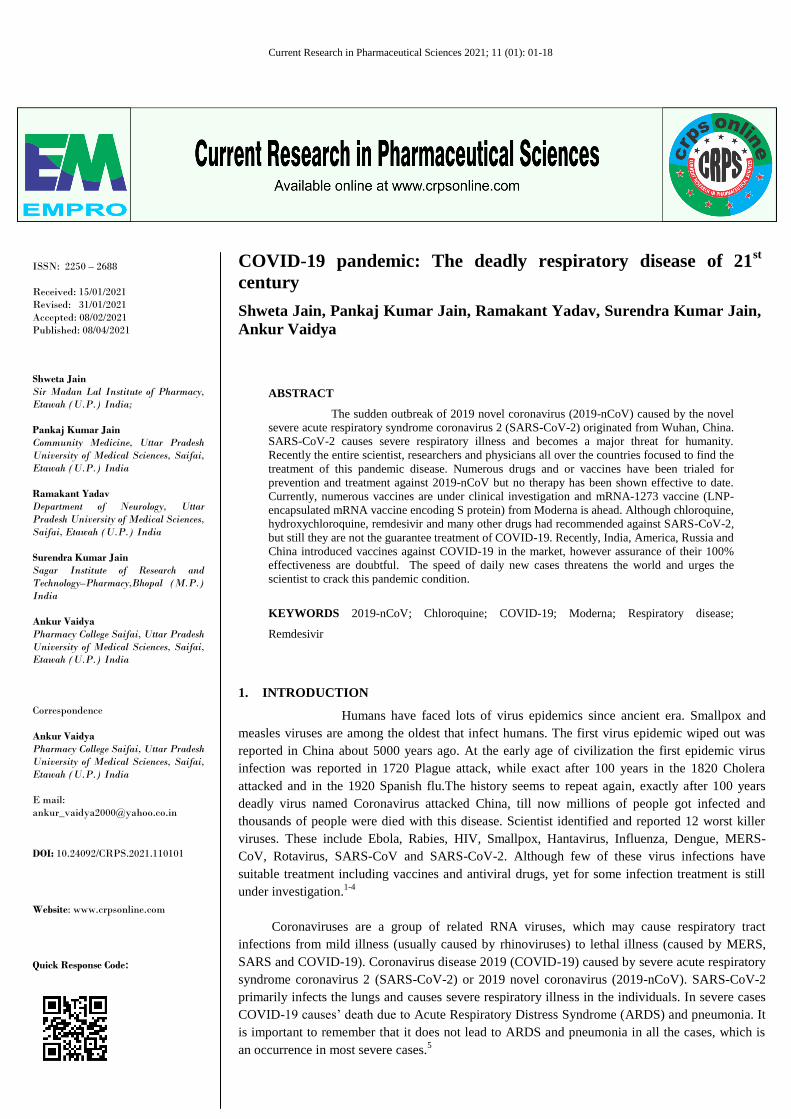
and ORF9) (Fig. 1). The structural proteins are essential for the
SARS-CoV-2 assembly and infection includes spike (S) surface
glycoprotein, membrane (M) protein (most abundant glycoprotein),
envelope (E) protein and nucleocapsid (N) protein. The genes for
all four structural proteins occur in the 5’-3’ order. Membrane
glycoprotein spans three times, resides NH2-terminal outside,
while COOH terminus inside the virion. Without entails an S
protein, M formulates virus particles.7 Envelope (E) and
Nucleocapsid (N) proteins are usually conserved, while Spike (S)
and Membrane (M) get widely mutational changess.
Spike protein has similar genomic sequence of fish
Myripristis murdjan and contains 39 nucleotide sequence (5’-aAT
GGT GTT GAA GGT TTT AAT TGT TAC TTT CCT TTA CAA
Tca-3’), responsible for it’s binding to host cells and cleaved by
host proteases into an membrane-bound C-terminal S2 region and a
N-terminal S1 subunit. This S1 subunit act as a molecular
chaperone, contributing to stabilize S2 in the prefusion state by
reducing its tendency to transition to the post fusion conformation.
The receptor-binding do-main (RBD) of S1 subunit endures hinge-
like conformational arrangements, which rapidly expose or hide
the determinants of receptor binding. These hides or expose states
are termed as ―down‖ (i.e. receptor-inaccessible state, more stable)
and ―up‖ (i.e. receptor-accessible state, less stable) conformation
respectively. These indispensable functions of S protein make it a
suitable target for antibody-mediated neutralization.8
Fig. 1 Structure of SARS-CoV-2 virus representing RNA genome,
surrounded by S, M, N and E proteins.
3. REPLICATION PROCESS OF SARS-COV-2
Hoffmann and co-workers reported that SARS-CoV-2
utilizes SARS-CoV-2 receptor angiotensin converting enzyme 2
(ACE2) for access and serine protease (i.e. TMPRSS2) for S
protein priming. SARS-CoV-2 attached to ACE2 by its Spike and
permits COVID-19 to go inside and infect cells. ACE2 is a type I
transmembrane metallocarboxy peptidase, expressed in the lung,
kidney, and gastrointestinal tract, and SARS-CoV-2 could utilize
ACE2 from humans, to add access into ACE2-expressing lung
HeLa cells.9 Since the SARS-CoV-2 spike binds more firmly
(usually10–20-fold higher) as compared to SARS-CoV spike to
human ACE2, responsible for easier spread in humans. After the
entry of virus into host cell, it uncoats and replicates rapidly and

Current Research in Pharmaceutical Sciences 2021; 11 (01): 01-18
3
elicits a burly immune response, ensuing in cytokine storm and
pulmonary tissue damage. Cytokine storm results
hypercytokinaemia and abrupt produces pro-inflammatory
cytokines, which is responsible for acute respiratory distress
syndrome (ARDS) and multiple organ failure. Continuous RNA
synthesis requires for replication of coronavirus genome. The
replicase complex encompasses number of cellular proteins and 16
viral subunits. Interestingly, coronavirus replication process requires some different RNA processing enzymes that are
exclusive for coronavirus and not reported in other RNA viruses. These rarely found coronaviruses RNA processing enzymes
includes 3’-to-5’ exoribonuclease, ADP ribose 1’-phosphatase, 2’-
O-ribose methyltransferase and cyclic phosphodiesterase. The
formed RNA genome incorporates in assembled proteins and form
mature particle to further invade flanking cell (Fig. 2).
Fig. 2 Schematic diagram of replication process of SARS
CoV-2 virus inside the human cell.
4. ORIGIN OF SARS-COV-2
Animals and bats have been reported as the natural
cistern hosts of numerous virus and these animals/bats play a
important role in transmitting numerous viruses. MERS, Ebola,
SARS, Nipah, Coronavirus and many other viruses have been
reported to originate through animals and or bats. Although SARS-
CoV-2 is believed to originate from bats, however still
confirmation requires. Very recently WHO announce that the first
SARS-CoV-2 case was linked to the human seafood market in
Wuhan city, China. Numerous studies suggested that bats are the
natural host of SARS-CoV-2.10
Zhou et al. reported that bats are
potential host of SARS-CoV-2 possibly due to 96% identical 2019-
nCoV genome to a bat coronavirus, whereas minks may be the
intermediate host for SARS-CoV-2.11
Furthermore, Lam et al.
reported 85.5%-92.4% similar 2019-nCoV genome to pangolin
coronavirus genomes and suggested pangolin as possible
transitional host for SARS-CoV-2.12
The above reported studies
need further research to confirm that whether SARS-CoV-2 is
directly transmitted from bats or by an intermediate host.
Discovering the source of SARS-CoV-2 will assist to manage its
spread (Fig. 3).
Fig. 3 Hypotheses of origin and transfer of SARS-CoV-2 virus
from bat to human.
5. TRANSMISSION ROUTE OF SARS-COV-2 VIRUS
According to WHO guidelines COVID-19 can be
transmitted primarily from human to human through aerosol
droplets when a COVID-19 positive patient coughs, sneezes, or
speaks. These droplets enter from the nose or mouth and people get
infected with COVID-19. The transmission resulted from direct or
indirect contact with mucous membranes in the eyes, nose or
mouth. Since the SARS-CoV-2 binds with ACE2, which is
overexpressed in the mucus membrane of the digestive tract, thus
digestive tract is impending route of SARS-CoV-2 infection
moreover to respiratory tract. Furthermore, SARS-CoV-2 can also
be transmitted by touching stained objects or surfaces. However,
the transmission of SARS-CoV-2 via breast milk or from pregnant
women to infant has not been reported.13
6. CLINICAL SYMPTOMS OF SARS-COV-2 INFECTION
The symptoms of COVID-19 can range from mild to
severe and seen from 2 - 14 days after exposure. Incubation period
is the time between exposures to symptoms and during the
incubation period, people may either symptom free or having few
symptoms. Direct correlations between COVID-19 and ARDS
have been reported. In severe cases of COVID-19 infection leads
to ARDS and pneumonia, which may be fatal for the infected
individual. ARDS causes dry cough, heavy breathing, breathing
difficulties and increased heart rate. Fever, cough and tiredness are
the common signs and symptoms, while difficulty in breathing or
shortness of breath, headache, chills, sore throat, muscle aches, loss

Current Research in Pharmaceutical Sciences 2021; 11 (01): 01-18
4
of smell or taste and chest pain are the other symptoms. Nausea,
vomiting and diarrhea are other less common symptoms.
Researchers and clinicians suggested alarming condition for old
age people, or the patients suffering from chronic diseases
including diabetes, heart disease, lung disease, kidney failure or
liver disease. Even immune-compromised patients also remain at
higher risk of serious illness. Guan et al. (2020) studied the data of
1099 hospitalized patients with Covid-19 confirmed cases of 30
provinces of 552 hospitals in China through January 29, 2020.14
Results showed that the fever (43.8% and 88.7% on admission and
during hospitalization respectively) and cough (67.8%) were
common symptoms, while diarrhea was rare (3.8%).
7. VULNERABLE POPULATION
In early December 2019, in Wuhan, China the first
pneumonia case of strange origin was reported. On the basis of
available data and clinical expertise, person of any age including
infants, adults or older people, remains at higher risk for infection
from COVID-19. Numerous clinical data reveals that the most
vulnerable are the elderly or the people who are suffering from
chronic disease. A U.S., virologist Lisa Gralinski, state that: ―If
you’re over 50 or 60 and you have some other health issues and if
you’re unlucky enough to be exposed to this virus, it could be very
bad.‖Around 1.3% mortality rate was reported in the U.S. recently.
Recently, White House COVID-19 Taskforce projections of
100,000 to 200,000 deaths this year from COVID-19 are made
with assumptions about the effectiveness of measures that are
currently in place. Similar to U.S. report, China study also
confirmed that only 1.0% of healthy people died from the disease,
but the high mortality rate was expected (about 6%) in people with
cancer, hypertension or chronic respiratory disease.15
8. DETECTION OF SARS-COV-2 INFECTION
Diagnostic testing to identify persons infected with
SARS–CoV-2 infection is essential to control the COVID-19
pandemic. Diagnosis of COVID-19 on a massive scale becomes a
key of pandemic control. However, majority of countries having
limited testing capacity, hampered to control the outbreak of
COVID-19. Reverse transcriptase–polymerase chain reaction (RT-
PCR) and IgM and IgG enzyme-linked immunosorbent assay
(ELISA) are currently available diagnostic tests for SARS-CoV-2
infections (Fig. 4).
RT-PCR is the reliable and usually used diagnostic test
for COVID-19. In RT-PCR test sample has been collected from
throat swab or, saliva and nasopharyngeal swabs or other upper
respiratory tract specimens. Numerous RNA gene targets are used
for RT-PCR. These include envelope (env), nucleocapsid (N),
spike (S), RNA-dependent RNA polymerase.
(RdRp), and ORF1 genes. All genes are equally sensitive for the
tests except the RdRp-SARSr (Charité) primer probe, which is less
sensitive owing to mismatch in the reverse primer. Usually, in
COVID-19 infected patients, in RT-PCR test, viral RNA is
measured by the cycle threshold (Ct). Cycle threshold represents
number of the replication cycles, which are require to produce a
fluorescent signal.
Fig. 4 Detection techniques of SARS–CoV-2 virus utilizing
reverse transcriptase–polymerase chain reaction (RT-PCR) and
IgM and IgG enzyme-linked immunosorbent assay (ELISA).
The lesser the value of Ct represents elevated viral RNA
loads and the value of Ct˂40, signifies PCR positive. However
from week 3, positivity starts to decline and consequently becomes
undetectable. Physician must keep in mind that RT-PCR signifies
viral RNA and not the viable virus. Meanwhile, scientists have
reported ―positive‖ PCR after 2 consecutive ―negative‖ PCRs tests
performed 24 hours apart. The reason behind this paradox is still
unclear. Furthermore, the different specimens respond differently
for timeline of PCR positivity. PCR positivity declines more
slowly in sputum as compared to nasopharyngeal swab. Wang et
al. performed the testing of SARS-CoV-2 infection in 205 patients
with varieties of specimens. RT-PCR positivity was reported
maximum in bronchoalveolar lavage specimens, followed by
sputum, then in nasal swab and least in pharyngeal swab.16
Lower
respiratory tract samples most often testing positive for the virus.
Researchers also reported live virus in feces and in blood samples.
Current Research in Pharmaceutical Sciences 2021; 11 (01): 01-18
5
Enzyme-linked immunosorbent assay (ELISA) is another method
to determine COVID-19 infection. It is based on the measuring the
host immune response to SARS-CoV-2 infection. This method is
based on the production of antibodies (e.g. IgM and IgG that
usually seen from the fourth day after symptom) that flag or
neutralise the virus. To et al. and Xiang et al. noticed that the IgM
and IgG seroconversion in all 23 and 85 patients respectively
between the 3rd
and 4th
week of clinical illness onset.17,18
Afterwards
IgM begins to decline and almost vanish by week 7, while IgG
remains beyond 7 weeks. For COVID-19diagnosis, ELISA-based
IgM and IgG antibody tests have 95% specificity. Guo et al.
investigated the time kinetics of various antibodies produced
against SARS-CoV-2, by collecting 208 plasma samples from 82
confirmed and 58 probable cases (qPCR negative, but with typical
expression) and examined the host humoral response against
SARS-CoV-2, with an ELISA-based assay.19
The results
demonstrated the median duration of IgM and IgA antibody
detection was 5 (IQR, 3–6) days, while IgG was detected 14 (IQR,
10–18) days after symptom onset, with a positive rate of 85.4%, 92.7% and 77.9% respectively. The positive rates of IgM
antibodies in confirmed and probable cases were 75.6% and 93.1%
respectively. After 5.5 days of symptom onset, the IgM ELISA
showed higher detection efficiency as compared to qPCR.
Furthermore, the combining of IgM ELISA assay with PCR
significantly increased (98.6%) positive detection rate as compared
with a single qPCR test (51.9%) for each patient.
Nowadays rapid antibody testkits for SARS-CoV-
2detection are available in the marketby numerous manufactures.
However, those manufacturers hide the nature of antigens used and
indicate only the presence or absence of SARS-CoV-2 antibodies.
9. TREATMENT OF SARS-COV-2 INFECTION
Recently numbers of natural and synthetic drugs have been
reported for symptomatic treatments.20-29
Numerous drug design
studies have been reported for search of novel compounds.30-38
Numbers of novel drug delivery systems have been utilizes for
effective drug delivery.39-48
Several agents being used for COVID-
19 are under clinical trial and few of them are available in market
for sale. Instead, treatment focuses on managing symptoms as the
virus runs its course. Similarly, other coronaviruses including
SARS and MERS are also treated by managing symptoms. In some
cases, experimental treatments are tested to see how effective they
are. In most cases, doctors suggested the patient for rest, being
hydrated and taking medications for symptomatic relief. Presently,
researchers are working hard to find out effective treatments, and
especially focus on the medicines that have already been used for
the treatment of malaria and autoimmune diseases. Antiviral drugs
are especially been investigated that were developed for other
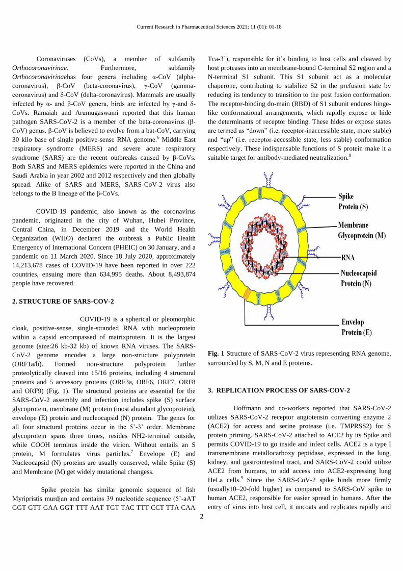
viruses(Fig.5).
Fig. 5 Numerous antiviral drugs act on different stages of SARS-
CoV-2 replication.
9.1 Antimalerial
Numerous drugs have been tested for COVID-
19treatment, among which chloroquine (CLQ) and
hydroxychloroquine (CLQ-OH), are the potential antimalarial
drugs showed promising effects in the treatment of COVID-19
clinical studies. Chloroquine is one of the most prescribed drug in
the world. The drug is supposed to exert potential antiviral effects
through inhibiting nucleic acid replication and also believed as an
inhibitor of endocytic pathways through elevation of endosomal
pH. Both CLQ and CLQ-OH have been shown inhibit novel
coronavirus SARS-CoV-2 in Vero E6 cells with an effective
concentration 50% (EC 50) of 1.1 μM and 0.72 μM respectively.
CLQ-OH was found to be superior over CLQ to inhibit SARS-
CoV-2.49
In China, till date, about twenty-three clinical trials for
COVID-19 treatment have been reported to examine safety and
efficacy of CLQ or CLQ-OH.
Current Research in Pharmaceutical Sciences 2021; 11 (01): 01-18
6
Gao et al. investigated the outcomes of numbers of clinical
trials conducted in China to determine safety and efficacy of CLQ
or CLQ-OH in the management of COVID-19 related pneumonia
in over 10 hospitals in Beijing, Chongqing, Guangzhou, Jingzhou,
Ningbo, Shanghai and Wuhan.50
Results showed that the over 100
patients, who received CLQ-phosphate merely control
exacerbation of pneumonia and improved lung stipulation.
Furthermore, patients were devoid of any severe adverse reactions
of CLQ- phosphate. Subsequently, National Health Commission of
the People's Republic of China, recommended the drug for addition
in the next version of the Guidelines for the Prevention, Diagnosis,
and Treatment of Pneumonia Caused by COVID-19. Department
of Science and Technology of Guangdong Province recommended
CLQ 500 mg b.d. for 10 days for COVID-19 positive patients.51
Gautret et al. evaluated the role of CLQ-OH at 600 mg
daily on respiratory viral loads.52
Results showed that at day 6
post-inclusion, 70% of CLQ-OH-treated patients were virologicaly
cured compared with 12.5% in the control group (p=0.001).
Moreover, 6 patients who were receiving the CLQ-OH were
virologically cured at day 6 (100%), when they were treated in
combination with azithromycin (AZT) (500 mg at day 1 followed
by 250 mg per day for the next 4 days) as compared with patients
treated with only CLQ-OH (57.1%) or without treatment (12.5%).
This result and many other studies revealed that the combination of
CLQ-OH with azithromycin an antiviral drug, improved the
efficacy of CLQ-OH against other RNA-viruses. The combination
of antimalarial drug with antiviral drug has the additional
advantage that this combination permit low dose of
chloroquine/hydroxychloroquine to omit severe side-effects,
including cardiac toxicity and allow this combination as
prophylactic with low dose to comorbidities who are at risk of
COVID-19infection.The promising results of numerous studies,
recommended CLQ at 100 mg daily or CLQ-OH at 300 mg weekly
in mass prophylaxis in individuals exposed to COVID-19.
Derendorf et al. investigated the physicochemical
properties of both CLQ-OH and AZT for the intracellular
lysosomal space.53
Both drugs were found to be accumulated about
50000 fold higher in the lysosomes as compared to cytosolic and
extracellular concentrations. Although CLQ antiviral mechanism
remains ambiguous, however, CLQ is believed to hamper the
terminal glycosylation of ACE2 to act as a plasma membrane
receptor for SARS-CoV-2. CLQ also block numerous steps of the
coronavirus replication cycle. These facts suggested that CLQ may
be a possible treatment for COVID-19 infections, but a lot of
studies are still required.
Matrosovich et al. identified and reported that for
binding of coronavirus to the respiratory tract, sialic-acid-
containing glycoproteins and gangliosides act as primary
attachment factors, besides their protein membrane receptor.54
Very recently, Fantini et al. performed molecular modelling studies
to examine the possible interaction between CLQ and sialic acids.55
Molecular modelling studies revealed that the presence of
ganglioside-binding domain (111–158) at the tip of the N-terminal
domain of the SARS- CoV-2 S protein improves attachment of the
virus to lipid rafts and assist contact with the ACE2 receptor.
Furthermore, in the presence of CLQ or CLQ-OH, the viral S
protein is no longer able to bind gangliosides and prevent the first
step of the viral replication cycle. These molecular modelling
results supported the use of CLQ and CLQ-OH, as initial therapy
for COVID-19 positive patients.
On the basis of above mentioned studies chloroquine plays
multiple mechanisms of action for antiviral therapy. These include
(Fig.5):56
1. Chloroquine inhibits a pre-entry step of the viral cycle by
interfering with viral particles binding to their cellular cell
surface receptor. Chloroquine interferes with sialic acid
biosynthesis, leadto impairment of binding of
viruses,including human coronavirus HCoV-O43 and the
orthomyxoviruses, which utilizes sialic acid moieties as
receptors. The potent anti-SARS-CoV-1 effects of chloroquine
in vitro were considered attributable to a deficit in the
glycosylation of a virus cell surface receptor, the ACE2 on
Vero cells.
2. Chloroquine can also impair virus replication by interfering
with the pH-dependent endosome-mediated viral entry of
enveloped viruses. Due to the alkalisation of endosomes,
chloroquine was an effective in vitro treatment against
Chikungunya virus when added to Vero cells prior to virus
exposure. The mechanism of inhibition likely involved the
prevention of endocytosis and/or rapid elevation of the
endosomal pH and abrogation of virus–endosome fusion. The
activation step that occurs in endosomes at acidic pH results in
fusion of the viral and endosomal membranes, leading to the
release of the viral SARS-CoV-1 genome into the cytosol.
3. Chloroquine- mediated inhibition of hepatitis A virus was
found to be associated with uncoating and thus blocks its
entire replication cycle.
4. Through reduction of cellular mitogen-activated protein
(MAP) kinase activation, chloroquine may also inhibit virus
replication.
5. Furthermore, chloroquine could alter the M protein maturation
and interfere with virion assembly and budding.
6. 9.2 Angiotensin-Converting Enzyme Inhibitors
7. Numerous experts believe treatments targeting angiotensin-
converting enzyme inhibitors (ACEIs) and angiotensin
receptor blockers (ARBs) could be aleadingapproachfor
Covid-19treatment. ACE2 inhibition or reducing its activity in
cell membranes probably reduces SARS-CoV-2 penetration
Current Research in Pharmaceutical Sciences 2021; 11 (01): 01-18
7
into cells. Nevertheless, ACE1 inhibitors do not inhibit
ACE2.ARBs have similar effects to ACE inhibitors, but ACE
inhibitors act by preventing the formation of angiotensin II
rather than by blocking the binding of angiotensin II.
Therefore, ARBs including losartan, valsartan, telmisartan, etc
can be a novel therapeutic approach to block the attachment of
SARS-CoV-2 to ACE2-expressing cells and ultimately its
penetration into host cells. It is well documented that, ACEIs
and ARBs protects the heart and kidney for patients with
hypertension and diabetes. However, the use of ACEIs and
ARBs will increase the expression of ACE2 andincrease
patient susceptibility to viral host cell entry and propagation.
Additionally, on prolong use of ACEIs might suppress the
adaptive immune response, which is a key defence against
viral infections. Similar effects have been noted with non-
steroid anti-inflammatory drugs against adaptive immune
response.57
8. These paradox responses of ACEI and ARBs may be
accountable for the high mortality rate in patients suffering
from hypertension, diabetes, heart disease and cerebrovascular
disease. Patients suffering from these diseases, have a long
term history to use ACEI and ARBs. Guan et al. extracted data
regarding 1099 patients to showed specific comorbidities
associated with increased risk of SARS-CoV-2 infection and
developing into a severe or fatal case.14
Results showed that
173 patients had severe disease, in whom the most common
comorbidities were hypertension (24%), diabetes (16%),
coronary heart disease (6%), and cerebrovascular disease
(2%). Similarly, Zhou and co-workers reported that
hypertension (30%; p=0·0008), diabetes (36%; p=0·0051), and
coronary heart disease (15%; p=0·0001) were the most
common comorbidities with significant effects on mortality, in
an analysis of 191 patients.58
9. However, still no scientific evidence has been reported to
claim the direct link between morbidity of COVID-19 with
previously reported ACEI and ARBs treatment. The European
Society of Cardiology state that ―The Council on Hypertension
strongly recommends that physicians and patients should
continue treatment with their usual anti-hypertensive therapy
because there is no clinical or scientific evidence to suggest
that treatment with ACEIs or ARBs should be discontinued
because of the COVID-19 infection‖. Furthermore, on March
17, 2020, the American College of Cardiology, the Heart
Failure Society of America and the American Heart
Association, jointly release the guidelines to continue ACEIs
and ARBs as prescribed in the setting of COVID-19.
10. Furthermore, on March 17, 2020, the American Heart
Association, the Heart Failure Society of America, and the
American College of Cardiology put out a joint statement
advocating for patients to continue ACEIs and ARBs as
prescribed and that changes in medications in the setting of
COVID-19 should be completed only after careful assessment.
9.3 Antiviral drugs
Numbers of antiviral drugs have been trial against
SARS-CoV-2 infections. Some of them showed promising effects,
but till date, none of antiviral drug has been approved officially for
COVID-19 treatment. The detail mechanism of antiviral drugs is
shown in Fig. 5.
9.3.1 Lopinavir/ Ritonavir/ Kaletra
Lopinavir and ritonavir are the two protease inhibitors,
antiretroviral drugs. Ritonavir add to half-life of lopinavir by
inhibiting the metabolising enzyme cytochrome P450 3A and thus
the combination prefer. Kaletra is the combination of lopinavir
(100 mg) and ritonavir (25 mg), approved by the U.S. Food and
Drug Administration (FDA) for the treatment of HIV infection in
adults and children. Previous studies reveal the success of
lopinavir/ritonavir against other coronaviruses. A molecular
docking study performed by Dayer et al. reported that lopinavir
significantly inhibit cronovirus proteinase.59
The inhibitory potency
of HIV 1 protease inhibitors to cronovirus proteinase was as
follows: lopinavir>ritonavir>amprenavir>tipranavir> saquinavir.
Chu and co-workers in year 2004 reported the success of
lopinavir/ritonavir against other coronaviruses.60
A significant
reduction in risk of severe hypoxia or death in 41 SARS-CoV
patients were reported who were treated with lopinavir/ritonavir
and ribavirin, as compared to those 111 historical controls treated
with ribavirin alone. Recently, Ye et al. proved that the
lopinavir/ritonavir combination and regular adjuvant medicine
against pneumoniashowed better recoveryof COVID-19 positive
patients as compared to treatment with adjuvant medicine alone.61
Study was performed on 47 admitted patients, who were divided
into the test group and the control group according to whether they
had been treated with lopinavir/ritonavir or not during
hospitalization. The treatment group who received
lopinavir/ritonavir and routine adjuvant medicine had a more
evident therapeutic effect in normalize the body temperature and
physiological mechanisms devoid of side effects, compared with
the treatment of pneumonia-associated adjuvant drugs alone. A
very recent clinical data published by Cao et al. on dated 7 may
2020 in The new England Journal of Medicine, revealed no
significantly accelerated clinical improvement, reduced mortality,
or diminished throat viral RNA detectability in serious SARS-
CoV-2 infected patients who were receiving lopinavir–ritonavir
treatment.62
No beneficial results were reported with lopinavir–
ritonavir treatment (on 99 patients) beyond standard care (on 100
patients).

Current Research in Pharmaceutical Sciences 2021; 11 (01): 01-18
8
Currently, numerous studies were reported showing no potential
efficacy of lopinavir/ritonavir in the treatment of
COVID-19.
9.3.2. Ribavirin
Ribavirin is a guanosine analog with antiviral activity.
The earliest report of in vitro efficacy of five FDA-approved drugs
namely ribavirin, penciclovir, nitazoxanide, nafamostat and
chloroquine were tested against the first CoV-19 viral strain i.e.
2019BetaCoV/Wuhan/WIV04/20192 (WIV04), which was isolated
from the lung fluid of one patient in a cohort of seven, six of whom
work in proximity of the Wuhan seafood market. The report of in
vitro direct-actinganti-viral activity against the CoV-19 established
the earliest basis for clinicalguidance. Certainly, treatment with
CLQ and ribavirin permits few benefits in an outbreak due to
instant drug availability.Certainly, due to the low cost of CLQ over
ribavirin, a CLQ phosphate multicenter trial was possible.
However, similar to chloroquine prophylaxis, the government of
China also recommended a 4- gram oral loading dose of ribavirin
followed by 1.2 gram orally every 8 hours after CoV-19
pneumonia diagnosis. This directive was further modified to 500
mg iv BID or TID in the revised Edition 5. Recently the numbers
of ribavirin in combination therapy are under clinical investigation.
At present a clinical trial is continuing to determine the efficacy
and safety of ribavirin in combination with interferon-alpha or
lopinavir/ritonavir or with interferon-alpha and lopinavir/ritonavir
in patients with coronavirus pneumonia (ChiCTR2000029387).63
Furthermore, a combination of ribavirin, lopinavir/ritonavir and
interferon beta-1b is under clinical investigation to compare with to
lopinavir/ ritonavir (NCT04276688) against SARS-CoV-2
infection.64
These studies revealed that ribavirin could be used in
COVID-19 positive patients, but more studies are required.
9.3.3 Remdesivir
Remdesivir developed by Gilead Sciences, is a
nucleoside analoguethat inhibits viral RNA polymerases.de Wit et
al. reported the antiviral efficacy of remdesivir against SARS-CoV
and MERS-CoV in vivo.65
In COVID-19, remdesivir inhibits RNA
polymerase (RdRp) and interfere with RNA replication.
Remdesivir cannot cause an immediate stop of growing chain (i
position) and allow to extend three more nucleotides down to stop
the strand at (i + 3) position. Remdesivir was first used in United
States for COVID-19 treatment. Remdesivir was administered
through intravenous route on the evening of day 7 and on day 8the
patient’s clinical condition was improved without any adverse
events.Due to improved oxygen saturation values to 96%, the
supplemental oxygen was discontinued. The symptomatic
treatment was continuing during remdesivir therapy.
Grein et al. gave remdesivir 53severe infected SARS-
CoV-2 patients during the period from January 25, 2020, through
March 7, 2020.66
The remdesivir was given for 10 days, started
with 200 mg intravenously administered on day 1, followed by 100
mg daily for the remaining 9 days of treatment. 36 patients (68%)
showed improvement in oxygen-support. Although 25 patients
(47%) were discharged and 7 were (13%) died. These results
supported the further clinical investigation of remdesivir against
severe Covid-19 patients.
Wang et al. also prescribed remdesivir in adults with
severe COVID-19 infection.67
Results showed that intravenous
remdesivir was effectively tolerated; however no significant
improvements were seen in seriously ill patients. Authors
suggested to conductscrupulous study of remdesivir in patients
with severe COVID-19. Recently, Wang and co-workers reported
in their study that the combination of remdesivir with CLQ is
efficient to control of SARS-CoV-2 infection.68
Since both drugs
are easily available and effective against diverse ailments, thus
authors suggested that this combination may be beneficial against
novel SARS-CoV-2 infection.
On May 1, 2020, The US FDA announced to use
remdesivir for severe COVID-19 (confirmed or suspected) cases in
hospitalized adults and children. Recently, US firm Gilead in talks
with Indian drug companies Cipla, Hetero and Dr. Reddy’s to
produce remdesivir for COVID-19 treatment.
9.3.4 Favipiravir
Favipiravir is pyrazinecarboxamide derivative; inhibit
RNA-dependent RNA polymerase and sold under the brand name
Avigan, used to treat influenza in Japan. It is being developed and
manufactured by Toyama Chemical (Fujifilm group) and was
approved in year 2014 for medical use. During outbreak of Ebola
virus (EBOV) in year 2014–2015 patients treated with favipiravir
showed improved survival.69
Alike of genome sequencing of the
SARS-CoV-2 to SARS-CoV and MERS-CoV, favipiravir is
recommended for COVID-19 treatment. Cai et al. performed
clinical trial of favipiravir for 80 COVID-19 positive patients in
The Third People’s Hospital of Shenzhen (ChiCTR2000029600).70
35 patients who received favipiravir showed rapid recovery over
45 patients in the control arm (median 4 (2.5– 9) d versus 11 (8–
13) d, P < 0.001). X-ray examinations also showed higher rate of
improvement in chest imaging in the favipiravir arm compared
with the control arm (91.43% vs. 62.22%). This study showed that
FPV significantly perform better COVID-19 treatment in terms of
disease progression and viral clearance. Furthermore, Chen et al.
also reported the clinical trial result, which was conducted by the
same research group to determine the efficacy of favipiravir
Current Research in Pharmaceutical Sciences 2021; 11 (01): 01-18
9
against COVID-19 treatment.71
With favipiravir treatment, clinical
recovery rate was increased from 55.86% to 71.43% within 7
day’s. Both the fever reduction time and cough relief duration,
decreased significantly after favipiravir treatment in ordinary
COVID-19 positive patients and in the patients suffering from
hypertension and/or diabetes. The National Medical Products
Administration of China approved favipiravir as the firstanti-
COVID-19 drug on March2020.
9.3.5 Umifenovir
Umifenovir is another antiviral compound developed
at the Russian Research Chemical and Pharmaceutical Institute and
marketed as Arbidol. Umifenovir had shown to possess antiviral
activity against both DNA and RNA viruses. Umifenovir inhibits
the membrane fusion between virus particles and plasma
membranes, and between virus particles and the membranes of
endosomes. Additionally umifenovir also reported for its
immunomodulatory activity, and induce interferon and/or
macrophage activation. In vitro studies of umifenovir, suggested
antiviral activity against SARS coronavirus.72
Recently, numerous
studies reported application of Arbidol for COVID-19 treatment,
but its efficacy and safety remained unclear
.
Xu and co-workers performed a retrospective cohort study
against 111 novel coronavirus-infected pneumonia patients (NCP)
in China, who received empirical antiviral regimens with or
without Arbidol.73
A total of 111 patients from two clinical
centersin China were enrolled. Results suggested that umifenovir
accelerated virologic clearance, improved focal absorption on
radiologic images and reduced the need for high flow nasal
catheter (HFNC) oxygen therapy in hospitalized patients. These
results provided the basis for the clinical use of Arbidoland
supported for further randomized controlled trials in patients with
NCP. Umifenovir combination with lopinavir/ritonavir increased
negative conversion rate of SARS-CoV-2 and improved chest CT
scan results. However, umifenovir has not been as effective as
favipiravir in clinical recovery rate and relief of fever and cough.
Recently two randomized clinical trials are ongoing inChina to
investigate efficacy and safety of umifenovir against COVID-19.
While umifenovir combination lopinavir/ritonavir is also under
clinical investigation (NCT04252885) against COVID-19.
9.3.6 Ivermectin
Ivermectin, a FDA-approved anti-parasitic agent, also
possess antiviral activity. Ivermectin,has been demonstrated
activity against RNA virus, including Dengue, influenza,
Venezuelan equine encephalitis virus (VEEV) and West Nile
Virus.74
Ivermectin inhibited replication of SARS-CoV-2 in in vitro
Vero-hSLAM cells, possibly due to the inhibition of importin-
α/β1-mediated nuclear import of viral proteins, as shown for other
RNA viruses. However, Schmith et al. performed successful
clinical trial using the approved dose of ivermectin, which showed
thativermectin recommended dose was insufficient for COVID-
19treatment.75
Momekov and Momekov investigated the
pharmacokinetics of ivermectin in patients used to surrogates for
juxtaposition with the in vitro SARS-CoV-2 inhibitory findings.76
Patri et al. hypothesized that the combination of CLQ-OH with
ivermectin could act in a consequential and synergistic manner.77
Indeed, CLQ-OH would behave as a first-level barrier by inhibiting
the entry of the virus into the host cell, while ivermectin could
reduce viral replication if the virus did get in, strengthening CLQ-
OH antiviral effects. However, it was just a hypothesis and no in
vitro or in vivo studies were conducted.
9.3.7 APEIRON
APEIRON (APN01) is the recombinant form of the
human angiotensin-converting enzyme 2 (rhACE2), and has the
potential to block the infection of cells caused by the novel SARS-
CoV-2 virus, and reduce lung injury. APN01 imitates the human
enzyme ACE2 and blocks virus entry into the cells. The virus binds
erroneously to ACE2/APN01 rather than cell surface ACE2 and no
longer infects the cells. Simultaneously, APN01 blocks
inflammatory reactions in the lungs and protects against acute lung
injury. Recently, Austria, Denmark and Germany approved to
initiate a Phase II clinical trial of APN01 to treat COVID-19.78
9.3.8 EIDD-2801
EIDD-2801 is an oral NHC-prodrug (β-D-N4-
hydroxycytidine-5’-isopropyl ester), showed efficacy against
SARS-CoV and MERS-CoV. Preliminary studies showed that
EIDD-2801 can be used as either a prophylactic or a therapeutic
for SARS-CoV-2. Similar to remdesivir, EIDD-2801 works by
mimicking ribonucleosides– causing debilitating errors and
prevents the spread of the virus. Senior researcher and eminent
professor of epidemiology at UNC-Chapel Hill Gillings School of
Global Public Health, Ralph Baric state that ―This new drug not
only has high potential for treating COVID-19 patients, but also
appears effective for the treatment of other serious coronavirus
infections‖. EIDD-2801 reduced the viral load in mice when given
as a treatment between 12 and 48 hours after infection began.
Though EIDD-2801 has small therapeutic window in mice, yet
researchers suggested large therapeutic window of this in humans
as compared to mice.79
Current Research in Pharmaceutical Sciences 2021; 11 (01): 01-18
10
9.3.9 Interferon
Interferons (IFNs) are natural proteins, secreted by
immune cells.Alfa, beta, and gamma are three classes of interferon.
Interferons do not directly kill bacteria or virus, but modulate
immune response against foreign substances that invade the body.
Interferon alfa (2b) and beta (1a) are under clinical investigation as
potential treatments for people with COVID-19 coronavirus
disease. Interferon may be able to make the immune system strong
by turning on quiescent parts and leading them toward the defense
against SARS-CoV-2 assault. Numerous studies around the world
is conducting, looking at different interferons to treat COVID-19
coronavirus. Zorzitto et al. reported that IFN-α/β are effective in
inhibiting SARS-CoV replication in in vitro.80
Furthermore,
Haagmans et al. reported that the pegylated interferon- α protects
type 1 pneumocytes against SARS coronavirus infection in
monkey.81
Thus IFN-α had proposed for COVID-19 therapy and
numbers of studies have been conducted and reported.
Lokugamage et al. study showed that IFN-α/β can be used as a
prophylaxis against SARS-CoV-2 similar to SARS-CoV.82
The
combination of IFN-β with ribavirin, remdesivir or
lopinavir/ritonavir improved pulmonary function, reduces viral
loads in vitro in other coronaviruses. In China, vapor inhalation of
5 million U of IFN-α in combination with ribavirin twice a day for
no more than 10 days has been recommend for COVID-19
treatment.83
Recently clinical trials for IFN-α2b in combination
with lopinavir/ritonavir (ChiCTR2000029387) orIFN-β1b with
ribavirin and lopinavir/ritonavir (NCT04276688) have been
recruited for COVID-19 treatment.84
Usually it has been seen that multiple antiviral
combinations are more effective against viral infection as
compared to single drug. Hung et al. reported phase 2 clinical trials
by utilizing IFN-β1b, lopinavir–ritonavir, and ribavirin
combinations against COVID-19 127 positive patients, who were
hospitalized in six hospitals of Hong Kong.85
Patients were
randomly assigned (2:1) to a 14-day combination of lopinavir 400
mg and ritonavir 100 mg every 12 h, ribavirin 400 mg every 12 h,
and three doses of 8 million international units of IFN-β1b on
alternate days (combination group; 86 patients) or to 14 days of
lopinavir 400 mg and ritonavir 100 mg every 12 h (control group;
41 patients). The combination therapy was found to be safe and
efficient as compared to lopinavir–ritonavir alone. These results
supported the future clinical study of a double antiviral therapy
with IFN-β1b as a backbone is warranted.
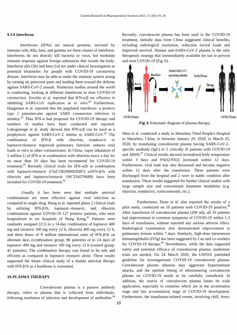
10. PLASMA THERAPY
Convalescent plasma is a passive antibody
therapy, refers to plasma that is collected from individuals,
following resolution of infection and development of antibodies.86
Recently, convalescent plasma has been used in the COVID-19
treatment. Initially data from China suggested clinical benefits,
including radiological resolution, reduction inviral loads and
improved survival. Human anti-SARS-CoV-2 plasma is the only
therapeutic strategy that isimmediately available for use to prevent
and treat COVID-19 (Fig. 6).
Fig. 6 Schematic diagram of plasma therapy.
Shen et al. conducted a study in Shenzhen Third People's Hospital
in Shenzhen, China, in between January 20, 2020, to March 25,
2020, by transfusing convalescent plasma having SARS-CoV-2–
specific antibody (IgG) to 5 critically ill patients with COVID-19
and ARDS.87
Clinical results showed normalized body temperature
within 3 days and PAO2/FIO2 increased within 12 days.
Furthermore, viral load was also decreased and became negative
within 12 days after the transfusion. Three patients were
discharged from the hospital and 2 were in stable condition after
transfusion. These results suggested for further clinical studies with
large sample size and concomitant treatment modalities (e.g.
ribavirin, remdesivir, corticosteroids, etc.).
Furthermore, Duan et al. also reported the results of a
pilot study, conducted on 10 patients with COVID-19 positive.88
After transfusion of convalescent plasma (200 ml), all 10 patients
had improvement in common symptoms of COVID-19 within 1-3
days of transfusion, without reported any serious adverse effect.
Radiological examination also demonstrated improvement in
pulmonary lesions within 7 days. Similarly, high-dose intravenous
immunoglobulin (IVIg) has been suggested by Cao and co-workers
for COVID-19 therapy.89
Nevertheless, while the data supported
safety and potential efficacy of convalescent plasma, randomize
trials are needed. On 24 March 2020, the USFDA published
guideline for investigational COVID-19 convalescent plasma.
Convalescent plasma infusion may aggravate hyperimmune
attacks, and the optimal timing of administering convalescent
plasma on COVID-19 needs to be carefully considered. In
addition, the source of convalescent plasma limits its wide
application, especially in countries which are in the acceleration
stage and late accumulation stage of COVID-19 development.
Furthermore, the transfusion-related events, involving chill, fever,
Current Research in Pharmaceutical Sciences 2021; 11 (01): 01-18
11
anaphylactic reactions also limits convalescent plasma use. The
convalescent plasma can be potentially use until an effective
treatment against COVID-19 is discovered.90
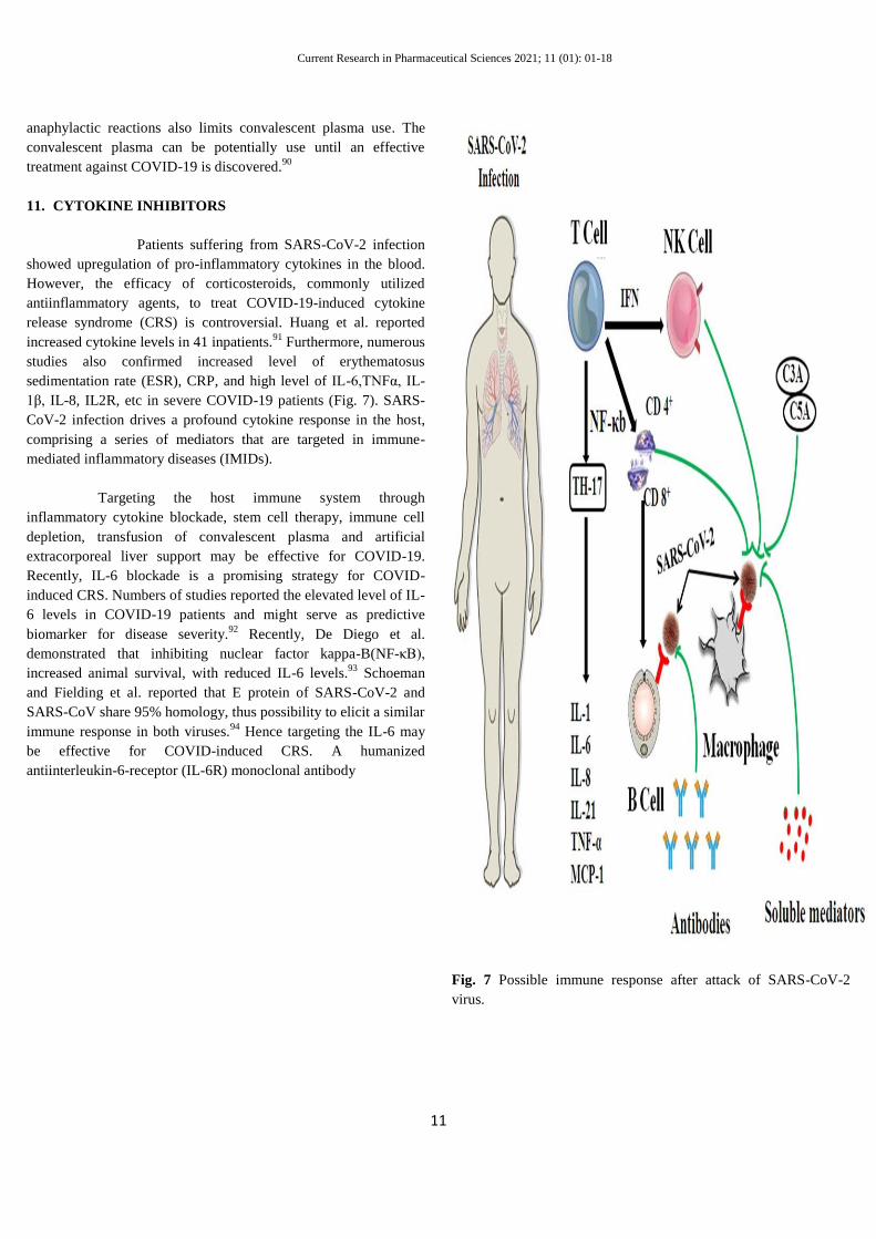
11. CYTOKINE INHIBITORS
Patients suffering from SARS-CoV-2 infection
showed upregulation of pro-inflammatory cytokines in the blood.
However, the efficacy of corticosteroids, commonly utilized
antiinflammatory agents, to treat COVID-19-induced cytokine
release syndrome (CRS) is controversial. Huang et al. reported
increased cytokine levels in 41 inpatients.91
Furthermore, numerous
studies also confirmed increased level of erythematosus
sedimentation rate (ESR), CRP, and high level of IL-6,TNFα, IL-
1β, IL-8, IL2R, etc in severe COVID-19 patients (Fig. 7). SARS-
CoV-2 infection drives a profound cytokine response in the host,
comprising a series of mediators that are targeted in immune-
mediated inflammatory diseases (IMIDs).
Targeting the host immune system through
inflammatory cytokine blockade, stem cell therapy, immune cell
depletion, transfusion of convalescent plasma and artificial
extracorporeal liver support may be effective for COVID-19.
Recently, IL-6 blockade is a promising strategy for COVID-
induced CRS. Numbers of studies reported the elevated level of IL-
6 levels in COVID-19 patients and might serve as predictive
biomarker for disease severity.92
Recently, De Diego et al.
demonstrated that inhibiting nuclear factor kappa-B(NF-κB),
increased animal survival, with reduced IL-6 levels.93
Schoeman
and Fielding et al. reported that E protein of SARS-CoV-2 and
SARS-CoV share 95% homology, thus possibility to elicit a similar
immune response in both viruses.94
Hence targeting the IL-6 may
be effective for COVID-induced CRS. A humanized
antiinterleukin-6-receptor (IL-6R) monoclonal antibody
Fig. 7 Possible immune response after attack of SARS-CoV-2
virus.
Current Research in Pharmaceutical Sciences 2021; 11 (01): 01-18
12
tocilizumab, inhibits interleukin-6 (IL-6) was administered
intravenously to test in the treatment of COVID-19 in China and
Italy with encouraging results. Even to evaluate the role of
tocilizumab in COVID-19 treatments, the pharmaceutical
manufacture company of tocilizumab has made the drug available
free of charge in Italy for COVID-19 pandemic. Michot et al.
reported first observation of a COVID-19 positive patient suffered
from lung disease successfully treated after two infusion with
tocilizumab.95
Xu et al. determined the efficacy of tocilizumab to
improve clinical symptoms and repress the deterioration of severe
COVID-19 patients.96
The 21 COVID-19 patients were admitted in
The First Affiliated Hospital of University of Science and
Technology of China, given tocilizumab in addition to routine
therapy between 5 and 14 February 2020.In all patients fever
normalized within one day and other symptoms improved
remarkably within a few days. 15 patients required less oxygen
supply and 1 patient needed no oxygen therapy within 5 days
treatment. Lung lesion opacity absorbed in 19 patients (90.5%) and
percentage of lymphocytes in peripheral blood also returned to
normal in 52.6% of patients on the fifth day after treatment. No
adverse reactions were observed and all patients were discharge on
average 15.1 day after giving tocilizumab. This preliminary study
supported to use tocilizumab in severe and critical COVID-19
patients.
Janus kinase (JAK) inhibitors inhibit type I/II cytokine
receptors, are currently being used for the treatment of COVID-19.
Baricitinib is a selective JAK1/JAK2 inhibitor, showed effective
treatment for ARDS in COVID-19 and decreased the virus
infectivity for lung cells. Furthermore, baricitinib also interrupts
the passage and intracellular assembly of SARS-CoV-2 into the
target cells via disruption of AP2-associated protein kinase 1
(AAK1) signaling and also reduced the inflammation in patients
with ARDS. In contrast, tofacitinib an effective oral JAK2/1/3
inhibitor does not significantly inhibit AAK1.97
Other JAK1/2
inhibitors including ruxolitinib, memolitinib, and oclacitinib can
potentially affect signaling pathways downstream of the receptors
involved in COVID-19 development.Ruxolitinib is currently
approved by the FDA for the treatment of patients with
myeloproliferative neoplasms, under phase III clinical trial to treat
patients with coronavirus disease 2019 (COVID-19)related
cytokine storm.
As reported on Sept 29 2019, ten US FDA approved and
four off-label indications for anti-TNF therapy, indicating that TNF
is a valid target in many inflammatory diseases. TNF is important
in nearly all acute inflammatory reactions and reported to present
in blood and disease tissues of patients with COVID-19. Anti-TNF
therapy was proposed to evaluate in patients with COVID-19 on
hospital admission to prevent progression to needing intensive care
support. Previously, Tobinick et al. reported that inhibition of
TNF-α has the potential therapeutic modulation of SARS
coronavirus infection.98
Anti-TNF antibodies infliximab or
adalimumab may be beneficial for COVID-19 treatment. However
adalimumab is the only TNF-α inhibitor undergoing evaluation, in
a trial registered in China (ChiCTR2000030089).99
12. STEM CELLS
Stem cells having the ability to generate other cells
called daughter cells possess specialized functions. Mesenchymal
stem cells (MSCs) are widely used in stem cellbased
therapyespecially in immune-mediated inflammatory diseases.
MSCs improve immunomodulatory mediated cytokines qualities
andshows antiviral activity. The immunomodulatory effects of
MSCs are triggered further by the activation of Toll-like receptors
(TLRs) in MSCs, which is stimulated by pathogen-associated
molecules.100
MSCs are expected to survive even if they are
transplanted into a patient with a confirmed COVID-19 and thus
trial in the treatment of COVID-19.Leng et al. transplanted MSC in
7 patients with COVID-19 pneumonia in Beijing You An Hospital,
China.101
All 7 patients showed significantly improvements
(including pulmonary function) without any adverse effects. The
gene expression profile revealed MSCs were free from COVID-19
infection. Both common and severe patients were recovered and
discharged within 10 days after treatment.MSC populations were
entrapped in the lung after intravenous administration and showed
improved lung functions and COVID-19 pneumonia. These results
ensure safer and effective use of MSCs against COVID-19 positive
patients and especially recommended for patients having COVID-
19 pneumonia. However, the major limitation of this approach is
the supplying source of clinical-grade MSCs and subsequently the
speed of preparation for clinical usage that here stem cell banks
can play an important role. Recently a case study was reported in
China on a 65-year-old female patient diagnosed in critical
condition with COVID-19. Initially patient was treated with
antiviral drugs, steroids and antibodies, but no significant
improvements were observed, even patients vital signs worsened
and then the patient was treated with MSCs and with thymosin α1;
5 × 10⁷ cells each three times. Remarkable improvements were
reported only after second injection and patient was removed from
the ventilator and able to walk. These results suggested that
umbilical cord mesenchymal stem cells could be an ideal treatment
option alone or in combination with other immune modulators for
acute COVID-19 patients.102
These studies suggested that the stem
cell therapy and especially MSCs may be a promising strategy for
COVID-19 treatment. However, the cost-effective and speed of
therapeutic preparation are the two major hurdles for MSC based
COVID-19therapy.Recently, China, USA, Jordon, Iran, and several
other countries have begun cell-based therapy clinical studies.
Current Research in Pharmaceutical Sciences 2021; 11 (01): 01-18
13
13. VACCINES
The genetic sequence of SARS-CoV-2 triggers intense
global R&D activity to develop a vaccine against the disease. The
first COVID-19 vaccine candidate entered human clinical testing
with unprecedented rapidity on 16 March 2020.
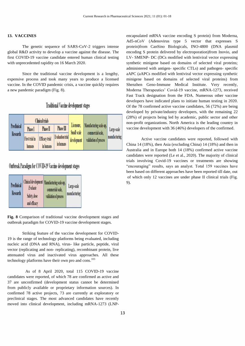
Since the traditional vaccine development is a lengthy,
expensive process and took many years to produce a licensed
vaccine. In the COVID pandemic crisis, a vaccine quickly requires
a new pandemic paradigm (Fig. 8).
Fig. 8 Comparison of traditional vaccine development stages and
outbreak paradigm for COVID-19 vaccine development stages.
Striking feature of the vaccine development for COVID-
19 is the range of technology platforms being evaluated, including
nucleic acid (DNA and RNA), virus- like particle, peptide, viral
vector (replicating and non- replicating), recombinant protein, live
attenuated virus and inactivated virus approaches. All these
technology platforms have their own pro and cons.103
As of 8 April 2020, total 115 COVID-19 vaccine
candidates were reported, of which 78 are confirmed as active and
37 are unconfirmed (development status cannot be determined
from publicly available or proprietary information sources). In
confirmed 78 active projects, 73 are currently at exploratory or
preclinical stages. The most advanced candidates have recently
moved into clinical development, including mRNA-1273 (LNP-
encapsulated mRNA vaccine encoding S protein) from Moderna,
Ad5-nCoV (Adenovirus type 5 vector that expresses S
protein)from CanSino Biologicals, INO-4800 (DNA plasmid
encoding S protein delivered by electroporation)from Inovio, and
LV- SMENP- DC (DCs modified with lentiviral vector expressing
synthetic minigene based on domains of selected viral proteins;
administered with antigen- specific CTLs) and pathogen- specific
aAPC (aAPCs modified with lentiviral vector expressing synthetic
minigene based on domains of selected viral proteins) from
Shenzhen Geno-Immune Medical Institute. Very recently,
Moderna Therapeutics’ Covid-19 vaccine, mRNA-1273, received
Fast Track designation from the FDA. Numerous other vaccine
developers have indicated plans to initiate human testing in 2020.
Of the 78 confirmed active vaccine candidates, 56 (72%) are being
developed by private/industry developers, with the remaining 22
(28%) of projects being led by academic, public sector and other
non-profit organizations. North America is the leading country in
vaccine development with 36 (46%) developers of the confirmed.
Active vaccine candidates were reported, followed with
China 14 (18%), then Asia (excluding China) 14 (18%) and then in
Australia and in Europe both 14 (18%) confirmed active vaccine
candidates were reported (Le et al., 2020). The majority of clinical
trials involving Covid-19 vaccines or treatments are showing
―encouraging‖ results, says an analyst. Total 159 vaccines have
been based on different approaches have been reported till date, out
of which only 12 vaccines are under phase II clinical trials (Fig.
9).

Current Research in Pharmaceutical Sciences 2021; 11 (01): 01-18
14
05
101520253035404550
Fig. 9 Varieties of vaccines
Very recently, Serum Institute of India’s ―Covishield‖ and Bharat
Biotech’s indigenous ―Covaxin‖ are the two vaccines against
Covid-19 that will drive the countries vaccination programme in
the first phase of inoculation. In September 2020, the first batch of
the 'Gam-Covid-Vac' [Sputnik V] vaccine for the prevention of the
new coronavirus infection, developed by the Gamaleya National
Research Center of Epidemiology and Microbiology of the
Ministry of Health of Russia, has passed the necessary quality tests
in the laboratories of Roszdravnadzor [medical device regulator]
and has been released into civil circulation. The US Food and Drug
Administration (FDA) authorized an emergency-use RNA
vaccine made by Pfizer on 17 December 2020. A week after US
regulators has followed with a second: another RNA vaccine, this
one made by Moderna of Cambridge, Massachusetts. Two more
vaccines of AstraZeneca and Johnson & Johnson are also in late-
stage testing and possibly soon available in the market.
14. Current Position
Current position is very critical and day by day increasing
numbers of cases worldwide. According to WHO report 106,000
new cases had been reported worldwide within 24 h in the date
between 20 to 21 May 2020. Up to 21 May 2020 5,213,678 total
cases have been reported, out of which 2,784,809 are active cases
and 2,093,874 patients have been recovered worldwide. The total
334,995 death have been reported till date. These data are the
serious threat for and under phase II clinical trials (red colour).
the world and need immediate recovery from this pandemic
condition.
reported (green colour)
15. Conclusion
In the present write up, we focus on deadly respiratory
diseases caused by SARS-CoV-2, its origin, replication,
transmission, clinical symptoms, detection techniques and
vulnerable population infected with SARS-CoV-2. Herein we
elaborate the numerous therapies for COVID-19 under clinical
investigation. Furthermore we also reported the worldwide data of
population suffering from COVID-19 pandemic disease. On 30
January 2021, there have been 101,561,219 confirmed cases of
COVID-19, including 2,196,944 deaths, reported by WHO.
Although in few countries condition is more decisive due to
COVID-19, while other countries showing remarkable recovery
rate especially in India wherein 96.04 recovery rate was reported
on 30 December 2020.
Acknowledgements
None
Conflict of Interest
The authors declare no conflict of interest, financial or otherwise.
Funding
None
Ethical Approval: Not required
Supplementary materials: None

Current Research in Pharmaceutical Sciences 2021; 11 (01): 01-18
15
REFERENCES
1. Herrington CS, Coates PJ, et al. Viruses and disease: emerging concepts for
prevention, diagnosis and treatment. J Pathol. 2015; 235: 149–152.
2. Jain AK, Sharma S, et al. 1,3,4-Thiadiazole and Its Derivatives: A Review on
Recent Progress in Biological Activities. Chem Biol Drug Des. 2013; 81(85):
557-576.
3. Vaidya A, Jain S, et al. Metabotropic Glutamate Receptors: A Review on
Prospectives and Therapeutic Aspects. Mini-Rev Med Chem. 2013; 12:
1967-1981.
4. Jain AK, Vaidya A, et al. Recent Developments and Biological Activities of
Thiazolidinone Derivatives: A Review. Bioorg Med Chem. 2012; 20: 3378-
3395.
5. Wong JEL, Leo YS, et al. COVID-19 in Singapore—current experience:
criti- cal global issues that require attention and action. JAMA. 2020;
323(13): 1243-1244.
6. Ramaiah A, Arumugaswami V. Insights into cross-species evolution of novel
human coronavirus 2019-nCoV and defining immune determinants for
vaccine development. bioRxiv. 2020; accessed on 30 June 2020.
7. Mousavizadeh L, Ghasemi S. Genotype and phenotype of COVID-19: Their
roles in pathogenesis. J Microbiol Immunol Infect. 2020; S1684-1182(20):
30082-30087.
8. Walls AC, Xiong X, et al. Unexpected receptor functional mimicry elucidates
activation of coronavirus fusion. Cell. 2019; 176: 1026–1039.
9. Hoffmann M, Kleine-Weber H, et al. SSARS-CoV-2 cell entry depends on
ACE2 and TMPRSS2 and is blocked by a clinically proven protease
inhibitor. Cell. 2020; 181(2): 271-280.
10. Lu R, Zhao X, et al. Genomic characterisation and epidemiology of 2019
novel coronavirus: implications for virus origins and receptor binding.
Lancet. 2020; 395: 565–574.
11. Zhou P, Yang XL, et al. A pneumonia outbreak associated with a new
coronavirus of probable bat origin. Nature. 2020; 579: 270–273.
12. Lam TT, Shum MH, et al. Identifying SARS-CoV-2 related coronaviruses in
Malayan pangolins. Nature. 2020; 583(7815): 282-285.
13. Zhang H, Kang Z, et al. The digestive system is a potential route of 2019-
nCov infection: a bioinformatics analysis based on single-cell transcriptomes.
bioRxiv. 2020; accessed on 05 July 2020.
14. Guan W, Ni Z, et al. Clinical Characteristics of Coronavirus Disease 2019 in
China. N Engl J Med. 2020; 382: 1708-1720.
15. Lauer SA, Grantz KH, et al. The Incubation Period of Coronavirus Disease
2019 (COVID-19) From Publicly Reported Confirmed Cases: Estimation and
Application. Ann Intern Med. 2020; accessed on 15 December 2020.
16. Wang W, Xu Y, et al. Detection of SARS-CoV-2 in different types of clinical
specimens. JAMA. 2020; 323(18): 1843–1844.
17. To KK, Tsang OT, et al. Temporal profiles of viral load in posterior
oropharyngeal saliva samples and serum antibody responses during infection
by SARS-CoV-2: an observational cohort study. Lancet Infect Dis. 2020; 20:
565-574.
18. Xiang F, Wang X, et al. Antibody detection and dynamic characteristics in
patients with COVID-19. Clin Infect Dis. 2020; 71(8): 1930-1934.
19. Guo L, Ren L, et al. Profiling early humoral response to diagnose novel
coronavirus disease (COVID-19). Clin Infect Dis. 2020; 71(15): 778-785.
20. Vaidya A, Jain S, et al. Pectin-metronidazole prodrug bearing microspheres
for colon targeting. J Saudi Chem Soc. 2012; 19(3): 257–264.
21. Vaidya A, Jain S, et al. Synthesis and Biological Activities of
Oxadiazole Derivatives: A Review. Mini-Rev Med Chem. 2016; 16(10): 825-
845.
22. Jain S, Pattnaik S, et al. 2017 Anticancer Potential of Thiazole Derivatives: A
Retrospective Review. Mini-Rev Med Chem. 2016; 18(8): 640-655.
23. Jain S, Pathak K, et al. Molecular therapy using siRNA: Recent trends and
advances of multi target inhibition of cancer growth International. J Biolo
Macromol. 2018; 116: 880-892.
24. Jain S, Jain S, et al. Gastrointestinal Protective Effect of Zizyphus xylopyrus
(Retz) Wild Leaf Extract Against Indomethacin and HCl-EtOH Induced
Ulcers. Curr Trad Med. 2019; 5(2): 140-146.
25. Vaidya A, Pathak D, et al. 1,3,4‐oxadiazole and its Derivatives: A Review on
Recent Progress in Anticancer Activities. Chem Biol Drug Des. 2020;
accessed on 24 October 2020.
26. Vaidya A, et al. Anticancer agents based on vulnerable components in a
signalling pathway. Mini-Rev Med Chem. 2020; 20(10): 886-907.
27. Jain S, Chandra V, et al. Comprehensive review on current developments of
quinoline-based anticancer agents. Arabian J Chem. 2019; 12(8): 4920-4946.
Current Research in Pharmaceutical Sciences 2021; 11 (01): 01-18
16
28. Jain S, Jain A, et al. Preliminary phytochemical, pharmacognostical and
physico-chemical evaluation of Cedrus deodara heartwood. J Pharmacog
Phytochem. 2014; 3(1): 91-95.
29. Jain S, Vaidya A, et al. Pharmacognostic and phytochemical investigations of
the leaves of zizyphusXylopyrus (retz) willd. Int J Pharm Pharmaceut Sci.
2011; 3: 122-125.
30. Vaidya A, Jain AK, et al. Predicting anti-cancer activity of quinoline
derivatives: CoMFA and CoMSIA approach. J Enzy Inhibit Med Chem.
2011; 26(6): 854-861.
31. Jain AK, Veerasamy R, et al. QSAR analysis of some novel sulfonamides
incorporating 1,3,5-triazine derivatives as carbonic anhydrase inhibitors. Med
Chem Res. 2010; 19: 1191-1202.
32. Agrawal RK, Jain AK, et al. QSAR analysis of B-ring-modified diaryl ether
derivatives as a InhA inhibitors. Med Chem Res. 2012; 21: 145-151.
33. Bhatiya R, Vaidya A, et al. QSAR analysis of furanone derivatives as potential
COX-2 inhibitors: kNN MFAapproach. J Saudi Chem Soc. 2014; 18(6): 977–
984.
34. Vaidya A, Jain S, et al. Quantitative Structure-Activity Relationships: A Novel
Approach of Drug Design and Discovery. J Pharm Sci Pharmacol. 2014;
1(3): 219-232.
35. Vaidya A, Jain S, et al. Computational Analysis of Quinoline Derivatives as
Potent Topoisomerase-II Inhibitors. Med Chem Res. 2015; 24(1): 383-393.
36. Vaidya A, Jain AK, et al. CoMFA, CoMSIA, kNN MFA and Docking studies
of 1,2,4-Oxadiazole derivatives as potent Caspase-3 activators. Arabian J
Chem. 2017; 10(2): S3936-S3946
37. Jain S, Vaidya A, et al. Computational analysis of benzyl vinylogous
derivatives as potent PDE3B inhibitors. Arabian J Chem. 2017; 10: S109-
S113
38. Vaidya A, Jain S, et al. Synthesis of 1,2,4-oxadiazole derivatives: anticancer
and 3D QSAR studies. Monatshefte für Chem- Chem Monthly. 2020; 151:
385-395.
39. Vaidya A, Jain S, et al. Simvastatin-Loaded PEGylated Solid Lipid
Nanoparticles: Lipid Functionalization to Improve Blood Circulation.
Bionanoscience. 2020; 10: 773–782.
40. Khan T, Vaidya A, et al. Meropenem Loaded Pectin Microspheres for Colon
Delivery. Asian J Biomat Res. 2018; 4(4): 8-20.
41. Vaidya A, Jain R, et al. Design and Development of Mucoadhesive Thiolated
Chitosan Microspheres for Colonic Drug Delivery. J Bionanosci. 2018;12(4):
590-598.
42. Pathak K, Vaidya A, et al. Confronting Penetration Threshold via Fluidic
Terpenoid Nanovesicles. Curr Drug Del. 2018; 15(6): 765-776.
43. Vaidya A, Jain S, et al. Dendrimers: Nanosized Multifunctional Platform for
Drug Delivery. Drug Deli Lett. 2018; 8(1): 3 – 19.
44. Jain P, Vaidya A, et al. Ethyl Cellulose Coated Chitosan Microspheres of
Metronidazole as Potential Anti-Amoebic Agent. J Bionanosci. 2018; 11(6):
599-607.
45. Vaidya A, Jain S, et al. Metronidazole loaded eudragit coated alginate beads
for colon targeting. Int J Pharma Healthcare Res. 2014; 2: 81 – 86.
46. Jain S, Jain AP, et al. Nanotechnology: An emerging area in the field of
dentistry. J Dental Sci. 2013; XX: 9-13.
47. Vaidya A, Agarwal A, et al. Bioconjugation as a novel platform for targeted
drug delivery: A Review. Curr Pharm Des. 2011; 17(11): 1108-1125.
48. Vaidya A, Jain A, et al. Metronidazole Loaded Pectin Microspheres for Colon
Targeting. J Pharm Sci. 2009; 98: 4229-4236.
49. Colson, P, Rolain, JM, et al. Chloroquine for the 2019 novel coronavirus
SARS-CoV-2. Int J Antimicrob Agents. 2020; 55(3): 105923.
50. Gao J, Tian Z, et al. Breakthrough: chloroquine phosphate has shown apparent
efficacy in the treatment of COVID-19 associated pneumonia in clinical
studies. Biosci Trends. 2020; 14: 72–73.
51. Zhonghua JHHHXZZ. Expert consensus on chloroquine phosphate for the
treatment of novel coronavirus pneumonia. Chinese. 2020; 43: 185–188.
52. Gautret P, Lagier JC, et al. Hydroxychloroquine and azithromycin as a
treatment of COVID-19: results of an open-label non-randomized clinical
trial. Int J Antimicrob Agents. 2020; 56(1): 105949.
53. Derendorf H, Excessive lysosomal ion-trapping of hydroxychloroquine and
azithromycin. Int J Antimicrob Agents. 2020; 55(6): 106007.
54. Matrosovich M, Herrler G, et al. Sialic acid receptors of viruses. Top Curr
Chem. 2015; 367: 1–28.
55. Fantini J, Scala CD, et al. Structural and molecular modelling studies reveal a
new mechanism of action of chloroquine and hydroxychloroquine against
SARS-CoV-2 infection. Int J Antimicrob Agents. 2020; 55(5): 105960.
56. Randolph VB, Winkler G, et al. Acidotropic amines inhibit proteolytic
processing of flaviviruspr M protein. Virology. 1990; 174: 450–458.

Current Research in Pharmaceutical Sciences 2021; 11 (01): 01-18
17
57. Fang L, Karakiulakis G, et al. Are patients with hypertension and diabetes
mellitus at increased risk for COVID-19 infection? Lancet Respir Med; 2020;
8(4): e21.
58. Zhou F, Yu T, et al. Clinical course and risk factors for mortality of adult in
patients with COVID-19 in Wuhan, China: a retrospective cohort study.
Lancet. 2020; 395: 1054-1062.
59. Dayer MR, Gassabi ST, et al. Lopinavir; A Potent Drug against Coronavirus
Infection: Insight from Molecular Docking Study. Arch Clin Infect Dis.
2017; 12(4): 1-7.
60. Chu CM, Cheng VC, et al. HKU/UCH SARS Study GroupRole of
lopinavir/ritonavir in the treatment of SARS: initial virological and clinical
findings. Thorax. 2004; 59: 252-256.
61. Ye XT, Luo YL, et al. Clinical Efficacy of Lopinavir/Ritonavir in the
Treatment of Coronavirus Disease 2019. Eur Rev Med Pharmacol Sci. 2020;
24: 3390-3396.
62. Cao B, Wang Y, et al. Trial of Lopinavir–Ritonavir in Adults Hospitalized
with Severe Covid-19. N Engl J Med. 2020; 382: 1787-1799.
63. ChiCTR2000029387 Comparison of efficacy and safety of three antiviral
regimens in patients with mild to moderate 2019-nCoV pneumonia: a
randomized controlled trial
64. NCT04276688 Lopinavir/ Ritonavir, Ribavirin and IFN-beta Combination for
nCoV Treatment
65. de-Wit E, Feldmann F, et al. Prophylactic and therapeutic remdesivir (GS-
5734) treatment in the rhesus macaque model of MERS-CoV infection. Proc
Natl Acad Sci USA. 2020; 117: 6771–6776.
66. Grein J, Ohmagari N, et al. A Compassionate Use of Remdesivir for Patients
with Severe Covid-19. N Engl J Med. 2020; 382: 24.
67. Wang Y, Zhang D, et al. Remdesivir in adults with severe COVID-19: a
randomised, double-blind, placebo-controlled, multicentre trial. Lancet.
2020; 395: 1569-1578.
68. Wang M, Cao R, et al. Remdesivir and chloroquine effectively inhibit the
recently emerged novel coronavirus (2019-nCov) in vitro. Cell Res. 2020; 30:
269–271.
69. Sissoko D, Laouenan C, et al. Experimental treatment with favipiravir for
Ebola virus disease (the JIKI trial): a historically controlled, single-arm
proof-of-concept trial in Guinea. PLoS Med. 2016; 13(3): e1001967.
70. Cai Q, Yang M, et al. Experimental treatment with Favipiravir for COVID-19:
an open-label control study. Engr. 2020; 6(10): 1192-1198.
71. Chen C, Zhang Y, et al. Favipiravir versus arbidol for COVID-19: a
randomized clinical trial. medRxiv. 2020; accessed on 23 November 2020.
72. Khamitov RA, Loginova S, et al. Antiviral activity of arbidol and its
derivatives against the pathogen of severe acute respiratory syndrome in the
cell cultures. Vopr Virusol. 2008; 53(4): 9-13.
73. Xu K, Chen Y, et al. Clinical efficacy of Arbidol in patients with 2019 novel
corona virus infected pneumonia: a retrospective cohort study. Lancet. 2020;
accessed on 16 November 2020.
74. Gotz V, Magar L, et al. Influenza A viruses escape from MxA restriction at
the expense of efficient nuclear vRNP import. Sci Rep. 2016; 6: 23138.
75. Schmith VD, Zhou J, et al. The Approved Dose of Ivermectin Alone is not the
Ideal Dose for the Treatment of COVID-19. medRxiv. 2020; 108(4): 762-
765.
76. Momekov G, Momekova D. et al. Ivermectin as a potential COVID-19
treatment from the pharmacokinetic point of view. medRxiv. 2020; (Epub
ahead of print).
77. Patri A. et al. Hydroxychloroquine and ivermectin: A synergistic combination
for COVID-19 chemoprophylaxis and treatment? J Am Acad Dermatol.
2020; accessed on 04 November 2020.
78. Zhang H, Baker A. Recombinant human ace2: acing out angiotensin II in ards
therapy. Crit Care. 2017; 21(1): 305.
79. Sheahan TP, Sims AC, et al. An orally bioavailable broad-spectrum antiviral
inhibits SARS-CoV-2 and multiple 1endemic, epidemic and batcoronavirus.
bioRxiv. 2020; accessed on 16 December 2020.
80. Zorzitto J, Galligan CL, et al. Characterization of the antiviral effects of
interferon- α against a SARS-like corona virus infection in vitro. Cell Res.
2006; 16: 220–229.
81. Haagmans BL, Kuiken T, et al. ADME Pegylated interferon- α protects type 1
pneumocytes against SARS coronavirus infection in macaques. Nat Med.
2004; 10: 290–293.
82. Lokugamage KG, Schindewolf C, et al. SARS-CoV-2 sensitive to type I
interferon pretreatment. bioRxiv. 2020; accessed on 10 December 2020.
83. Dong L, Hu S, et al. Discovering drugs to treat coronavirus disease 2019
(COVID-19). Drug Discov Ther. 2020; 14: 58–60.

Current Research in Pharmaceutical Sciences 2021; 11 (01): 01-18
18
84. NCT04315948 Trial of Treatments for COVID-19 in Hospitalized Adults
(DisCoVeRy)
85. Hung IFN, Lung KC, et al. Triple combination of interferon beta-1b,
lopinavir–ritonavir and ribavirin in the treatment of patients admitted to
hospital with COVID-19: an open-label, randomised, phase 2 trial. Lancet.
2020; 395(10238), 1695‐1704.
86. Casadevall A, Dadachova E, et al. Passive antibody therapy for infectious
diseases. Nat Rev Microbiol. 2004; 2(9): 695-703.
87. Shen C, Wang Z, et al. Treatment of 5 Critically Ill Patients With COVID-19
With Convalescent Plasma. JAMA. 2020; 323(16): 1582-1589.
88. Duan K, Liu B, The feasibility of convalescent plasma therapy in severe
COVID-19 patients: a pilot study. medRxiv. 2020; accessed on 14 December
2020.
89. Cao W, Liu X, et al. High-Dose Intravenous Immunoglobulin as a Therapeutic
Option for Deteriorating Patients With Coronavirus Disease 2019. Open
Forum Infect Dis. 2020; 7(3): 1-6.
90. Bloch EM, Shoham S, et al. Deployment of convalescent plasma for the
prevention and treatment of COVID-19. J Clin Invest. 2020; 130(6): 2757-
2765.
91. Huang C, Wang Y, et al. Clinical features of patients infected with 2019 novel
corona virus in Wuhan China. Lancet. 2020; 395(10223): 497-506.
92. Chen N, Zhou M, et al. Epidemiological and clinical characteristics of 99
cases of 2019 novel corona virus pneumonia in Wuhan, China:a descriptive
study. Lancet. 2020; 395(10223): 507-513.
93. De Diego ML, Nieto-Torres JL, et al. Inhibition of NF-kappa B-mediated in-
flammation in severe acute respiratory syndrome corona virus-infected mice
in-creases survival. J Virol. 2014; 88: 913–924.
94. Schoeman D, Fielding BC. Corona virus envelope protein: current
knowledge. Virol J. 2019; 16: 69.
95. Michot JM, Albiges L, et al. Tocilizumab, an anti-IL6 receptor antibody, to
treat Covid-19-related respiratory failure: a case report. Ann Oncol. 2020;
31(7):961-964.
96. Xu X, Han M, et al. Effective Treatment of Severe COVID-19 Patients with
Tocilizumab. PNAS. 2020; 117(20): 10970-10975.
97. Sanchez GA, Reinhardt A, et al. JAK1/2 inhibition with baricitinib in the
treatment of auto inflammatory interferonopathies . J Clin Invest. 2018;
128(7): 3041–3052.
98. Tobinick E. TNF-αinhibition for potentialtherapeutic modulation of
SARScoronavirus infection. Curr Med Res Opin. 2004; 20(1): 239-240.
99. ChiCTR2000030089 Chinese Trial Clinical Registry A randomized, open-
label, controlled trial for the efficacy and safety of Adalimumab Injection in
the treatment of patients with severe novel coronavirus pneumonia (COVID
19)
100. Li W, Ren G, et al. Mesenchymal stem cells: a double-edged sword in
regulating immune responses. Cell Death Differ. 2012; 19: 1505-15013.
101. Leng Z, Zhu R, et al. Transplantation of ACE2- Mesenchymal Stem Cells
Improves the Outcome of Patients with COVID-19 Pneumonia. Aging Dis.
2020; 11(2): 216-228.
102. Liang B, Chen J, et al. Clinical remission of a critically ill COVID-19 patient
treated by human umbilical cord china. Medicine. 2020; 99(31): e21429.
103. Traggiai E, Becker S, et al. An efficient method to make human monoclonal
antibodies from mem- ory B cells: potent neutralization of SARS
coronavirus. Nat Med. 2004; 10: 871–875.