cover letter - scielo · sical co-morbidity associated with increased fatality rate. our patient...
TRANSCRIPT
213
COV
ERLE
TTER
Atypical case of Mediterranean spotted fever
AuthorsJoão Figueira-Coelho, MD1
Teresa Martins, MD2
João Machado, MD2
Fernando Maltez, MD2
1Department of Internal Medicine I - Hospital Curry Cabral, Lisbon, Portugal.2Department of Infectious Diseases - Hospital Curry Cabral, Lisbon, Portugal.
Submitted on: 02/25/2010Approved on: 03/02/2010
Correspondence to:
João Figueira-Coelho, MDRua Domingos Sequeira,Emp. Amorosa PlaceEd. 2, Bloco B, 6º A2675-338Odivelas – PortugalPhone: +351-933260906E-mail: [email protected]
Dear Editor,The authors present a case of atypical severe (malignant) Mediterranean spotted fever, with a brief review on the subject. Although not previously described in Brazil, the possibility of imported cases, especially from Portuguese tourists, is real. This case report highlights the severe form of the disease and the possibility of atypical presentation with confounding differential diagnosis. A brief review of classical presentation is also done. The authors believe it is a valid paper and a good contribution to your Journal of Infectious Diseases. The content of the manuscript represents the views of the coauthors, and neither the corresponding author nor the coauthors have submitted duplicate or overlapping manuscripts elsewhere.Best regards,
João Figueira-Coelho
[Braz J Infect Dis 2010;14(3):213-216]©Elsevier Editora Ltda.
INTRODUCTION
Mediterranean spotted fever is an endemic zoo-nosis in the Mediterranean area (Southern Eu-rope and Northern Africa) caused by Rickettsia conorii, an intracellular gram-negative bacteria. The main vector for the disease is the dog tick Rhipicephalus sanguineus, which occurs mainly in the summer months, with 87% of cases be-tween July and September.1,2 Due to the affi nity to endothelial cells, Ricketsia conorii infection causes a small to medium vessel vasculitis, lead-ing to disease clinical manifestations.3 After an average incubation period of 6 days, fever and fl u-like symptoms are followed, usually within 3 to 5 days, by a maculopapular rash with ten-dency to generalization, involving palms and plants.4,5 An inoculation scar (“tache noire”) is usually identifi ed but Portuguese data report a 12-61% possibility of not fi nding it.1,6,7,8 Treat-ment relies mainly on doxycyline 200 mg/day, with macrolides josamycine, clarithromycin and azithromycin, and the fl uoroquinolone ciprofl oxacin as possible alternatives;3 the dura-tion of treatment should be guided by clinical response, being usually safe to conclude it 24 h after fever ending. Although the early reports revealed a low mortality rate of 1-3%,9 since the fi rst description of severe cases of the disease
made by Raoult et al. in 1982,10 its reported frequency has increased in many countries. We present the case of a 48-year-old women with an atypical severe form of the disease.
CASE REPORT
A 48-year-old caucasian women came to the Emergency Room with a ten-day history of fever (38.5-39.5° C), fronto-occipital head-aches and polyarthralgia, associated with odinophagia, nausea, vomiting, generalized abdominal pain, adinamia and asthenia. Four days before the beginning of these symptoms she had been harvesting grapes at her home-town in the Portuguese region of Ribatejo, and she reported to have been bitten by a tick in the right inguinal region. She denied any other recent travelling, ingestion of unpasteurized milk/milk-derivative or non-drinkable water, and direct contact with animals. The patient had a past history of rheumatoid arthritis, currently medicated with salazopirine and etoricoxib, and a depressive disorder with trazodone, bromazepam and propranolol. On clinical observation, the patient was pale, blood pressure 103/65 mmHg, heart rate 100 bpm, febrile (38.5° C), normal cardiac and pulmonary auscultation, painful abdomen on
JUN-2010.indd 213 28/7/2010 10:35:13
214
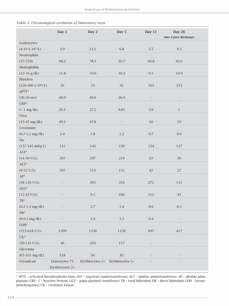
Table 1. Chronological evolution of laboratory tests
Day 1 Day 2 Day 5 Day 11 Day 26
(day 8 post discharge)
Leukocytes
(4-10 x 109/L) 3.9 12.1 6.8 5.5 9.3
Neutrophils
(37-72%) 88.2 78.5 50.7 60.8 65.6
Hemoglobin
(12-16 g/dL) 11.8 10.9 10.3 9.1 10.9
Platelets
(150-400 x 109/L) 56 23 91 503 353
aPTT*
(26-36 sec) 40.9 40.6 26.9 - -
CRP*
(< 1 mg/dL) 26.1 27.2 9.81 5.6 1
Urea
(15-45 mg/dL) 49.1 47.8 - 10 19
Creatinine
(0.7-1.2 mg/dL) 2.4 1.8 1.2 0.7 0.9
Na+
(137-145 mEq/L) 131 143 150 134 137
AST*
(14-36 U/L) 165 297 219 33 30
ALT*
(9-52 U/L) 107 153 151 42 27
AP*
(38-126 U/L) - 205 310 272 131
GGT*
(12-43 U/L) - 9-2 106 112 45
TB*
(0.2-1.3 mg/dL) - 2.7 1.4 0.6 0.3
DB*
(0-0.3 mg/dL) - 2.3 1.2 0.4 -
LDH*
(313-618 U/L) 1209 1336 1156 697 417
CK*
(30-135 U/L) 46 103 117 - -
Glycemia
(65-105 mg/dL) 118 94 85 - -
Urynalysis Leucocytes 75 Erythrocytes 2+ Erythrocytes 1+ - -
Erythrocytes 2+
* aPTT – activated thromboplastin time; AST – aspartate aminotransferase; ALT – alanine aminotransferase; AP – alkaline phos-phatase; CRP - C - Reactive Protein, GGT – gama glutamyl transferase; TB – total bilirrubin; DB – direct bilirrubin; LDH – lactate dehydrogenase; CK – creatinine kinase.
Atypical case of Mediterranean spotted fever
JUN-2010.indd 214 28/7/2010 10:35:13

215Braz J Infect Dis 2010; 14(3):213-216
the epigastric region without hepatomegaly or splenom-
egaly, no lymphadenopathy, no edemas, no articular
inflammatory signs, and a normal neurologic examina-
tion, including absence of meningitis signs. On the right
inguinal region, an evanescent 3 cm purple macula was
identified.
Laboratory work-up results were as seen on Table 1.
Chest X-ray showed an interstitial infiltrate and enlarge-
ment of the cardiac area. The diagnosis of Mediterranean
spotted fever was, thus, assumed and the patient was ad-
mitted in the Infectious Diseases Department and began
doxicycline 100 mg 12/12h PO. On day 2, the patient pre-
sented mental confusion and disorientation, high fever
(39º C), tachypnea, hemodynamic instability (blood pres-
sure 86/40 mmHg, heart rate 114 bpm), conjuctival injec-
tion. Due to urinary retention, the patient was undergone
to catheterization, revealing a yellowish and fetid vaginal
discharge. The presence of meningitis signs led to a lum-
bar puncture, which revealed no pleocytosis and a nor-
mal protein count. A discrete macular erythematous rash,
progressing to a petechial aspect, was visible on trunk and
arms, without involvement of palms and plants, and an
atypical minimal papular lesion was identified on the left
shoulder. A laboratory evaluation revealed leukocytosis
with neutrofilia, and aggravation of liver enzymes (Table
1). Arterial blood gas revealed respiratory alkalemia (pH
7.64, pCO2 21, pO
2 79, HCO
3- 19.2). Chest X-ray showed
bilateral pleural effusion, mainly on the right side, with-
out adenomegaly or pulmonary parenquimal lesions
(supported on CT scan). Head CT-scan, abdominal and
renal ultrasound and echocardiogram were normal. With
the hypothesis of sepsis of unknown origin, possibly a
staphylococcal toxic syndrome (although the patient de-
nied the use of vaginal tampons or presence of an intrau-
terine contraceptive device), ceftriaxone 2 gm IV q24 and
vancomycin 1 gm IV q12 were added to doxicycline. Blood
cultures were negative as well as the vaginal exsudate cul-
ture. PCR for Chlamydia trachomatis and Neisseria gonor-
rhoeae was negative. Serologic evaluation was negative for
HIV 1 and 2. Biopsy of the papular lesion revealed a dense
infl ammatory infi ltrate, composed by histiocytes, lym-
phocytes and neutrophils, with diffuse and perivascular
distribution, without the typical vasculitic signs present in
a “tache noire” (Figure 1). Identifi cation of the species was
not possible due to technical diffi culties in the processing
of skin tissue samples. The clinical course of the patient was
positive, despite a diffi cult effervescence period (Figure 2),
and discharge was possible on day 18, fully recovered. The
levels of erythrocyte glucose-6-phosphate-dehydrogenase
(G6PD) were normal - 369 U/1012 erythrocytes (normal
reference range = 146-376 U/1012 erythrocytes).
The serology for Rickettsia conorii was positive, with
IgM > 256 and IgG > 1024.
Figure 1: Biopsy of the inoculation scar.
Figure 2: Temperature graphic.
Figueira-Coelho, Martins, Machado et al.
JUN-2010.indd 215 28/7/2010 10:35:14

216
DISCUSSION
Mediterranean spotted fever was fi rst described by Conor and Bruch in Tunisia in 1910.11 In Portugal, the fi rst de-scription of this exanthematic disease was made by Delfi m Pinheiro in 1923,6 but it was the work of Ricardo Jorge, re-viewing several cases in 1930, that brought this disease into attention, naming it “febre escaro-nodular”.12 In 1982, Raoult et al. described the fi rst severe cases (malignant form) of this apparent benign disease. This form results from a diffuse vasculitic process, involving several organs – kidney, lung, liver, pancreas, heart, spleen, skin, brain – eventually leading to multiorgan failure. Identifi ed risk factors are: advanced age, immunosuppression, diabetes, cardiac insuffi ciency, chronic alcoholism, respiratory insuffi ciency, G6PD defi -ciency, delay in treatment and inadequate antibiotic thera-py.3 The incidence of severe cases varies according to time. In Salamanca, Spain, the disease was more severe in 1983, with an incidence of complications of 19%, in contrast to 3.7% in 1981 and 4.3% in 1982.13 Severity also seems to vary according to geographical region. In Beja, a southern dis-trict of Portugal, during 1997, the mortality of hospitalized cases with Mediterranean spotted fever was 32.3%, while in Bragança, a northern district, the mortality rate was almost nil, despite having the highest incidence of cases in that country.14 Of further interest is the fact that, in Portugal, cases caused by the strain “Israeli tick typhus” were also fi rst described in 1997. A study by de Sousa et al., published in 2008, with 140 patients admitted in 13 Portuguese hos-pitals from 1994 to 2006 with documented identifi cation of the ricketsial strain causing the infection, revealed that this strain appears to be more virulent than the other strain described in the country: the Malish strain.7 In the same study, case fatalities were statistically more frequently as-sociated with symptoms of abdominal pain, nausea, vom-iting, diarrhea, prostration, altered mental status, evidence of tachypnea, hepatomegaly and a petechial rash, labora-tory fi ndings of leukocytosis (> 11300 cells/mm3, partial thromboplastin time > 35 s, glucose level > 110 mg/dL, urea level > 50 mg/dL, creatinine level > 1.2 mg/dL, sodium < 145 mEq/ L, total bilirrubin level > 1.2 mg/dL, and el-evated levels of gama-glutamyl transferase, alkaline phos-phatase and creatine kinase. Alcoholism was the only clas-sical co-morbidity associated with increased fatality rate.
Our patient had an atypical presentation in several as-pects: the late occurrence of the rash without involvement of palms and plants, the doubtful presence of two inocula-tion scars, the absence of classic vasculitic fi ndings in the histological examination of the inoculation scar, the ex-tended decline of fever (18 days of treatment). The presence of abdominal pain, nausea, vomiting, prostration, altered
mental status, tachypnea, a petechial rash, elevated partial thromboplastin time, hyponatremia, elevated glucose, urea, creatinine, total bilirrubin, gama-glutamyl transferase, and alkaline phosphatase levels predicted a fatal outcome. Al-though identifi cation was not possible, the “Israeli tick ty-phus” strain might have been the responsible strain. Apart from the delay in beginning antibiotic treatment, the patient had no identifi able classic risk factor, which probably ex-plains the successful outcome.
CONCLUSION
Severe cases of Mediterranean spotted fever can present with atypical signs. The eventual patient instability can lead to the consideration of a different diagnosis, usually of infec-tious origin, until the confi rmation is done by serology.
REFERENCES
1. Maltez F, Machado J, Morgado A, Proença R. Febre escaro-nodular: casuística de 10 anos (1977-1986). Estudo Clínico e Epidemiológico de 247 casos. O Médico 1989; 20:459-64.
2. Tavares L., Botas J, Antunes F, Araújo FC. A febre escaro-nodular em Portugal. I – Análise estatístico-epidemiológica nos últimos 30 anos (1955-1984). O Médico 1985; 20:838-40.
3. Rovery C, Raoult D. Mediterranean Spotted Fever. Infect Dis Clin N Am 2008; 22:515-30.
4. Colomba C, Saporito L, Polara VF et al. Mediterranean spot-ted fever: clinical and laboratory characteristics of 415 Sicilian children. BMC Infect Dis 2006; 6:60.
5. Anton E, Font B, Munoz T et al. Clinical and laboratory char-acteristics of 144 patients with Mediterranean spotted fever. Eur J Clin Microbiol Infect Dis 2003; 22:126-8.
6. Carmo G, Caixeiro IS, Uva AS, Paiva JED. Febre escaro-nod-ular: actualização teórica e análise retrospectiva de 231 casos. Rev Port Doenças Infecciosas 1981; 4:13-25.
7. Sousa R, França A, Dória Nóbrega S et al. Host- and mi-crobe-related risk factors for and pathophysiology of Fa-tal Rickettsia conorii Infection in Portuguese Patients. J Infect Dis 2008; 198(4):576-85.
8. Oliveira J, Côrte-Real R. Rickettsioses em Portugal. Acta Med Port 1999; 12:313-21.
9. Rovery C, Brouqui P, Raoult D. Questions on mediterranean spotted fever a century after its discovery. Emerg Infect Dis 2008; 14:1360-7.
10. Raoult D, Kohler JL, Gallais H et al. Fatal rickettsiosis. Nouv Presse Med 1982; 11:607.
11. Conor A, Bruch A. Une fi èvre éruptive observée en Tunisie. Bull Soc Pathol Exot Filiales 1910; 8:8492-6.
12. Jorge R. La Fièvre exanthématique (Fièvre escharo-nodulaire) et son apparition au Portugal. Jornal Lisboa Médica 1930; 8:433-54.
13. Ruiz Beltran R, Herrero-Herrero JI, Martin-Sanchez AM. Formas graves de fi ebre exantemática mediterrânea. Analisis prospectivo de 71 enfermos. Ann Med Interna 1985; 2:365-8.
14. Sousa R, Dória Nóbrega S, Bacellar F, Torgal J. Sobre a real-idade da febre escaro-nodular em Portugal. Acta Med Port 2003; 16:429-36.
Atypical case of Mediterranean spotted fever
JUN-2010.indd 216 28/7/2010 10:35:14