course code: anp 201 comparative anatomy and physiology …
TRANSCRIPT

http://www.unaab.edu.ng
1
COURSE CODE: ANP 201
COURSE TITLE: Comparative Anatomy And Physiology of Farm Animals NUMBER OF UNITS: 3 units
COURSE DURATION: Three hours per week C Course Coordinator: Dr. A. O. Ladokun B.Sc., M.Sc., Ph.D (Ibadan) Email: [email protected] Office Location: ANP Staff Office, COLANIM Other Lecturers: Prof. O. A. Osinowo, Prof. O. M. Onagbesan, Dr. O. F. Smith, Dr. O. S. Sowande, Dr. A. Akinloye Skeletal system, Reproduction, Digestion, Excretion, Nervous, Circulatory, Respiratory Systems, Growth and Development.
This is a compulsory course for 200 Level Agricultural Students in the University. In view of this, students are expected to participate in all the course activities and have minimum of 75% attendance to be able to write the final examination.
1. R. H. S. Carpenter Neurophysiology 2nd edition. Edward Arnold (Publishers) London
2. William O. Reece Physiology of Domestic Animals 2nd edition. Williams & Wikins
(Publisher) London, Paris
COURSE DETAILS:
COURSE CONTENT:
COURSE REQUIREMENTS:
READING LIST:

http://www.unaab.edu.ng
2
THE CIRCULATORY SYSTEM Table of Contents Types of Circulatory Systems | Vertebrate Cardiovascular System | Vertebrate Vascular Systems The Heart | The Vascular System | Blood | The Lymphatic System | Learning Objectives | Links Types of Circulatory Systems | Back to Top Living things must be capable of transporting nutrients, wastes and gases to and from cells. Single-celled organisms use their cell surface as a point of exchange with the outside environment. Multicellular organisms have developed transport and circulatory systems to deliver oxygen and food to cells and remove carbon dioxide and metabolic wastes. Sponges are the simplest animals, yet even they have a transport system. Seawater is the medium of transport and is propelled in and out of the sponge by ciliary action. Simple animals, such as the hydra and planaria (shown in Figure 1), lack specialized organs such as hearts and blood vessels, instead using their skin as an exchange point for materials. This, however, limits the size an animal can attain. To become larger, they need specialized organs and organ systems. Figure 1. Structures that serve some of the functions of the circulatory system in animals that lack the system. Image from Purves et al., Life: The Science of Biology, 4th Edition, by Sinauer Associates (www.sinauer.com) and WH Freeman (www.whfreeman.com), used with permission.
LECTURE NOTE:

http://www.unaab.edu.ng
3
Multicellular animals do not have most of their cells in contact with the external environment and so have developed circulatory systems to transport nutrients, oxygen, carbon dioxide and metabolic wastes. Components of the circulatory system include
blood: a connective tissue of liquid plasma and cells heart: a muscular pump to move the blood blood vessels: arteries, capillaries and veins that deliver blood to all tissues
There are several types of circulatory systems. The open circulatory system, examples of which are diagrammed in Figure 2, is common to molluscs and arthropods. Open circulatory systems (evolved in insects, mollusks and other invertebrates) pump blood into a hemocoel with the blood diffusing back to the circulatory system between cells. Blood is pumped by a heart into the body cavities, where tissues are surrounded by the blood. The resulting blood flow is sluggish. Figure 2. Circulatory systems of an insect (top) and mollusc (middle). Images from Purves et al., Life: The Science of Biology, 4th Edition, by Sinauer Associates (www.sinauer.com) and WH Freeman (www.whfreeman.com), used with permission.

http://www.unaab.edu.ng
4
Vertebrates, and a few invertebrates, have a closed circulatory system, shown in Figure 2. Closed circulatory systems (evolved in echinoderms and vertebrates) have the blood closed at all times within vessels of different size and wall thickness. In this type of system, blood is pumped by a heart through vessels, and does not normally fill body cavities. Blood flow is not sluggish. Hemoglobin causes vertebrate blood to turn red in the presence of oxygen; but more importantly hemoglobin molecules in blood cells transport oxygen. The human closed circulatory system is sometimes called the cardiovascular system. A secondary circulatory system, the lymphatic circulation, collects fluid and cells and returns them to the cardiovascular system. Vertebrate Cardiovascular System | Back to Top The vertebrate cardiovascular system includes a heart, which is a muscular pump that contracts to propel blood out to the body through arteries, and a series of blood vessels. The upper chamber of the heart, the atrium (pl. atria), is where the blood enters the heart. Passing through a valve, blood enters the lower chamber, the ventricle. Contraction of the ventricle forces blood from the heart through an artery. The heart muscle is composed of cardiac muscle cells. Arteries are blood vessels that carry blood away from heart. Arterial walls are able to expand and contract. Arteries have three layers of thick walls. Smooth muscle fibers contract, another layer of connective tissue is quite elastic, allowing the arteries to carry blood under high pressure. A diagram of arterial structure is shown in Figure 3. Figure 3. Structure of an artery. Image from Purves et al., Life: The Science of Biology, 4th Edition, by Sinauer Associates (www.sinauer.com) and WH Freeman (www.whfreeman.com), used with permission.
The aorta is the main artery leaving the heart. The pulmonary artery is the only artery that carries oxygen-poor blood. The pulmonary artery carries deoxygenated blood to the lungs. In the lungs, gas exchange occurs, carbon dioxide diffuses out, oxygen diffuses

http://www.unaab.edu.ng
5
in. Arterioles are small arteries that connect larger arteries with capillaries. Small arterioles branch into collections of capillaries known as capillary beds, an exampe of one is shown in Figure 4. Figure 4. Structure and blood flow through a vein. The above illustration is from http://www.prs.k12.nj.us/schools/PHS/Science_Dept/APBio/pic/capillary.gif.
Figure 5. Capillary with Red Blood Cell (TEM x32,830). This image is copyright Dennis Kunkel at www.DennisKunkel.com, used with permission.
Capillaries, shown in Figures 4 and 5, are thin-walled blood vessels in which gas exchange occurs. In the capillary, the wall is only one cell layer thick. Capillaries are concentrated into capillary beds. Some capillaries have small pores between the cells of the capillary wall, allowing materials to flow in and out of capillaries as well as the passage of white blood cells. Changes in blood pressure also occur in the various vessels of the circulatory system, as shown in Figure 6. Nutrients, wastes, and hormones are exchanged across the thin walls of capillaries. Capillaries are microscopic in size, although blushing is one manifestation of blood flow into capillaries. Control of blood flow into capillary beds is done by nerve-controlled sphincters.

http://www.unaab.edu.ng
6
Figure 6. Changes in blood pressure, velocity, and the area of the arteries, capillaries, and veins of the circulatory system. Image from Purves et al., Life: The Science of Biology, 4th Edition, by Sinauer Associates (www.sinauer.com) and WH Freeman (www.whfreeman.com), used with permission.
The circulatory system functions in the delivery of oxygen, nutrient molecules, and hormones and the removal of carbon dioxide, ammonia and other metabolic wastes. Capillaries are the points of exchange between the blood and surrounding tissues. Materials cross in and out of the capillaries by passing through or between the cells that line the capillary, as shown in Figure 7. Figure 7. Capillary structure, and relationships of capillaries to arteries and veins. Image from Purves et al., Life: The Science of Biology, 4th Edition, by Sinauer Associates (www.sinauer.com) and WH Freeman (www.whfreeman.com), used with permission.

http://www.unaab.edu.ng
7
The extensive network of capillaries in the human body is estimated at between 50,000 and 60,000 miles long. Thoroughfare channels allow blood to bypass a capillary bed. These channels can open and close by the action of muscles that control blood flow through the channels, as shown in Figure 8. Figure 8. Capillary beds and their feeder vessels. Image from Purves et al., Life: The Science of Biology, 4th Edition, by Sinauer Associates (www.sinauer.com) and WH Freeman (www.whfreeman.com), used with permission.
Blood leaving the capillary beds flows into a progressively larger series of venules that in turn join to form veins. Veins carry blood from capillaries to the heart. With the exception of the pulmonary veins, blood in veins is oxygen-poor. The pulmonary veins

http://www.unaab.edu.ng
8
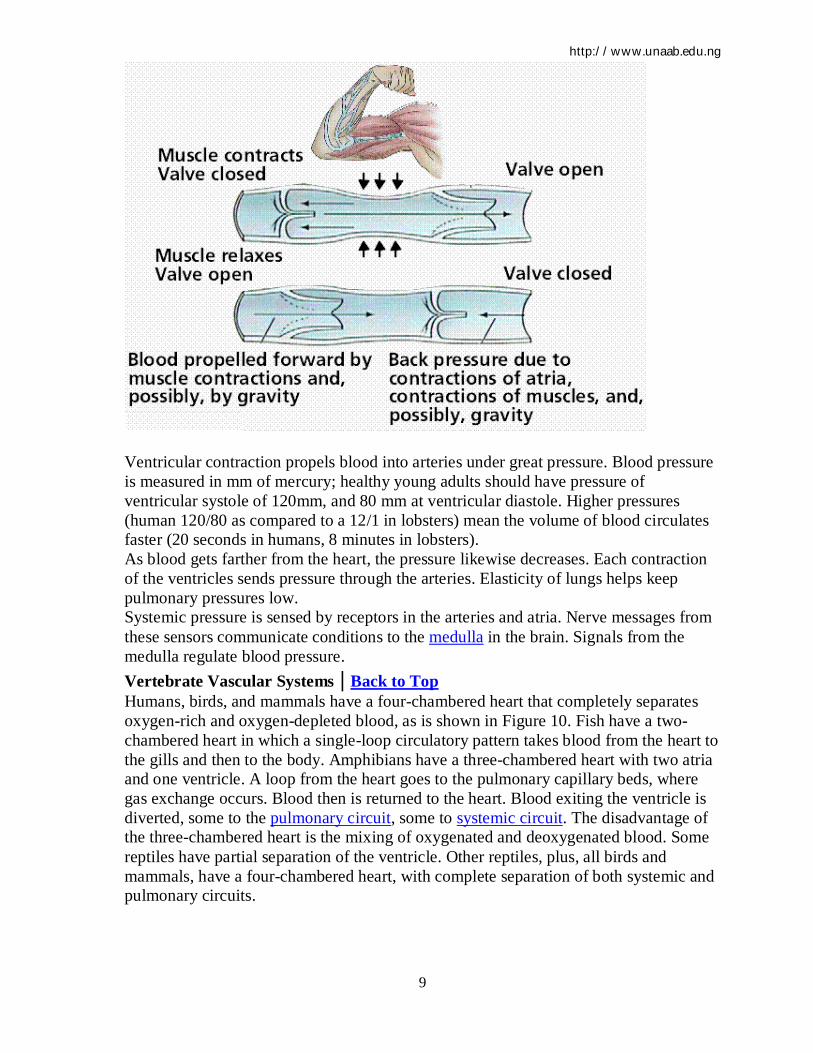
carry oxygenated blood from lungs back to the heart. Venules are smaller veins that gather blood from capillary beds into veins. Pressure in veins is low, so veins depend on nearby muscular contractions to move blood along. The veins have valves that prevent back-flow of blood, as shown in Figure 9. Figure 9. Structure of a vein (top) and the actions of muscles to propel blood through the veins. Images from Purves et al., Life: The Science of Biology, 4th Edition, by Sinauer Associates (www.sinauer.com) and WH Freeman (www.whfreeman.com), used with permission.
http://www.unaab.edu.ng
9
Ventricular contraction propels blood into arteries under great pressure. Blood pressure is measured in mm of mercury; healthy young adults should have pressure of ventricular systole of 120mm, and 80 mm at ventricular diastole. Higher pressures (human 120/80 as compared to a 12/1 in lobsters) mean the volume of blood circulates faster (20 seconds in humans, 8 minutes in lobsters). As blood gets farther from the heart, the pressure likewise decreases. Each contraction of the ventricles sends pressure through the arteries. Elasticity of lungs helps keep pulmonary pressures low. Systemic pressure is sensed by receptors in the arteries and atria. Nerve messages from these sensors communicate conditions to the medulla in the brain. Signals from the medulla regulate blood pressure. Vertebrate Vascular Systems | Back to Top Humans, birds, and mammals have a four-chambered heart that completely separates oxygen-rich and oxygen-depleted blood, as is shown in Figure 10. Fish have a two-chambered heart in which a single-loop circulatory pattern takes blood from the heart to the gills and then to the body. Amphibians have a three-chambered heart with two atria and one ventricle. A loop from the heart goes to the pulmonary capillary beds, where gas exchange occurs. Blood then is returned to the heart. Blood exiting the ventricle is diverted, some to the pulmonary circuit, some to systemic circuit. The disadvantage of the three-chambered heart is the mixing of oxygenated and deoxygenated blood. Some reptiles have partial separation of the ventricle. Other reptiles, plus, all birds and mammals, have a four-chambered heart, with complete separation of both systemic and pulmonary circuits.

http://www.unaab.edu.ng
10
Figure 10. Circulatory systems of several vertebrates showing the progressive evolution of the four-chambered heart and pulmonary and systemic circulatory circuits. Images from Purves et al., Life: The Science of Biology, 4th Edition, by Sinauer Associates (www.sinauer.com) and WH Freeman (www.whfreeman.com), used with permission.

http://www.unaab.edu.ng
11
The Heart | Back to Top The heart, shown in Figure 11, is a muscular structure that contracts in a rhythmic pattern to pump blood. Hearts have a variety of forms: chambered hearts in mollusks and vertebrates, tubular hearts of arthropods, and aortic arches of annelids. Accessory hearts are used by insects to boost or supplement the main heart's actions. Fish, reptiles, and amphibians have lymph hearts that help pump lymph back into veins. The basic vertebrate heart, such as occurs in fish, has two chambers. An auricle is the chamber of the heart where blood is received from the body. A ventricle pumps the blood it gets through a valve from the auricle out to the gills through an artery. Amphibians have a three-chambered heart: two atria emptying into a single common ventricle. Some species have a partial separation of the ventricle to reduce the mixing of oxygenated (coming back from the lungs) and deoxygenated blood (coming in from the body). Two sided or two chambered hearts permit pumping at higher pressures and the

http://www.unaab.edu.ng
12
addition of the pulmonary loop permits blood to go to the lungs at lower pressure yet still go to the systemic loop at higher pressures. Figure 11. The relationship of the heart and circulatory system to major visceral organs. Below: the structure of the heart. Images from Purves et al., Life: The Science of Biology, 4th Edition, by Sinauer Associates (www.sinauer.com) and WH Freeman (www.whfreeman.com), used with permission.

http://www.unaab.edu.ng
13
Establishment of the four-chambered heart, along with the pulmonary and systemic circuits, completely separates oxygenated from deoxygenated blood. This allows higher the metabolic rates needed by warm-blooded birds and mammals. The human heart, as seen in Figure 11, is a two-sided, four-chambered structure with muscular walls. An atrioventricular (AV) valve separates each auricle from ventricle. A semilunar (also known as arterial) valve separates each ventricle from its connecting artery. The heart beats or contracts approximately 70 times per minute. The human heart will undergo over 3 billion contraction cycles, as shown in Figure 12, during a normal lifetime. The cardiac cycle consists of two parts: systole (contraction of the heart muscle) and diastole (relaxation of the heart muscle). Atria contract while ventricles relax. The pulse is a wave of contraction transmitted along the arteries. Valves in the heart open and close during the cardiac cycle. Heart muscle contraction is due to the presence of nodal tissue in two regions of the heart. The SA node (sinoatrial node) initiates heartbeat. The AV node (atrioventricular node) causes ventricles to contract. The AV node is sometimes called the pacemaker since it keeps heartbeat regular. Heartbeat is also controlled by nerve messages originating from the autonomic nervous system.