corrosion and mercury release from dental...
TRANSCRIPT

Corrosion and Mercury Release from Dental Amalgam
Jaro Pleva, Ph.D.1
Abstract Corrosion attacks on twenty-two dental
amalgam restorations after in vivo service have been studied by Scanning Electron Microscopy together with the Energy Dispersive X-Ray Technique, and by optical microscopy. From the measured depth and type of corrosion attack, estimates of released mercury amounts are made.
The amalgam fillings have been obtained from members of a group of 250 individuals, who suspected their health troubles potentially to be chronic mercury poisoning from amalgam and were to have all amalgam fillings removed. Three typical patient cases are presented.
Model calculations of released mercury, based on previously published measurements of corrosion currents with and without abrasion are also given.
The investigations show, that the long-term release of mercury from a few amalgam fillings will often reach or exceed the recommended limits for daily intake of mercury. Hence, mercury from corroding amalgam fillings represents a potential health hazard. Danger of galvanic contact between amalgam and gold restorations is particularly emphasized.
1. Introduction The impact of corrosion on operation of
industrial appliances and personal security is well recognized and subject to a large number of investigations. Corrosion can also be a cause of environmental contamination, for instance by heavy metals in pharmaceutical or food industry.
Hitherto, the possible danger from the internal source of corrosion products, dental amalgam fillings, has been mostly overlooked. Presently, this problem is devoted special attention.1 Though a proof of amalgam biocompatibility has never
1. P1 3079, S - 68300 Hagfors, Sweden.
been presented, amalgam is still by far the most extensively used material for dental restorations.2 Since the investigations and warnings of the outstanding German chemist Alfred Stock, 1920-19453 4 5 little epidemiological studies of mercury release and its effects have been made. One of the possible reasons may be the very interdisciplinary character of the problem. For correct answer, specialist competence in the following fields is required: materials science, corrosion/electrochemistry, toxicology, medicine/diagnostics, physical biology, analytical chemistry.
The common type of dental amalgam is an alloy containing typically in weight-%; 50 Hg, 35 Ag, 10 Sn, Cu, Zn.
Reported types of amalgam degradation are crevice corrosion,2 6 7 8 selective corrosion,9 galvanic corrosion in contact with dissimilar alloys10 11 and mechanical wear.12 33 Besides selective attack, stress corrosion has been proposed to be responsible for the marginal breakdown of amalgam restorations.13 Further, cyclic loading, simulating chewing, strongly promotes corrosion of the amalgam surface.14 15
Published investigations of amalgam corrosion are mostly concerned about the mechanical performance of dental fillings and neglect to make estimates of mercury amounts released by corrosion attack and wear. Nevertheless, the unfounded and simplified argument that "dental amalgam releases very small amounts of mercury" is often used, even in scientific and official reports. In some cases, the exposure for released mercury has been confused with mercury in urine and blood of exposed patients.
Laboratory experiments, described in the odontologic literature, often neglect a number of important factors, which will influence corrosion of fillings in vivo. These are for instance wear, pressure fluctuations from biting contact, temperature
141

Journal of Orthomolecular Medicine Vol. 4, No. 3, 1989
increase, acidic and salt food, galvanic contacts with dissimilar metals.
The work of Mayer and Diehl16 provides an example. In their experiments, the investigators eliminated any cathodic depolarizer, which is most often oxygen from the air. The use of oxygen free double distilled water effectively stopped possibility of any corrosion process and implied a wrong conclusion about high corrosion resistance of amalgam.
The aim of the present work was to investigate a number of amalgam fillings, which have served in vivo for several years. Estimates of mercury amounts, corresponding to measured intensity of corrosion attack, are given. The amounts of mercury are compared to recommended limits for maximum daily exposure in food and in air.
For a number of patient cases, the removal of the corroded fillings is linked to a distinct alleviation of symptoms, typical for chronic mercury poisoning. Three of the cases are described in more detail.
2. Experimental
2.1 Material From 15 persons, parts of 22 removed
amalgam fillings have been gathered and investigated. The material also included extracted teeth with amalgam fillings, and gold restorations in direct metallic contact with amalgam.
The donators were selected from a group of 250 persons, all of them having large amounts of amalgam, often in combination with gold restorations in direct or intermittent contact. With a questionnaire, the following information has been gathered: - age, sex, profession, anamnesis, - history of dental treatments and oral status:
number of amalgam fillings, other metals, contact to gold, permanent or intermittent,
- root treated teeth, - all health complaints, symptoms classified as
fair or severe. Summary of the most frequent symptoms in the whole patient group is listed in Table 2. Below, three patient cases are described
in more detail. The dental restorations, whose details are shown in Figures 1 to 11 belonged to these three patients.
Persons with possible occupational exposure to mercury and those consuming fresh water fish more than twice a week have been excluded from the investigation.
Control group: Ten persons have been found, age 30-60 years, who have never had any amalgam restoration. No person in the control group complained about more than three symptoms given in Table 2, and none of the symptoms was classified as severe.
Inversely, none of the patients with the syndrome was free of dental amalgam, and all complained about at least 5 symptoms, mostly more than ten. Most severely ill, till invalidity, were bearers of galvanic cells gold - amalgam. A statistical evaluation of the patient material will be the subject of another paper.
2.2 Case Histories PATIENT A: Male, 41 years Profession: corrosion engineer Oral status: 20 amalgam fillings, 1 gold bridge over 5 teeth (2 molars extracted) Root fillings: 3 Investigated restorations: amalgam in the gold bridge; 5 year old filling, intermittent contact to gold; 20 year old large filling, no contact to dissimilar metal Number of symptoms: 25 After root filling, hole in the gold bridge has been filled with amalgam. A few months later the patient suffered increasing symptoms of both physical, psychical and neurological kind, most of them listed in Table 2. No dysfunction could be found by common diagnostic methods on heart muscle (ECG) or on gastrointestinal tract (X-ray) or elsewhere. As a corrosion engineer, the patient pointed at the black and rough amalgam filling in the galvanic cell gold - amalgam, but no doctor took the idea of poisoning from corroding amalgam seriously. First a look into a toxicology handbook identified the symptoms as chronic mercury poisoning. Removal of amalgam in the galvanic cell broke the trend and the strongest symptoms went
142
Corrosion and Mercury Release from Dental Amalgam
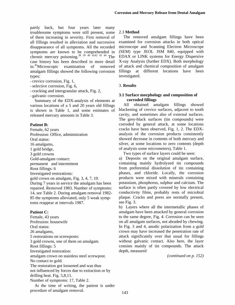
partly back, but four years later many troublesome symptoms were still present, some of them increasing in severity. First removal of all fillings resulted in alleviation and successive disappearance of all symptoms. All the recorded symptoms are known to be comprehended in chronic mercury poisoning.38 39 40 4142 43 44 The case history has been described in more detail in.45Microscopic examination of removed amalgam fillings showed the following corrosion types: - crevice corrosion, Fig. 1, - selective corrosion, Fig. 6, - cracking and intergranular attack, Fig. 2, - galvanic corrosion.
Summary of the EDX-analysis of elements at various locations of a 5 and 20 years old fillings is shown in Table 1, and some estimates of released mercury amounts in Table 3.
Patient B: Female, 62 years Profession: Office, administration Oral status: 16 amalgams, 1 gold bridge, 3 gold crowns Gold-amalgam contact: permanent and intermittent Root fillings: 6 Investigated restorations: gold crown on amalgam, Fig. 3, 4, 7, 10. During 7 years in service the amalgam has been repaired. Removed 1983. Number of symptoms: 14, see Table 2. During amalgam removal 1982-85 the symptoms alleviated, only 5 weak symp-toms reappear at intervals 1987.
Patient C: Female, 43 years Profession: housewife Oral status: 26 amalgams, 5 restorations on screwposts: 3 gold crowns, one of them on amalgam. Root fillings: 5 Investigated restoration: amalgam crown on stainless steel screwpost. No contact to gold. The restoration got loosened and was thus not influenced by forces due to extraction or by drilling heat. Fig. 5,8,11. Number of symptoms: 17, Table 2.
At the time of writing, the patient is under procedure of amalgam removal.
2.3 Method The removed amalgam fillings have been
examined for corrosion attacks in both optical microscope and Scanning Electron Microscope (SEM) type JEOL JSM 840, equipped with EDAX or LINK systems for Energy Dispersive X-ray Analysis (further EDX). Both morphology of attack and chemical composition of amalgam fillings at different locations have been investigated.
3. Results
3.1 Surface morphology and composition of corroded fillings
All obtained amalgam fillings showed blackening of crevice surfaces, adjacent to tooth cavity, and sometimes also of external surfaces. The grey-black surfaces (tin compounds) were corroded by general attack, at some locations cracks have been observed, Fig. 1, 2. The EDX-analysis of the corrosion products consistently showed decrease in contents of both mercury and silver, at some locations to zero contents (depth of analysis some micrometers), Table 1.
Two types of surface layers could be seen: a) Deposits on the original amalgam surface, containing mainly hydrolyzed tin compounds from preferential dissolution of tin containing phases, and chloride. Locally, the corrosion products were mixed with minerals containing potassium, phosphorus, sulphur and calcium. The surface is often partly covered by low electrical conductivity films, probably rests of microbial plaque. Cracks and pores are normally present, see Fig. 3. b) Layers where all the intermetallic phases of amalgam have been attacked by general corrosion to the same degree, Fig. 4. Corrosion can be seen on all amalgam surfaces, not abraded by chewing. In Fig. 3 and 4, anodic polarization from a gold crown may have increased the penetration rate of attack significantly over that usual for fillings without galvanic contact. Also here, the layer consists mainly of tin compounds. The attack depth, measured
(continued on p. 152)
143
Journal of Orthomolecular Medicine Vol. 4, No. 3, 1989
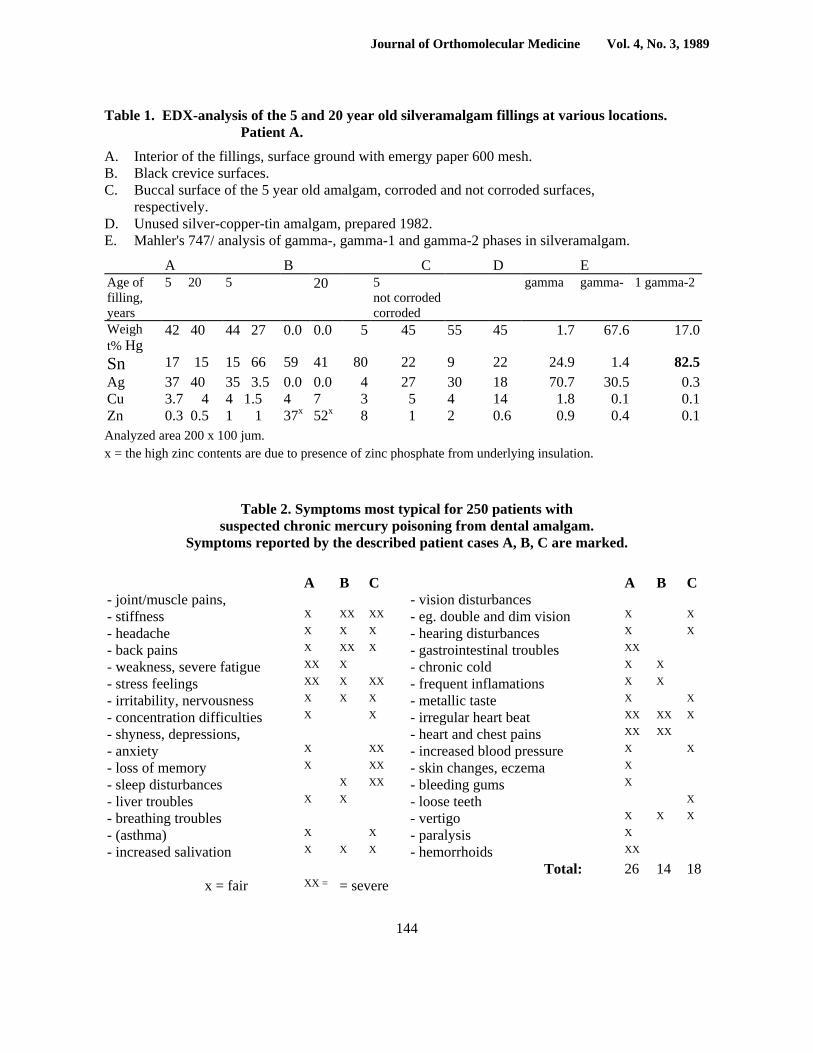
Table 1. EDX-analysis of the 5 and 20 year old silveramalgam fillings at various locations. Patient A.
A. Interior of the fillings, surface ground with emergy paper 600 mesh. B. Black crevice surfaces. C. Buccal surface of the 5 year old amalgam, corroded and not corroded surfaces,
respectively. D. Unused silver-copper-tin amalgam, prepared 1982. E. Mahler's 747/ analysis of gamma-, gamma-1 and gamma-2 phases in silveramalgam.
A B C D E Age of filling, years
5 20 5 20 5 not corroded corroded
gamma gamma- 1 gamma-2
Weigh t% Hg
42 40 44 27 0.0 0.0 5 45 55 45 1.7 67.6 17.0
Sn 17 15 15 66 59 41 80 22 9 22 24.9 1.4 82.5Ag 37 40 35 3.5 0.0 0.0 4 27 30 18 70.7 30.5 0.3Cu 3.7 4 4 1.5 4 7 3 5 4 14 1.8 0.1 0.1Zn 0.3 0.5 1 1 37x 52x 8 1 2 0.6 0.9 0.4 0.1Analyzed area 200 x 100 jum. x = the high zinc contents are due to presence of zinc phosphate from underlying insulation.
Table 2. Symptoms most typical for 250 patients with suspected chronic mercury poisoning from dental amalgam.
Symptoms reported by the described patient cases A, B, C are marked.
A B C A B C - joint/muscle pains, - vision disturbances - stiffness X XX XX - eg. double and dim vision X X - headache X X X - hearing disturbances X X - back pains X XX X - gastrointestinal troubles XX - weakness, severe fatigue XX X - chronic cold X X - stress feelings XX X XX - frequent inflamations X X - irritability, nervousness X X X - metallic taste X X - concentration difficulties X X - irregular heart beat XX XX X - shyness, depressions, - heart and chest pains XX XX - anxiety X XX - increased blood pressure X X - loss of memory X XX - skin changes, eczema X - sleep disturbances X XX - bleeding gums X - liver troubles X X - loose teeth X - breathing troubles - vertigo X X X - (asthma) X X - paralysis X - increased salivation X X X - hemorrhoids XX Total: 26 14 18
x = fair XX = = severe
144

Corrosion and Mercury Release from Dental Amalgam
Table 3. Summary of estimates of released mercury from Amalgam fillings from patients A, B and C. Available measured corrosion attacks, literature data are given.
Calculated from Patient Fig. Attack Depth jxm/serv. time
Surface, invest. filling
cm2
total Free Mercury: /Kg/day invest. filling all fillings
Present Investigation Galvanic corr. A — 1000/1 yr. 0.16 10 250 250 + 160') Galvanic corr. B 4 80/7 yrs. 1 8 19 19 + 105» General corr. C 5 30/5 yrs. 1 13 10 1302) Selective corr. B 7 1000/7 yrs. 1 8 15 1202) Evaporation A — 45 — 42% Hg in
5 years 1 gram 10 16 250*> + 160
Literature Reference General corr. Gasser8 50-90/? yrs. General corr. Mateer24 30/? yrs. Selective corr. Espevik6 200/3 yrs. 1 7
1) Released by general corrosion of the remaining fillings, not in contact to gold. 2) Galvanic effects with gold restorations not included. 3) 250 /ug/day released from gold-amalgam galvanic cell.
Table 4. Released mercury, calculated according to Faradays law from corrosion currents, measured in vitro by Marek33. The released amounts should be compared to the following recommended maximum Hg-intake: 216 /xg/day for occupational exposure (Sweden) 43 /jg/day for occupational exposure (Switzerland and Soviet)
13 jug/day for general population (EPA) 4 )ug/day for general population (Soviet) 30 /ug/day in food (Sweden)
Amalgam Type Abrasion Corrosion Current fiA.cnv2
Free Mercury Mg/day
Conventional (lathe-cut) no yes 2 hrs./day
2.0 87 87
1365900
490Conventional (spherical) no
yes 2 hrs./day
2.3 130 130 1568840
735Non-gamma-2 (high copper)
no yes 2 hrs./day
2.5 11 11 170750
62
145

Journal of Orthomolecular Medicine Vol. 4, No. 3, 1989
Figure 1. Crevice corrosion of a five year old amalgam filling. Bright area is the buccal surface. Case A.
Figure 2. Detail of Figure 1, showing cracks in the corroded surface.
146

Corrosion and Mercury Release from Dental Amalgam
Figure 3. Corrosion products on surface of silveramalgam. Dark areas contain mainly tin compounds, white areas are probably rests of microbial plaque. Case B.
Figure 4. Cross section of corroded amalgam surface, which has been in contact with a gold crown. Optical micrograph. Case B.
147

Journal of Orthomolecular Medicine Vol. 4, No. 3, 1989
Figure 5. Cross section of amalgam crown with corrosion attack on buccal surface. Case C.
Figure 6. Selective attack of free amalgam surface at the margin. Five year old filling, intermittent contact to gold. Case A.
148

Corrosion and Mercury Release from Dental Amalgam
Figure 7. Cross section showing selective corrosion of gamma-2 phase. The cavities are filled with corrosion products containing tin and chlorine atoms. Case B.
Figure 8. Segregation of mercury to certain areas of the occlusal surface. Point of EDX-analysis is indicated. Note also corrosion attacks of the surface. Case C.
149

Journal of Orthomolecular Medicine Vol. 4, No. 3, 1989
Figure 9. Comparison of EDX-spectra of the bright lines in Figure 8 (left spectra, point analysis) with average spectra of a ground surface (analyzed surface 300x200 pm2).
Figure 10. Galvanic corrosion of silveramalgam (Amg) under a gold crown (Au). Case B.
150

Corrosion and Mercury Release from Dental Amalgam
Figure 11. Occlusal surface of an amalgam crown with emerging stainless steel screwpost. Galvanic corrosion occurred in vicinity of the stainless area. Case C.
Figure 12. Potentiokinetic polarization curve of silver-tin-mercury dental amalgam alloy at 25° C - a) in 0.2% NaCl (after Abadir, 8), b) in synthetic saliva (after Ross et al, 19).
151

Journal of Orthomolecular Medicine Vol. 4, No. 3, 1989
on transversal sections, varied considerably, as shown in Table 3.
The selective corrosion of external surfaces, Fig. 6, reached depths up to 1.4 mm. The attack propagated into the filling, and was sometimes, but not always, filled with corrosion products, as shown in Fig. 7. The products contained only tin and chloride atoms. The subsurface selective attacks were connected to the bulk saliva electrolyte through the pores in the surface layer.
In order to check the mercury contents in the interior of fillings, SEM-analysis of freshly prepared cross section surfaces has been performed. Grinding was done carefully on emery paper 600 mesh under 10° C cold water.
In no case the mercury content was higher than in an unused dentist-made filling. A 5 year old filling from patient A contained 42% mercury (standard deviation ± 1.28%) and a 20 year old filling 40% mercury (s.d. ±1.11%), whereas the unused amalgam contained 45 (s.d. ± 1.39%) weight% mercury. In contrast, the outer buccal surface of the 5 year old filling contained 55% mercury. Analysis data from the mentioned fillings of patient A and comparison with literature are given in Table 1.
3.2 Mercury segregation On the occlusal surface of the amalgam crown
from patient C segregation of mercury to certain areas has been observed. The secondary electrons image in SEM is shown in Fig. 8 as bright lines reminding of grain boundaries.
The EDX-spectra of the bright areas has been compared to the average composition of the filling on a ground surface. The spectras in Fig. 9 are based on the same number of counts and are thus comparable. Whereas the interior of the filling shows about the expected relationship between mercury, silver, tin and copper contents, the bright areas contain only mercury and silver in clear favour of the former element.
3.3 Corrosion potentials Corrosion potentials of removed gold bridge
(patient A) and of the amalgam fillings from the same patient have been measured in aerated 0.2% sodium chloride
solution at 25° C. Gold: +190 mV SCE (Saturated
Calomel Electrode) Amalgam: -310 mV SCE These values have been compared to polarization curves measured by Abadir18 and Ross19 on the same type of amalgam, Fig. 12. The impact of an anodic polarization of amalgam in the galvanic couple on increase of the corrosion current and amounts of released mercury will be discussed in one of the sections below.
3.4 Galvanic corrosion Accelerated corrosion attack of amalgam
occurred in contact with gold restorations and in contact with a stainless steel screw-post.
As expected, amalgam under gold crown in Fig. 10 suffered strongest attack near the contact to gold. Depth of corrosion is around 100 um.
The visible cavity under the crown may be an imperfection from tooth reconstruction, but there are signs that corrosion has also been involved. The cavity is connected to the bulk environment through channels in the amalgam. The channels are partly filled with corrosion products of low electrical conductivity, giving high brightness due to charging in SEM. Fig. 11 shows four quadrants of a stainless steel screw-post, emerging in the occlusal surface due to abrasion of amalgam. The vicinity of the stainless area suffered increased corrosion, compared to the rest of the occlusal surface.
No sign of corrosion has been observed on the screwpost itself. The EDX-analysis indicated a chromium-nickel-manganese alloyed stainless steel with molybdenum addition.
4. Discussion Corrosion and tarnish of amalgam restorations
have been studied and described in a number of papers.2 20 21 Most of the papers published in dental journals are concerned about the influence of corrosion on mechanical strength of the fillings and their service life. Though it is often stated, that free mercury is released during corrosion, it has not been shown that the free mercury does not evaporate. The question if, and under what circumstances the re-
152
Corrosion and Mercury Release from Dental Amalgam
leased mercury can be toxic to dental patients is still not unambiguously answered.
The estimates of released mercury, given in the present work, should be compared to the following maximum recommended daily exposures: - 0.4 ug per kilogram of body weight, i.e. about
30 ug mercury in food for an adult. - 50 ug/m3 air for industrial exposure 8 hours a
day, 40 hours a week (Sweden). - 25 ug/m3 air for industrial exposure 8 hours a
day, 40 hours a week (World Health Organization WHO).
- 10 ug/m3 air for industrial exposure 8 hours a day, 40 hours a week (Switzerland and Soviet).
- 1 ug/m3 air, U.S. national emission standard (U.S. Environmental Protection Agency EPA).
- 0.3 ug/m3 air for general population (Soviet). The large variations in the official limits
express the difficulty to establish a generally valid threshold for harmful mercury doses, as these are dependent on individual sensitivity and prevalence of non-specific symptoms.
Mercury vapour is effectively absorbed in the lungs.12 51 At 80% absorption, respiration volume 0.75 litre and respiration rate 15 per minute, the absorbed amounts will be, at the limit concentrations given above: - at 50 ug mercury /m3: 216 ug mercury in 8
hours. - at 10 ug mercury /m3: 43 ug mercury in 8 hours. - at 0.3 ug mercury /m3 : 4 ug mercury in 24
hours. Table 3 shows, that the normal long term
release in vivo from conventional amalgam without galvanic contact is 10-20 micrograms mercury a day from 1 cm2 surface. For a person with total filling surface of 10 cm2 the possible intake may reach 100-200 Mg a day.
Hence, many dental patients will be at risk and should be under regular medical control in the same way as workers with occupational exposure to mercury.
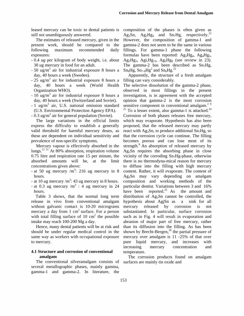
4.1 Structure and corrosion of conventional amalgam
The conventional silveramalgam consists of several metallographic phases, mainly gamma, gamma-1 and gamma-2. In literature, the
composition of the phases is often given as Ag3Sn, Ag3Hg4 and Sn7Hg, respectively.22 However, the composition of gamma-1 and gamma-2 does not seem to be the same in various fillings. For gamma-1 phase the following formulas have been reported: Ag3Hg4, Ag4Hg5, Ag2Hg3, Agi,Hg15, Ag5Hg8 (see review in 23). The gamma-2 has been described as Sn7Hg, Sn8Hg, Sn7.8Hg2 and Sn6Hg.22
Apparently, the structure of a fresh amalgam filling can vary considerably. The selective dissolution of the gamma-2 phase, observed in most fillings in the present investigation, is in agreement with the accepted opinion that gamma-2 is the most corrosion sensitive component in conventional amalgam.2 9 21 To a lesser extent, also gamma-1 is attacked.24 Corrosion of both phases releases free mercury, which may evaporate. Hypothesis has also been proposed, that the released mercury may partly react with Ag3Sn, to produce additional Sn7Hg, so that the corrosion cycle can continue. The filling becomes porous and can lose most of its strength.9 An absorption of released mercury by Ag3Sn requires the absorbing phase in close vicinity of the corroding Sn7Hg-phase, otherwise there is no thermodyna-mical reason for mercury to diffuse into the filling with high mercury content. Rather, it will evaporate. The content of Ag3Sn may vary depending on amalgam composition and working methods of the particular dentist. Variations between 3 and 16% have been reported.25 As the amount and distribution of Ag3Sn cannot be controlled, the hypothesis about AgjSn as a sink for all mercury released by corrosion is not substantiated. In particular, surface corrosion such as in Fig. 4 will result in evaporation and abrasion of major part of free mercury, rather than its diffusion into the filling. As has been shown by Brecht-Bergen,26 the partial pressure of mercury over amalgam is 11 -25% of that over pure liquid mercury, and increases with increasing mercury concentration and temperature.
The corrosion products found on amalgam surfaces are mainly tin oxide and
153

Journal of Orthomolecular Medicine Vol. 4, No. 3, 1989
hydroxichloride27 and silver chloride.21 In the present investigation, no silver has been found in solid corrosion products by EDX-analysis. Instead, as reported by Brune,28 silver has been found in the solution already after a few days exposure to artificial saliva. Though ionized mer-cury also has been found among the dissolved corrosion products by radioactive tracer method,14 the main part is released as metallic mercury, which can be found as droplets on freshly corroded amalgam surfaces.49 The evaporated mercury can be measured in expired air.12 29
4.2 Galvanic corrosion gold-amalgam In spite of warnings,113031 galvanic cells gold-amalgam are still frequently put by dentists in oral cavities of dental patients. For the amalgam filling in the occlusal surface of a large 24-carat gold restoration of patient A, the ratio of cathode to anode surfaces was C/A = (670 : 16) mm2 = 42. According to Holland,32 this C/A-ratio corresponds maximum corrosion currents of 100 juA/cm2, as measured on silver-amalgam in artificial saliva at 37°C. Hence, from the amalgam surface 16 mm2 it is 16 fiA. The anodic polarization by the gold bridge caused general corrosion of the amalgam filling, rather than selective dissolution of a certain phase. Similar example of general attack was observed on the restoration of patient B and is shown in Fig. 4. According to Faradays law, a constant current of 16 fiA will give an equivalent amount of dissolved metal: m = i.t. M z. F where: m = dissolved metal (g) i = current density (A.cnv2) t = time of current (s) M = molecular weight (g.mol1) z = number of electrons in reaction F = Faraday charge 96485 Coulomb As mentioned above, tin and silver preferentially dissolve to metal ions.
To render the calculation of released mercury possible, the following assumptions will be made: I. The charge transfer factor z = 1.5 (a
compromise between the ionization reactions Sn — Sn2+ + 2e and Ag — Ag+ +
e-). 2. The molecular weight M = 113 (average of tin
= 119 and silver = 108).
3. The galvanic current is evenly distributed over the amalgam surface. This assumption is based on own microscopic observations and on literature data.26 Then, the current of 16 juA.cnr2 will result in
release of 1080 micrograms tin and silver a day. As amalgam contains essentially 50 weight % of tin + silver and 50 weight % mercury, an equivalent amount of metallic mercury will be set free, i.e. 1080 /xg Hg/day. Whereas this weight loss corresponds to a corrosion rate of about 4.1 mm/year (at amalgam density 11.5 g.cnv3), inspections of the filling revealed a corrosion rate in vivo 1 mm/year, which will give 250 fxg mercury and 250 jug dissolved tin and silver a day.
Obviously, the average galvanic current in vivo was about one fourth of the peak current measured by Holland.32 The same is true for comparison with Fraunhofers measurements11 in natural saliva, in spite of the low C/A-ratio 1:1.
Fluctuations of corrosion current occur due to break-through of surface corrosion products. The average current under oral conditions strongly depends on variations in electrolyte conductivity, access of catho-dic depolarizer, temperature, removal of corrosion products by chewing or tooth-brushing, and presence of inhibiting species.
The values of measured corrosion potentials have been compared with the polarization curves measured by Abadir18 and by Ross,19 Fig. 12. Anodic polarization to over -200 mV caused primarily release of tin, at slightly nobler potentials also silver.18 The polarization curves indicate, that anodic polarization of amalgam may increase the corrosion rate to over 1000 IxA/cmr2.
This high corrosion rate would result in release of as much mercury as 67,000 micrograms/cm2 and day.
The current densities at -310 mV SCE, measured by Ross and Abadir in synthetic saliva were 200 and 50 juA.cm-2, respectively. This corresponds to mercury release of 14,800 and 3,700 ng per cm2 and day. Even if the long term corrosion rate in
154

Corrosion and Mercury Release from Dental Amalgam
vivo is likely to be lower than in vitro, metallographic investigation of in vivo corroded fillings indicates, that the release of mercury in vivo is still rather high. As summarized in Table 2, general corrosion 80 fim in 7 years will release 19 fig mercury from 1 cm2 each day.
Though the combination of gold and amalgam is the most frequent case of oral galvanic cell, galvanic corrosion can occur between many other metals used for dental restorations. An example is shown in Fig. 11. Due to wear of the occlusal surface, the stainless steel screwpost emerged in the surface. Increased corrosion of amalgam is observed in close vicinity of the stainless surface. Crevice corrosion may also have been involved.
4.3 Mercury release from gamma-2-phase The content of Sn7Hg may vary within large
limits. Brecht-Bergen26 found contents between 0.06 an 59 weight %, but in most cases 11 -36 will be more realistic.22 In Fig. 7, the content of gamma-2 has been roughly estimated to 20 volume %. Using specific densities for gamma-, gamma-1 and gamma-2 phases given by Marxkors,22 the content of gamma-2 can be calculated to 15 weight % Sn7Hg. The mercury content of Sn7Hg is about 20 weight %.
Then, for the average selective attack depth of 1 mm the following amount of mercury will be released from 1 cm2 of surface: (0.2 x 1 x 1) cm3 x 15% x 20% = 3 mm3 Hg = 40.5 milligram mercury during 7 years service, which gives 15.8 micrograms mercury a day.
For comparison, Espevik6 reported corrosion depth of Sn7Hg 200 /um in 3 years in vivo. Also from his micrographs, the content of gamma-2 can be estimated to about 15 weight %. In this case, the corrosion rate corresponds to the release of 0.6 mm3 mercury, i.e. 7.1 fig free mercury per cm2 and day. From a number of such fillings the daily intake may exceed the maximum daily limit, compare Table 3.
4.4 Mercury release from non-gamma-2 amalgam
To eliminate the undesired gamma-2 phase, modified types of amalgam with increased copper content have appeared on the market. In high copper amalgams, the silver-mercury-tin phase is most prone to corrosion.54 The more resistant
phases Ag3Sn, Cu3Sn and Cu6Sn5 do not contain mercury. It follows, that at overall mercury content of 50%, the silver-mercury-tin phase will contain more than 50% mercury, or mercury must be enriched at some depots, such as grain boundaries.
Observations made by Bengtsson50 and Schneider51 support the idea of depots: After slight grinding or polishing, dentist-condensed specimens of non-gamma-2 amalgam+ showed growing mercury droplets, "sweating out" on the ground surface, without applying pressure and in absence of any corrosive environment. The droplets were 1 -2 fim in diameter and appeared after each of ten repeated polishings, even on one year old specimens. An average frequency of the mercury droplets was 160,000 on 1 mm2 surface. For an average diameter 1 fim and 90 percent mercury in the liquid phase, the calculation gives 100 fig free mercury on 1 cm2 surface.
In Mareks33 investigation of abrasive corrosion on a number of dental alloys, a high copper "single composition alloy"++ showed the least influence of abrasion on the corrosion current density. The corrosion current with and without abrasion was 2.5 and 11 /xA.cnr2, respectively. With approximations used in the previous sec-tion, these lowest current densities still release from 1 cm2 a day 170 and 750 /tig mercury, respectively. For a total chewing time of two hours a day abrasion will release 60 fig, so that the total daily release from 1 cm2 will be about 200 fig mercury. As shown in Table 4, the other tested amalgam types showed current densitites up to 4.8 fiA.cm-2 without and up to 130 fiA.cm2 with abrasion.
Mercury amounts released by corrosion currents of this magnitude are likely to be a hazard to health of dental patients.
4.5 Evaporation of mercury from fillings Beside mercury depletion in corroded
surface areas, also the average mercury content in interior of fillings has been rather low, compared to the normal initial content of 50% in fresh fillings.
Table 1 shows summary of measurements on section surfaces, made immediately after careful grinding under cooling
155

Journal of Orthomolecular Medicine Vol. 4, No. 3, 1989
water. Instead of point analysis, some EDX-analysis have been performed on a larger surface to check the average filling composition.
In the 5 and 20 year old fillings of patient A, the mercury content was 42% (s.d. ± 1.28%) and 40% (s.d. ± 1.11%), respectively. The interior of an unused, freshly condensed amalgam specimen contained 45% mercury (s.d. ± 1.39%). The results are statistically significant. With a decrease of mercury content from 45 to 42% in 5 years, a one gram filling will give off 16 fig mercury each day, which is the half of the maximum daily dose in food, compare Tables 3 and 4.
Mercury maintains a measurable partial vapour pressure over amalgam. Brecht-Bergen26 found for amalgam with 45 and 54% mercury vapour pressure 11% and 25%, respectively, of vapour pressure of pure mercury. Consistently, mercury vapour has been measured in expired air of amal-gam bearers in contrast to amalgam free controls.12 29
In no case the mercury content in interior of used amalgams was higher than 45% (in the fresh filling). An increase of average mercury content would be consistent with the hypothesis that mercury, released by surface corrosion, diffuses into fillings and does not escape into the body. An enrichment of certain areas by mercury has been suggested by Jorgensen9 25 to be a cause of s.c. mercurioscopic expansion and marginal breakdown. Jorgensen stated that corrosion of amalgam results in mercury droplets on the surface and evaporation in free air, but the studies did not give an answer about amounts of evaporated mercury and its toxicological impact.
Nevertheless, other measurements have shown that the expired air does contain mercury vapour, and that chewing and elevated temperature substantially increase evaporation of mercury from amalgam fillings in vivo.12 29
In the present work, the evaporated mercury from the investigated filling of patient A has been estimated to 16 Mg a day, which will give 160 /xg from all fillings, Table 3.
4.6 Mercury segregation On the amalgam crown from patient C, segregation of mercury to grain boundaries has
been observed on the occlusal surface, Fig. 8. The segregation seems to be irreversible. Increased mercury content in the grain boundaries of conventional amalgam, caused by biting pressure, has been previously noticed by Walter.17
4.7 Toxic effects of mercury It should be realized, that each of the model
calculations above gives only a part of the total released mercury. In real life, the different partial mechanisms of amalgam deterioration will be encountered and must be summed up to give the total body burden: - galvanic cells between dissimilar metals, also
between amalgams, - general and selective corrosion of gamma-1 and
-2 phases, - crevice corrosion, - wear during chewing, teeth brushing or bruxism
have a very strong effect on deterioration of fillings and amounts of released mercury.14 33 Mercury can be taken up by the body in three ways:
- as vapour in the lungs,12 - as salts or complexes in gastrointestinal tract
and through mucous membranes,46 - on bottom of fillings through dental tissue and
nerve ends. Mercury contents up to 1200 ppm in teeth roots have been found.47 Mercury is accumulated in many organs, the
most important being the brain. In particular, enrichment occurs in the pituitary gland of both dentists and amalgam bearing patients.35 On this basis, it is possible to explain a number of typical poisoning symptoms in Table 2 as nervous disorders in pituitary gland.
Another important feature is increased mercury content in red and white blood cells of amalgam bearers.36 Removal of amalgam fillings may result in an increase of T4-lymphocytes count by as much as 55%. The T4-cells are responsible for proper response of body immune system to antigens.37
At present, Sweden has no obligatory threshold values for daily intake of mercury. The varying recommended limit values mentioned in a previous section express the uncertainty regarding sensitivity to mercury for various individuals.
156

Corrosion and Mercury Release from Dental Amalgam
5. Conclusions Connection between depth of corrosion, corrosion
currents and amounts of mercury released from dental amalgam has been elucidated. - Amalgam fillings, obtained from a number of
individuals, have shown the following types of deterioration in vivo:
- selective corrosion - galvanic corrosion - crevice corrosion - wear and general corrosion - cracking - The corroded surfaces are depleted in mercury. - On the basis of depth of corrosion, the typical
estimated release from 1 cm2 surface is 10-20 /xg mercury a day. The three presented patient cases obtained between 120 and 160 ng mercury a day from fillings not in contact to gold.
- Contact to a gold restoration caused a release of additional 250 ng mercury a day. Galvanic contact to gold may increase the amounts of released mercury by an order of magnitude.
- When chewing is considered, mercury release computed on the basis of Faradays law from corrosion currents, may reach several hundreds micrograms per 1 cm2 and day.
- The hypothetical absorption of mercury, released by corrosion, in the interior of fillings has not been confirmed.
- Segregation of mercury to grain boundaries in an occlusal surface of an amalgam crown has been observed.
- Compared to known toxic values, mercury from dental amalgam fillings presents a substantial contribution to the body mercury load.
- Amalgam removal results in alleviation of symptoms, known to occur upon chronic exposure to mercury.
Literature 1. Nystrand A: Amalgam: Mercury — a threat to our
health? Lakartidningan, J. of Swedish Physicians 83:505-522, 1986.
2. Espevik S: Dental amalgam. Ann. Rev. Mater. Sci. 7:55, 1977.
3. Stock A: Die chronische Quecksilber- und Amalgamvergiftung. Arch. Gewerbepath. 7:388-413, 1936.
4. Stock A: Der Quecksilbergehalt des mensch-lichen Organismus. Biochem. Zeitschr. 340: 73-80, 1940.
5. Stock A, Cucuel F: Der Quecksilbergehalt der
menschlichen Ausscheidungen und des
menschlichen Blutes. Angew. Chemie 47: 641-647, 1934.
6. Espevik S, Mjor IA: Corrosion and Degradation of Implant Materials. ASTM STP 648, pp. 316-327.
7. Radics J, Schwander H, Gasser F: Die kristallinen Komponenten der Silberamal-game. Untersuchungen mit der elektroni-schen Rontgenmikrosonde. Zahnarztl. WeltCE Rdsch. 79(23/24): 1031, 1970.
8. Gasser F: Neue Untersuchungsergebnisse uber Amalgam. Die Quintessenz 12:47-53, 1976.
9. Jorgensen KD: The Mechanism of Marginal Fracture of Amalgam Fillings. Acta Odont. Scand. 23:347-389, 1965.
10. Schriever W, Diamond LE: Electromotive Forces and Electric Currents Caused by Metallic Dental Fillings. /. Dent. Res. 31: 205-229, 1952.
11. Fraunhofer JA, Staheli PJ: Gold-Amalgam Galvanic Cells. Brit. Dent. J. 132:357-362, 1972.
12.Sware CW, Peterson LC, Reinhardt JW, Boyer DB, Frank CW, Gay DD, Cox RD: The Effect of Dental Amalgams on Mercury Levels in Expired Air. /. Dent. Res. 60:1668-1671, 1981.
13. Granet I: Modern Materials Science. Reston Publ. Co. 1980, p. 474.
14. Brune D, Evje DM: Man's Mercury Loading From a Dental Amalgam. Sci. Total Environ. 44:51-63, 1985.
15. Brune D: A Model for Recording Mercury Release From an Amalgam Surface. Bioma-terials 6:357-359, 1985.
16. Mayer R, Diehl W: Abgabe von Quecksilber aus Amalgamfullungen in den Speichel. Deutsche zahnarztl. Z. 31:855-859, 1976.
17. Walter FJ: Nuclear Sci. 17:196, 1970. 18. Abadir BS: Potentiostatic Behaviour of Dental
Amalgam Alloy in Saliva. Werkst. Korr. 29:522-525, 1978.
19. Ross TK, Carter DA, Smith DC: A Potentiostatic Study of the Corrosion of Dental Silver-Tin Amalgam. Corr. Sci. 7:373-376, 1967.
20. Schoonover IC, Souder W: Corrosion of Dental Alloys. /. Amer. Dent. Ass. 28:1278-1291, 1941.
21.Guthrow CE, Johnson LB, Lawless KR: Corrosion of Dental Amalgam and its Component Phases. /. Dent. Res. 46(6): 1372-1381, 1967.
22. Marxkors R: Korrosionserscheinungen an Amalgamfullungen und deren Auswirkun-gen auf den menschlichen Organismus. Das Deutsche Zahnarzteblatt 24(2):53-65, 24(3): 117-127, 24(4): 170-174.
157

Journal of Orthomolecular Medicine Vol. 4, No. 3, 1989
23. Vrijhoef MMA, Vermeersch AG, Spanauf AJ: Dental Amalgam. Quintessence Publ. Co., Inc., 1980.
24. Mateer RS, Reitz CD: Corrosion of Amalgam Restorations. /. Dent. Res. 49:399-407, 1970.
25. Jorgensen KD: Dentale amalgamer (in Da nish), 2nd ed., Odontologisk Boghandels Forlag 1976, pp. 63, 141.
26. Brecht-Bergen N: Korrosionsuntersuchun-gen an Zinn-Silber-Amalgamen. Zeitschr. Elektrochem. 39(12):927-935, 1933.
27. Sarkar NK, Marshall GW, Moser JB, Greener EH: In Vivo and in Vitro Corrosion Products of Dental Amalgam. /. Dent. Res. 54:1031-1038, 1975.
28. Brune D, Evje DM: Initial Corrosion of Amalgams in Vitro. Scand. J. Dent. Res. 92:165-171, 1984.
29. Patterson JE, Weissberg BG, Dennison PJ: Mercury in Human Breath from Dental Amalgams. Bull. Environ. Contam. Toxicol. 34:459-468, 1985.
30. Skinner EW: The Science of Dental Mater-ials, 3rd ed. W. B. Saunders Co., 1948, p. 328.
31. Till T, Wagner G: Uber elektrochemische Untersuchungen an verschiedenen metalli-schen Zahnreparaturmaterialien. Zahnarztl. Welt/Reform 80(8):334-339, 1971. 81(10): 490-494,1972. Untersuchungen zurLoslich-keitder Bestandteile von Amalgamfullungen wahrend des Kau- und Trinkaktes. dito 82(19):945-948, 1004-1006, 1973.
32. Holland RI: Galvanci Currents Between Gold and Amalgam. Scand. J. Dent. Res. 88:269-272, 1980.
33. Marek M: Acceleration of Corrosion of Dental Amalgam by Abrasion. /. Dent. Res. 63(7):1010-1013, 1984.
34. Marek M, Okabe T: Corrosion Behaviour of Structural Phases in High Copper Dental Amalgam. /. Biomed. Mater. Res. 12:857-866, 1978.
35. Nylander M: High Mercury Content in the Pituitary Glands of Dentists. Lancet 8478( 1): 442, 1986.
36. Ahlrot-Westerlund B, Carlmark B, Gronq-vist S-O, Johansson E, Lindh U, Thebrell H, deVahl K: Altered Distribution Patterns of Macro and Trace Elements in Human Tissues of Patients With Decreased Levels of Blood Selenium. Nutrition Res., Suppl. 1:442-450, 1985.
37. Eggleston DW: Effect of Dental Amalgam and Nickel Alloys on T-Lymphocytes: Preliminary report. /. Prosthetic Dent. 51:617-623, 1984.
38. Schulz H: Wirkung und Anwendung der unorganischen Arzneistoffe. G. Thieme Verlag, Leipzig 1907, pp. 302-324.
39. Burgener P, Burgener A: Erfahrungen uber chronische Quecksilbervergiftungen. Schweiz. Med. Woehenschr. 8:204-210,1952.
40. Oettingen WF von: Poisoning. A Guide to
Clinical Diagnosis and Treatment. 2nd ed., W. B. Saunders Co., 1958, p. 420 ff.
41.Poulsson E: Lehrbuch der Pharmakologie.6. Ausgabe 1922,16. Ausgabe 1949, p. 483 ff. 42. Nordin J: Occupational diseases (in Swedish).
Mercury, chronic poisoning. Almqvist & Wiksell, 1943, p. 184.
43. Baader E: Quecksilbervergiftung. Handbuch der gesamten Arbeitsmedizin. Vol. 2, 1961.
44. Moeschlin S: Klinik und Therapie der Ver-giftungen. 6. Ausgabe 1980, G. Thieme Verlag.
45. Pleva J: Mercury Poisoning From Dental Amalgam. /. Orthomol. Psych. 12(3): 184-193, 1983.
46. Wranglen G, Berendson J: Corrosion Processes in Oral Cavity From Electrochemical Point of View With Special Respect to Amalgam Fillings (in Swedish). Royal Inst. Technol., Publ. Series of Dept. Electrochemistry and Corrosion, No. 31, Stockholm, April 1983.
47. Till T, Maly K: Zum Nachweis der Lyse von Hg aus Silber-Amalgam von Zahnfullun-gen. Derpraktische Arzt 32:1042-1056,1978.
48. Mahler DB, Adey JD, Eysden Jvon: Quantitative Microprobe Analysis of Amalgam. /. Dent. Res. 54(2):218-226, 1975.
49. Fredin B: Studies on the Mercury Release From Dental Amalgam Fillings. Univ. of Lund, Dept. Physiol. Chemistry, Sweden, 1985.
50. Bengtsson U: Diffusion and Precipitation of Mercury From Dental Amalgams. Preliminary study, Linkoping Univ., Sweden 1986.
51. Schneider PE, Sarkar NK: Mercury Release From Dispersalloy Amalgam. IADR Abstract 630, 1982.
52.Hursh JB, Clarkson TW, Cherian MB, Vostal J, Mallie R van der: Clearance of Mercury (Hg-197, Hg-203) vapour inhaled by human subjects. Arch. Environ. Health 31:302.
53.Gasser F: Nebenwirkungen zahnarztlicher Behandlungsstoffen. Fortschritte der Medi-zin 86(10):438-444, 1968.
Literature 1. Nystrand A: Amalgam: Mercury — a threat toour
health? Lakartidningen, J. of Swedish Physicians 83:505-522, 1986.
2. Espevik S: Dental amalgam. Ann. Rev. Mater. Sci. 7:55, 1977.
3. Stock A: Die chronische Quecksilber- und Amalgamvergiftung. Arch. Gewerbepath. 7:388-413, 1936.
4. Stock A: Der Quecksilbergehalt des mensch-lichen Organismus. Biochem. Zeitschr. 304:1 73-80, 1940.
158