corallivorous reef fishes as potential vectors of coral
TRANSCRIPT
SIT Graduate Institute/SIT Study AbroadSIT Digital Collections
Independent Study Project (ISP) Collection SIT Study Abroad
Fall 2008
Corallivorous Reef Fishes as Potential Vectors ofCoral Disease Based on a Study of DietaryPreferencesTanya RogersSIT Study Abroad
Follow this and additional works at: https://digitalcollections.sit.edu/isp_collection
Part of the Aquaculture and Fisheries Commons, Biology Commons, and the EnvironmentalHealth and Protection Commons
This Unpublished Paper is brought to you for free and open access by the SIT Study Abroad at SIT Digital Collections. It has been accepted forinclusion in Independent Study Project (ISP) Collection by an authorized administrator of SIT Digital Collections. For more information, pleasecontact [email protected].
Recommended CitationRogers, Tanya, "Corallivorous Reef Fishes as Potential Vectors of Coral Disease Based on a Study of Dietary Preferences" (2008).Independent Study Project (ISP) Collection. 560.https://digitalcollections.sit.edu/isp_collection/560
brought to you by COREView metadata, citation and similar papers at core.ac.uk
provided by World Learning
Corallivorous reef fishes as potential vectors of coral disease based on a study of dietary preferences
Tanya Rogers
Academic Director: Tony Cummings Advisor: Morgan Pratchett
ARC Centre of Excellence for Coral Reef Studies James Cook University
University of Puget Sound Biology
Lizard Island Research Station Submitted in partial fulfillment of the requirements for
Australia: Natural and Cultural Ecology SIT Study Abroad Fall semester 2008
i
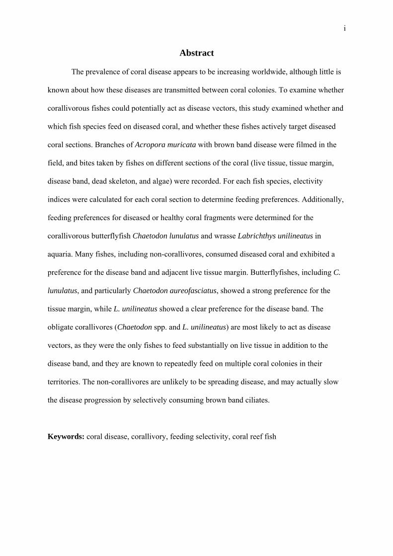
Abstract
The prevalence of coral disease appears to be increasing worldwide, although little is
known about how these diseases are transmitted between coral colonies. To examine whether
corallivorous fishes could potentially act as disease vectors, this study examined whether and
which fish species feed on diseased coral, and whether these fishes actively target diseased
coral sections. Branches of Acropora muricata with brown band disease were filmed in the
field, and bites taken by fishes on different sections of the coral (live tissue, tissue margin,
disease band, dead skeleton, and algae) were recorded. For each fish species, electivity
indices were calculated for each coral section to determine feeding preferences. Additionally,
feeding preferences for diseased or healthy coral fragments were determined for the
corallivorous butterflyfish Chaetodon lunulatus and wrasse Labrichthys unilineatus in
aquaria. Many fishes, including non-corallivores, consumed diseased coral and exhibited a
preference for the disease band and adjacent live tissue margin. Butterflyfishes, including C.
lunulatus, and particularly Chaetodon aureofasciatus, showed a strong preference for the
tissue margin, while L. unilineatus showed a clear preference for the disease band. The
obligate corallivores (Chaetodon spp. and L. unilineatus) are most likely to act as disease
vectors, as they were the only fishes to feed substantially on live tissue in addition to the
disease band, and they are known to repeatedly feed on multiple coral colonies in their
territories. The non-corallivores are unlikely to be spreading disease, and may actually slow
the disease progression by selectively consuming brown band ciliates.
Keywords: coral disease, corallivory, feeding selectivity, coral reef fish
ii
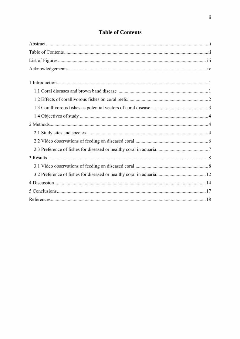
Table of Contents Abstract .......................................................................................................................................i
Table of Contents.......................................................................................................................ii
List of Figures .......................................................................................................................... iii
Acknowledgements...................................................................................................................iv
1 Introduction.............................................................................................................................1
1.1 Coral diseases and brown band disease ...........................................................................1
1.2 Effects of corallivorous fishes on coral reefs...................................................................2
1.3 Corallivorous fishes as potential vectors of coral disease ...............................................3
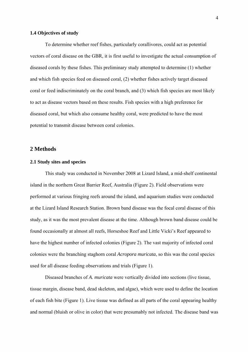
1.4 Objectives of study ..........................................................................................................4
2 Methods...................................................................................................................................4
2.1 Study sites and species.....................................................................................................4
2.2 Video observations of feeding on diseased coral.............................................................6
2.3 Preference of fishes for diseased or healthy coral in aquaria...........................................7
3 Results.....................................................................................................................................8
3.1 Video observations of feeding on diseased coral.............................................................8
3.2 Preference of fishes for diseased or healthy coral in aquaria.........................................12
4 Discussion .............................................................................................................................14
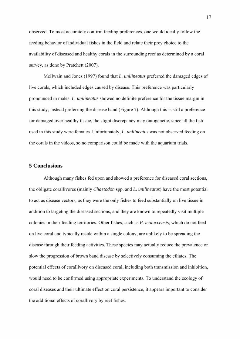
5 Conclusions...........................................................................................................................17
References................................................................................................................................18
iii
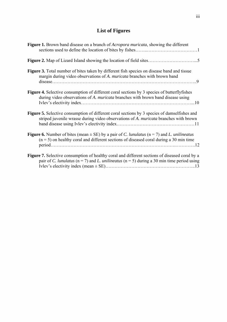
List of Figures Figure 1. Brown band disease on a branch of Acropora muricata, showing the different
sections used to define the location of bites by fishes……..……………………………1 Figure 2. Map of Lizard Island showing the location of field sites…………………………...5 Figure 3. Total number of bites taken by different fish species on disease band and tissue
margin during video observations of A. muricata branches with brown band disease…………………………………………………………………………………..9
Figure 4. Selective consumption of different coral sections by 3 species of butterflyfishes
during video observations of A. muricata branches with brown band disease using Ivlev’s electivity index………………………………………………………………...10
Figure 5. Selective consumption of different coral sections by 3 species of damselfishes and
striped juvenile wrasse during video observations of A. muricata branches with brown band disease using Ivlev’s electivity index……………………………………………11
Figure 6. Number of bites (mean ± SE) by a pair of C. lunulatus (n = 7) and L. unilineatus
(n = 5) on healthy coral and different sections of diseased coral during a 30 min time period……………………………………………………………………….………….12
Figure 7. Selective consumption of healthy coral and different sections of diseased coral by a
pair of C. lunulatus (n = 7) and L. unilineatus (n = 5) during a 30 min time period using Ivlev’s electivity index (mean ± SE)…………………………………………………..13
iv
Acknowledgements Thank you foremost to my advisor Dr. Morgan Pratchett of James Cook University
and honors student Karen Chong-Seng, without whom this project could not exist. Their
knowledge of the study system and guidance with methodology and analysis was invaluable.
I thank the entire Pratchett lab – Morgan, Karen, Darren Coker, Rebecca Lawton, Andy Cole,
and others – for their assistance in data collection, including driving the boats, putting out the
cameras, collecting fish and coral, helping observe fish in the lab and field, providing advice
about methodology, and allowing me to accompany and assist them at various sites around
the island. I would gladly work with any of them again if given the chance. My immense
thanks to Drs. Anne Hoggett and Lyle Vail, Lizard Island Research Station directors, for
assisting and organizing my stay, for their incredible helpfulness and hospitality, and for
allowing me some of the most amazing and unforgettable marine experiences of my life.
Thank you to Marianne and Lance Pearce, LIRS caretakers, for keeping the station running
fabulously, and to all of the Lizard Island researchers for their interest, company, and
hospitality. I cannot think of a more fabulous place to conduct an ISP. I learned an incredible
amount about coral reef ecological research and am so glad I was able to spend time here. I
hope I will be able to return someday. Thank you also to Tony Cummings, SIT Cairns
academic director, for his support, logistical assistance, and for organizing and running this
great program.
1
1 Introduction
1.1 Coral diseases and brown band disease
The prevalence of coral disease appears to be increasing worldwide, perhaps because
of the increasing environmental stresses corals are facing (Willis et al. 2004). In the
Caribbean, coral diseases are a major cause of reef deterioration, known to decrease coral
abundance and reproductive potential and to change community composition (Boyett 2006).
Much less in known about the effects of coral disease in the Indo-Pacific, including the Great
Barrier Reef (GBR), where disease has only recently become an important area of research.
Coral diseases can be caused by a range of fungi, bacteria, cyanobacteria, and
protozoans, and of the more 29 described coral diseases, very few have been examined in
detail (Willis et al. 2004). On the GBR, the most common scleractinian (hard) coral diseases
are black band disease (caused by filamentous cyanobacteria), skeletal eroding band disease
(caused by the ciliate Halofolliculina corallasia), brown band disease (also caused by a
ciliate), and white syndrome (multiple potential causes) (Willis et al. 2004).
Brown band
disease was first described
from the GBR in 2004,
and appears as a brown
band on the coral sur
bordered on one side by
healthy tissue, and the
other side by white, dead
skeleton (Willis et al.
2004, Figure 1). The band
moves along the branch in Figure 1. Brown band disease on a branch of Acropora muricata, showing the different sections used to define the location of bites by fishes. (Photo: Morgan Pratchett)
face
2
the direction of the healthy tissue at variable but potentially rapid rates of 0.3 to 6.1 cm/day in
the northern GBR (Boyett 2006). The brown color of the band comes from a high density of
ciliates (class Oligohymenophora, subclass Scuticociliatia), which are filled with
zooxanthellae from coral tissue they have consumed (Boyett 2006). In addition to the ciliates,
an array of bacteria are associated with brown band disease that may compromise the coral
tissue before the ciliates invade (Boyett 2006, Bourne et al. 2008). Not much is known about
the disease, including how it is transmitted, and how it affects interactions between the corals
and the fish that feed on them.
1.2 Effects of corallivorous fishes on coral reefs
The impact of corallivory, or the consumption is live coral, is predicted to increase as
coral cover decreases in response to stressors such as coral disease, as well as rising water
temperature and bleaching events, increased storm intensity, pollution, sedimentation, and
eutrophication (Rotjan & Lewis 2008). Although corallivory by fishes was not considered
important historically, as it often causes little apparent damage to reefs, chronic tissue
removal by fish can be energetically costly to prey corals and have a significant influence on
their distribution, abundance, growth, fitness, and competitive ability (Cole et al. 2008). In
addition to directly affecting coral condition by mechanical damage and tissue removal,
corallivory can have indirect effects on coral colonies, including the facilitation of algal
competitors, boring organisms, or disease pathogens (Rotjan & Lewis 2008). Synergistic
effects with other stressors may also have important consequences for corals. For instance,
the recovery of corals affected by a bleaching or storm event can be highly impaired by
predation (Cole et al. 2008). In turn, declines in coral cover can negatively affect populations
of corallivorous fishes, especially obligate feeders (Cole et al. 2008).
Fishes in 11 families are known to consume coral, and the damage they inflict on
coral colonies varies with the amount of coral tissue and skeleton the fish removes (Rotjan &

3
Lewis 2008). Corallivorous fishes can be obligate of facultative, and most target scleractinian
corals. Fish are often very selective in the corals they consume (Pratchett 2005, 2007),
perhaps because of differences in coral morphology, physical or chemical defenses, or
nutriment (Cole et al. 2008). Some fish species have a known preference for physically
damaged coral, perhaps because of increased mucous production (Pratchett 2005, McIlwain
& Jones 1997), but the preference of fishes for other types of stressed corals, such as those
with disease, and the consequences for those corals, has not been examined in depth.
1.3 Corallivorous fishes as potential vectors of coral disease
Many coral diseases, such as black band disease, are known to be spread by direct
contact and possibly by prevailing currents, but animal vectors have yet to be thoroughly
investigated (Aeby & Santavy 2006). Corallivorous fish, snails, worms, and nudibranchs
have been recently suggested as potential vectors of coral bacterial infection (Rotjan & Lewis
2008). Butterflyfishes (family Chaetodontidae), which comprise 69 of the 128 known
corallivorous fish species (Cole et al. 2008), are the most well studied corallivorous fishes
and are likely candidates for disease vectors. Butterflyfishes feed on corals in home territories
by removing coral tissue with fleshy, pointed mouths, generally without damaging the coral
skeleton (Rotjan & Lewis 2008). In Florida, the butterflyfish Chaetodon capistratis was
observed feeding on coral with black band disease, and the presence of a C. capistratis
individual enabled infection to spread from diseased to healthy coral fragments in the same
aquarium, suggesting oral and/or fecal transmission (Aeby and Santavy 2006). Since
corallivorous fishes are known to feed preferentially on damaged coral (Pratchett 2005,
McIlwain & Jones 1997), they may also prefer diseased tissue or tissue on the edge of disease
band. After feeding on diseased coral, a fish may be able to transmit the pathogen to other
colonies (particularly damaged or stressed parts of non-diseased corals, which may be more
prone to infection) on which it subsequently feeds or defecates.
4
1.4 Objectives of study
To determine whether reef fishes, particularly corallivores, could act as potential
vectors of coral disease on the GBR, it is first useful to investigate the actual consumption of
diseased corals by these fishes. This preliminary study attempted to determine (1) whether
and which fish species feed on diseased coral, (2) whether fishes actively target diseased
coral or feed indiscriminately on the coral branch, and (3) which fish species are most likely
to act as disease vectors based on these results. Fish species with a high preference for
diseased coral, but which also consume healthy coral, were predicted to have the most
potential to transmit disease between coral colonies.
2 Methods
2.1 Study sites and species
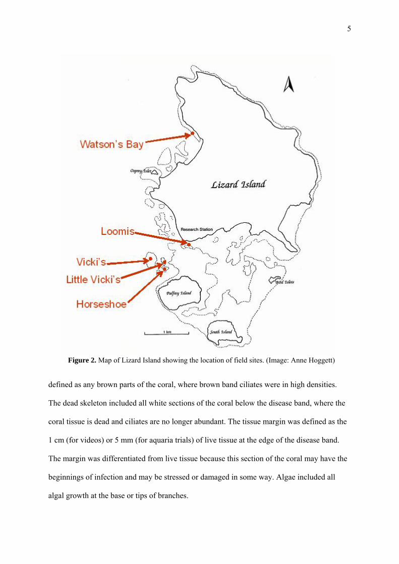
This study was conducted in November 2008 at Lizard Island, a mid-shelf continental
island in the northern Great Barrier Reef, Australia (Figure 2). Field observations were
performed at various fringing reefs around the island, and aquarium studies were conducted
at the Lizard Island Research Station. Brown band disease was the focal coral disease of this
study, as it was the most prevalent disease at the time. Although brown band disease could be
found occasionally at almost all reefs, Horseshoe Reef and Little Vicki’s Reef appeared to
have the highest number of infected colonies (Figure 2). The vast majority of infected coral
colonies were the branching staghorn coral Acropora muricata, so this was the coral species
used for all disease feeding observations and trials (Figure 1).
Diseased branches of A. muricata were vertically divided into sections (live tissue,
tissue margin, disease band, dead skeleton, and algae), which were used to define the location
of each fish bite (Figure 1). Live tissue was defined as all parts of the coral appearing healthy
and normal (bluish or olive in color) that were presumably not infected. The disease band was
5
Figure 2. Map of Lizard Island showing the location of field sites. (Image: Anne Hoggett)
defined as any brown parts of the coral, where brown band ciliates were in high densities.
The dead skeleton included all white sections of the coral below the disease band, where the
coral tissue is dead and ciliates are no longer abundant. The tissue margin was defined as the
1 cm (for videos) or 5 mm (for aquaria trials) of live tissue at the edge of the disease band.
The margin was differentiated from live tissue because this section of the coral may have the
beginnings of infection and may be stressed or damaged in some way. Algae included all
algal growth at the base or tips of branches.
6
Corallivorous butterflyfishes (family Chaetodontidae) and wrasses (family Labridae),
including Chaetodon aureofasciatus (golden-striped butterflyfish), Chaetodon baronessa
(triangular butterflyfish), Chaetodon lunulatus (redfin butterflyfish), Chaetodon plebeius
(bluespot butterflyfish), Chaetodon rainfordi (Rainford’s butterflyfish), and Labrichthys
unilineatus (tubelip wrasse) were observed to consume A. muricata and the diseased portion
of the coral during preliminary field observations. These fish species are all obligate
corallivores, and generally rove between coral colonies within a feeding territory (Pratchett
2005, McIlwain & Jones 1997), so these were the focal species of the study and primary
disease vector candidates. Territorial, herbivorous damselfishes (family Pomacentridae), such
as Stegastes spp., frequently defended algal turfs at the bases of A. muricata branches,
including infected colonies, and planktivorous damselfishes, such as Chromis atripectoralis
and Pomacentrus moluccensis, often swam above and within the branches.
2.2 Video observations of feeding on diseased coral
To determine whether and which fish species feed on diseased coral, and whether
these fishes actively target the diseased coral sections, diseased branches of A. muricata
(n = 17) were located and filmed at Horseshoe Reef, Little Vicki’s Reef, Vicki’s Reef, and
Loomis Reef (Figure 2). A video camera (Sony Handycam DCR-SR300E) in underwater
housing was positioned approximately 0.5 m from the diseased branch so the disease band
and surrounding healthy branches were in view. The camera was left to record unattended for
at least 70 minutes. The first 10 minutes of footage were ignored to allow fish to acclimatize
to the presence of the camera. For the next 60 minutes of footage, all fishes seen feeding on
the coral were identified, and the number of bites taken by fishes on each section of the coral
was recorded (Figure 1). Live tissue included the healthy coral on the diseased branch and on
healthy neighboring branches. Bites were only recorded when the location of the bite was
clear and within the frame of the camera.
7
Preference or avoidance of each section of the coral was calculated for each fish
species using Ivlev’s electivity index (McIlwain & Jones 1997), which is defined as:
E = (r – p) / (r + p)
where r is the proportion of a food type consumed and p is the proportion of this food type
available in the environment. E values range from -1 to +1. A value of 0 indicates no
selection (proportion consumed equal to proportion available), positive values indicate
preference, and negative values indicate avoidance. E values were calculated for all videos
combined, rather than each video, because of high variation in feeding rates. Total number of
bites on each section was used to obtain the proportional consumption by each fish species. E
values were only calculated for fishes that took more than 30 bites in total. Proportional
availability of each coral section was calculated by placing a transparent grid of evenly-
spaced points on the viewing screen, and counting the number of points intersecting each
section for each video. There were 558 points covering the entire viewing screen.
Background areas and background branches not examined for bites were excluded from the
count. The number of points in each section was added across all videos to obtain the
proportion available.
2.3 Preference of fishes for diseased or healthy coral in aquaria
To supplement observations from the video recordings, the feeding preferences of two
obligate corallivores, C. lunulatus and L. unilineatus, were also measured in aquaria. C.
lunulatus and L. unilineatus individuals were collected from Watson’s Bay (Figure 2) using
barrier nets and clove oil, and then kept in large tanks for 6-8 days before the experiment with
an ample supply of A. muricata and Pocillopora damicornis for food. One day before the
experiment, diseased and healthy fragments of A. muricata approximately 10-15 cm in length
were collected from Loomis Reef (Figure 2). Two healthy fragments or two diseased
fragments were affixed upright to the opposite corners of a 10 × 10 cm tile with plasticine
8
clay and superglue. Healthy fragments were kept in tanks in the lab, and diseased fragments
were kept on racks near Loomis Reef before retrieval. No algae were present on any of the
fragments.
Fish were starved for at least 2 hours, and then a pair of C. lunulatus (n = 7) or L.
unilineatus (n = 5) was placed into one side of a glass aquarium (60 × 26 × 38 cm, 59 l) with
flow-through seawater divided in half by a removable partition. Fish were paired because
they typically do not feed when alone (Pratchett, personal comm.). A tile with diseased coral
fragments and a tile with healthy coral fragments were placed into the other half of the tank.
Fish were allowed to acclimate for at least one hour before the partition was removed and the
tiles with corals were moved to opposite ends of the tank. All bites on the corals taken by
both fish (as a pair) were recorded for 30 minutes starting at the first bite. The number of
bites and the location of each bite was recorded (Figure 1).
Preference for each section of coral was calculated using Ivlev’s electivity index (see
Section 2.2). E values were calculated for each pair of fish and then averaged for the species.
Proportional consumption was based on the number of bites taken from each section. The
vertical length (mm) of each section relative to the length of the coral fragment was used to
determine the proportional availability. Lengths from the two corals on each tile were
combined. Measuring only the linear dimensions did not consider the absolute surface area of
each section available to the fish, but provided an approximate index.
3 Results
3.1 Video observations of feeding on diseased coral
A total of 15 identifiable fish species from 4 families were observed feeding on any
part of the coral branch: 5 butterflyfish (Chaetodontidae), 8 damselfish (Pomacentridae), 1
wrasse (Labridae), and 1 leatherjacket (Monacanthidae). 12 species took at least one bite
9
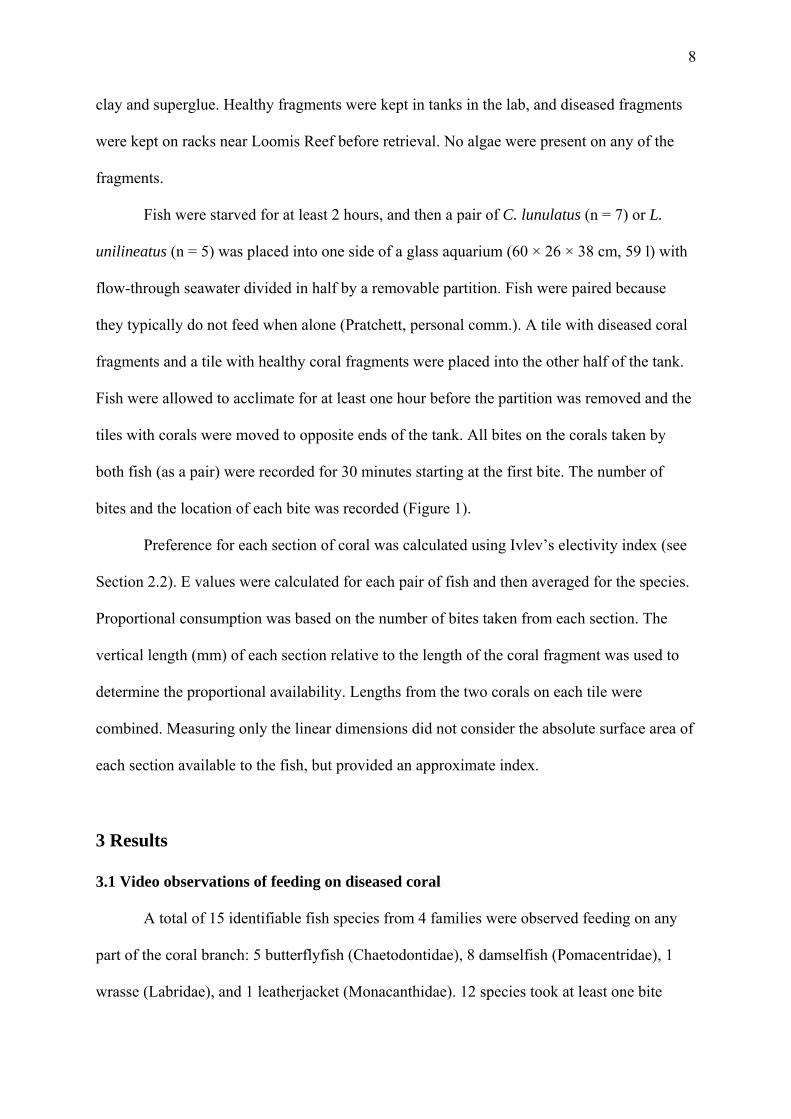
from the disease band or tissue margin (the diseased sections). Only 6 species took more than
a total of 30 bites.
The two species with the most number of bites on the disease band and tissue margin
were Pomacentrus moluccensis and C. aureofasciatus (Figure 3). P. moluccensis took more
bites on the disease band than the tissue margin, while C. aureofasciatus did the opposite.
Unidentified, juvenile striped wrasses took the third most number of bites on these two
sections, followed by Stegastes spp., Cheiloprion labiatus, C. lunulatus, and C. rainfordi.
Juvenile wrasse took almost no bites from the tissue margin.
0
50
100
150
200
250
300
350
Pomac
entru
s molu
ccen
sis
Chaeto
don a
ureofa
sciat
us
juven
ile st
riped
wras
se
Segas
tes sp
p.
Cheilo
prion
labia
tus
Chaeto
don l
unula
tus
Chaeto
don r
ainfor
di
Chaeto
don b
aron
essa
Halich
oeres
mela
nurus
Chaeto
don p
lebeiu
s
tota
l num
ber o
f bite
s disease bandtissue margin
The proportional availability of each coral section was 67% live tissue, 20% algae,
8% dead skeleton, 4% disease band, and 1% tissue margin. All fish species for which
electivity indices were calculated showed selective consumption of different sections. The
Figure 3. Total number of bites taken by different fish species on disease band and tissue margin during video observations of A. muricata branches with brown band disease. Data were combined from 17 different videos, each 1 hour long.
10
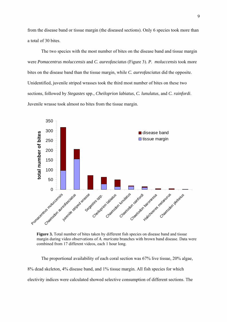
three butterflyfish C. aureofasciatus, C. rainfordi, and C. lunulatus all exhibited a strong
preference for the tissue margin (E ≈ 0.9), and avoidance of dead skeleton and algae
(E < -0.5) (Figure 4). Of the three butterflyfish, C. aureofasciatus showed the most
preference for the disease band and the least preference for live tissue, followed by C.
rainfordi, and then C. lunulatus, which showed the least preference for the disease band and
the most preference for live tissue.
Chaetodon aureofasciatus
-1
-0.5
0
0.5
1
live tissue tissuemargin
diseaseband
deadskeleton
algae
Chaetodon rainfordi
-1
-0
0.5
.5
0
1
live tissue tissuemargin
diseaseband
deadskeleton
algaeChaetodon lunulatus
-1
-0.5
0
0.5
1
live tissue tissuemargin
diseaseband
deadskeleton
algae
Ivle
v’s E
lect
ivity
Inde
x (E
)
Figure 4. Selective consumption of different coral sections by 3 species of butterflyfishes during video observations of A. muricata branches with brown band disease using Ivlev’s electivity index. Positive values indicate preference and negative values indicate avoidance. Data were combined from 17 different videos, each 1 hour long.
11
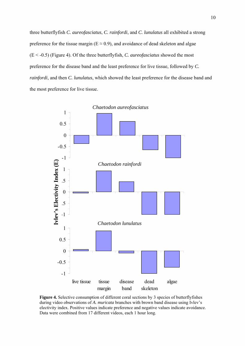
Three damselfish species all showed a strong preference for the tissue margin and
disease band, and avoidance of live tissue (Figure 5). Cheiloprion labiatus (biglip damsel), a
corallivore, additionally preferred the dead skeleton and avoided algae. Conversely, the
herbivorous Stegastes spp. preferred algae and avoided the dead skeleton. P. moluccensis, a
planktivore, avoided both the dead skeleton and algae. Juvenile striped wrasses tended to
Ivle
v’s E
lect
ivity
Inde
x (E
)
Cheiloprion labiatus
-1
-0.5
0
0.5
1
live tissue tissuemargin
diseaseband
deadskeleton
algaeSegastes spp.
-1
-0.5
0
0.5
1
live tissue tissuemargin
diseaseband
deadskeleton
algaePomacentrus moluccensis
-1
-0.5
0
0.5
1
live tissue tissuemargin
diseaseband
deadskeleton
algaejuvenile striped wrasse
-1
-0.5
0
0.5
1
live tissue tissuemargin
diseaseband
deadskeleton
algae
Figu
re 5
. Se
lect
ive
cons
umpt
ion
of d
iffer
ent c
oral
sect
ions
by
3 sp
ecie
s of d
amse
lfish
es a
nd st
riped
ju
veni
le w
rass
e du
ring
vide
o ob
serv
atio
ns o
f A. m
uric
ata
bran
ches
with
bro
wn
band
dis
ease
usi
ng
Ivle
v’s e
lect
ivity
inde
x. P
ositi
ve v
alue
s ind
icat
e pr
efer
ence
and
neg
ativ
e va
lues
indi
cate
avo
idan
ce.
Dat
a w
ere
com
bine
d fr
om 1
7 di
ffer
ent v
ideo
s, ea
ch 1
hou
r lon
g.
12
avoid live tissue and tissue margin, preferring the disease band, dead skeleton, and algae.
Gobies (probably Eviota sp.) were often observed sitting on and biting the disease
band and tissue margin in the videos. It was difficult to count the number of bites taken, and
difficult to spot the gobies on healthy branches for comparison, so they were not included in
the electivity analysis, but they appeared to have a strong preference for the diseased sections.
3.2 Preference of fishes for diseased or healthy coral in aquaria
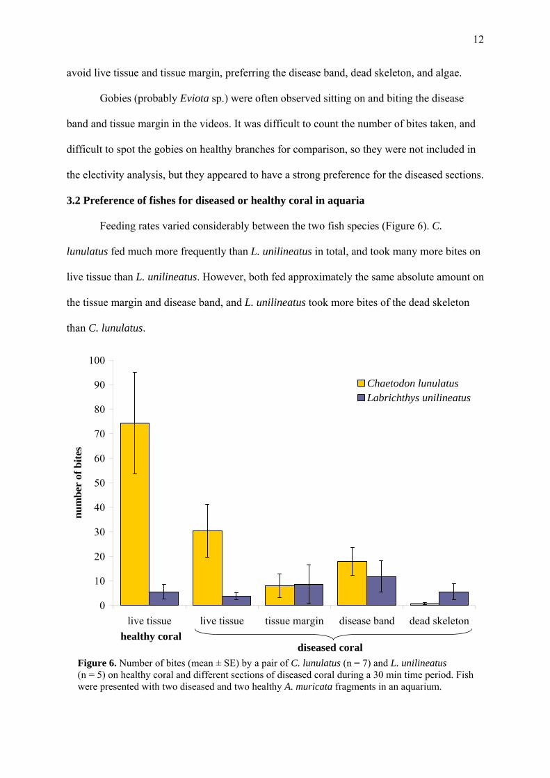
Feeding rates varied considerably between the two fish species (Figure 6). C.
lunulatus fed much more frequently than L. unilineatus in total, and took many more bites on
live tissue than L. unilineatus. However, both fed approximately the same absolute amount on
the tissue margin and disease band, and L. unilineatus took more bites of the dead skeleton
than C. lunulatus.
0
10
20
30
40
50
60
70
80
90
100
live tissue live tissue tissue margin disease band dead skeleton
num
ber
of b
ites
Chaetodon lunulatusLabrichthys unilineatus
healthy coraldiseased coral
Figure 6. Number of bites (mean ± SE) by a pair of C. lunulatus (n = 7) and L. unilineatus (n = 5) on healthy coral and different sections of diseased coral during a 30 min time period. Fish were presented with two diseased and two healthy A. muricata fragments in an aquarium.
13
Chaetodon lunulatus
-1
-0.8
-0.6
-0.4
-0.2
0
0.2
0.4
0.6
0.8
1
live tissue live tissue tissue margin disease band dead skeleton
Ivle
v's E
lect
ivity
Inde
x (E
)
healthy coraldiseased coralLabrichthys unilineatus
-1
-0.8
-0.6
-0.4
-0.2
0
0.2
0.4
0.6
0.8
1
live tissue live tissue tissue margin disease band dead skeleton
Ivle
v's E
lect
ivity
Inde
x (E
)
healthy coraldiseased coral
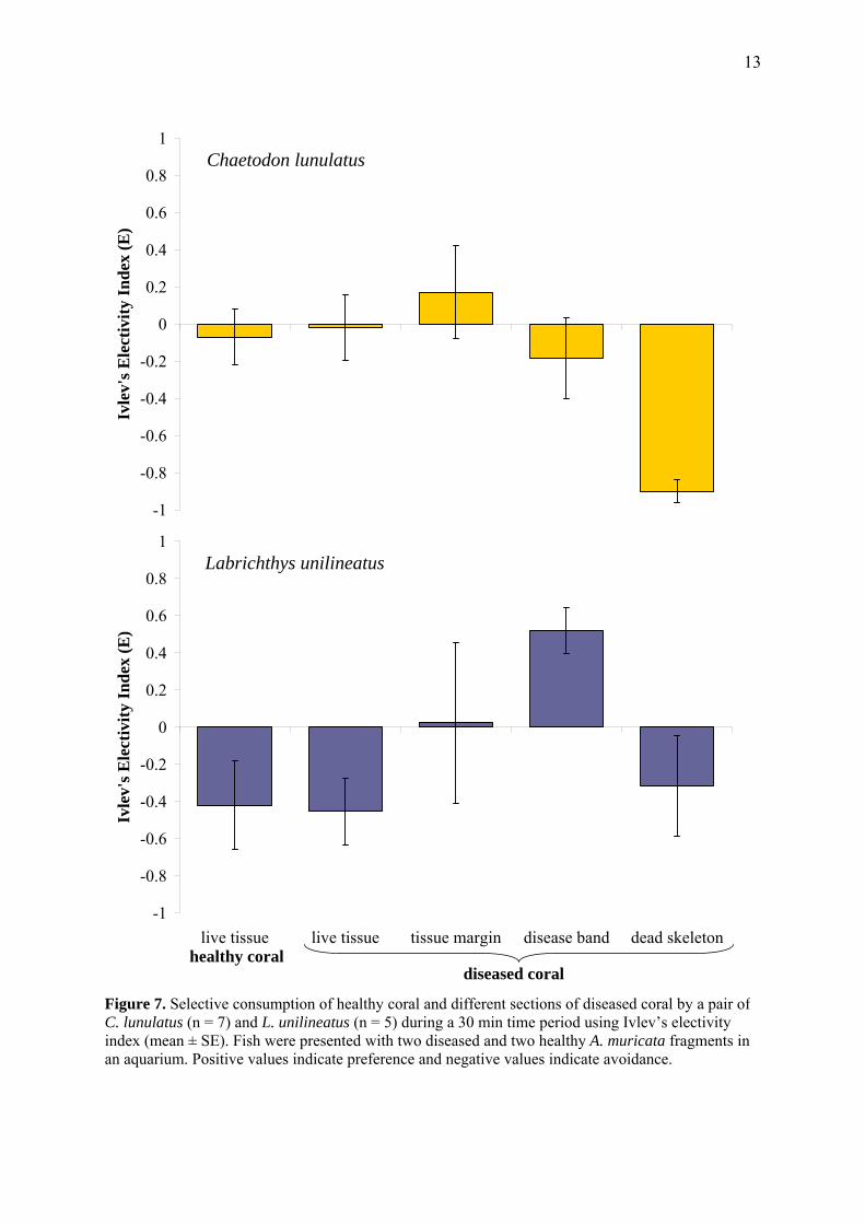
Figure 7. Selective consumption of healthy coral and different sections of diseased coral by a pair of C. lunulatus (n = 7) and L. unilineatus (n = 5) during a 30 min time period using Ivlev’s electivity index (mean ± SE). Fish were presented with two diseased and two healthy A. muricata fragments in an aquarium. Positive values indicate preference and negative values indicate avoidance.
14
The proportional availability of each coral section was 20% live tissue on healthy
coral, 15% live tissue on diseased coral, 18% disease band, 16% dead skeleton, and 2% tissue
margin. C. lunulatus showed preferences in the aquarium trials (Figure 7) similar to those it
exhibited in the video observations (Figure 4). It had a close to neutral preference for live
tissue, both on healthy and diseased coral, a slight avoidance of the disease band, and a strong
avoidance of the dead skeleton. C. lunulatus had a potential preference for the tissue margin,
although it was not as extreme as in the video observations (E = 0.17 ± 0.25 as opposed to
0.88). Because of variation in the data from the aquarium trials, however, zero is included in
the SE range for all sections except the dead skeleton.
The preferences of L. unilineatus were very different from those of C. lunulatus
(Figure 7). L. unilineatus avoided live tissue on both healthy and diseased coral, and may or
may not prefer the tissue margin, as there was large variation. Like C. lunulatus, it avoided
the dead skeleton, but unlike C. lunulatus, it showed a definite preference for the disease
band.
4 Discussion
Most fish species, including non-corallivores, took bites from the disease band and/or
tissue margin and showed a preference for at least one of these two sections. However, it was
only corallivores (Chaetodon spp. and L. unilineatus) that appeared to feed on live tissue in
addition to these diseased sections, and so would be mostly likely to spread disease to healthy
corals. The disease band is known to contain the brown band ciliates (Boyett 2006), and
fishes may be picking them up by feeding on this section. The fish might then transmit the
pathogens to other corals they feed on, as found by Aeby and Santavy (2006). By feeding on
the tissue margin, which may have the onset of infection by ciliates or associated bacteria, the
fish may also pick up pathogens, or the fish might further stress this tissue and increase the

15
rate of progression of the disease. It is also possible that by feeding on the disease band, the
fish (including non-corallivores) could remove the disease from the coral. In Hawaii, the
butterflyfish Chaetodon multicinctus preferentially fed on coral polyps infected with a
parasitic trematode (Aeby 2002). Feeding removed the parasite from a colony, but fish then
propagated the parasite to more colonies through their feces. Oral and fecal transmission, and
the effect consuming the disease band or margin on the coral itself, are all worthy of future
investigation.
The preference of fishes for the disease band and tissue margin is consistent with
other studies showing a preference for damaged corals (Pratchett 2005, McIlwain & Jones
1997), but why fishes prefer these diseased sections is uncertain. It has been suggested that
damaged coral tissue emits olfactory attractants or increases mucous production in a way that
makes it more appealing to fish (McIlwain & Jones 1997). Perhaps the ciliates themselves
offer some nutritional value, or the damaged tissue is easier to remove.
It is interesting that many non-corallivores, such as P. moluccensis, fed so frequently
on the disease band and tissue margin (Figure 5). Perhaps they were targeting the ciliates, as
opposed to the coral tissue, which would be interesting to examine in the laboratory. It is
possible that the small gobies and juvenile wrasse seen in the videos were consuming ciliates,
as both fish seemed to bite preferentially on the disease band with their tiny mouths. The
herbivorous Stegastes spp. were naturally one of the only fishes to prefer algae, but it was
unexpected that they also preferred the diseased sections. Perhaps they thought the brown
band resembled algae, or were biting at potentially weakened coral tissue to try and promote
algal growth. Because many of the damselfishes and other non-corallivores did not consume
live coral, and often resided within well-defended territories in a single coral colony, these
species are not likely to be transmitting disease. It is possible they may even reduce the
prevalence of disease by selectively feeding on and removing the ciliates.
16
The corallivores (Chaetodon spp. and L. unilineatus) are known to feed repeatedly on
the multiple colonies within their feeding territories (Pratchett 2005, McIlwain & Jones
1997), which makes them more likely to transmit disease between colonies. To better
understand which of these corallivores are most capable of acting as disease vectors, it would
be useful to investigate the number of coral colonies and coral species visited by these fishes
after feeding on the diseased coral. Assuming fishes can act as oral or fecal disease vectors
after feeding on a diseased colony in their territory, fishes that then visit a large number of
different coral species and colonies would have the most potential to spread the disease. Of
particular interest would be the number of different A. muricata colonies a fish feeds from,
since this species appears most susceptible to disease. Why A. muricata is affected so much
by brown band disease is another subject that could be examined. Being a rapidly growing
coral species, perhaps it has fewer defenses against damage and infection.
Of the butterflyfishes, C. aureofasciatus has the most potential to be a disease vector.
Not only did this species take the most bites of the disease band and tissue margin (Figure 3),
it also showed the most preference for the disease band relative to live tissue (Figure 4). C.
rainfordi would follow as the next most likely vector, as it showed the next most preference
for the disease band relative to live tissue, and then C. lunulatus (Figure 4). The other
butterflyfishes observed feeding on diseased coral, C. baronessa and C. plebeius, fed only
rarely on the diseased sections in the videos (Figure 3), so it was not possible to determine
their feeding preferences and potential to transmit disease. It is likely that their preferences
are similar to those of the other butterflyfishes.
Although the feeding preferences of C. lunulatus were similar for video and aquarium
observations, suggesting reliability, it is possible that both trials were biased. Diseased coral
was likely represented in higher proportions than would be available to the fish in its natural
environment. The feeding behavior of fishes away from the diseased branches was not
17
observed. To most accurately confirm feeding preferences, one would ideally follow the
feeding behavior of individual fishes in the field and relate their prey choice to the
availability of diseased and healthy corals in the surrounding reef as determined by a coral
survey, as done by Pratchett (2007).
McIlwain and Jones (1997) found that L. unilineatus preferred the damaged edges of
live corals, which included edges caused by disease. This preference was particularly
pronounced in males. L. unilineatus showed no definite preference for the tissue margin in
this study, instead preferring the disease band (Figure 7). Although this is still a preference
for damaged over healthy tissue, the slight discrepancy may ontogenetic, since all the fish
used in this study were females. Unfortunately, L. unilineatus was not observed feeding on
the corals in the videos, so no comparison could be made with the aquarium trials.
5 Conclusions
Although many fishes fed upon and showed a preference for diseased coral sections,
the obligate corallivores (mainly Chaetodon spp. and L. unilineatus) have the most potential
to act as disease vectors, as they were the only fishes to feed substantially on live tissue in
addition to targeting the diseased sections, and they are known to repeatedly visit multiple
colonies in their feeding territories. Other fishes, such as P. moluccensis, which do not feed
on live coral and typically reside within a single colony, are unlikely to be spreading the
disease through their feeding activities. These species may actually reduce the prevalence or
slow the progression of brown band disease by selectively consuming the ciliates. The
potential effects of corallivory on diseased coral, including both transmission and inhibition,
would need to be confirmed using appropriate experiments. To understand the ecology of
coral diseases and their ultimate effect on coral persistence, it appears important to consider
the additional effects of corallivory by reef fishes.
18
References
Aeby GS. 2002. Trade-offs for the butterflyfish Chaetodon multicinctus, when feeding on coral prey infected with trematode metacercariae. Behavioral Ecology and Sociobiology 52:158-165.
Aeby GS, Santavy DL. 2006. Factors affecting susceptibility of the coral Montastrea
faveolata to black-band disease. Marine Ecology Progress Series 318:103-110. Bourne DG, Boyett HV, Henderson ME, Muirhead A, Willis BL. 2008. Identification of a
ciliate (Oligohymenophora: Scuticociliatia) associated with brown band disease on corals of the Great Barrier Reef. Applied and Environmental Microbiology 74:883-888.
Boyett HV. 2006. The ecology and microbiology of black band disease and brown band
syndrome on the Great Barrier Reef. M.Sc. thesis, James Cook University. Cole AJ, Pratchett MS, Jones GP. 2008. Diversity and functional importance of coral feeding
fishes on tropical coral reefs. Fish and Fisheries 9:286-307. McIlwain JL, Jones GP. 1997. Prey selection by an obligate coral-feeding wrasse and its
response to small-scale disturbance. Marine Ecology Progress Series 155: 189-198. Pratchett MS. 2005. Dietary overlap among coral-feeding butterflyfishes (Chaetodontidae) at
Lizard Island, northern Great Barrier Reef. Marine Biology 148:373-382. Pratchett MS. 2007. Dietary selection by coral-feeding butterflyfishes (Chaetodontidae) on
the Great Barrier Reef, Australia. The Raffles Bulletin of Zoology 14:171-176. Rotjan RD, Lewis SM. 2008. Impact of coral predators on tropical reefs. Marine Ecology
Progress Series 367:73-91. Willis BL, Page CA, Dinsdale EA. 2004. Coral disease on the Great Barrier Reef. In
Rosenberg E, Loya Y (eds) Coral heath and disease, p.69-104.