copyright © the mcgraw-hill companies, inc. permission
TRANSCRIPT

1
2

Copyright © The McGraw-Hill Companies, Inc. Permission required for reproduction or display.
Diencephalon
Mesencephalon
Telencephalon Forebrain
Pons
Cerebellum Metencephalon
Spinal cord
Hindbrain
Telencephalon
Optic vesicle
Diencephalon
Metencephalon
Myelencephalon
Spinal cord
Rhombencephalon
Mesencephalon
Prosencephalon
(a) 4 weeks (b) 5 weeks
(c) Fully developed
Midbrain
Myelencephalon (medulla oblongata)
3
4

Copyright © The McGraw-Hill Companies, Inc. Permission required for reproduction or display.
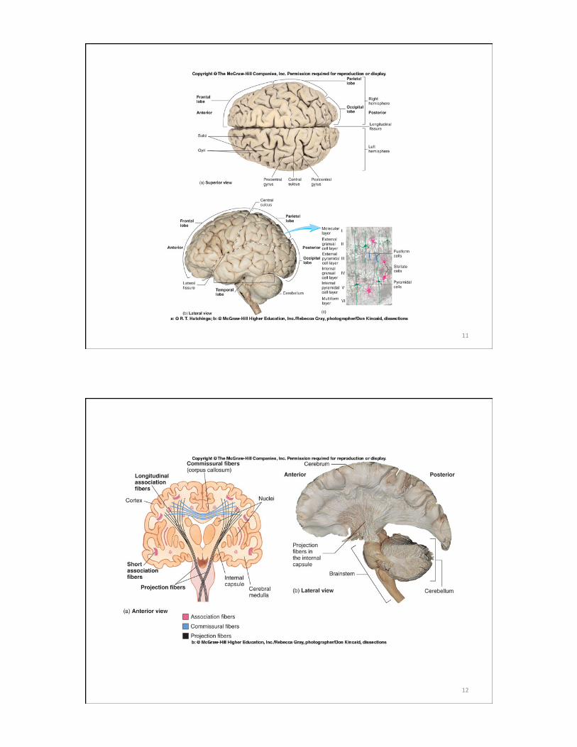
Brainstem
Cerebellum
Cerebrum
Spinal cord
Frontal lobe
Rostral Caudal
Occipital lobe
Central sulcus
Longitudinal fissure
Parietal lobe
Central sulcus
Lateral sulcus Gyri
(a) Superior view (b) Lateral view
Central sulcus
Frontal lobe
Insula
Occipital lobe
Cerebellum
(c) Lateral view
Parietal lobe
Blood vessels
Cerebral hemispheres
Temporal lobe
Postcentral gyrus
Arachnoid mater
Medulla oblongata
Precentral gyrus
Temporal lobe
c: © The McGraw-Hill Companies, Inc./Rebecca Gray, photographer/Don Kincaid, dissections
5
6

7
8

9
10
11
12
13
14

Copyright © The McGraw-Hill Companies, Inc. Permission required for reproduction or display.
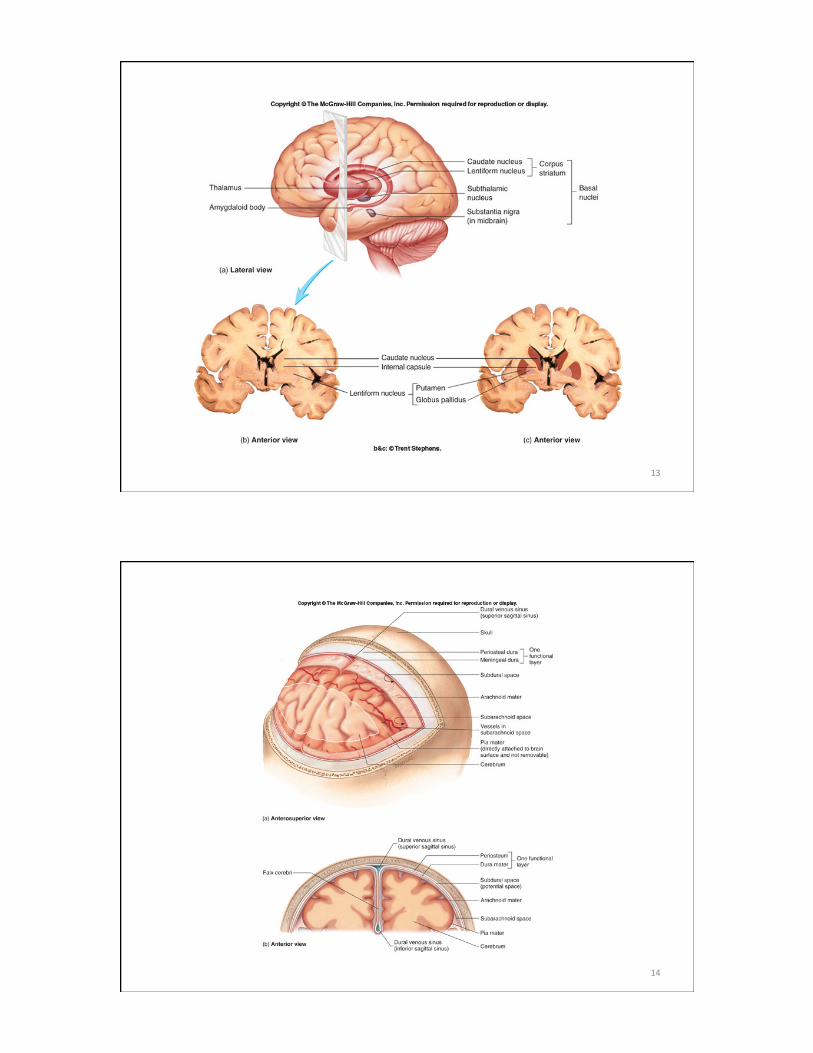
Subdural space
Skull
Pia mater
Blood vessel
Dura mater: Periosteal layer Meningeal layer
Arachnoid mater
Brain: Gray matter White matter
Arachnoid villus Subarachnoid space
Superior sagittal sinus
Falx cerebri (in longitudinal fissure only)
15
Copyright © The McGraw-Hill Companies, Inc. Permission required for reproduction or display.
Lateral ventricles
Central canal
Lateral aperture Fourth ventricle
Third ventricle
Median aperture
(a) Lateral view
Caudal
Interventricular foramen
Cerebral aqueduct
Rostral
16

Copyright © The McGraw-Hill Companies, Inc. Permission required for reproduction or display.
Lateral ventricle
Third ventricle
Cerebrum
Lateral aperture
Fourth ventricle
Median aperture
(b) Anterior view
Interventricular foramen
Cerebral aqueduct
17
Copyright © The McGraw-Hill Companies, Inc. Permission required for reproduction or display.
Choroid plexus in fourth ventricle adds more CSF.
CSF flows out two lateral apertures and one median aperture.
CSF fills subarachnoid space and bathes external surfaces of brain and spinal cord.
At arachnoid villi, CSF is reabsorbed into venous blood of dural venous sinuses.
1
2
3
4
5 6
7
7
8
1
2
3
4
5
6
7
8
CSF is secreted by choroid plexus in each lateral ventricle.
CSF flows through Interventricular foramina into third ventricle.
Choroid plexus in third ventricle adds more CSF.
CSF flows down cerebral aqueduct to fourth ventricle.
Arachnoid villus
Superior sagittal sinus
Arachnoid mater
Subarachnoid space
Dura mater
Choroid plexus
Third ventricle
Cerebral aqueduct
Lateralaper ture
Fourth ventricle
Median aperture
Centralcanal of spinal cord
Subarachnoid space of spinal cord
18

19
20

21
22

23
24

25
26

27
28

29
30

31