copyright © the mcgraw-hill companies, inc.. chapter 10 and 11 nervous system test preparation
TRANSCRIPT

Copyright © The McGraw-Hill Companies, Inc..
Chapter 10 and 11
Nervous System
Test Preparation

Know the basic structure of a neuron
Neurons are the structural and functional units.
Neuroglial Cells surround the neuron
Dendrites receive input
Axons (nerve fibers) carry information away from the cell as nerve impulses

Know the direction and function of sensory and motor neurons.
What is efferent and afferent?
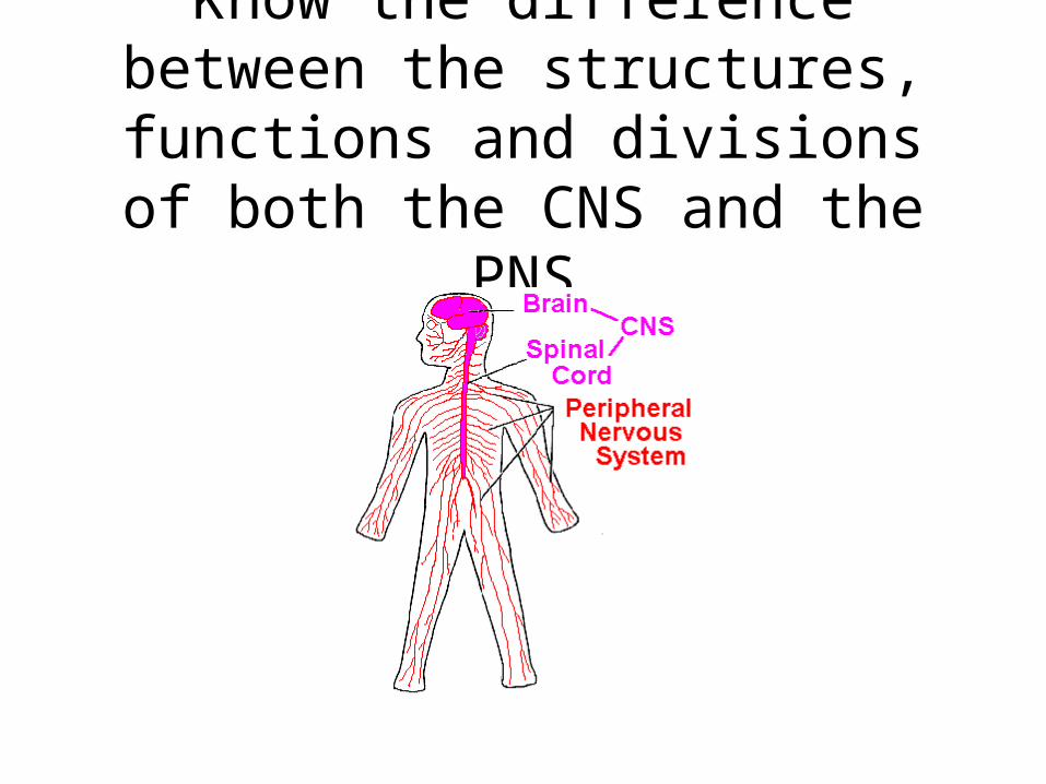
Know the difference between the structures, functions and divisions of
both the CNS and the PNS

Levels of Organization of the NERVOUS SYSTEM
CNS (Brain and Spinal Chord)
(Interneurons)
PNS(Cranial Nerves &
Spinal Nerves)
Sensory(Input into CNS)
(Afferent Neurons)
Motor (Output from CNS)(Efferent Neurons)
Somatic(Effectors: Skeletal Muscle)
(Conscious Control)
Autonomic(Effectors: Smooth Muscle,
Cardiac Muscle, Glands)(Unconscious Control)
Parasympathetic(Homeostasis)
(NT: Acetylcholine)
Sympathetic(Fight or Flight)
(NT: Norepinephrine)

Myelination of Axons• Know how and why
certain neurons are myelinated.
• Know what cell are involved in myelination.
• Know the structure of a myelinated neuron.

Classification of Neurons – Functional Classification
Know the three different types of neurons, where they work and how they work.

Classification of Neuroglial Cells
•What is a neuroglial cell?•What are the 4 types of neuroglial cells?

The Synapse•Know the structure and basic functions of a synapse.
•What are the functions of neurotransmitters?
•How and under what circumstances are they released?

Nerve Impulse
• What is RMP?• What ions are involved?• What is an all or nothing
response?• What is hyperpolarization
and depolarization?• What is summation?• What is a refractory
period?

Synaptic Potentials•What is an Excitatory Postsynaptic Potential (EPSP) and what happens to the postsynaptic neuron?
•What is an Inhibitory Postsynaptic Potential (IPSP) and what happens to the postsynaptic neuron?

12
Meninges
•What are the 3 layers of the meniges?•What are the locations and characteristics of each?
Copyright © The McGraw-Hill Companies, Inc. Permission required for reproduction or display.
Scalp
Cranium
Cerebrum
Cerebellum
Spinal cord
Meninges
Meninges
Cerebrum
(b)(a)
Gray matterWhite matter
Subarachnoid space
Falx cerebri
Pia mater
Dura mater
Bone of skull
Subcutaneous tissue
Skin
Tentoriumcerebelli
Vertebra
Dural sinus (superiorsagittal sinus)
Arachnoidgranulation
Arachnoidmater

13
11.3: Ventricles and Cerebrospinal Fluid
• Name the four (4) ventricles• What is there basic function?
Lateral ventricle (2)
Third ventricle
Fourth ventricle
(a)
Interventricularforamen
Cerebralaqueduct
To central canalof spinal cord
Copyright © The McGraw-Hill Companies, Inc. Permission required for reproduction or display.
Third ventricle
(b)
Cerebralaqueduct
To central canalof spinal cord
Fourthventricle
Lateralventricle
Interventricularforamen

14
Cerebrospinal Fluid
• Where is it secreted•What is its function?•What happens to excess CSF?
Copyright © The McGraw-Hill Companies, Inc. Permission required for reproduction or display.
Third ventricle
Fourth ventricle
Cerebral aqueductSubarachnoid space
Arachnoid mater
Dura mater
Pia mater
Pia materCentral canal of spinal cord
Subarachnoid space
Filum terminaleArachnoid mater
Dura mater
ArachnoidGranulationsOr Villi
Choroid plexusesof third ventricle
Blood-filleddural sinus
Choroid plexus offourth ventricle

15
11.4: Spinal Cord
• Where does it begin and end?•What are its basic functions?
Copyright © The McGraw-Hill Companies, Inc. Permission required for reproduction or display.
(a) (b)
Brainstem
Spinal cord
Foramenmagnum
Cervicalenlargement
Vertebralcanal
LumbarenlargementConusmedullarisCaudaequina
Filumterminale
Conusmedullaris
Lumbarenlargement
Cervicalenlargement

16
Reflex Arcs• Know the two basic types of reflex arcs.
12
Unipolar Neuron

17
Reflex Arcs
Usually same neuron (Unipolar)

18
General Components of a Spinal Reflex
Receptor
Sensory neuron
Motor neuron
White matter
Gray matter
Spinal cord
DorsalInterneuron
4
5
3
2
1
(b)
Cell bodyof sensoryneuron
Effector(muscleor gland)
Centralcanal
Ventral
Copyright © The McGraw-Hill Companies, Inc. Permission required for reproduction or display.

19
Patellar Reflex
• Example is the knee-jerk reflex• Simple monosynaptic reflex (Simple Reflex)• Helps maintain an upright posture & prevents overstretching
Copyright © The McGraw-Hill Companies, Inc. Permission required for reproduction or display.
Spinal cord
Patella
Patellar TendonDirection of impulse
Axon of sensoryneuron
Cell body ofsensory neuron Cell body of
motor neuron
Axon of motorneuron
Effector (quadriceps femorismuscle group)Receptor associated withdendrites of sensory neuron

20
Withdrawal Reflex
• Prevents or limits tissue damage (sensory-association-motor)Copyright © The McGraw-Hill Companies, Inc. Permission required for reproduction or display.
Interneuron
Spinal cord
Axon of sensory neuron
Cell body of sensory neuron
Dendrite ofsensoryneuron
Painreceptorin skin
Directionof impulse
Cell body ofmotor neuron
Axon ofmotor neuron
Effector (flexormuscle contractsand withdraws partbeing stimulated)
Tack

21
Crossed Extensor Reflex
•Contralateral (on the other side) reflex•Maintain balance
Copyright © The McGraw-Hill Companies, Inc. Permission required for reproduction or display.
= Stimulation
= Inhibition
Interneuron
Flexor contracts
Sensory neuron
+
+
+
–
–
–
Motorneurons
Extensorcontracts
Flexorrelaxes
Motorneurons
Extensorrelaxes
17

22
Tracts of the Spinal Cord
• Know the difference between ascending and descending tracts
Copyright © The McGraw-Hill Companies, Inc. Permission required for reproduction or display.
Posterior spinocerebellar tract
Lateral corticospinal tract
Lateral reticulospinal tract
Rubrospinal tract
Anterior spinocerebellar tract
Lateral spinothalamic tract
Anterior reticulospinal tract
Medial reticulospinal tract
Fasciculus cuneatus
Fasciculus gracilisDorsal column
Anterior spinothalamic tractAnterolateralsystem
Anteriorcorticospinaltract

23
Nervous System Subdivisions

24
11.6: Peripheral Nervous System
• Know the difference between cranial and spinal nerves and basic functions

25
Structure of a Peripheral NerveCopyright © The McGraw-Hill Companies, Inc. Permission required for reproduction or display.
Peripheral nerve
Epineurium
Axon
Neurilemma
Myelin sheathSchwann cell
Node of Ranvier
Endoneurium
Perineurium
Fascicle
Sensory receptor
Motor neuron ending

26
Nerve and Nerve Fiber Classification
• Know the difference between sensory, motor and mixed nerves and the direction of nerve impulse conduction.

27
Nerve Fiber Classification
•Know the basic difference between General and Special fibers.•Both efferent and afferent•Somatic and visceral

28
Spinal Nerves
• Know that most spinal nerves are mixed•Know how many pairs come form each region
Copyright © The McGraw-Hill Companies, Inc. Permission required for reproduction or display.
Cauda equina
C1C2C3C4C5C6C7C8T1T2
T3
T4
T5
T6
T7
T8
T9
T10T11
T12
L1
L2
L3L4
L5
S2S3
S4
S1
S5Co
Posteriorview
Cervicalnerves
Thoracicnerves
Lumbarnerves
Sacralnerves
Coccygealnerve
29
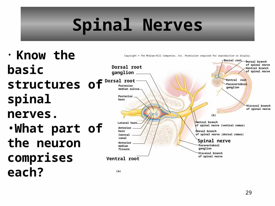
Spinal Nerves
• Know the basic structures of spinal nerves.•What part of the neuron comprises each?
Copyright © The McGraw-Hill Companies, Inc. Permission required for reproduction or display.
Lateral horn
Ventral root
(a)
(b)
Dorsal root
Dorsal root
Spinal nerve
Dorsal rootganglion
Posteriormedian sulcus
Posteriorhorn
Anteriorhorn
Centralcanal
Anteriormedianfissure
Dorsal branchof spinal nerveVentral branchof spinal nerve
Visceral branchof spinal nerve
Paravertebralganglion
Ventral branchof spinal nerve (ventral ramus)
Dorsal branchof spinal nerve (dorsal ramus)
Paravertebralganglion
Visceral branchof spinal nerve
Ventral root

30
Dermatome• What is Dermatome?
C2C3
C4C5
C6
T1
C6
C7
S2
S3
C8
L1
L2
L3
L4
L5
T12
T1
S1
(a) (b)
S5C0
S4S3
S2S1
L5
L4
L3
L2
L1
L5
L1
C8T1
T12
C7C6
C5C4
C3
C2
Copyright © The McGraw-Hill Companies, Inc. Permission required for reproduction or display.

31
Nerve Plexuses
• Know the 3 nerve plexuses and where they innervate.

32
Plexuses
C1C2C3C4C5C6C7C8T1
T2T3
T4
T5
T6
T8
T9
T10T11
T12
L1
L2
L3
L4
L5
S2S3S4S5
Co
Posterior view
Cervical plexus(C1–C4)
Lumbosacral plexus(T12–S5)
Sciatic nerve
Brachial plexus(C5–T1)
Obturator nerve
Phrenic nerve
Ulnar nerveMedian nerveRadial nerveAxillary nerve
T7
S1
Cauda equina
Musculocutaneousnerve
Femoralnerve
Intercostalnerves
Copyright © The McGraw-Hill Companies, Inc. Permission required for reproduction or display.

33
11.5: Brain
•What are the major functions of the brain?•What are the 4 major parts of the brain?•What are the 5 lobes of the cerebrum?•What three structures make up the brainstem?

34
Structure of the Cerebrum• Be able to name the basic structure of the cerebrum
Copyright © The McGraw-Hill Companies, Inc. Permission required for reproduction or display.
Central sulcus
Gyrus
SulcusFrontal lobe
Lateral sulcus
Parietal lobe
Occipital lobe
Central sulcus
Parietal lobe
Occipital lobe
(a)
(b) (c)
Parietal lobe
Central sulcus
Occipital lobeFrontal lobeInsula
Temporal lobe
Longitudinalfissure
TransversefissureCerebellarhemisphere
Retractedtemporal lobe

35
Lobes of the Cerebrum• Five (5) lobes bilaterally:
• Frontal lobe• Parietal lobe• Temporal lobe• Occipital lobe• Insula
Copyright © The McGraw-Hill Companies, Inc. Permission required for reproduction or display.
(c)
Parietal lobe
Central sulcus
Occipital lobe
Frontal lobe
Insula
Retractedtemporal lobe

36
Functional Regions of theCerebral Cortex
• What is the cerebral cortex?•What is the basic function of each lobe?
Copyright © The McGraw-Hill Companies, Inc. Permission required for reproduction or display.
Frontal eye field
Central sulcus
Parietal lobe
Occipital lobe
Cerebellum
Brainstem
Interpretation of auditory patterns
Lateral sulcus
Auditory area
Sensory areas involved withcutaneous and other senses
Sensory speech area( Wernicke’s area)
Combiningvisual images,visual recognitionof objects
Visual area
Temporal lobe
Motor speech area(Broca’s area)
Motor areas involved with the controlof voluntary muscles
Concentration, planning,problem solving
Front lobe

37
Functions of the Cerebral Lobes

38
Sensory Areas(post-central sulcus)
• Cutaneous sensory area• Parietal lobe• Interprets sensations on skin
• Visual area• Occipital lobe• Interprets vision
• Auditory area• Temporal lobe• Interprets hearing
• Sensory area for taste• Near base of the central sulcus
• Sensory area for smell• Arises from centers deep within the cerebrum
Copyright © The McGraw-Hill Companies, Inc. Permission required for reproduction or display.
Frontal eye field
Central sulcus
Parietal lobe
Occipital lobe
Cerebellum
Brainstem
Interpretation of auditory patterns
Lateral sulcus
Auditory area
Sensory areas involved withcutaneous and other senses
Sensory speech area( Wernicke’s area)
Combiningvisual images,visual recognitionof objects
Visual area
Temporal lobe
Motor speech area(Broca’s area)
Motor areas involved with the controlof voluntary muscles
Concentration, planning,problem solving
Front lobe

39
Association Areas
• What are association areas?•What is the basic function of each?
Copyright © The McGraw-Hill Companies, Inc. Permission required for reproduction or display.
Frontal eye field
Central sulcus
Parietal lobe
Occipital lobe
Cerebellum
Brainstem
Interpretation of auditory patterns
Lateral sulcus
Auditory area
Sensory areas involved withcutaneous and other senses
Sensory speech area( Wernicke’s area)
Combiningvisual images,visual recognitionof objects
Visual area
Temporal lobe
Motor speech area(Broca’s area)
Motor areas involved with the controlof voluntary muscles
Concentration, planning,problem solving
Front lobe

40
Association Areas
• Frontal lobe association areas• Concentrating• Planning• Complex problem solving
• Parietal lobe association areas• Understanding speech• Choosing words to express thought
• Temporal lobe association areas• Interpret complex sensory experiences • Store memories of visual scenes, music, and complex patterns
• Occipital lobe association areas• Analyze and combine visual images with other sensory experiences

41
Hemisphere Dominance
•What is the dominant hemisphere in most humans?•What does the dominant and dominant sides of the brain control?

42
Memory
• What is the basic differences between short and long term memory?

43
Brainstem
Know the three structure of the brainstem and the functions that make them unique.
Copyright © The McGraw-Hill Companies, Inc. Permission required for reproduction or display.
Spinal cord
Thalamus
HypothalamusDiencephalon
Pons
Midbrain
Corpuscallosum
Corporaquadrigemina
Cerebralaqueduct
Reticularformation
Medullaoblongata

44
Types of Sleep
• What are the major differences between the two types of sleep?

45
Cerebellum
• What is the cerebellum?•What are its major functions?
Copyright © The McGraw-Hill Companies, Inc. Permission required for reproduction or display.
Thalamus
Superior peduncle
Middle peduncleInferior peduncle
Pons
Medulla oblongata
Cerebellum
Corpus callosum
Longitudinalfissure

46
Major Parts of the Brain

47
Cranial NervesCopyright © The McGraw-Hill Companies, Inc. Permission required for reproduction or display.
Olfactory bulb
Hypoglossal (XII)
Optic tract
Olfactory tract
Olfactory (I)
Optic (II)
Oculomotor (III)
Abducens (VI)
Facial (VII)
Glossopharyngeal (IX)
Accessory (XI)
Trochlear (IV)
Trigeminal (V)
Vestibulocochlear (VIII)
Vagus (X)

48
Functions of Cranial Nerves

49
Sympathetic Division
• Fight or Flight

50
Parasympathetic Division
• Rest and Digest
