copyright © 2018 · blood transfusion is an essential part of ... the who developed a bedside...
TRANSCRIPT


Copyright © 2018Safe Blood Transfusion Programme, Government of Pakistan
All rights reserved. No part of this publication may be reproduced, stored in a retrieval system, or transmitted, in any form or by any means, electronic, mechanical, photocopying, recording or otherwise, without the prior written permission of the publisher.
Published by:
Safe Blood Transfusion ProgrammeMinistry of National Health Services, Regulation & CoordinationGovernment of Pakistan Website: www.sbtp.gov.pk Facebook: https://www.facebook.com/sbtp.pk
Editors:
Prof. Hasan Abbas Zaheer Usman Waheed Kaenat Nasir Saira Tahir
ISBN 978 969 9881 35 0
Printed in Islamabad, Pakistan

PrefaceBlood transfusion is an essential part of modern health care. Used correctly, it can save life and improve health. However, the transmission of infectious agents by blood and blood products has focused particular attention on the potential risks of transfusion. The World Health Organization (WHO) has developed an integrated strategy to promote global blood safety and minimize the risks associated with transfusion. A key component of this strategy is reduction in unnecessary transfusions through the appropriate clinical use of blood and blood products, and the use of simple alternatives to transfusion, wherever possible. Accordingly, the WHO developed a bedside transfusion handbook ‘The Clinical Use of Blood’ to provide a set of comprehensive learning materials that can be used by individual clinicians and blood transfusion specialists. The materials have been written by an international team of clinical and blood transfusion specialists and have been reviewed by a wide range of specialists throughout the world.
The Safe Blood Transfusion Programme has derived the ‘Handbook of Clinical Transfusion Practices’ from the WHO’s The Clinical Use of Blood with the permission of the WHO. The pocket handbook is designed for quick reference to promote rational use of blood components and avoid unnecessary blood transfusions. The Handbook is not designed to replace the conventional textbooks or to provide a definitive text on the clinical use of blood. Rather, its purpose is to provide an easily accessible learning tool that will assist prescribers of blood to make appropriate clinical decisions on transfusion and contribute to wider efforts to minimize the unnecessary use of blood and blood products.
The SBTP appreciates Prof. Syed Mohammad Irfan, Liaquat National Hospital and Medical College, Karachi for motivating the Programme to compile this Handbook and also for reviewing and endorsing the adopted document. The SBTP technical team (Usman Waheed, Kaenat Nasir and Saira Tahir) also deserve special acknowledgement for their tireless efforts to adopt the original WHO document into a Handbook well suited to the national needs and requirements.
It is expected that the use of this document in the Pakistan healthcare system will significantly improve the blood transfusion practices in our hospitals. It will also help overcome to a large extent the chronic shortage of blood and blood components in our blood centers and promote the rational and judicious use of blood and blood components.
Prof. Dr. Hasan Abbas Zaheer National Coordinator
Safe Blood Transfusion Programme Government of Pakistan
April 2018
AcronymsAHF Antihaemophilic Factor APTT Activated Partial Thromboplastin Time DAT Direct Antiglobulin Test DIC Disseminated Intravascular Coagulation FFP Fresh Frozen Plasma Hb Haemoglobin HBV Hepatitis B Virus Hct Haematocrit HCV Hepatitis C Virus HDN Haemolytic Disease of the Newborn HIV Human Immunodeficiency Virus ITP Idiopathic Autoimmune Thrombocytopenic Purpura MTP Massive Transfusion Protocol lPC Platelet Concentrates PRBC Packed Red Blood Cells PT Prothrombin Time PTT Partial Thromboplastin Time SRO Statutory Regulation Order TACO Transfusion Associated Circulatory Overload TA‐GVHD Transfusion Associated Graft‐Versus‐Host Disease TR Transfusion Reaction TRALI Transfusion Related Acute Lung Injury TTIs Transfusion Transmissible Infections TTP Thrombotic Thrombocytopenic Purpura

Contents
Acronyms ...................................................................................4
1. Introduction .................................................................................. 81.2 Principles of Clinical Transfusion Practice ........................ 81.2 Safe Blood ............................................................................... 9
2. Replacement Fluids .................................................................... 102.1 Intravenous Replacement Therapy.................................... 102.2 Intravenous Replacement Fluids ....................................... 102.3 Maintenance Fluids ............................................................. 132.4 Safety ..................................................................................... 132.5 Other Routes of Fluid Administration.............................. 13
3. Blood Components ..................................................................... 193.1 Whole blood ......................................................................... 193.2 Red Cell Concentrates (RCC) ............................................ 203.3 Platelet Concentrates (PC) ................................................. 213.4 Fresh Frozen Plasma (FFP) ................................................ 233.5 Cryoprecipitates ................................................................... 253.6 Plasma Derivative ................................................................ 26
4. Storage of blood components ................................................... 32
5. Clinical Transfusion Procedure ............................................... 335.1 Indications For Blood Transfusion .................................... 335.2 Transfusion Trigger (Adults) .............................................. 345.3 Responsibilities of Attending Physician ........................... 34
6. Administration of Blood Products ......................................... 356.1 Blood Request Form ............................................................ 356.2 Blood Samples ...................................................................... 366.3 Red Cell Compatibility Testing .......................................... 366.4 Collection and Receipt of Blood ........................................ 37

6.5 Performing the Transfusion ............................................... 396.6 Monitoring the Transfusion ............................................... 416.7 Documentation of the Transfusion ................................... 426.8 Other aspects of Transfusion ............................................. 44
7. Adverse Effects of Transfusion................................................. 467.1 Guidelines for Recognition and Management of Acute Transfusion Reactions ............................................................... 487.2 Investigating Acute Transfusion Reactions ...................... 517.3 Haemolytic Transfusion Reaction ..................................... 527.4 Bacterial Contamination and Septic Shock...................... 537.5 Transfusion Associated Circulatory Overload................. 547.6 Anaphylactic Reaction ........................................................ 547.7 Transfusion Related Acute Lung Injury ............................ 557.8 Delayed Complications of Transfusion ............................. 55
8. Clinical decisions on Transfusion ........................................... 58Assessing the Need for Transfusion ........................................ 58
9. Massive Blood Transfusion ...................................................... 619.1 Massive Transfusion Protocol ............................................ 619.2 Management ......................................................................... 63
Annexures ......................................................................................... 65Annexure 1: Bedside Transfusion Notes ................................ 66Annexure 2: Management of Adverse Transfusion Reaction: Physician’s Notes ........................................................................ 67Annexure 3: Blood Request Form .......................................... 68Annexure 4: Notification of an Adverse Reaction in Recipient ..................................................................................... 69

8Handbook of Clinical Transfusion Practices
1. IntroductionIt is well known that errors in blood transfusion practices can lead to serious consequences for the recipient in terms of morbidity and mortality. The majority of errors occur due to incorrect sampling of blood from a patient, fetching the wrong unit of blood for a patient and transfusing blood inappropriately. These clinical transfusion guidelines describe protocols for the collection of blood samples for blood grouping and cross matching, and for the collection, storage and administration of blood and blood products. The guidelines provide a standardized approach to transfusion so that the potential for errors is minimized and the administration of safe and efficacious blood products in the health care setting is maximized. It also contain protocols for the investigation and treatment of adverse transfusion reactions and provide guidelines for the use of specialized blood products.
1.2 Principles of Clinical Transfusion Practice• The patient with acute blood loss should receive effective
resuscitation (intravenous replacement fluids, oxygen and other medication) immediately and the need for transfusion is estimated thereafter.
• The patient’s haemoglobin (Hb) value, although important, should not be the sole deciding factor in the decision to transfuse blood. This decision should be supported by the need to relieve clinical signs and symptoms and to prevent significant morbidity or mortality.
• Clinicians should be aware of the risk of transfusion transmissible infections in blood products prescribed for patients.
• Transfusion should be prescribed only when the benefits to the patient are likely to outweigh the risks.
• Clinicians should clearly record the reason for ordering a transfusion (clinical diagnosis).
• Trained staff should monitor a patient undergoing transfusion and respond immediately if there are signs of an adverse effect.

9Handbook of Clinical Transfusion Practices
1.2 Safe bloodBlood for transfusion is considered safe when it is:
• Donated by a carefully selected, healthy donor• Free from infections that could be harmful to the recipient• Processed by reliable methods of testing, component
production, storage and transportation. Transfused only upon need and for the patient’s health and wellbeingThe quality and safety of blood and blood products must be assured throughout the process from the selection of blood donors to the administration of blood to the patient. This is described in the WHO Blood Safety Initiative:
• Establishment of a well‐organized blood transfusion service with quality system in all areas.
• Collection of blood only from voluntary non‐remunerated donors from low‐risk populations, using rigorous procedures for donor selection.
• Screening of all donated blood for transfusion transmissible infections i.e. HIV, HBV, HCV, syphilis and malaria.
• Good laboratory practice in all aspects of blood grouping, compatibility testing, component preparation and the storage and transportation of blood and blood products.
• Reduction of unnecessary transfusions through appropriate clinical use of blood and blood products and the use of simple alternatives to transfusion when possible.Transfusion of blood and products should be undertaken only to treat a condition that would lead to significant morbidly or mortality which cannot be prevented or managed effectively by other means.

10Handbook of Clinical Transfusion Practices
2. Replacement Fluids
2.1 Intravenous Replacement TherapyThe administration of intravenous replacement fluids restores the circulating blood volume and so maintains tissue perfusion and oxygenation. In severe haemorrhage, initial treatment (resuscitation) with intravenous replacement fluids may be life-saving and provide time to control the bleeding and order blood for transfusion, if necessary
2.2 Intravenous Replacement Fluids
Crystalloid solutions• Contain a similar concentration of sodium to plasma• Are excluded from the intracellular compartment because
the cell membrane is generally impermeable to sodium• Cross the capillary membrane from the vascular
compartment to the interstitial compartment• Are distributed through the whole extracellular compartment• Normally, only a quarter of the volume of crystalloid
infused remains in the vascular compartment.
Composition of Crystalloid replaCement solutions
FluidNa+
mmol/LK+
mmol/LCa2+
mmol/LCl–
mmol/L Base– mEq/L
Colloid osmotic pressure mmHg
Norm
al sa
line(
sodi
um
chlo
ride
0.9%
)
154 0 0 154 0 0
Balan
ced
salt
solu
tions
(Rin
ger’s
lac
tate
or H
artm
ann’s
so
lutio
n)
130–140 4–5 2–3 109–110 28–30 0
Table 1: Composition of crystalloid replacement solutions
11Handbook of Clinical Transfusion Practices
To restore circulating blood volume (intravascular volume), crystalloid solutions should be infused in a volume at least three times the volume lost.
Dextrose (glucose) solutions do not contain sodium and are poor replacement fluids. Do not use to treat hypovolaemia unless there is no alternative.
Colloid Solution• Contain a similar concentration of sodium to plasma• Are excluded from the intracellular compartment because
the cell membrane is generally impermeable to sodium• Cross the capillary membrane from the vascular
compartment to the interstitial compartment • Are distributed through the whole extracellular
compartment • Normally, only a quarter of the volume of crystalloid
infused remains in the vascular compartment.• Colloids require smaller infusion volumes than crystalloids.
They are usually given in a volume equal to the blood volume deficit.However, when the capillary permeability is increased, they may leak from the circulation and produce only a short-lived volume expansion. Supplementary infusions will be needed to maintain blood volume in conditions such as:
• Trauma • Acute and chronic sepsis• Burns• Snake bite (haemotoxic and cytotoxic).

12Handbook of Clinical Transfusion Practices
Co
mpo
siti
on o
f Co
llo
id r
epla
Cem
ent s
olu
tio
ns
Flui
dNa
+ m
mol
/L
K+ m
mol
/L
Ca2+
mm
ol/L
Cl–
mm
ol/L
Ba
se–
mEq
/L
Collo
id
osm
otic
pres
sure
m
mHg
Gela
tin (u
rea
linke
d): e
.g.
Haem
acce
l14
55.
16.
2514
5Tr
ace
am
ount
s27
Gela
tin (s
ucci
nyla
ted)
: e.g
. Ge
lofu
sine
154
<0.4
<0.4
125
Trac
e a
mou
nts
34
Dext
ran
70 (6
%)
15
40
015
40
58
Dext
ran
60 (3
%)
13
04
211
030
22
Hydr
oxye
thyl
star
ch
450/
0.7
(6%
)15
40
015
40
28
Albu
min
5%
130–
160
<1V
VV
27
Ioni
c co
mpo
sition
of
norm
al p
lasm
a 1
35–1
453.
5–5.
52.
2–2.
697
–110
38–4
427
V =
varie
s be
twee
n di
ffere
nt b
rand
s
Tabl
e 2: C
ompo
sitio
n of
collo
id re
plac
emen
t sol
utio
ns

13Handbook of Clinical Transfusion Practices
Crys
tallo
ids Advantages Disadvantages
• Few side-effects• Low cost• Availability
• Short duration of action• May cause oedema• Less weighty and bulky
Collo
ids
• Longer duration of action• Less fluid required to
correct hypovolaemia• Less weighty and bulky
• No evidence that they are more clinically effective
• Higher cost• May cause volume over-load• May interfere with clotting• Risk of anaphylactic reaction
Table 3: Advantages and disadvantages of crystalloid and colloid solution
There is no evidence that colloid solutions are superior to normal saline (sodium chloride 0.9%) or balanced salt solutions (BSS) for resuscitation.
2.3 Maintenance Fluids• Used to replace normal physiological losses through skin,
lung, faeces and urine • The volume of maintenance fluids required by a patient will
vary, particularly with pyrexia, high ambient temperature or humidity, when losses will increase
• Composed mainly of water in a dextrose solution; may contain some electrolytes
• All maintenance fluids are crystalloid solutions.
Examples of maintenance fluids• 5% dextrose• 4% dextrose in sodium chloride 0.18%.
2.4 SafetyBefore giving any intravenous infusion:
• Check that the seal of the infusion bottle or bag is not broken.• Check the expiry date.• Check that the solution is clear and free from visible particles.
2.5 Other Routes of Fluid AdministrationThere are other routes of fluid administration in addition

14Handbook of Clinical Transfusion Practices
to the intravenous route. However, with the exception of the intraosseous route, they are generally unsuitable in the severely hypovolaemic patient.
Intraosseous• Can provide the quickest access to the circulation in a
shocked child in whom venous cannulation is impossible • Fluids, blood and certain drugs can be administered by this
route• Suitable in the severely hypovolaemic patient.
Oral and nasogastric • Can often be used in patients who are mildly hypovolaemic
and in whom the oral route is not contraindicated • Should not be used in patients if:
— Severely hypovolaemic — Unconscious — Gastrointestinal lesions or reduced gut motility — General anaesthesia and surgery is planned
imminently.
WHo/uniCef formula for oral reHydration fluid
Dissolve in one litre of potable water Sodium chloride (table salt) 3.5 g
Sodium bicarbonate (baking soda) 2.5 g
Potassium chloride or substitute (banana or degassed cola drink) 1.5 g
Glucose (sugar) 20.0 g
Resulting concentrationsNa+ 90 mmol/L K+ 20 mmol/L CI– 80 mmol/L Glucose 110 mmol/L
Table 4: WHO/UNICEF formula for oral rehydration fluid
Rectal • Unsuitable in the severely hypovolaemic patient • Ready absorption of fluids• Absorption ceases with fluids being ejected when hydration
is complete• Administered through plastic or rubber enema tube
inserted into the rectum and connected to a bag or bottle

15Handbook of Clinical Transfusion Practices
of fluid• Fluid rate can be controlled by using a drip infusion set, if
necessary• Fluids used do not have to be sterile: a safe and effective
solution for rectal rehydration is 1 litre of clean drinking water to which is added a teaspoon of table salt.
Subcutaneous• Can occasionally be used when other routes of
administration of fluids are unavailable• Unsuitable in the severely hypovolaemic patient• A cannula or needle is inserted into the subcutaneous tissue
(the abdominal wall is a preferred site) and sterile fluids are administered in a conventional manner
• Dextrose-containing solutions can cause sloughing of tissues and should not be given subcutaneously.
Crystalloid solutions
NORMAL SALINE (Sodium chloride 0.9%)
Infection risk Nil
Indications Replacement of blood volume and other extracellular fluid losses
Precaution Caution in situations where local oedema may aggravate pathology: e.g. head injury may precipitate volume overload and heart failure
Contraindications Do not use in patients with established renal failure
Side-effects Tissue oedema can develop when large volumes are used
Dosage At least 3 times the blood volume lost
BALANCED SALT SOLUTIONS
Examples Ringer’s lactateHartmann’s solution
Infection risk Nil
Indications Replacement of blood volume and other extracellular fluid losses

16Handbook of Clinical Transfusion Practices
Precautions Caution in situations where local oedema may aggravate pathology: e.g. head injury may precipitate volume overload and heart failure
Contraindications Do not use in patients with established renal failure
Side-effects Tissue oedema can develop when large volumes are used
Dosage At least 3 times the blood volume lost
DEXTROSE AND ELECTROLYTE SOLUTIONS
Examples 4.3% dextrose in sodium chloride 0.18%2.5% dextrose in sodium chloride 0.45%2.5% dextrose in half-strength Darrow’s solution
Note2.5% dextrose in half-strength Darrow’s solution is commonly used to correct dehydration and electrolyte disturbances in children with gastroenteritis. Several products are manufactured for this use. Not all are suitable. Ensure that the preparation you use contains:• Dextrose• Sodium 60 mmol/L• Potassium 17 mmol/L • Chloride 52 mmol/L• Lactate
Table 5: Crystalloid Solutions
plasma-derived (natural) Colloid solutions
Plasma-derived colloids are all prepared from donated blood or plasma. They include:• Plasma• Fresh frozen plasma• Liquid plasma• Freeze-dried plasma• Albumin
These products should not be used simply as replacement fluids. They can carry a similar risk of transmitting infections, such as HIV and hepatitis, as whole blood. They are also generally more expensive than crystalloid or synthetic colloid fluids.
Synthetic colloid solutions
GELATINS (Haemaccel, Gelofusine)
Infection risk None known at present

17Handbook of Clinical Transfusion Practices
Indications Replacement of blood volume
Precautions
May precipitate heart failureCaution in renal insufficiencyDo not mix Haemaccel with citrated blood because of its high calcium concentration
Contraindications Do not use in patients with established renal failure
Side-effects Minor allergic reactions due to histamine releaseTransient increase in bleeding time may occurHypersensitivity reactions may occur including, rarely, severe anaphylactic reactions
Dosage No known dose limit
DEXTRAN 60 and DEXTRAN 70
Infection risk Nil
Indications Replacement of blood volumeProphylaxis of postoperative venous thrombosis
Precautions
Coagulation defects may occurPlatelet aggregation inhibitedSome preparations may interfere with compatibility testing of blood
Contraindications Do not use in patients with pre-existing disorders of haemostasis and coagulation
Side-effects
Minor allergic reactionsTransient increase in bleeding time may occurHypersensitivity reactions may occur including, rarely, severe anaphylactic reactions. Can be prevented with injection of 20 mL of Dextran 1 immediately before infusion, where available
Dosage
Dextran 60: should not exceed 50 mL/kg body weight in 24 hoursDextran 70: should not exceed 25 mL/kg body weight in 24 hours
DEXTRAN 40 and DEXTRAN 110
Not recommended as replacement fluids
HYDROXYETHYL STARCH (Hetastarch or HES)
Infection risk Nil

18Handbook of Clinical Transfusion Practices
Indications Replacement of blood volume
Precautions Coagulation defects may occurMay precipitate volume overload and heart failure
Contraindications Do not use in patients with pre-existing disorders of haemostasis and coagulationDo not use in patients with established renal failure
Side-effects Minor allergic reactions due to histamine releaseTransient increase in bleeding time may occurHypersensitivity reactions may occur including, rarely, severe anaphylactic reactionsSerum amylase level may rise (not significant)HES is retained in cells of the reticuloendothelial system; the long-term effects of this are unknown
Dosage Should not usually exceed 20 mL/kg body weight in 24 hours
Table 6: Plasma derived Natural and Synthetic colloid solution

19Handbook of Clinical Transfusion Practices
3. Blood ComponentsA blood component is a constituent of blood, separated from whole blood, such as:
• Red cell concentrate• Plasma• Platelet concentrate• Cryoprecipitate, prepared from fresh frozen plasma; rich in
Factor VIII and fibrinogenA plasma derivative is made from human plasma proteins prepared under pharmaceutical manufacturing conditions, such as:
• Albumin• Coagulation factor concentrates• ImmunoglobulinIn Pakistan, blood is generally collected in CPDA1 blood bags that contain 70 mL anticoagulant solution which provides a shelf life for red cells of 35 days when stored at +2°C to +6°C. For collection of blood components in the country, double or triple bags are used. Components are prepared using a centrifuge, a piece of equipment that is available in most blood transfusion departments of medical colleges.
3.1 Whole Blood
Description:500 mL whole blood in 70 mL anticoagulant‐preservative solution of which Hb will be approximately 1.2 g/dL and haematocrit (Hct) 35‐45% with no functional platelets or labile coagulation factors (V and VIII) when stored at +2°C to +6°C.
Infection risk:Capable of transmitting an agent present in cells or plasma which was undetected during routine screening for TTIs, i.e. HIV, hepatitis B & C, syphilis and malaria.

20Handbook of Clinical Transfusion Practices
Storage:Between +2°C and +6°C in an approved blood bank refrigerator, fitted with a temperature monitor and alarm.
Indications:• Red cell replacement in acute blood loss with hypovolaemia.• Exchange transfusion.
Contraindications:Risk of volume overload in patients with:
• Chronic anaemia.• Incipient cardiac failure.
Administration:• Must be ABO and RhD compatible with the recipient.• Never add medication to a unit of blood.• Complete transfusion within 4 hours of commencement.
3.2 Red Cell Concentrates (RCC)
Description:150‐200 mL red blood cells from which most of the plasma has been removed. Hb concentration will be approximately 20 g/100 mL (not less than 45 g per unit) and Hct 55‐75%.
Infection risk: Same as for whole blood.
Storage: Same as for whole blood.
Indications: Replacement of red cells in anaemic patients.

21Handbook of Clinical Transfusion Practices
3.3 Platelet Concentrates (PC)
Description:PCs are prepared from units of whole blood that have not been allowed to cool below +20°C. A single donor unit consists of 50‐60 mL plasma that should contain ≥55 x 109 platelets.
Unit of issue:PCs may be supplied as a pooled unit, i.e. platelets prepared from 4‐6 donor units containing at least 240 x 109 platelets.
Infection risk: Bacterial contamination affects about 1% of pooled units.
Storage:PCs may be stored for up to 5 days at +20°C to +24°C (with agitation). PCs require continuous agitation during storage, on a platelet shaker and in an incubator that maintains the required storage temperature.
Dosage:1 unit of platelet concentrate/10 kg; for an adult of 60‐70 kg, 4‐6 single donor units containing at least 240 x 109 platelets should raise the platelet count by 20‐40 x 109/L. Increment will be less if there is splenomegaly, disseminated intravascular coagulation (DIC) or septicaemia.
Indications:Treatment of bleeding due to:
• Thrombocytopenia.• Platelet function defects.• Prevention of bleeding due to thrombocytopenia as in bone
marrow failure.
Contraindications:• Idiopathic autoimmune thrombocytopenic purpura (ITP).

22Handbook of Clinical Transfusion Practices
• Thrombotic thrombocytopenic purpura (TTP).• Untreated DIC.• Thrombocytopenia associated with septicaemia, or in cases
of hypersplenism.Use in bone marrow failure:
• Treatment of bleeding, thrombocytopenic patients.• Prophylactic use in thrombocytopenic patients.• Maintain platelet count >10 x 109/L in non‐bleeding, non‐
infected patient.• Maintain platelet count >20 x 109/L in infected/pyrexial
patient.
Use in DIC:For acute DIC, where bleeding is associated with thrombocytopenia, maintain platelet count above 20 x 109/L even in the absence of overt bleeding.
Use in massive blood transfusion:Maintain platelet count >50 x 109/L in patients receiving massive transfusions (dilutional thrombo‐ cytopenia occurs when >1.5 x blood volume of patient is transfused).
Use in cardiopulmonary bypass surgery:• Platelet function defects and thrombocytopenia often
occur after cardiac bypass surgery. Platelet transfusion is recommended for patients with bleeding not due to surgically correctable causes (closure time provides global indication of platelet function).
• Prophylactic platelet transfusions are not required for all bypass procedures.
Prophylaxis for surgery:• Ensure platelet count is >50 x 109/L for procedures such
as lumbar puncture, epidural anaesthesia, insertion of indwelling lines, trans‐bronchial biopsy, liver biopsy, renal biopsy and laparotomy.
• Maintain platelet count >100 x 109/L for neurological and

23Handbook of Clinical Transfusion Practices
ophthalmic surgery.
Administration:
Platelet concentrates after pooling should be infused as soon as possible because of the risk of bacterial proliferation. Depending on the condition of the recipient, a unit should be infused over a period of not more than 30 minutes. Do not give platelet concentrates prepared from RhD positive donors to an RhD negative female with childbearing potential. Give platelet concentrates that are ABO compatible, whenever possible.
Complications:Febrile non‐haemolytic and allergic urticarial reactions are not uncommon, especially in patients receiving multiple transfusions.
3.4 Fresh Frozen Plasma (FFP)
Description:FFP is plasma prepared from whole blood, either from the primary centrifugation of whole blood into red cells and plasma or from a secondary centrifugation of platelet rich plasma. The plasma is rapidly frozen to –25°C or colder within 8 hours of collection and contains normal plasma levels of stable clotting factors, albumin, immunoglobulin and Factor VIII at a level of at least 70% of normal fresh plasma.
Unit of issue:200‐300 mL.
Infection risk:Capable of transmitting any agent present in cells or plasma which was undetected by routine screening TTIs, including HIV, hepatitis B and C, syphilis and malaria.

24Handbook of Clinical Transfusion Practices
Storage:FFP is stored at –25°C or colder for up to 1 year. Before use, it should be thawed in the blood transfusion centre between +30°C and +37°C.
Definite indications:Replacement of a single coagulation factor deficiency, where a specific or combined factor concentrate is unavailable or contraindicated.
• Immediate reversal of warfarin effect where prothrombin complex concentrate is unavailable.
• Thrombotic thrombocytopenic purpura.• Inherited coagulation inhibitor deficiencies where specific
concentrate is unavailable.• C1 esterase inhibitor deficiency where specific concentrate
is unavailable.
Conditional indications:• Massive blood transfusion.• Acute DIC if there are coagulation abnormalities and
patient is bleeding.• Liver disease, with abnormal coagulation and bleeding
– prophylactic use to reduce prothrombin time (PT) to 1.6‐1.8 x normal for liver biopsy.
• Cardiopulmonary bypass surgery – use in the presence of bleeding but where abnormal coag‐ ulation is not due to heparin. Routine perioperative use is not indicated.
• Severe sepsis, particularly in neonates (independent of DIC).
• Plasmapheresis.
Precautions:• Acute allergic reactions are not uncommon, especially with
rapid infusions.• Severe life‐threatening anaphylactic reactions occasionally
occur.

25Handbook of Clinical Transfusion Practices
Dosage: 15 mL/kg.
Administration:• Should be ABO compatible.• Infuse as soon as possible after thawing.• Labile coagulation factors rapidly degrade; use within 6
hours of thawing.• FFP may be beneficial if PT and/or partial thromboplastin
time (PTT) >1.5 times normal.• FFP for volume expansion carries a risk of infectious
disease transmission and other transfusion reactions (e.g. allergic) that can be avoided by using crystalloid or colloid solutions.
3.5 Cryoprecipitates
Description:Cryoprecipitates are prepared from FFP by collecting the precipitate formed during controlled thawing at +4°C and re‐suspending in 10‐20 mL plasma. It is stored at –25°C or colder for up to 1 year after the date of phlebotomy. Cryo‐AHF contains about half the Factor VIII and fibrinogen as a pack of fresh whole blood: e.g. Factor VIII: 80‐100 iu/ pack; fibrinogen: 150‐300 mg/ pack.
Infection risk: As for plasma, but a normal adult dose involves at least 6 donor exposures.
Storage: At –25°C or colder for up to 1 year.
Indications:As an alternative to Factor VIII concentrate in the treatment of inherited deficiencies of:
• von Willebrand Factor (von Willebrand’s disease).

26Handbook of Clinical Transfusion Practices
• Factor VIII (haemophilia A).• As a source of fibrinogen in acquired coagulopathies; e.g.
DIC.• Can be used in isolated Factor XIII deficiency.• Ameliorate platelet dysfunction associated with uraemia.• Used topically as a fibrin sealant.
Administration:• ABO compatible product should be used.• After thawing, infuse as soon as possible.• Must be transfused within 6 hours of thawing.
3.6 Plasma Derivative
3.6.1 Human albumin solutionDescriptionPrepared by fractionation of large pools of donated plasma
Preparations• Albumin 5%: contains 50 mg/mL of albumin• Albumin 20%: contains 200 mg/mL of albumin• Albumin 25%: contains 250 mg/mL of albumin• Stable plasma protein solution (SPPS) and plasma
protein fraction (PPF): similar albumin content to albumin 5%
Infection risk: No risk of transmission of viral infections if correctly manufactured
Indications• Replacement fluid in therapeutic plasma exchange:
use albumin 5%• Treatment of diuretic-resistant oedema in
hypoproteinaemic patients: e.g. nephrotic syndrome or ascites. Use albumin 20% with a diuretic

27Handbook of Clinical Transfusion Practices
• Although 5% human albumin is currently licensed for a wide range of indications (e.g. volume replacement, burns and hypoalbuminaemia), there is no evidence that it is superior to saline solution or other crystalloid replacement fluids for acute plasma volume replacement
PrecautionsAdministration of 20% albumin may cause acute expansion of intravascular volume with risk of pulmonary oedema
ContraindicationsDo not use for IV nutrition: it is an expensive and inefficient source of essential amino acids
Administration• No compatibility testing required• No filter needed
3.6.2 Coagulation factorsFactor VIII concentrateDescription
• Partially purified Factor VIII prepared from large pools of donor plasma
• Factor VIII ranges from 0.5–20 iu/mg of protein.Preparations with a higher activity are available
• Products that are licensed in certain countries (e.g. USA and European Union) are all heated and/or chemically treated to reduce the risk of transmission of viruses
Unit of issue Vials of freeze-dried protein labelled with content, usually about 250 iu of Factor VIII
Infection riskCurrent virus ‘inactivated’ products do not appear to transmit HIV, HTLV, hepatitis C and other

28Handbook of Clinical Transfusion Practices
viruses that have lipid envelopes: the inactivation of non-enveloped viruses such as hepatitis A and parvovirus is less effective
Storage: +2°C to +6°C up to stated expiry date, unless otherwise indicated in manufacturer’s instructions
Indications• Treatment of haemophilia A• Treatment of von Willebrand’s disease: use only
preparations that contain von Willebrand Factor
Dosage: As per severity of bleed
Administration• Reconstitute according to manufacturer’s
instructions• Once the powder is dissolved, draw up the solution
using a filter needle and infuse through a standard infusion set within 2 hours
Alternatives• Cryoprecipitate, fresh frozen plasma• Factor VIII prepared in vitro using recombinant
DNA methods is commercially available. It is clinically equivalent to Factor VIII derived from plasma and does not have the risk of transmitting pathogens derived from plasma donors
3.6.3 Plasma derivatives containing factorsProthrombin complex concentrate (PCC)Factor IX concentratesDescription Contains:
• Factors II, IX and X (in PCC and Factor IX)• Factor IX only (in Factor IX)

29Handbook of Clinical Transfusion Practices
• Some preparations also contain Factor VIII (in PCC)
Unit of issueVials of freeze-dried protein labelled with content, usually about 350–600 iu of Factor IX
Infection risk: As Factor VIII
Storage: As Factor VIII
Indications• Treatment of Haemophilia B (Christmas disease)
(for PCC and factor IX)• Immediate correction of prolonged prothrombin
time (for PCC)
ContraindicationsPCC is not advised in patients with liver disease or thrombotic tendency
Dosage: As per severity of bleed
Administration: As Factor VIII
Alternatives: Plasma
Factor IX produced in vitro by recombinant DNA methods will soon be available for the treatment of haemophilia B

30Handbook of Clinical Transfusion Practices
3.6.4 Coagulation factor products for patients with factor VIII inhibitors
Description: A heat-treated plasma fraction containing partly-activated coagulation factors
Infection risk: Probably the same as other heat-treated factor concentrates
Indications: Only for use in patients with inhibitors to Factor VIII
Administration: Should be used only with specialist advice
3.6.5 Immunoglobulins
3.6.5.1 Immunoglobulin for intramuscular useDescriptionConcentrated solution of the IgG antibody component of plasma
PreparationsStandard or normal immunoglobulin: prepared from large pools of donations and contains antibodies against infectious agents to which the donor population has been exposed
Infection risk Transmission of virus infections has not been reported with intramuscular immunoglobulin
Indications • Hyper-immune or specific immunoglobulin: from
patients with high levels of specific antibodies to infectious agents: e.g. hepatitis B, rabies, tetanus

31Handbook of Clinical Transfusion Practices
• Prevention of specific infections• Treatment of immune deficiency states
Administration Do not give intravenously as severe reactions occur
3.6.5.2 Anti-RhD immunoglobulin (Anti-D RhIG)DescriptionPrepared from plasma containing high levels of anti-RhD antibody from previously immunized persons
Indications: Prevention of haemolytic disease of the newborn in RhD- negative mothers
3.6.5.3 Immunoglobulin for intravenous useDescriptionAs for intramuscular preparation, but with subsequent processing to render product safe for IV administration
Indications• Idiopathic autoimmune thrombocytopenic
purpura and some other immune disorders• Treatment of immune deficiency states • Hypogammaglobulinaemia• HIV-related disease

32Handbook of Clinical Transfusion Practices
4. Storage of Blood ComponentsBlood must not be stored in a ward refrigerator under any circumstances.
The blood cold chain from collection to transfusion
Prep
erat
ion
of
com
pone
nt
Qua
rant
ine
stor
age
Avai
labl
e st
ock
stor
age
Hos
pita
l Blo
od
Bank
Don
ated
who
le b
lood
or p
lasm
a
Red
cel
l com
pone
nt
Bloo
d re
frige
rato
r +2
0 C to
+6
0 C
Bloo
d re
frige
rato
r +2
0 C to
+6
0 C
Tran
spor
t box
T0 r
ange
: +2
0 C to
+10
0C
Figu
re 1:
The
blo
od co
ld ch
ain fr
om co
llect
ion
to tr
ansf
usio
n
Plas
ma
Com
pone
nt
Plas
ma
freez
er
-30
0 C o
r low
er
Plas
ma
freez
er
-30
0 C o
r low
er
Tran
spor
t box
T0 r
ange
: le
ss th
an -2
0 0C
Bloo
d R
ecip
ient
(pat
ient
)
Plat
elet
Com
pone
nt
Plat
elet
agi
tato
r +2
0 0 C
to +
24 0 C
Plat
elet
agi
tato
r +2
0 0 C
to +
24 0 C
Tran
spor
t box
T0 r
ange
: +2
0 0 C
to +
24 0C
Tara
nspo
rt bo
x at
20
0 C to
+24
0 C fo
r max
. 6 h
ours

33Handbook of Clinical Transfusion Practices
5. Clinical Transfusion Procedure
5.1 Indications for blood transfusion• To increase the oxygen capacity of blood by giving red cells.• To restore the blood volume to maintain effective tissue
perfusion.• To replace platelets, coagulation factors and other plasma
proteins.
Blood may be needed in the following circumstances:
• Blood loss:– Bleeding– Trauma
• Inadequate production:–Diseases such as thalassemia, leukaemia
• Excessive destruction of cells:– Disease– Mechanical
Transfusion of blood and products should be undertaken only to treat a condition that would lead to significant morbidly or mortality that cannot be prevented or managed effectively by other means.
Blood is more often needed under the following circumstances:
• Maternity: women during pregnancy and at the time of delivery– Anaemia of pregnancy; bleeding in pre- or post-partum
stage of delivery.• 5-29 years
– Vulnerability during this age range due to infancy on the one hand (e.g. malnutrition, malaria) and youth on the other (e.g. nature of work which may be more physical and more likely to expose individual to accidents).
• Patients with chronic blood disease

34Handbook of Clinical Transfusion Practices
–e.g. thalassemia, leukaemia.
5.2 Transfusion trigger (adults)• One unit of whole blood/PRBC can increase Hb by 1g/dL in
an adult or Hct by 3% (Hb of unit must be >75%).• Perioperative transfusion:
– 8g/dL for patient undergoing cardiovascular surgery, orthopaedics and acute GI bleeding.
• Chronic anaemia:– 7g/dL in adults.
• Acute blood loss:– 30% of volume of blood.
5.3 Responsibilities of Attending Physician• Assess patient’s clinical need for blood, and when required.• Inform patient and/or relatives about proposed transfusion
and record in patient’s notes.• Select blood product and quantity required (i.e. whole blood/
PRBC/FFP/PC) and complete request form accurately and legibly.
• Enter the reason for transfusion on the form, so that the blood centre can check that the product ordered is the most suitable with regard to diagnosis.
• Obtain and correctly label a blood sample for compatibility testing.
• Send the blood request form and blood sample to the blood bank.
• When the blood product that was ordered arrives, transfuse it as soon as possible to avoid having to store it. However, if the blood product is not used immediately, store it under the correct storage conditions.
• Cross check the identity of the patient and the blood product:– Patient and documentation.– Blood / blood products.

35Handbook of Clinical Transfusion Practices
6. Administration of Blood ProductsWhen blood is transfused, it is important to keep detailed re-cords including the following in the patient’s notes:Type and volume of each unit transfused.
• Unique donation number of each unit transfused.• Blood group of each unit transfused.• Time at which the transfusion of each unit commenced.• Signature of the individual responsible for administration of
the blood.• Monitor the patient before, during and on completion of the
transfusion.• Record the time of completion of the transfusion.• Identify and respond immediately to any adverse effect, by
stopping the transfusion.• Record the details of any transfusion reaction.
The process starts with the request for blood, followed by the selection of the correct blood product for compatibility testing and finally the issuing of compatible blood for infusion into the patient.
6.1 Blood Request FormWhen blood is required for transfusion, the prescribing clinician should complete and sign a blood request form that is designed to provide all necessary information. All details requested on the blood request form must be completed accurately and legibly.
• The blood request form should always be accompanied by the patient’s blood sample. The sample is placed in a sample tube that is correctly labelled and is uniquely identifiable with the patient.
• The blood sample shall not be submitted in a syringe, as this could lead to errors when transferring to a test tube for grouping and compatibility testing. It may also cause haemolysis.
• For a routine case, the sample and request form should be submitted to the transfusion department at least 24 hours before required, to ensure availability of blood.
• Physicians may request those, who accompany the patient, to
36Handbook of Clinical Transfusion Practices
consider becoming blood donors if they are healthy and lead a healthy lifestyle.
6.2 Blood SamplesThe taking of a blood sample from the patient needs supervision. If the patient is conscious at the time of taking the sample, ask him/her to identify himself/herself by given name and all other appropriate information.
A 5 mL blood sample should be collected into a dry test tube and then correctly and clearly labelled with the patient’s details, and submitted to the blood centre for testing. The specimen label must include the following information:
• Patient’s full name, age and sex.• Registration number.• Ward/bed number.• Date and time of specimen collection.• Phlebotomist’s signature/initials.
– Use positive patient identification to identify the patient. NEVER pre‐label the sample tube before phlebotomy.
– Use the blood product request form, write legibly and fill in all appropriate details.
– When taking a blood sample for cross match, complete the whole procedure before any other task is undertaken
– it is important that there are no interruptions during the process.
– The signature of the individual who took the sample must appear on the specimen label.
Retention of blood samples:• Blood samples from recipient and donor(s) must be
retained for 7 days at +2°C to +8°C after each transfusion.• Should another transfusion be necessary 72 hours after the
earlier transfusion, a fresh sample shall be requested for cross match. Collection of a second 5 mL blood sample is required for re‐ checking and further cross matching and must be retained in case of investigation of transfusion reaction.

37Handbook of Clinical Transfusion Practices
6.3 Red Cell Compatibility TestingThe laboratory performs:
• ABO and RhD grouping on patient and donors.• Antibody screening on patient.• Cross matching between serum of patient and red cells of
donor.These procedures normally take about an hour or more to complete. Shortened procedures are possible in case of emergency, but may fail to detect some incompatibilities.
• The term compatibility test and cross match are sometimes used interchangeably; they should be clearly differentiated.
• The cross match is the part of pre‐transfusion test known as compatibility testing.
• The compatibility test includes:– ABO and RhD grouping of donor and recipient.– Screening for unexpected antibodies on donor and
patient.– Cross match.
All pre‐transfusion test procedures should provide information on ABO and RhD grouping of both patient and units of blood to be transfused.
Purpose of compatibility testing:• To select blood components that will cause no harm to
the recipient and will have acceptable survival rates when transfused.
• When correctly performed, compatibility tests will confirm ABO compatibility between component and recipient and will detect the most clinically significant unexpected antibodies.
• Compatibility (cross match) must be performed before blood is transfused. The cross match is incompatible if there is a reaction between the patient’s serum and donor’s red cells.
6.4 Collection and Receipt of Blood• ALWAYS take a complete patient documentation label to

38Handbook of Clinical Transfusion Practices
the issue room of the blood transfusion department when collecting the first unit of blood.
• MATCH the details on the blood request form against the blood compatibility label (tag), the bag unit number and the patient documentation label.
• If everything matches, sign out the unit with the date and time.
• If there is any discrepancy, DO NOT sign out the unit; contact the staff member of the blood transfusion department immediately.
• When receiving the unit of blood in the clinical area, check that it is the right unit for the right patient.
Always check patient/component compatibility/identity. Inspect pack and contents for signs of deterioration or damage.
Are ther any leaks?Have you squeezed the bag? Look for blood here
Look for haemolysis in the plasma. Is the plasma pink?
Look for for large clots in plasma
Plasma
Red cells
Look for haemolysis here on the line between red cells and plasma Look at the red cells. Are
they normal, or are they purple or black
Figure 2: Check points for signs of deterioration in blood and plasma
Discoloration or signs of any leakage may be the only warning that the blood has been contaminated by bacteria and could cause a severe or fatal reaction if transfused.
Blood bag should be checked for:• Any sign of haemolysis in the plasma indicating that the
blood has been contaminated, allowed to freeze or to warm.

39Handbook of Clinical Transfusion Practices
• Any sign of haemolysis on the line between the red cells and plasma during storage.
• Any sign of contamination, such as a change of colour in the red cells, which often look darker/ purple/ black when contaminated.
• Any clot, which may mean that the blood was not mixed properly with the anticoagulant when it was collected or might also indicate bacterial contamination due to the utilization of citrate by proliferating bacteria.
• Any sign that there is a leak in the bag or that it has already been opened.
The blood unit must be discarded if:• It has been out of the refrigerator for longer than 30
minutes, or• The seal is broken, or• There is any sign of haemolysis, clotting or contamination.
6.5 Performing the TransfusionOnce issued by the blood centre, the transfusion of whole blood, red cells, platelet concentrate and thawed fresh frozen plasma should be commenced within 30 minutes of removal from the optimal storage conditions.
If the transfusion cannot be started within this period, the unit(s) must be stored under approved optimal storage conditions. The temperature inside every blood bank refrigerator used for whole blood/ red cell storage should be monitored and recorded daily to ensure that the temperature remains between +2°C and +6°C. If the ward or operating room does not have a blood bank refrigerator that is appropriate for storing blood, the blood should only be released from the blood centre just before transfusion.
Checking the patient’s identity and the blood bag before transfusionBefore starting the transfusion, it is vital to make the final identity check in accordance with the hospital’s standard
40Handbook of Clinical Transfusion Practices
operating procedure. The final identity check should be undertaken at the patient’s bedside immediately before commencing the administration of the blood product. It should be undertaken by two people, at least one of whom should be a registered nurse or doctor.
The final check at the patient’s bedside is the last opportunity to detect an identification error and prevent a potentially incompatible transfusion, which may be fatal.
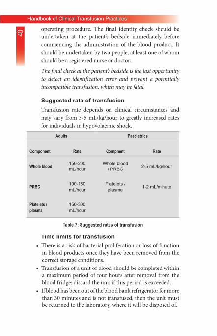
Suggested rate of transfusionTransfusion rate depends on clinical circumstances and may vary from 3‐5 mL/kg/hour to greatly increased rates for individuals in hypovolaemic shock.
Adults Paediatrics
Component Rate Compnent Rate
Whole blood 150-200 mL/hour
Whole blood / PRBC 2-5 mL/kg/hour
PRBC 100-150 mL/hour
Platelets / plasma 1-2 mL/minute
Platelets / plasma
150-300 mL/hour
Table 7: Suggested rates of transfusion
Time limits for transfusion• There is a risk of bacterial proliferation or loss of function
in blood products once they have been removed from the correct storage conditions.
• Transfusion of a unit of blood should be completed within a maximum period of four hours after removal from the blood fridge: discard the unit if this period is exceeded.
• If blood has been out of the blood bank refrigerator for more than 30 minutes and is not transfused, then the unit must be returned to the laboratory, where it will be disposed of.

41Handbook of Clinical Transfusion Practices
Table 8: Duration times for transfusionBlood products Start transfusion Complete transfusion
Whole blood / PRBC Within 30 minutes of removing from refrigerator
≤ 4 hours
Discard unit if this period is exceeded
Platelet concentrate Immediately Within 30 minutes
FFP As soon as possible
Within 30 minutes
Cryoprecipitate As soon as possible
Within 30 minutes
Table 8: Duration times for transfusion
Blood administration set:• Use a new, sterile blood administration set containing an
integral 170‐200µ filter.• Change the set at least 12‐hourly during blood transfusion.• In a very warm climate, change the set more frequently and
usually after every four units of blood, if given within a 12‐hour period.
• Use a fresh blood administration set or special platelet transfusion set, primed with saline. All blood components can be slowly infused through small‐bore cannulas or butterfly needles, e.g. 21 to 25 G. For rapid infusion, large‐bore cannulas, e.g. 14 G, are needed.
6.6 Monitoring the Transfusion• It is essential to take baseline observations and to ensure
that the patient is monitored during the transfusion in order to detect any adverse event as early as possible. Before commencing the transfusion, it is essential to encourage the

42Handbook of Clinical Transfusion Practices
patient to notify a nurse or doctor immediately if he or she becomes aware of any discomfort such as shivering, flushing, pain or shortness of breath or begins to feel anxious.
• Ensure that the patient is in a setting where he or she can be directly observed.
• For each unit of blood transfused, monitor the patient:– Before starting the transfusion (baseline observation).– 15 minutes after starting the transfusion.– At least every hour during transfusion.– Carry out a final set of observations 15 minutes after each
unit has been transfused.
6.7 Documentation of the TransfusionMonitor the patient before, during and on completion of the transfusion.
• At each of these stages, record the following information on the patient’s chart:
– Patient’s general appearance.– Temperature.– Pulse.– Blood pressure.– Respiratory rate.
• Make note of the following:– Time when transfusion started.– Time when transfusion was completed.– Volume and type of blood products transfused.– Unique donation number of all products transfused.– Any adverse effect.
• Record in the patient’s notes:– Type and volume of each unit transfused.– Unique donation number of each unit transfused.– Blood group of each unit transfused.– Time at which the transfusion of each unit commenced.– Signature of the individual responsible for
administration of the blood.– Record the time of completion of the transfusion.– Record the details of transfusion reaction
• Identify and respond immediately to any adverse effect, by

43Handbook of Clinical Transfusion Practices
stopping the transfusion and inform the doctor on duty.Severe reactions most commonly present during the first 15 minutes of a transfusion. All patients and in particular, unconscious patients should be monitored during this period and for the first 15 minutes of each subsequent unit.
Specific instructions concerning possible adverse events shall be provided to the patient.
The transfusion of each unit of the whole blood or red blood cells should be completed within four hours of the start of the transfusion. If a unit is not fully transfused within four hours, discontinue its use and dispose of the remainder through the clinical waste system.
Check that the following information has also be recorded in the patient’s notes.
• Whether the patient and/or relatives were informed about the transfusion.
• The reason for transfusion.• Signature of the prescribing clinician.• Pre‐transfusion checks of:
– Patient’s identity.– Blood bag.– Compatibility label.– Signature of individual performing the pre‐transfusion
identity check.• The transfusion:
– Type and volume of each product transfused.– Unique donation number of each unit transfused.– Blood group of each unit transfused.– Time at which the transfusion of each unit commenced.– Signature of the person administering the blood
component.– Monitoring of the patient before, during and on
completion of transfusion.– All other details related to the transfusion process.– Informed consent in case of planned transfusion.– Administration of the unit.

44Handbook of Clinical Transfusion Practices
– Set‐up time of each unit transfused.– Time of transfusion.
6.8 Other Aspects of Transfusion
6.8.1 Warming bloodThere is no evidence that warming blood is beneficial to the patient when transfusion is slow. At transfusion rates of greater than 100 mL/minute, cold blood may be a contributing factor in cardiac arrest. However, keeping the patient warm is probably more important than warming the blood. Warmed blood is most commonly required in:
• Large volume rapid transfusions:– Adults: more than 50 mL/kg/hour.– Children: more than 15 mL/kg/hour.
• Exchange transfusion in infants.• Patients with clinically significant cold agglutinins.
– Blood should only be warmed in a blood warmer. Blood warmers should have a visible thermometer and an audible warning alarm and should be properly maintained.
– Blood should never be warmed in a bowl of hot water as this could lead to haemolysis of the red cells which could be life‐threatening when transfused
6.8.2 Use of medication at time of transfusionIt is generally not recommended to routinely use pre‐medication like anti‐histamines, steroids or other medication before transfusion. This practice may mask or delay the signs and symptoms of an acute transfusion reaction and therefore delay recognition and action to stop the transfusion.
Addition of medicine or other fluids with blood and blood components
• Medicines or other fluids should never be infused within the same line as blood and blood components. The exception is normal saline
45Handbook of Clinical Transfusion Practices
(sodium chloride 0.9%) which may be used in special circumstances, e.g. when the flow is slow due to increased Hct, or when saline is used to prepare washed red cells.
• Use a separate IV line if an intravenous fluid has to be given at the same time as blood transfusion.
6.8.3 Use of fresh bloodStored blood less than 7 days old is termed “fresh blood”
• Uses ( to avoid biochemical overload) to raise Hb:– Renal and liver dysfunction.– Patient requiring massive blood transfusion.– Patient with raised plasma potassium due to extensive
burns, or intravascular haemolysis.– Neonate requiring exchange transfusion.
There is no justification in transfusing whole blood to stop bleeding due to coagulopathies in adults, as it does not contain sufficient viable platelets, or fibrinogen, or other coagulation factors.
To stop bleeding, the specific component is needed. FFP, PCs or cryoprecipitate, are the treatments of choice to stop bleeding.
Disadvantages of using blood that has not been stored between +2°C and +6°C:
• Increased risk of disease transmission:– Intracellular pathogens (CMV, HTLV) survive in
leukocytes present in fresh blood.– Syphilis transmission: Treponema should not
survive >96 hours in stored blood.– Malaria transmission: malarial parasite should
not survive > 7 hours in stored blood.

46Handbook of Clinical Transfusion Practices
7. Adverse Effects of Transfusion• The very first step is to stop the transfusion immediately.
If the reaction is severe, the needle should be removed to prevent any further transfusion of blood.
• All suspected acute transfusion reactions should be reported immediately to the blood transfusion centre and to the doctor responsible for the patient. With the exception of urticarial allergic reactions and febrile non‐haemolytic reactions, all are potentially fatal and require urgent treatment.
• Acute reactions may occur in 1% to 2% of transfused patients. Rapid recognition and management of the reaction may save the patient’s life. Once immediate action has been taken, careful and repeated clinical assessment is essential to identify and treat the patient’s problems.
• Errors and failure to adhere to correct procedures are the common causes of life‐threatening acute haemolytic transfusion reactions.
• Bacterial contamination in red cells or platelet concentrates is an under‐recognized cause of acute transfusion reaction.
• Patients who receive regular transfusions are particularly at risk of acute febrile reactions. These should be recognized so that transfusion is not delayed or stopped unnecessarily.
• Transfusion transmitted infections are the serious delayed complications of transfusion. Since a delayed transfusion reaction may occur days, weeks or months after the transfusion, the association with the transfusion may not be recognised.
• The transfusion of a large volume of blood and intravenous fluids may cause haemostatic defects or metabolic disturbances in the patient. There should be an availability of emergency trolly including oxygen, adrenaline, corticosteroids, bronchodilators, diuretics and an emergency team.
In an unconscious or anaesthetized patient, hypotension and uncontrolled bleeding may be the only sign of an incompatible (mismatched) transfusion. In a conscious patient undergoing an acute severe haemolytic transfusion reaction, signs and symptoms

47Handbook of Clinical Transfusion Practices
may appear within minutes of transfusion of 5-10 mL of blood. Close observation at the start of the transfusion of each unit is therefore essential.
TRAN
SFUS
ION
REAC
TION
S
Imm
unol
ogic
Hae
mol
ytic
Febr
ileno
nhae
mol
ytic
Alle
rgic
Tras
fusi
on-R
elat
ed
Acut
e Lu
ng In
jury
(T
RAL
I)
Hae
mol
ytic
Tran
sfus
ion
asso
ciat
edgr
aft-v
ersu
s-ho
st d
isea
se
Post
-tran
sfus
ion
Purp
ura
Tran
sfus
ion-
Indu
ced
haem
osid
eros
is
Dis
ease
trans
mis
sion
Bact
eria
l co
ntam
inat
ion
Circ
ulat
ory
over
load
Phys
ical
/che
mic
alha
emol
ysis
Imm
unol
ogic
Non
imm
unol
ogic
Non
imm
unol
ogic
ACUT
EDE
LAYE
D
Figu
re 3:
Haz
ards
of b
lood
tran
sfus
ion

48Handbook of Clinical Transfusion Practices
Transfusion reaction (TR)• Acute TR (<24 hours)
– Wrong blood, primed immunological recipient– Poor quality blood, faulty assessment
• Delayed TR (>24 hours)– Diseases, other delayed immunologic reactions, metabolic
effect (5‐10 days)
Investigation of a suspected TRTRs may be acute or delayed. Acute reactions range from a non‐specific febrile episode to life threatening intravascular haemolysis. All suspected transfusion reactions should therefore be assessed and treated appropriately.
• If an acute transfusion reaction is suspected, stop the transfusion immediately.
• Check the blood bag label against the patient’s identity.• If the reaction is severe or misidentification is confirmed on
checking, remove the needle.• If the reaction is mild, keep the IV line open with an infusion
of 0.9% sodium chloride.• At the same time, call for assistance; notify the blood bank
and the senior in charge of the ward.• With the exception of urticarial allergic and febrile non‐
haemolytic reactions, all are potentially fatal and require urgent treatment. The severity of the reaction and the degree of morbidity is usually related to the volume of blood transfused.
The only sign in an unconscious or anesthetized patient may be hypotension and uncontrolled bleeding or oozing. In a conscious patient this may occur within minutes of transfusion of as little as 5-10 mL blood.
49Handbook of Clinical Transfusion Practices
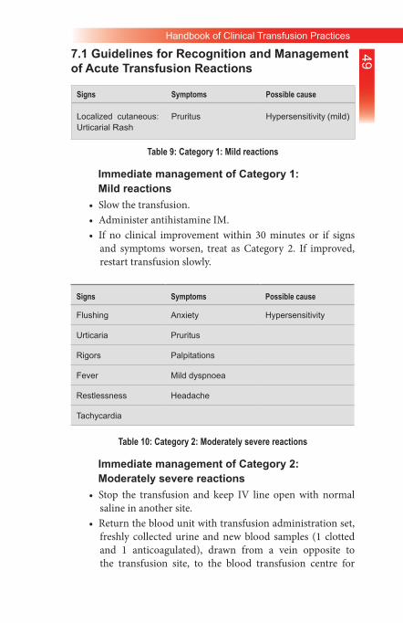
7.1 Guidelines for Recognition and Management of Acute Transfusion Reactions
Signs Symptoms Possible cause
Localized cutaneous:Urticarial Rash
Pruritus Hypersensitivity (mild)
Table 9: Category 1: Mild reactions
Immediate management of Category 1: Mild reactions
• Slow the transfusion.• Administer antihistamine IM.• If no clinical improvement within 30 minutes or if signs
and symptoms worsen, treat as Category 2. If improved, restart transfusion slowly.
Table 10: Category 2: Moderately severe reactions
Immediate management of Category 2: Moderately severe reactions
• Stop the transfusion and keep IV line open with normal saline in another site.
• Return the blood unit with transfusion administration set, freshly collected urine and new blood samples (1 clotted and 1 anticoagulated), drawn from a vein opposite to the transfusion site, to the blood transfusion centre for
Signs Symptoms Possible cause
Flushing Anxiety Hypersensitivity
Urticaria Pruritus
Rigors Palpitations
Fever Mild dyspnoea
Restlessness Headache
Tachycardia
50Handbook of Clinical Transfusion Practices
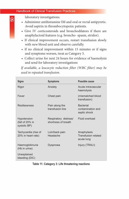
laboratory investigations.• Administer antihistamine IM and oral or rectal antipyretic.
Avoid aspirin in thrombocytopenic patients.• Give IV corticosteroids and bronchodilators if there are
anaphylactoid features (e.g. broncho‐ spasm, stridor).• If clinical improvement occurs, restart transfusion slowly
with new blood unit and observe carefully.• If no clinical improvement within 15 minutes or if signs
and symptoms worsen, treat as Category 3.• Collect urine for next 24 hours for evidence of haemolysis
and send for laboratory investigationsIf available, a leucocyte reduction filter (WBC filter) may be used in repeated transfusion.
Signs Symptoms Possible cause
Rigor Anxiety Acute intravascular haemolysis
Fever Chest pain (mismatched blood transfusion)
Restlessness Pain along the transfusion line
Bacterial contamination and septic shock
Hypotension (fall of 20% insystolic BP)
Respiratory distress/shortness of breath
Fluid overload
Tachycardia (rise of 20% in heart rate)
Loin/back painHeadache
AnaphylaxisTransfusion related acute lung
Haemoglobinuria (Hb in urine)
Dyspnoea Injury (TRALI)
Unexplained bleeding (DIC)
Table 11: Category 3: Life threatening reactions

51Handbook of Clinical Transfusion Practices
Immediate management of Category 3: Life-threatening reactions
• Stop the transfusion and keep IV line open with normal saline in another site.
• Infuse normal saline to maintain systolic BP.• Maintain airway and give high flow oxygen by mask.• Give adrenaline (as 1:1000 solution) 0.01 mg/kg body
weight by slow intramuscular injection.• Give IV corticosteroids and bronchodilators if there are
anaphylactoid features.• Give diuretic: e.g. frusemide 1 mg/kg IV or equivalent.• Check a fresh urine specimen visually for signs of
haemoglobinuria.• Notify the superior or senior doctor attending the patient,
and the blood centre immediately.• Send blood unit with transfusion set, fresh urine sample
and new blood samples (1 clotted and 1 anticoagulated), drawn from a vein opposite the infusion site, with the appropriate request form to the blood transfusion centre for investigation.
• Start a 24‐hour urine collection and record all intake and output. Maintain fluid balance chart.
• Assess for bleeding from puncture sites or wounds. If there is clinical or laboratory evidence of DIC, give platelets (adult: 4‐6 units) and either cryoprecipitate (adult: 12 units) or FFP (adult: 3 units).
• Reassess. If hypotensive:– Give further saline.– Give inotrope, if available.
• If urine output falls or there is laboratory evidence of acute renal failure (rising K+, urea, creatinine):– Maintain fluid balance accurately.– Give further diuretic: e.g. frusemide 1 mg/kg IV or
equivalent.– Consider dopamine infusion, if available.– Seek expert help: the patient may need renal dialysis.
• If bacteraemia is suspected (rigor, fever, collapse, no

52Handbook of Clinical Transfusion Practices
evidence of a haemolytic reaction), start a broad‐spectrum antibiotic IV.
7.2 Investigating Acute Transfusion ReactionsImmediately report all acute transfusion reactions, with the exception of mild hypersensitivity (Category 1) to the doctor responsible for the patient and to the blood transfusion centre that supplied the blood. If a severe life‐threatening reaction is suspected, seek help immediately from the blood transfusion expert/anaesthetist/emergency team/whoever is available and skilled to assist.
• Record the following information on the patient’s notes:– Type of transfusion reaction.– Time lapse between start of transfusion and transfusion
reaction.– Volume, type and bag number of blood products
transfused.• Immediately take post‐transfusion blood samples (1 clotted
and 1 anti‐coagulated) from the vein opposite the transfusion site and forward to the blood centre for investigation of the following:
– Repeat ABO and RhD group.– Repeat antibody screen and cross match.– Full blood count.– Coagulation screen.– Direct antiglobulin test.– Urea and creatinine.– Electrolytes.
• Also return the following to the blood centre:– Blood bag and transfusion set containing red cell and
plasma residues from the transfused unit.– Blood culture in a special blood culture bottle.– First specimen of the patient’s urine following the reaction.– Completely filled transfusion reaction report form.
• After the initial investigation of the reaction, send patient’s 24‐hour urine sample to the blood transfusion centre for laboratory investigation.
• Record the results of the investigations for future follow‐up.

53Handbook of Clinical Transfusion Practices
7.3 Haemolytic Transfusion ReactionAn acute haemolytic transfusion reaction is the result of a mismatched blood transfusion, and causes acute intravascular haemolysis.
• Acute intravascular haemolytic reaction is caused by the transfusion of incompatible red cells, i.e. mismatched blood. Antibodies in the patient’s plasma haemolyse the incompatible red cells transfused.
• Even a small volume (5‐10 mL) of incompatible blood can cause a severe reaction and larger volume increases the risk.
• The most common cause of reaction is ABO incompatible transfusion. This almost always arises from:
– Errors in the blood request form.– Taking blood from the wrong patient into a pre‐labelled
sample tube.– Incorrect labelling of the blood sample tube sent to the
blood transfusion centre.– Inadequate checking of the blood label against the patient’s
identity.• Antibodies in the patient’s plasma against other red cell
antigens present on transfused blood, such as those of the Kidd, Kell or Duffy blood group systems, can also cause acute haemolysis.
• In the conscious patient, signs and symptoms usually appear within minutes of commencing the transfusion, sometimes when <10 mL blood has been given.
• In an unconscious or anaesthetized patient, hypotension and uncontrollable bleeding, from the transfusion site, may be the only sign of an incompatible transfusion.
• It is therefore essential to monitor the patient from the commencement of the transfusion up to its completion.
Prevention:• Correctly label blood sample and request form.• Place the patient’s blood sample in the correct sample tube.• Always check the blood unit against the identity of the
patient at the bedside before transfusion.

54Handbook of Clinical Transfusion Practices
7.4 Bacterial Contamination and Septic Shock• Bacterial contamination affects up to 0.4% of red cells and
1‐2% of platelet concentrates.• Blood may become contaminated by:
– Bacteria from the donor’s skin entering the blood unit during collection (usually staphylococci).
– Bacteraemia present in the blood of the donor during collection (e.g. Yersinia).
– Improper handling during blood processing.– Defect or damage to the blood bag.– Thawing FFP or cryoprecipitate in a water‐bath (often
contaminated).• Some contaminants, particularly Pseudomonas species, grow
at +2°C to +6°C and can survive or multiply in refrigerated red cell units.
• Staphylococci grow in warmer conditions and are able to proliferate in PCs which are stored at +20°C to +24°C.
• Signs usually appear rapidly after starting infusion, but may be delayed for a few hours.
• A severe reaction may be characterized by sudden onset of high fever, rigors and hypotension.
• Urgent supportive care and high‐dose intravenous antibiotics are required.
7.5 Transfusion Associated Circulatory Overload• Transfusion associated circulatory overload (TACO), i.e.
fluid overload, can result in heart failure and pulmonary oedema.
– May occur when:– Too much fluid is transfused.– The transfusion is given too rapidly.– Renal function is impaired.
• Fluid overload is particularly likely to happen in patients with:
– Chronic severe anaemia.– Underlying cardiovascular disease.
55Handbook of Clinical Transfusion Practices
7.6 Anaphylactic Reaction• This is a rare complication of transfusion of blood
components or plasma derivatives.• The risk is increased by rapid infusion, typically when fresh
frozen plasma is used.• IgA deficiency in the recipient is a rare cause of very severe
anaphylaxis. This can be caused by any blood product since most contain traces of IgA.
• Cytokines in the plasma may occasionally cause broncho‐constriction and vaso‐constriction in recipients.
• Occurs within minutes of starting the transfusion and is characterized by:
– Cardiovascular collapse.– Respiratory distress.– No fever.
• Anaphylaxis is likely to be fatal if it is not managed rapidly and aggressively.
7.7 Transfusion Related Acute Lung Injury• Transfusion related acute lung injury (TRALI) is usually
caused by donor plasma that contains antibodies against the patient’s leucocytes.
• Rapid failure of pulmonary function usually presents within 1‐4 hours of starting transfusion, with diffuse opacity on the chest X‐ray.
• There is no specific therapy. Intensive respiratory and general support in an intensive care unit is required.
7.8 Delayed Complications of Transfusion
7.8.1 Delayed haemolytic transfusion reactionSigns appear 5‐10 days after transfusion:
• Fever.• Anaemia.• Jaundice.• Occasionally haemoglobinuria.
Severe, life‐threatening delayed haemolytic transfusion reactions with shock, renal failure and DIC are rare.

56Handbook of Clinical Transfusion Practices
7.8.2 Post-transfusion purpura• This is a rare but potentially fatal complication of
transfusion of red cells or platelet concentrates, caused by antibodies directed against platelet‐specific antigens in the recipient.
• Most commonly seen in multigravida female patients.• Signs and symptoms:
– Signs of bleeding.– Acute, severe thrombocytopenia 5‐10 days after
transfusion, defined as a platelet count of <100 x 109/L.ManagementManagement becomes clinically important at a platelet count of 50 x 109/L, with a danger of hidden (occult) bleeding at 20 x 109/L.
• Give high dose corticosteroids.• Give high dose IV immunoglobulin, 2 g/kg or 0.4
g/kg for 5 days.• Plasma exchange.• Monitor the patient’s platelet count: normal range
is 150 x 109/L to 440 x 109/L.• If available, give platelet concentrates that are
negative for the platelet‐specific antigen against which the antibodies are directed.
• Unmatched platelet transfusion is generally ineffective.
• Recovery of platelet count after 2‐4 weeks is usual.
7.8.3 Transfusion associated graft-versus-host disease (TA-GVHD)
• Unlike transplant associated GVHD, TA‐GVHD is usually a fatal condition.
• Occurs in patients such as:– Immuno‐deficient recipients of bone marrow
transplants.– Immuno‐competent patients transfused with blood from
individuals with whom they have a compatible HLA tissue type, usually blood relatives particularly 1st degree.

57Handbook of Clinical Transfusion Practices
• Signs and symptoms typically occur 10‐12 days after transfusion and are characterized by:– Fever.– Skin rash and desquamation.– Diarrhoea.– Hepatitis.– Pancytopenia.
ManagementTreatment is supportive; there is no specific therapy.
PreventionDo not use 1st degree relatives as donors, unless gamma irradiation of cellular blood components is carried out to prevent the proliferation of transfused lymphocytes.
7.8.4 Delayed complications: Transfusion transmitted infections
• The following infections may be transmitted by transfusion:• HIV, Hepatitis B and C, syphilis (Treponema pallidum),
malaria.• Cytomegalovirus (CMV).• Other TTIs include human parvovirus B19, brucellosis,
Epstein‐Barr virus, toxoplasmosis, Chagas disease, infectious mononucleosis and Lyme’s disease.Since a delayed transfusion reaction may occur days, weeks or months after the transfusion, the association with the transfusion may easily be overlooked. It is essential to record all transfusions accurately in the patient’s case notes and to consider transfusion in the differential diagnosis.

58Handbook of Clinical Transfusion Practices
8. Clinical Decisions on Transfusion
Assessing the need for transfusionThe decision to transfuse blood or blood products should always be based on a careful assessment of clinical and laboratory indications that transfusion is necessary to save life or prevent significant morbidity.
Transfusion is only one element of the patient’s management. See below for a summary of the main factors in determining whether transfusion may be required in addition to supportive management and treatment of the underlying condition.
faCtors determining tHe need for transfusion
Blood loss• External bleeding• Internal bleeding – non-traumatic: e.g.
— Peptic ulcer— Varices— Ectopic pregnancy— Antepartum haemorrhage— Ruptured uterus
• Internal bleeding – traumatic:— Chest— Spleen— Pelvis— Femur
• Red cell destruction: e.g. malaria, sepsis, HIV
Haemolysis: e.g.• Malaria• Sepsis• Disseminated intravascular coagulation
Cardiorespiratory state and tissue oxygenation• Pulse rate• Blood pressure• Respiratory rate• Capillary refill

59Handbook of Clinical Transfusion Practices
• Peripheral pulses• Temperature of extremities• Dyspnoea• Cardiac failure• Angina• Conscious level• Urine output
Assessment of anaemia Clinical• Tongue• Palms• Eyes• Nails
Laboratory• Haemoglobin or haematocrit
Patient’s tolerance of blood loss and/or anaemia• Age• Other clinical conditions: e.g.
— Pre-eclampsic toxaemia— Renal failure— Cardiorespiratory disease— Chronic lung disease— Acute infection— Diabetes— Treatment with beta-blockers
Anticipated need for blood• Is surgery or anaesthesia anticipated?• Is bleeding continuing, stopped or likely to recur?• Is haemolysis continuing?
Table 12: Factors determining the need for transfusion
Prescribing decisions should be based on national guidelines on the clinical use of blood, taking individual patient needs into account. They should also be based on knowledge of local patterns of illness, the resources available for managing patients and the safety and availability of blood and intravenous replacement fluids. However, responsibility for the decision to transfuse ultimately rests with individual clinicians.

60Handbook of Clinical Transfusion Practices
presCribing blood: a CHeCklist for CliniCians
1. Before prescribing blood or blood products for a patient, ask yourself the following questions.
2. What improvement in the patient’s clinical condition am I aiming to achieve?
3. Can I minimize blood loss to reduce this patient’s need for transfusion?
4. Are there any other treatments I should give before making the decision to transfuse, such as intravenous replacement fluids and oxygen?
5. What are the specific clinical or laboratory indications for transfusion for this patient?
6. What are the risks of transmitting HIV, hepatitis, syphilis or other infectious agents through the blood products that are available for this patient?
7. Do the benefits of transfusion outweigh the risks for this particular patient?
8. What other options are there if no blood is available in time?
9. Will a trained person monitor this patient and respond immediately if any acute transfusion reactions occur?Have I recorded my decision and reasons for transfusion on the patient’s chart and the blood request form?
10. Finally, if in doubt, ask yourself the following question:
11. If this blood was for myself or my child, would I accept the transfusion in these circumstances?
Table 13: Prescribing blood: A checklist for clinicians

61Handbook of Clinical Transfusion Practices
9. Massive Blood TransfusionMassive blood transfusion may be defined as the replacement of one blood volume (equivalent to 10 units of blood) in any 24 hour period, or half of the blood volume (5 units of blood) in any four‐ hour period in an adult.
• Replacement of a blood volume equivalent within 24 hours.• >10 units within 24 hours.• Transfusion >4 units in 1 hour.-• Replacement of 50% of blood volume in 3‐4 hours.• A rate of loss >150 ml/hour.
Massive transfusion occurs in settings such as severe trauma, ruptured aortic aneurysm, surgery and obstetric complications. The goals to the management of massive transfusion include early recognition of blood loss, maintenance of tissue perfusion, oxygenation by restoration of blood volume and Hb, and the cessation of bleeding by several means including early surgical or radiological intervention, and the judicious use of blood component therapy to correct coagulopathy.
• Plasma undergoes progressive loss of coagulation factors during storage, particularly Factors V and VIII, unless stored at –30°C or colder.
• Dilution of coagulation factors and platelets will occur following administration of large volumes of replacement fluids. Massive or large volume transfusions can therefore result in disorders of coagulation.
9.1 Massive Transfusion ProtocolA massive transfusion protocol (MTP) should be used in critically bleeding patients anticipated to require massive transfusion. The parameters in Table 6 below should be measured early and frequently (every 30‐60 minutes, or after transfusion of blood component).

62Handbook of Clinical Transfusion Practices
Parameter Values for which to aim
Temperature >35°C
Acid-base status pH >7.2, base excess < 6, lactate <4 mmol/L
Ionised calcium (Ca)
>1.1 mmol/L
Haemoglobin (Hb)
This should not be used alone as a transfusion trigger; and, should be interpreted in context with haemodynamic status, organ and tissue perfusion
Platelets (Plt) ≥50 x 109 /L
PT/APTT (activated partial thromboplastin time)
≥1.5 x of normal
Fibrinogen ≥1.0 g/L
Table 14: Parameters in massive transfusion – investigation and monitoringMortality is high in massive transfusion and its aetiology is multifactorial, which includes hypotension, acidosis, coagulopathy, shock and the underlying condition of the patient. The lethal triad, i.e. patients with acidosis, hypothermia, and coagulopathy have the highest mortality rate. It is often the underlying cause and consequences of major haemorrhage that result in complications, rather than the transfusion itself. However, administering large volumes of blood and intravenous fluids may itself give rise to the following complications.
AcidosisAcidosis in a patient receiving a large volume transfusion is more likely to be the result of inadequate treatment of hypovolaemia than due to the effects of transfusion. Under normal circumstances, the body can readily neutralize this acid load from transfusion. The routine

63Handbook of Clinical Transfusion Practices
use of bicarbonate or other alkalizing agents, based on the number of units transfused, is unnecessary.
Hyperkalaemia
The storage of blood results in a small increase in extra‐cellular potassium concentration, which will increase the longer it is stored. This rise is rarely of clinical significance, other than in neonatal exchange transfusions. Fresh blood (up to 7 days old) should be requested from the blood centre.
Citrate toxicity and hypocalcaemiaCitrate toxicity is rare, but is most likely to occur during the course of a large volume transfusion of whole blood. Hypocalcaemia, particularly in combination with hypothermia and acidosis, can cause a reduction in cardiac output, bradycardia, and other dysrhythmias. Citrate is usually rapidly metabolized to bicarbonate. It is therefore unnecessary to attempt to neutralize the acid load of transfusion. There is very little citrate in red cell concentrates.
9.2 Management• If there is prolongation of PT, give ABO compatible fresh
frozen plasma in a dose of 15 mL/kg.• If the APTT is also prolonged, Factor VIII/fibrinogen
concentrate is recommended in addition to FFP. If none is available, give 10‐15 units of ABO compatible cryoprecipitate, which contains Factor VIII and fibrinogen.
• Give PCs only when:– The patient shows clinical signs of microvascular
bleeding: i.e. bleeding and oozing from mucous membranes, wounds, raw surfaces and catheter sites.
– The patient’s platelet count falls below 50 x 109/L.• Give sufficient PCs to stop microvascular bleeding and
maintain an adequate platelet count.

64Handbook of Clinical Transfusion Practices
Consider PC transfusion in cases where the platelet count falls below 20 x 109/L, even if there is no clinical evidence of bleeding, because there is a danger of occult bleeding, such as into brain tissue.
The prophylactic use of platelet concentrates in patients receiving large volume blood transfusions is not recommended.
65Handbook of Clinical Transfusion Practices
ANNEXURES

66Handbook of Clinical Transfusion Practices
Patient ID Date
Patient Name Age Sex
Diagnosis Ward
Bed Number
Blood Group Pre-transfusion Hb g/dL
Date
Transfusion Start time
Doctor’s Signature
Source of Blood Bag number
Collection Date Type of component
Amount of blood Cross match and Screening Lab ID
Parameter Pre- transfusion
15 min
30 min 1 hr 2 hrs 3 hrs 4 hrs
Pulse
BP
Temperature
Respiration
Colour of urine
Drop rate/min
Time transfusion completed: √
Completion of transfusion note (check answer)
Transfusion was successful without any adverse reactionTransfusion was deferred due to adverse reaction of severe category
Transfusion was completed with management of febrile/allergic reaction
Name of HospitalBedside Transfusion Notes
Logo
Annexure 1: Bedside Transfusion Notes

67Handbook of Clinical Transfusion Practices
Name of HospitalBedside Adverse Transfusion Reaction Form
Logo
Annexure 2: Management of Adverse Transfusion Reaction: Physician’s Notes
Reported by________________ Date reaction occured_____________
Name of recipient_____________________ Hospital ID_____________
Age of recipient______years Sex of recipient Male Female
Date of transfusion (day/month/year/) _____________
Adverse reaction in relation with Whole blood Platelets
Packed red cells
Fresh frozen plasma
Others (Specify) ____________________
Chief Complaint/Concern Treatment:
On Examination:• BP• Respiratory rate • Pulse• Sign of bleeding
Investigation:Blood samples:• blood bag• patient urine• culture• electrolytes• BT• CT• APTT• Bilirubin• direct antiglobulin test
Report of blood transfusion centre
Instruction of Experts: (trans-fusion specialist/ Anaesthetic/ Departmental head)
Treatment:
Signature of Doctor

68Handbook of Clinical Transfusion Practices
Patient’s Name ____________________________________________
W/O, S/O, D/O ____________________________________________
Age /Sex _______ / _______ Date __________Ward ______________
Bed No. ________________
Diagnosis ________________________________________________
Indication for transfusion_____________________________________
Previous history of transfusions _______________________________
Any transfusion reaction _____________________________________
Previous pregnancies _______________________________________
Haemoglobin ______________________________________________
Platelet count (if platelets required) _____________________________
Request for Red Cell Concentrates
Fresh Frozen Plasma Platelet Concentrates
Whole Blood
No. of blood units required ___________________________________
Blood group (if done earlier) __________________________________
Date and time when required _________________________________
Date and time blood sample taken _____________________________
Name and signatures of requesting doctor _______________________
Name of HospitalBlood Request Form
Logo
Annexure 3: Blood Request Form
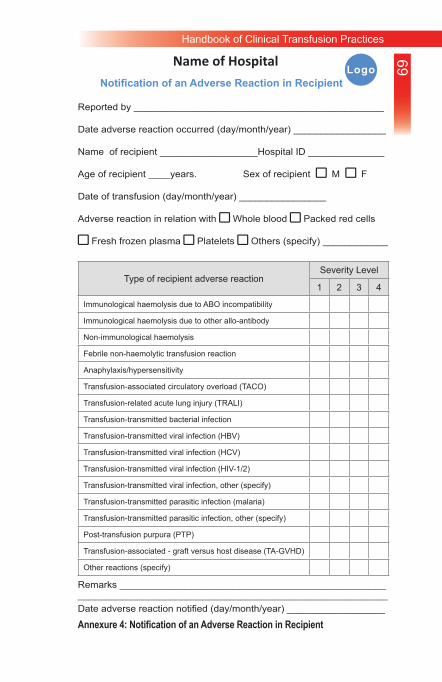
69Handbook of Clinical Transfusion Practices
Reported by ______________________________________________
Date adverse reaction occurred (day/month/year) _________________
Name of recipient __________________Hospital ID ______________
Age of recipient ____years. Sex of recipient square M square F
Date of transfusion (day/month/year) ________________
Adverse reaction in relation with square Whole blood square Packed red cells
square Fresh frozen plasma square Platelets square Others (specify) ____________
Type of recipient adverse reactionSeverity Level
1 2 3 4
Immunological haemolysis due to ABO incompatibility
Immunological haemolysis due to other allo-antibody
Non-immunological haemolysis
Febrile non-haemolytic transfusion reaction
Anaphylaxis/hypersensitivity
Transfusion-associated circulatory overload (TACO)
Transfusion-related acute lung injury (TRALI)
Transfusion-transmitted bacterial infection
Transfusion-transmitted viral infection (HBV)
Transfusion-transmitted viral infection (HCV)
Transfusion-transmitted viral infection (HIV-1/2)
Transfusion-transmitted viral infection, other (specify)
Transfusion-transmitted parasitic infection (malaria)
Transfusion-transmitted parasitic infection, other (specify)
Post-transfusion purpura (PTP)
Transfusion-associated - graft versus host disease (TA-GVHD)
Other reactions (specify)
Remarks __________________________________________________________________________________________________________Date adverse reaction notified (day/month/year) __________________
Name of HospitalNotification of an Adverse Reaction in Recipient
Logo
Annexure 4: Notification of an Adverse Reaction in Recipient
