copyright and use of this thesisses.library.usyd.edu.au/bitstream/2123/4054/4/lachlan... · ·...
TRANSCRIPT

Copyright and use of this thesis
This thesis must be used in accordance with the provisions of the Copyright Act 1968.
Reproduction of material protected by copyright may be an infringement of copyright and copyright owners may be entitled to take legal action against persons who infringe their copyright.
Section 51 (2) of the Copyright Act permits an authorized officer of a university library or archives to provide a copy (by communication or otherwise) of an unpublished thesis kept in the library or archives, to a person who satisfies the authorized officer that he or she requires the reproduction for the purposes of research or study.
The Copyright Act grants the creator of a work a number of moral rights, specifically the right of attribution, the right against false attribution and the right of integrity.
You may infringe the author’s moral rights if you:
- fail to acknowledge the author of this thesis if you quote sections from the work
- attribute this thesis to another author
- subject this thesis to derogatory treatment which may prejudice the author’s reputation
For further information contact the University’s Copyright Service.
sydney.edu.au/copyright
9. Manuscript
The preventive effects of systemic casein phosphopeptides
on the resorption of roots in rats
The condensed version of this manuscript is to be submitted to the American
Journal of Orthodontics and Dentofacial Orthopedics
76

The preventive effects of systemic casein phosphopeptides on the resorption
of roots in rats
Lachlan Crowther BDSc Postgraduate student Discipline of Orthodontics Faculty of Dentistry University of Sydney Nour Eldin Tarraf BDS Postgraduate student Discipline of Orthodontics Faculty of Dentistry University of Sydney Gang Shen BDS, MDS, PhD Associate Professor Discipline of orthodontics Faculty of Dentistry University of Sydney Rema Oliver PhD Surgical and Orthopaedic Research Laboratories University of New South Wales Prince of Wales Hospital William R Walsh PhD Professor Surgical and Orthopaedic Research Laboratories University of New South Wales Prince of Wales Hospital Alan Jones PhD Senior Lecturer Electronic Microscopic Unit University of Sydney.
77
Peter Petocz PhD Associate Professor Department of Statistics Macquarie University, Sydney Honorary Associate Discipline of Orthodontics Faculty of Dentistry University of Sydney M.Ali Darendeliler BDS. PhD, DipOrth, CertifOrtho, PrivDoc Professor and Chair Discipline of Orthodontics Faculty of Dentistry University of Sydney. Address for correspondence: Professor M.Ali Darendeliler Discipline of Orthodontics Faculty of Dentistry The University of Sydney Level 2, 2 Chalmers Street Surry Hills NSW 2010 Australia Phone +61 2 93518314 Fax +61 2 9351 8336 Email: [email protected]
78
9.1 Abstract
Introduction: Root resorption is a well documented side effect of orthodontic treatment.
The aim of this study is to assess the potential for casein phosphopeptides (CPPs) to
create an environment in which teeth are less susceptible to root resorption following
orthodontic tooth movement.
Method: Thirty one eleven week old, Wistar rats, were used in this experiment.
Experimental and control animals were given the same diet of nutrigel (Troy Laboratories
Pty. Ltd, NSW, Australia) mixed with rat chow with the same frequency and had free
access to water throughout the whole experimental period. The 14 experimental rats
received a diet supplemented with 40mg CPP per 100g of rat while the 17 control rats did
not receive any supplements in their diet. The rats underwent a 2-week period of
acclimatization with their respective diets followed by a two week experimental period
after which the animals were euthanized and the samples were harvested. The tooth
movement appliance was a 150cN sentalloy® (GAC Australia) closed coil spring (wire
diameter 0.22mm, eyelet diameter 0.56 mm) applied from the maxillary incisor randomly
to the right and left first maxillary molar. A volumetric analysis of the extent of root
resorption on the mesial root of the first maxillary molar was examined using a micro CT
scan.
Results: The rats which received dietary CPP did not have a significant reduction in
overall root resorption (p=0.28) although the results did reveal that dietary CPP appeared
to lower the average root resorption by about a 15%. Dietary CPP was also found to have
no statistically significant effect on decreasing tooth movement (p=0.42) although the
average tooth movement was reduced by about 15%.
79
Conclusions: CPP seems to have a variable effect on the volumetric quantification of
root resorption. While on average, there was less root resorption observed in rats fed
dietary CPP, individual variability makes this effect statistically insignificant. Similarly,
CPP did not have a statistically significant effect on reducing tooth movement. Although
it is possible that CPP may have a beneficial effect on reducing cementum solubility, this
may be counteracted by its anabolic effect on bone mass which explains the variability
observed in this study.
Key words: Casein Phosphopeptieds, Root resorption, 3D Micro CT, Volumetric
analysis, Orthodontic tooth movement, Wistar rats
80
9.2 Introduction
Casein Phosphopeptides (CPPs) are multi-phosphorylated peptides from an enzymatic
digest of the bovine milk protein casein.1 In milk, casein stabilizes the structure of the
liquid in order for it to maintain its high calcium phosphate concentration without
allowing precipitation.2
Bioactivity of phosphopeptides yielded after tryptic hydrolysis of casein was reported
more than 50 years ago by Mellander who discovered that CPPs were found to improve
calcium balance in rachitic newborns.3
CPPs have been shown to enhance calcium absorption by increasing calcium solubility in
vitro and in situ.4 CPPs have been shown to enhance paracellular transport of calcium in
the distal small intestine in elderly female rats at least under conditions of marginal
Calcium levels.5 It has been found that rats fed a diet supplemented with CPP have less
bone loss than rats given Ca and P as pure minerals.4
The positive effects of CPP supplementation on the skeleton has also been demonstrated
in humans. Mellander demonstrated that calcium bound to phosphopeptides could be
absorbed from the digestive tract and promote bone calcification in rachitic children.6
Heaney et al7 studied the effects of administering 87.5mg of CPP to 35 normal post-
menopausal women as a part of a standard test meal containing a calcium load of 250mg.
Their findings suggested that casein phosphopeptide supplementation is particularly
useful for persons with low basal absorptive performance.
81
In recent years there has been significant interest in the potential for casesin
phosphopeptides to prevent and remineralise carious lesions. Casein Phosphopeptides
(CPP) stabilize amorphous calcium phosphate (ACP), localize ACP in dental plaque and
are anticariogenic in animal and in vivo human caries models.1
The use of a remineralizing solution containing calcium and phosphate ions have not
been clinically successful due to the low solubility of calcium and phosphates.1 Calcium
and phosphate ions at low concentrations do not incorporate to any significant degree into
plaque or localize at the tooth surface.1 In contrast, dairy products are a food group
which are widely recognized to exhibit anti-caries activity.8,9 Animal and in situ caries
models have demonstrated that the components responsible for the anticariogenic nature
of dairy products are the casein, calcium and phosphate.1
Reynolds has proposed that the anticariogenic mechanism for CPP-ACP is the
localization of ACP at the tooth surface which buffers the free calcium and phosphate ion
activities, thereby helping to maintain a state of supersaturation with respect to tooth
enamel depressing demineralization and enhancing remineralization.1 CPP-ACP has
proved to be easy to deliver in the diets of both humans and rats. Reynolds et al1,10 have
exposed the rats to CPP-ACP solution in their drinking water while in humans the CPP-
ACP complex has been delivered in solution,1 in sugar-free gum11,12 and it has also been
incorporated into a self-cured glass-ionomer cement (GIC).13 CPP is not considered to be
a hazardous product.14 Toxicological tests on GC tooth mousse which is a CPP
82
containing product showed no death in rats following oral consumption at a concentration
of 200mg/kg.14
Root resorption is a physiologic or pathologic process that results in a loss of substance
from dentine or cementum.15 Physiological root resorption occurs during the exfoliation
of deciduous teeth. Pathological root resorption may be either internal or external in
origin. Internal root resorption is initiated from within the pulp while external root
resorption arises from the periodontium affecting the external surface of the tooth.
The mineralized tissues of the permanent dentition are normally protected in the root
canal by dentine and osteoblasts, and on the root surface by cementum and
cementoblasts.16,17 Resorption of the root surface is not a normal process.18 However if
the cementum is mechanically damaged, multi nucleated cells will arrive near the surface
and resorption will occur.19 In the field of orthodontics, this process has been termed,
Orthodontically Induced Inflammatory Root Resorption (OIIRR).20 OIIRR occurs on the
cemental surface of the tooth root. Although cementum is more resistant to resorption
relative to bone it is still possible for both the cementum and dentine to resorb as a result
of this inflammatory process. This process is an unavoidable consequence of orthodontic
tooth movement.
Risk factors for OIIRR may be classified as either systemic, local or treatment related.
Systemic factors include genetic predisposition,21-24 ethnicity,25,26 chronological age,27
dental age,28 endocrine imbalance,29-32 nutirion,28,33 alveolar bone density and
83

turnover.33,34 Local factors include habits,28,35 history of trauma,28,36 root resorption prior
to orthodontic treatment,37,38 abnormal or dilacerated roots,35,39 a hypofunctional
periodontium40,41 and increased overjet and openbite.25,28,38,42. Treatment related factors
included extended duration of treatment,28,43-48 the use of rapid maxillary expanders,49,50
heavy orthodontic forces,50-53 orthodontic intrusion,54,55 contact with the cortical plate56
and the distance moved by the tooth roots.25,26
Like bone, teeth also require a balance in mineral content in order to prevent resorption.
The ability for CPP to effectively provide bioavailable calcium for maintaining
mineralization of both the skeleton and the dental tissues has been extensively
demonstrated in both rat and human models.
The association between bone metabolism and root resorption has also been
demonstrated in studies which have looked at the effects of medication which increase
the amount of bone resorption. A study by Kalia et al57 on rats demonstrated that
subjects which received long term corticosteroids had a significant increase in the relative
extent of root resorption in rats who received both chronic and acute corticosteroid
treatment. This can be explained by the evidence that long term side effects of steroid
therapy which include disturbances in mineralized tissue metabolism and wound healing,
discrepancies in chondrogenesis and osteogenesis, bone loss and osteoporosis.58 This
lends support to the theory that mineralization of the bones and the teeth have an effect
on the levels of root resorption during orthodontic tooth movement.
84
Based on the association between bone resorption, tooth movement and root resorption it
follows that if the mineralization of the bone and dental tissues were to be increased, then
there is a potential to protect teeth against orthodontically induced inflammatory root
resorption. The importance of minerals with respect to root resorption has been
demonstrated by Rex et al59 who performed an electron probe micoranalysis (EPMA) on
the concentrations of calcium (Ca), phosphorous (P), and fluoride (F) concentrations in
human first premolar cementum after the application of light and heavy orthodontic
forces. The application of heavy forces caused a significant decrease in the calcium
concentration of cementum at certain areas of periodontal ligament tension which
demonstrates the dematerializing nature of orthodontic force.
Seifi et al60 demonstrated how the provision of calcium can potentially protect against
root resorption. They studied the effect of submucosal injections of Prostaglandins in
association with intraperiotoneal injections of Calcium Gluconate on root resorption in
rats. They showed that while the prostaglandins increased tooth movement, the
combination with Calcium Gluconate led to a decrease in root resorption.60 Goldie and
King33 studied the association of parathyroid hormone with root resorption. The main
action of parathyroid hormone is to mobilize calcium from bone. They showed that there
is an increased risk of root resorption associated with hyperparathyroidism.33 These
studies lend support to the suggestion that protection against root resorption may be
achieved if calcium can be made available to the periodontium during orthodontic tooth
movement.
85

The ability for CPP to effectively provide bioavailable calcium for maintaining the
mineralization of both the skeleton and the dental tissue4,5,61 is of significance due to the
importance of calcium in the maintenance of tooth structure. The influence of diet on the
structure of teeth has been studied in depth. Of particular interest to this study are the
factors which influence the hardness of the teeth. Bielacyc and Golebiewska in 1997
demonstrated the importance of calcium and vitamin D in roots of teeth in rats.62
Scanning microscope observations showed the increased cementolysis and decreased
mineralization of cementum and dentin in rats fed a low calcium and vitamin D-deficient
diet. Some of the earliest studies in this field were conducted by Mellanby.63,64 Mellanby
concluded that a low dietary calcium content may result in teeth being soft to the point
that they can be cut with a scalpel.63 Mellanby demonstrated that well and badly calcified
teeth can be produced at will by altering in the diet the relative amounts of calcifying
vitamin, found in milk, egg-yolk, cod-liver oil, etc., and anti-calcifying substances found
chiefly in cereals.64 These studies demonstrate how dietary CPP, which increases
bioavailable calcium, may be of benefit in increasing the hardness of teeth which may
help to decreasing their susceptibility to root resorption.
Dairy products are particularly effective in storing calcium because the milk protein
casein stabilizes the structure of the liquid which allows it to maintain its high calcium
phosphate concentration without allowing precipitation.2 It follows that the provision of
dietary casein would be of potential benefit in making calcium available for the
protection of tooth roots against orthodontically induced inflammatory root resorption.
86

The question therefore follows as to whether or not CPP can offer potential benefits to
teeth which are at risk of losing mineral content as is the case in teeth which are loaded
with orthodontic force. CPP has been demonstrated to be a safe, natural substance when
delivered topically or systemically in both rat and human models.7,10-14 CPP could
therefore be a potentially effective and safe way of preventing root resorption. This study
was undertaken to determine whether or not casein phosphopeptides have the potential to
protect teeth against orthodontically induced inflammatory root resorption in a rat
experimental model.
87

9.3 Materials and Methods
Ethical approval was sought and approved by the University of New South Wales (ACEC
Number: 07/32A). Forty, 11 week old female Wistar rats were used in this study. The
rats were allowed 2 weeks to acclimatise to their new laboratory environment and were
housed with two animals per cage.
CPP delivery
Casein Phosphopeptides can be obtained in a powdered form and this can be easily mixed
in the diet.5 Both the experimental and control rats received water, ad libitum and a diet
of nutrigel (Troy Laboratories Pty. Ltd, NSW, Australia) mixed with rat chow. Nutrigel
is an oral supplement which is used in veterinary medicine to provide partial or full
nutritional support for mammals.
The rats received 5ml of nutrigel per day mixed with 5g of ground rat chow on a daily
basis. The rat chow was mixed with the nutrigel to provide a source of fibre in the diet.
The rat chow was ground so as not to pose a risk of damage to the appliances. The
experimental rats were given 40mg CPP/100g body weight on a daily basis for 29 days.5
A visual estimation of the consumption of the nutrigel was made on a daily basis.
Orthodontic tooth movement
Orthodontic tooth movement was implemented by use of a Nickel Titanium closed coil
(GAC USA), with a force of 150g. The NiTi coils were assigned randomly to the left and
right rat maxillary first molars. The coils were ligated from the first maxillary molar to
88

both maxillary incisors with steel ligiatures. Figure 1 The wires were then bonded to
both the incisor anchor unit and the first maxillary molar with composite resin. Figure 2
Attachment of the nickel titanium coils to the teeth was performed under general
anaesthesia. The rats were weighed, and they were constantly monitored for changes in
vital areas (e.g. breathing) during surgery. The animals were induced and maintained
using isoflurane (2%) and oxygen (2%) inhalation during surgery. The rats were given
the analgesic Temgesic (Buprenorphine) at a dose of 0.01mg/kg post-operatively.
The rats were divided into 2 groups Figure 3
Group 1 Control
Group 1 (n=20) had orthodontic tooth movement but did not receive any CPP
supplements
Group 2 Experimental
Group 2 (n=20) had orthodontic tooth movement and received a dietary supplement of
40mg CPP /100g body weight.
Two experimental rats were lost due to anaesthetic deaths and seven rats were eliminated
from the study due to appliance failure. 17 control rats and 14 experimental rats were left
at the completion of the study.
The acclimatisation period was 2 weeks in which the rats received their diet with or
without supplements. The experimentation period during which the tooth movement
appliances were active was 2 weeks. Figure 3 After this period, the rats were euthanized
89
using carbon dioxide. The maxillae were then dissected and sectioned to include only the
segment of the palate with the three molars on the side where the orthodontic force was
applied. The samples were then stored in formalin.
SkyScan 1172 Micro CT
The maxillae were scanned with a SkyScan 1172 Micro CT which is a compact, desktop
x-ray system used for the non destructive three dimensional reconstructions of samples.
Figure 4 There is a significant risk of root fracture and damage to tooth roots when
extracting rat molars and therefore the ability to digitally remove the bone from the teeth
is of advantage because it eliminates their need for their extraction. The software
package enables non destructive evaluation of the molar root surfaces and digital
measurements of the molar movements.65
The scanner is a cone beam x-ray source with a spatial resolution of between 2 and 5
micrometres. The recommended sample size is a diameter of 1.5 cm and a height of 3
cm. The sample is placed on a rotating platform, and depending on its proximity to the x-
ray beam, there is a magnification factor. A high resolution Charged Coupled Device
with a resolution of 1024 x 1024 pixels detected the incoming x-rays. Figure 5 The
scanned segments of the maxillae included the three molars on the side of the palate on
which the orthodontic force was applied. Figure 6 The samples were scanned at a
resolution of 6 µm. Throughout the scanning procedure, the samples were rotated 360
degrees with a scanning period of 2 seconds per degree of rotation. The average scanning
time per sample was around 1 hours and 15 minutes.
90

Non destructive three dimensional reconstruction of the objects’ inner structure from two
dimensional x-ray shadow projections were achieved with the software program NRecon
version 1.4.2. Approximately 1800 cross sections were collected per sample and the
reconstructions took approximately 6 hours to complete. Once this was completed, the
raw data was converted to 8 bit, Bit Mapped Picture files, with a resolution of 1024x1024
pixels. The software package, VGStudioMax v1.2 was then used to collate all the axial
slices to form a three dimensional reconstruction of the scanned images.
VGStudioMax 1.2
VGStudioMax v1.2 was used to collect the data to create viewable three dimensional
reconstructions of the samples with 256 shades of gray. Only the first maxillary molar
and the mesial portion of the second molar were reconstructed for the purposes of data
collection. Figure 7 The bone was not removed from the images for the purposes of data
collection. However for the purposes of visualisation for the sample, the bone can be
digitally removed and the image of the tooth Figure 8 can be manipulated to allow for
visualisation of resorption craters. Figure 9
VGStudioMax v1.2 crater isolation
Due to the variability and size of the molar roots it was decided that only the mesial root
of the maxillary molar was large enough to ensure consistent recording of the location of
the root resorption craters. Furthermore it was found that the apical section of the molar
roots was too porous to ensure a clear differentiation between normal tooth anatomy and
91
root resorption craters. Figure 10 It was found that there was a definite delineation of the
porous section of the molar root and this could be delineated accurately to within a few
axial sections. It was therefore decided that for the purposes of data collection we would
only use the section of the most mesial, first maxillary molar tooth between the
cementoenamel junction and the start of the porous apex. Figure 10
For the purposes of collecting root resorption data, the images were viewed in axial
slices. A ‘mask’ was created for the volumetric analysis of the crater images. The
procedure was as follows. Figure 11
1. The section of the mesial molar root to be analysed between the CEJ and the
porous apex was defined.
2. A map was created of the crater locations and coordinates.
3. A ‘mask’ was created for each crater. The mask was created as follows:
a. The axial section at which the crater began was located
b. An outline of the crater was drawn using the VGstudiosMax segmentation
tool. The outline was drawn to follow the internal contours of the crater
while the external margin of the crater was an estimate of the continuation
of the convexity of the root surface. The continuation in convexity was
estimated by connecting the 2 points at the edge of the break in convexity
in a straight line.
c. The mask was then propagated through the axial section of the tooth for
no more than 6 slices.
92
d. The propagation of the slice was discontinued if the limits of the crater
were reached or if the mask no longer followed the outline of the crater. If
the mask ceased to resemble the crater a new outline was drawn.
4. The masks for all of the craters in the tooth were cumulated in a single
segmentation. Figure 12
5. VG StudioMax then calculated the volume of the segmented mask which was
recorded as the amount of root resorption.
Tooth Movement
Tooth movement was measured digitally utilizing the 3D micro-CT images. It was
assumed that the first and second molars were in contact prior to appliance placement and
therefore the tooth movement was measured as the distance between the first and second
molars at the end of the experimental period. The measurement was taken with a
software tool that is designed to identify and measure the closest distance between two
parallel or nearly parallel surfaces or lines. Figure 7
Due to variations in the angles at which the samples were scanned, there was
occasionally a difference in the axial and sagittal measurements of tooth movement
which was due to parallax error. Figure 13 The measurements were therefore taken in a
stepwise fashion. Firstly the observer scrolled down through the axial sections of the
contact area between the first and second molars and visually identified the axial section
that appeared to show the shortest distance between the two surfaces. This axial section
was used as a starting point. The tool was then employed to identify the line of shortest
93
distance between the distal surface of the first molar contact area to the second molar
contact area in the axial plane. Figure 14 The sagittal sections were then viewed and the
same software tool was used to identify the narrowest point between the heights of
contour of the proximal surfaces of the first and second molars in the occluso-gingival
dimension. Figure 15 These measurements were repeated until the measurements of the
axial and sagittal sections corresponded to the same position between the molar surfaces.
The axial and sagittal tooth movement measurements were not always identical due to
parallax errors associated with variations in the angle at which the samples were scanned.
In these cases the sagittal measurements and the last axial measurements were averaged.
94
9.4 Results
Weight
The average weight of rats which were included in the scanned samples at the start of
force application for group 1 control was 204 ± 26g and group 2 experimental was 213 ±
21g. The average finish weight for group 1 control was 243 ± 46g and group 2
experimental was 249 ± 19g. The average weight gained for the control group was 39g
and the average weight gained for the experimental group was 35.5g. Only one rat
experienced severe weight loss. This weight loss was attributed to appliance failure and
the rat was eliminated from the experiment.
CPP intake
It was anticipated that the average weight of the rats would reach 250g. This proved to
be a fair assumption as the average weight of the control group reached 243g while the
average weight of the experimental group reached 249g. The experimental rats were
therefore supplied with 100mg of CPP per day in accordance with the regime of
administering 40mg CPP/100g of rat.5 ie. 100mg of CPP mixed with 5ml of nutrigel per
day.
Nutrigel and Rat Chow consumed
The water consumed by the rats was given ad libitum and was not measured. Both the
experimental and control rats consumed the nutrigel/rat chow blend in entirety
throughout the duration of the experiment.
95
Accuracy of Measurements
Root Resorption
An intraoperator study was conducted whereby the same operator measured the same 10
samples on two separate occasions. The coefficient of variation (CV) was found to be
7.2%. Table I
Tooth Movement
The intraoperator error associated with the tooth movement measurement tool had a CV
of 1.5% in the sagittal sections and 2.4% in the axial sections. Table I
Distribution of root resorption cratering
Root resorption was only measured for the cervical portion of the most mesial root of the
first maxillary molar. Figure 10 The distribution of cratering in this section of the tooth
was almost entirely localised to the mesial aspect of the teeth.
Effects of CPP on the overall volume of root resorption
A comparison of the orthodontically loaded molars in the control group (Group 1) which
did not receive CPP with the orthodontically loaded molars in the experimental group
(Group 2) that received dietary CPP revealed that there was no significance in the
decrease of overall root resorption in the experimental group. (p=0.28) Table II,
Table III, Figure 16
96

Although the results did not reveal enough evidence to suggest that CPP had a
statistically significant effect on root resorption, it did appear to lower the average
volume of root resorption by about a 15%. Table II, Table III, Figure 16
Effect of CPP on Tooth Movement
A comparison of the orthodontically loaded molars in the control group (Group 1) which
did not receive CPP with the orthodontically loaded molars in the experimental group
(Group 2) that received dietary CPP revealed that there was no significant decrease in the
overall amount of tooth movement in the experimental group.(p=0.42) Table IV,
Table V, Figure 17
Although the results did not reveal enough evidence to suggest that CPP had a
statistically significant effect on tooth movement, it did appear to lower the average
amount of tooth movement by about a 15%. Table IV, Table V, Figure 17
97

9.5 Discussion
Methodology
This study aimed to investigate the influence of CPP on root resorption. An animal
model was chosen so it would be possible to control the level of dietary CPP. Wistar rats
were chosen because they have been successful subjects for root resorption experiments
using similar methodologies to those used in this experiment65,66 and they have also been
used in experiments involving CPP.5 All samples used were of the same age at the start
of the experiment (11 weeks), and therefore the development of their teeth was the same.
Matias et al67 indicated that by 8 weeks of age, there is complete development of root
dentine, cementum, periodontal ligaments and alveolar bone and for this reason mature
11 week old rats were chosen for this experiment.
The samples were divided into two groups. Both the control and experimental groups
received orthodontic force. It has been shown that orthodontically loaded teeth show
more root resorption than unloaded teeth and therefore the study was limited to
evaluating the effect of the CPP dietary supplement.65
The experimental and control groups had orthodontic force applied to the maxillary first
molars to initiate root resorption using a continuous force implemented by use of a
Nickel Titanium closed coil (GAC) spring.65,66,68-74 A heavy force of 150g was used
because it was thought that if a reduction in root resorption was to be seen with dietary
CPP, it would be more likely to be clearly seen with a heavy force. Figure 2 The aim of
the study was to analyze the maximum effect of the resorptive defects before any
98
reparative processes occurred. Hellsing and Hammarstrom75 looked at resorption sites
using a scanning electron microscope and found definitive resorption sites forming within
a week of appliance placement while Owman Moll et al76 showed cementum repair as
soon as 2 weeks after stopping orthodontic tooth movement. It was therefore decided
that a two week experimental period would be sufficient to produce resorption sites for
analysis. This methodology allowed us to study the maximum resorptive sites after the 2
week experimentation period, without the possibility of the reparative phase.
While CPP has been proven to be effective in remineralising early carious lesions as a
topical agent10 it is not practical in either an animal or human model to administer CPP to
root surfaces. CPP has been demonstrated to be safe when administered in the diet of
both human and rat studies.4,5,61 40mg of CPP per 100g of rat has proven to be effective
in preventing bone loss in aged rats and therefore this dosage of dietary CPP was chosen.5
It would be of interest to look at the effect of different doses of CPPs on root resorption
and different durations of supplementation prior to orthodontic tooth movement. This
would give some insight as to when the effects of CPPs start becoming manifest.
The root resorption sites were measured using images from the SkyScan 1172 micro CT
scanner. The non-destructive nature of these scans facilitated the avoidance of the
physical extraction of the rat molars which carries the risk of creating artifacts on the root
surface being analyzed. The scanned images were three dimensional in nature and
therefore a volumetric analysis of the resorption craters could be quantified. Figure 12
Previous volumetric analyses using scanning electron microscopy have been limited by
99
the two dimensional nature of the images which require extensive manipulation in order
to achieve a volumetric quantification of root resorption.77 The three dimensional
reproduction of the micro CT images can be manipulated to visualise the location of the
craters. Figure 9 Once localised, software analysis can be undertaken to calculate the
volume of the craters. Figure 11
In this experiment, all measurements were taken by the same operator in order to
eliminate the interoperator variability. The analysis of the volume of root resorption was
performed using a new method which was a modification of the method used by Foo et
al.65 It was therefore felt that it would be appropriate to determine the error of the
measurement. The intraoperator error of the method used for quantification of root
resorption was found to have a coefficient of variation (CV) of 7.2% which was
considered to be acceptable. Table I The tool used to measure tooth movement in this
study also involved a new protocol. The error of this tool was again determined using 10
samples which were measured on two separate occasions. The error in this measurement
was lower than the root resorption measurement with a CV of 1.5% in the sagittal
sections and 2.4% in the axial sections. Table I The use of the digital tool for the
measurement of tooth movement proved to be useful in reducing the parallax error
associated with measuring tooth movement in one plane as is the case when measuring
tooth movement on a lateral cephalogram. Figure 1, Figure 13
100
Root Resorption
Although the results did not reveal enough evidence to suggest that dietary supplements
of casein phosphopepetides had a statistically significant effect on root
resorption(p=0.28) it did appear to reduce the average amounts of root resorption in
orthodontically loaded teeth by about 15%. Table II, Table III, Figure 16 While this
trend in reduction of root resorption is encouraging, the trend was not statistically
significant due to individual variability.
The effect of CPP on orthodontic tooth movement, root resorption and the periodontium
is unclear as there are no previous investigations. Studies on the provision of dietary CPP
to osteoporotic rats and calcium deficient humans give some insight into its effects on
bone metabolism.4,5
Although cementum is more resistant to resorption relative to bone it is still possible for
both the cementum and dentine to resorb as a result of inflammation associated with
orthodontic tooth movement. This process is an unavoidable consequence of orthodontic
tooth movement. The mineralized tissues of the permanent dentition are normally
protected in the root canal by dentine and osteoblasts, and on the root surface by
cementum and cementoblasts.16,17
A number of studies have shown that medications which prevent resorption of bone are
associated with a reduction in both tooth movement and root resorption. Non-steroidal
anti-inflammatory drugs (NSAIDs), have been shown to affect the sequence of tooth
101
movement by reducing the associated inflammatory and bone resorptive process.78 This
results in a decrease in tooth movement which in turn translated into a decrease in root
resorption.79 Mavragani et al80 showed a similar association in a study which looked at
the effect of low-dose systemic administration of the tetracycline, doxycycline (DC). The
results revealed a significant reduction in root and bone resorption for the DC-
administered group.80 Similarly, Liu et al81 showed this association between bone and
root resorption in a study which looked at the effects of the local administration of the
bisphosphonate clodronate on orthodontic tooth movement and root resorption in rats.
Clodronate strongly inhibits bone resorption and consequently they found that there was a
dose-dependent reduction in tooth movement in the rats and again there was found to be a
reduction in root resorption. It is possible that the trend of decreased tooth movement in
the rats which received dietary CPP in this study may be related to a decrease in the
amount of bone resorption which ultimately may have contributed to the trend in
decreased root resorption seen in the experimental animals.
Tooth Movement
With respect to tooth movement, the results revealed that CPP did not have a statistically
significant effect on the reduction in the average tooth movement between the
experimental and control groups.(p=0.42) Table IV, Table V, Figure 17 However there
was a reduction in the average tooth movement by about 15%.
The trend towards a decrease in tooth movement associated with dietary supplements of
CPP may be attributed to the mineralizing effect that it has on the skeleton.4,5,7,61 It is
102
possible that the increase in bone retention observed in subjects fed CPP may have an
inhibitory effect on orthodontic tooth movement because tooth movement relies on the
removal of bone.82,83
Increases in tooth movement have been demonstrated in studies which have reduced bone
mineralisation. Kale et al84 looked at the effect of 1,25 dihydroxycholecalciferol on
orthodontic tooth movement and found that it increased tooth movement. 1,25
dihydroxycholecalciferol is a biologically active Vitamin D derivative which induces the
differentiation of osteoclasts, and increases the activity of existing osteoclasts. Kale et al
suggested that the increase in the resorption of bone promotes tooth movement.
At present the interaction between bone metabolism and cementum remodelling is
unclear. While there is evidence to suggest that CPP has a positive effect on bone
retention, there is only anecdotal evidence to suggest that it could possibly have a
positive effect on inhibiting cementum resorption. The variability of the results in this
study can be explained in part by the complex nature of the interaction between bone and
cementum remodelling and resorption. While the results were not statistically
significant, the general trend in reduction of root resorption suggests that further
investigation is warranted to clarify what effect casein phosphopeptides may have on the
reduction in the solubility of the alveolar bone and the dental tissues.
Previous research on aged ovariectomized rats4 and post-menopausal women7 has
demonstrated the anabolic effect that CPPs have on bone retention in both rats and
103
humans with low basal absorptive performance. This suggests that CPPs may have a
more significant effect on subjects with low basal absorptive performance.
Due to the lack of statistical significance found in this study it is not possible to say that
dietary CPP provides significant protection against root resorption. However the results
lend some support for the argument that dairy products, from which CPPs are derived,
may have a role in protecting against root resorption due to their ability to make calcium
available to the mineralized tissues of the body.
104

9.6 Conclusions
The results of this study show that
1. CPP seems to have a variable effect on the volumetric quantification of root
resorption. While on average, the amount of resorption observed in rats fed
dietary CPP was less, individual variability makes this effect statistically
insignificant.
2. CPP did not have a statistically significant effect on reducing tooth movement
however there was an overall decrease in the average tooth movement.
3. Although it is possible that CPP may have a beneficial effect on reducing
cementum solubility it may be counteracted by its anabolic effect on bone mass
which explains the variability observed in this study.
9.7 Acknowledgement
This study was supported by the Australian Society of Orthodontics Foundation for
Research and Education Inc, The Australian Dental Research Foundation and GAC
Australia Pty Ltd.
105

9.8 References
1. Reynolds EC. Remineralization of early enamel caries by anticariogenic casein phosphopeptide - amorphous calcium phosphate nanocomplexes. Dental Practice 2001;Nov/Dec:21-22.
2. Rose RK. Binding Characteristics of Streptococcus mutans for Calcium and Casein Phosphopeptide. Caries Research 2000;34:427-431.
3. Scholz-Ahrens KE, Schrezenmeir J. Effects of bioactive substances in milk on mineral and trace element metabolism with special reference to casein phosphopeptides. British Journal of Nutrition 2000;84 Suppl 1:S147-153.
4. Tsuchita H, Goto T, Shimizu T, Yonehara Y, Kuwata T. Dietary Casein Phosphopeptides Prevent Bone loss in Aged Ovariectomized Rats. Journal of Nutrition 1995;126:86-93.
5. Ma JM, Yamaguchi M. Synergistic Effect of Genistein and Casein Phosphopeptides on Bone Components in Youn and Elderly Female Rats. Journal of Health Science 2000;46:474-479.
6. Mellander O. The physiological importance of the casein phosphopeptide calcium salts. II. Peroral calcium dosage of infants. Acta Soc. Med Ups 1950;55:247-255.
7. Heaney RP, Saito Y, Orimo H. Effect of Casein Phosphopeptides on absorbability of co-ingested calcium in normal postmenopausal women. Journal of Bone Mineral Metabolism 1994;12:77-81.
8. Rosen S MD, Harper DS, Harper WJ, Beck EX, Beck FM. Effect of Cheese, With and Without Sucrose, on Dental Caries and Recovery of Streptococcus mutans in Rats. Journal of Dental Research 1984;63:894-896.
9. Harper DS OJ, Clayton R, Hefferren JJ. Modification of Food Cariogenicity in Rats by Mineral-rich Concentrates from Milk. Journal of Dental Research 1987;66:42-45.
10. Reynolds EC, Cain CJ, Webber FL, Black CL, Riley PF, Johnson IH et al. Anticariogenicity of calcium phosphate complexes of tryptic casein phosphopeptieds in the rat. Journal of Dental Research 1995;74:1272-1279.
106
11. Iijima Y, Cai F, Shen P, Walker G, Reynolds C, Reynolds EC. Acid Resistance of Enamel Subsurface Lesions Remineralized by a Sugar-Free Chewing Gum Containing Casein Phosphopeptide-Amorphous. Caries Research 2004;38:551-556.
12. Shen P, Cai F, Nowicki A, Vinvent J, Reynolds EC. Remineralization of Enamel Subsurface Lesions by Sugar-free Cewing Gum Containing Casein Phsophopeptide-Amorphous Calcium Phosphate. Journal of Dental Research 2001;80:2066-2070.
13. Mazzaoui SA, Burrow MF, Tyas MJ, Dashper SG, Eakins D, Reynolds EC. Incorporation of Casein Phosphopeptie-Amorphous Calcium Phosphate into a Glass-ionomer Cement. Dental Research 2003;82:914-918.
14. Matsuka E. GC Tooth Mousse. Material Safety Data Sheet. Tokyo, GC Corporation; 2002: p. 1-3.
15. Rygh P. Orthodontic root resorption studied by electron microscopy. Angle Orthodontist 1977;47:1-16.
16. Hammarstrom L, Lindskog S. General morphological aspects of teeth and alveolar bone. International Endodontic Journal 1985;18:93-108.
17. Wedenberg C. Evidence for a dentine-derived inhibitor of macrophage spreading. Scandinavian Journal of Dental Research 1987;95:381-388.
18. Tronstrad L. Root resorption - a multidisciplinary problem in dentistry. In: Davidovitch Z, editor. The Biological Mechanisms of Tooth Eruption and Root Resorption. Birmingham: EBSCO Media; 1988. p. 293-301.
19. Lindskog S, Blomlof L, Hammarstrom L. Cellular colonization of denuded root surfaces in vivo: cell morphology in dentin resorption and cementum repair. Journal of Clinical Periodontology 1987;12:390-385.
20. Brezniak N, Wasserstein A. Root resorption after orthodontic treatment: Part 1. Literature review. American Journal of Orthodontics & Dentofacial Orthopedics. 1993;103:62-66.
21. Harris EF, Kineret SE, Tolley EA. A heretible component for external apical root resorption in patients treated orthodontically. American Journal of Orthodontics and Dentofacial Orthopaedics 1997;111:301-309.
22. Al-Qawasmi RA, Hartsfiled JK Jr, Everett ET. Genetic predisposition to external apical root resorption. American Journal of Orthodontics and Dentofacial Orthopaedics 2003;123:242-252.
107

23. Al-Qawasmi RA, Hartsfiled JK Jr, Everett ET. Genetic predisposition to external apical root resorption in orthodontic patients: linkage of chromosome- 18 marker. Journal of Dental Research 2003;82:356-360.
24. Hartsfield JK Jr, Everett ET, Al-Qawasmi RA. Genetic Factors in External Apical Root Resorption and Orthodontic Treatment. Crit Rev Oral Biol Med 2004;15:115-122.
25. Sameshima GT, Sinclair PM. Predicting and preventing root resorption: Part I. Diagnostic factors. American Journal of Orthodontics and Dentofacial Orthopedics. 2001;119:505-510.
26. Sameshima GT, Sinclair PM. Characteristics of patients with severe root resorption. Orthodontic and Craniofacial Research 2004;7:108-114.
27. Reitan K. Biomechanical principles and reactions. St Louis: CV Mosby; 1985.
28. Linge BO, Linge L. Apical root resorption in upper anterior teeth. European Journal of Orthodontics 1983;5:173-183.
29. Goultschin J, Nitzin D. Root resorption. Oral Surgery, Oral Medicine, Oral Pathology 1982;54:586-590.
30. Smith NHH. Monostotic Paget's disease of the mandible, presenting with progressive resorption of the teeth. Oral Surgery, Oral Medicine, Oral Pathology 1978;46:246-253.
31. Tangney NJ. Hypophosphotasia: a case report and literature review. Journal of the Irish Medical Associaiton 1979;72:530-531.
32. Hemley S. The incidence of root resorption of vital teeth. Journal of Dental Research 1941;20:133-141.
33. Goldie RS, King GJ. Root resorption and tooth movement in orthodontically treated, calcium deficient, and lactating rats. American Journal of Orthodontics 1984:424-430.
34. Horiuchi A, Hotokezaka H, Kobayashi K. Correlation between cortical plate proximity and apical root resorption. American Journal of Orthodontics and Dentofacial Orthopaedics 1998:311-318.
35. Newman WG. Possible etiologic factors in external root resorption. American Journal of Orthodontics 1975;67:522-539.
108

36. Linge L, Linge BO. Patient characteristics and treatment variable associated with apical root resorption during orthodontic treatment. American Journal of Orthodontics and Dentofacial Orthopaedics 1991;99:35-43.
37. Massler M, Malone AJ. Root resorption in human permanent teeth. A roentgenographic study. American Journal of Orthodontics 1954;40:619-633.
38. Harris EF, Butler ML. Patterns of incisor root resorption before and after orthodontic correction in cases with anterior open bites. American Journal of Orthodontics and Dentofacial Orthopedics. 1992;102:112-119.
39. Mirabella AD, Artun J. Risk factors for apical root resorption of maxillary anterior teeth in adult orthodontic patients. American Journal of Orthodontics and Dentofacial Orthopaedics 1995;108:48-55.
40. Cooper SM, Sims MR. Evidence of acute inflammation in the periodontal ligament subsequent to orthodontic tooth movement in rats. Australian Orthodontic Journal 1989;11:107-109.
41. Selliseth NJ, Selvig KA. The vasculature of the periodontal ligament: a scanning electron microscopic study using corrosion casts in the rat. Journal of Periodontology 1994;65:1079-1087.
42. Hendrix I, Carels C. A radiographic study of posterior apical root resorption in orthodontic patients. American Journal of Orthodontics and Dentofacial Orthopedics. 1994;105:345-349.
43. Baumrind S, Korn EL, Boyd RL. Apical root resorption in orthodontically treated adults. American Journal of Orthodontics and Dentofacial Orthopaedics 1996;110:311-320.
44. Brezniak N, Wasserstein A. Root resorption after orthodontic treatment: Part 1. Literature review. American Journal of Orthodontics and Dentofacial Orthopedics. 1993;103:62-66.
45. Levander E, Malmgren O. Evaluation of the risk of root resorption during orthodontic treatment: a study of upper incisors. European Journal of Orthodontics 1988;10:30-38.
46. Sameshima GT, Sinclair PM. Predicting and preventing root resorption: Part II. Treatment factors. American Journal of Orthodontics and Dentofacial Orthopedics. 2001;119:511-515.
109
47. Kurol JP, Owman-Moll P, Lundgren D. Time-related root resorption after application of a controlled continuous orthodontic force. American Journal of Orthodontics and Dentofacial Orthopedics 1996;110:303-310.
48. Vlaskalic V, Boyd RL, Baumrind S. Etiology and sequelae of root resorption. Seminars in Orthodontics 1998;4:124-131.
49. Barber AF, Sims MR. Rapid maxillary expansion and external root resorption in man: A scanning electron microscope study. American Journal of Orthodontics 1981;79:630-652.
50. Vardimon AD, Graber TM, Pitaru S. Repair process of external root resorption subsequent to palatal expansion treatment. American Journal of Orthodontics and Dentofacial Orthopedics. 1993;103:120-130.
51. King GJ, Fischlshweiger W. The Effect of Force Magnitude on Extractable Bone Resorptive Activity and Cemental Cratering in Orthodontic Tooth Movement. Journal of Dental Research 1982;61:775-779.
52. Harry MR, Sims MR. Root resorption in bicuspid intrusion. A scanning electron microscope study. Angle Orthodontist 1982;52:235-258.
53. Darendeliler MA, Kharbanda OP, Chan EK, Srivicharnkul P, Rex T, Swain MV et al. Root resorption and its association with alterations in physical propeties, mineral contents and resorption craters in human premolars following application of light and heavy controlled orthodontic forces. Orthodontics and Craniofacial Research 2004;7:79-97.
54. Reitan K. Initial tissue behaviour during apical root resorption. Angle Orthodontist 1974;44:68-82.
55. Dermaut LR, De Munck. Apical root resorption of upper inciors caused by intrusive tooth movemnt: a radiographic study. American Journal of Orthodontics and Dentofacial Orthopaedics 1986;90:321-326.
56. Kaley J, Philips C. Factors related to root resorption in edgewise practice. Angle Orthodontist 1991;61:125-132.
57. Kalia S, Melsen B, Verna C. Tissue reaction to orthodontic tooth movement in acute and chronic corticosteroid treatment. Orthodontic and Craniofacial Research 2004;7:26-34.
58. Krishnan V, Davidovitch Z. The effect of drugs on orthodontic tooth movement. Orthodontic and Craniofacial Research 2006;9:163-171.
110

59. Rex T, Kharbanda OP, Petocz P, Darendeliler MA. Physical properties of root cementum: part 6. A comparative quantitative analysis of the mineral composition of human premolar cementum after the application of orthodontic forces. . American Journal of Orthodontics and Dentofacial Orthopaedics 2006;129:358-367.
60. Seifi M, Eslami B, Saffar AS. The effect of prostaglandin E2 and calcium gluconate on orthodontic tooth movement and root resorption in rats. European Journal of Orthodontics 2003;25:199-204.
61. Saito Y, Lee YS, Kimura S. Minimum effective dose of casein phosphopeptides (CPP) for enhancement of calcium absorption in growing rats. International Journal of Vitamin Nutritional Research 1998:335-340.
62. Bielacyc A GM. Ultrastructural changes of a tooth root in young rats fed a low calcium and vitamin D-deficient diet. Roczniki Akademii Medyccznej W Bialymstoku. 1997;42:153-158.
63. Mellanby M. The Influence of Diet on Teeth Formation. The Lancet
1918;ii:767 -770.
64. Mellanby M KE. A Preliminary Study of Factors Influencing Calcification Processes in The Rabbit. Journal of Physiology 1926;61:906 - 926.
65. Foo M, Jones A, Darendeliler MA. Physical properties of root cementum: Part 9. Effect of systemic fluoride intake on root resorption in rats. American Journal of Orthodontics and Dentofacial Orthopaedics 2007;131:34-43.
66. Brudvik P, Rygh P. Root resorption after local injection of prostaglandin E2 during experimental tooth movement. European Journal of Orthodontics 1991;13:255-263.
67. Matias MA, Li H, Young WG, Bartold PM. Immunohistochemical localisation of fibromodulin in the periodontium during cementogenesis and root formation in the rat molar. Journal of Periodontal Research. 2003;38:502-507.
68. Brudvik P, Rygh P. Non-clast cells start orthodontic root resorption in the periphery of hyalinized zones. European Journal of Orthodontics. 1993;15:467-480.
69. Brudvik P, Rygh P. The initial phase of orthodontic root resorption incident to local compression of the periodontal ligament. European Journal of Orthodontics. 1993;15:249-263.
111

70. Brudvik P, Rygh P. Multi-nucleated cells remove the main hyalinized tissue and start resorption of adjacent root surfaces. European Journal of Orthodontics. 1994;16:265-273.
71. Brudvik P, Rygh P. Root resorption beneath the main hyalinized zone. European Journal of Orthodontics. 1994;16:249-263.
72. Brudvik P, Rygh P. The repair of orthodontic root resorption: an ultrastructural study. European Journal of Orthodontics. 1995;17:189-198.
73. Brudvik P, Rygh P. Transition and determinants of orthodontic root resorption-repair sequence. European Journal of Orthodontics. 1995;17:177-188.
74. Mavragani M, Amundsen OC, Selliseth NJ, Brudvik P, Selvig KA. Early root alterations after orthodontic force application studied by light and scanning electron microscopy. European Journal of Orthodontics. 2004;26:119-128.
75. Hellsing E, Hammarstrom L. The hyaline zone and associated root surface changes in experimental orthodontics in rats: a light and scanning electron microscope study. European Journal of Orthodontics. 1996;18:11-18.
76. Owman-Moll P, Kurol J. The early reparative process of orthodontically induced root resorption in adolescents--location and type of tissue. European Journal of Orthodontics 1998;20:727-732.
77. Chan EK, Darendeliler MA, Petocz P, Jones AS. A new method for volumetric measurement of orthodontically induced root resorption craters. European Journal of Oral Sciences. 2004;112:134-139.
78. Kehoe MJ, Cohen SM, Zarrinnia K, Cowan A. The effect of acteoaminophen, ibuprofen and misoprotol on prostaglandin E2 synthesis and the degree and rate of orthodontic tooth movement. Angle Orthodontist 1996;66:339-350.
79. Villa PA, Oberti G, Moncada CA, Vasseur O, Jaramillo A, Tobon D. Pulp-dentine complex changes and root resorption during intrusive orthodontic tooth movement in patients prescribed nabumetone. Journal Endodontics 2005;31:61-66.
80. Mavragani M, Brudvik P, Selvig KA. Orthodontically induced root and alveolar bone resorption: inhibitory effect of systemic doxycycline administration in rats. European Journal of Orthodontics 2005;27:215-225.
81. Liu L, Igarashi K, Haruyama N, Saeki S, Shinoda H. Effects of local administration of clodronate on orthodontic tooth movement and root resorption in rats. European Journal of Orthodontics 2004;26:469-473.
112

82. Davidovitch Z. Tooth movement. Critical Reviews in Oral Biology and Medicine 1991;2:411-450.
83. Storey E. The nature of tooth movement. American Journal of Orthodontics 1973;63:292-315.
84. Kale S, Kocadereli I, Atilla P, Asan E. Comparison of the effects of 1,25 dihydroxycholecalciferol and prostaglandin E2 on orthodontic tooth movement. American Journal of Orthodontics & Dentofacial Orthopedics. 2004;125:607-614.
113
9.9 LIST OF TABLES
Table I: Intraoperator error for the measurements of the total volume of root resorption,
tooth movement in the sagittal plane and tooth movement in the axial plane................ 115
Table II: Univariate analysis of variance on the effect of CPP on the total volume of root
resorption craters............................................................................................................. 115
Table III: Estimated Marginal Means ............................................................................. 115
Table IV: Univariate analysis of variance on the effect of CPP on tooth movement ..... 116
Table V: Estimated marginal means ............................................................................... 116
114

Table I: Intraoperator error for the measurements of the total volume of root resorption, tooth movement in the sagittal plane and tooth movement in the axial plane
RR TMs TMa mean 12106.95 38.3105 36.6145mse 757759 0.337815 0.773585SE mt 870 0.581 0.880CV(%) 7.19 1.52 2.40
Table II: Univariate analysis of variance on the effect of CPP on the total volume of root resorption craters
Tests of Between-Subjects Effects
Dependent Variable: RRvol
Source Type III Sum of Squares df Mean Square F Sig.
Corrected Model 64577948.843(a) 1 64577948.84
3 1.207 .281
Intercept 3209088090.263 1 3209088090.
263 59.975 .000
CPP 64577948.843 1 64577948.84
3 1.207 .281
Error 1551697189.093 29 53506799.62
4
Total 4945254171.000 31
Corrected Total 1616275137.936 30
a R Squared = .040 (Adjusted R Squared = .007) Table III: Estimated Marginal Means
CPP
Dependent Variable: RRvol
95% Confidence Interval
CPP Mean Std. Error Lower Bound Upper Bound no 11672.52
9 1774.108 8044.071 15300.987
yes 8772.286 1954.972 4773.920 12770.652
115

Table IV: Univariate analysis of variance on the effect of CPP on tooth movement
Tests of Between-Subjects Effects
Dependent Variable: TMav
Source Type III Sum of Squares df Mean Square F Sig.
Corrected Model 175.700(a) 1 175.700 .677 .418
Intercept 26222.201 1 26222.201 100.987 .000 CPP 175.700 1 175.700 .677 .418 Error 7270.441 28 259.659 Total 34728.877 30 Corrected Total 7446.141 29 a R Squared = .024 (Adjusted R Squared = -.011)
Table V: Estimated marginal means
CPP
Dependent Variable: TMav 95% Confidence Interval
CPP Mean Std. Error Lower Bound Upper Bound no 32.273 3.908 24.267 40.279 yes 27.389 4.469 18.234 36.544
116

9.10 LIST OF FIGURES
Figure 1: Lateral Cephalogram showing a 150g Nickel Titanium coil attached from the
maxillary incisors to the first molar ................................................................................ 119
Figure 2: Nickel Titanium coil attached from the maxillary incisors to the first molar and
bonded with composite resin. ......................................................................................... 119
Figure 3: Experiment Flow Chart ................................................................................... 120
Figure 4: SkyScan 1172 Desk Top microCT X-ray scanner (SkyScan, Aartsellaar,
Belgium) ......................................................................................................................... 121
Figure 5: Diagram of the Micro CT Scanner .................................................................. 121
Figure 6: Scanned segment of the maxilla including three maxillary molars. The first
maxillary molar is highlighted in yellow. ....................................................................... 122
Figure 7: First maxillary molar and the mesial portion of the second maxillary molar.
The thin red line between the contacts of the first and second molars represents the
shortest distance between the two surfaces as detected by the software tool. ................ 122
Figure 8: Digital Extraction of Rat Molar....................................................................... 123
Figure 9: Manipulation of the three dimensional image of the first maxillary rat molar 124
Figure 10: The mesial root of the maxillary first molar. The area defined by the red lines
is the segment of the root which was analysed for root resorpiton. The porous apical
region can be seen in this view. ...................................................................................... 125
Figure 11: Crater isolation method ................................................................................. 125
117

Figure 12: Total volume of root resorption. These images demonstrate the sequence of
crater identification and isolation. The volume highlighted in green represents the total
volume of root resorption which be calculated using the software. ............................... 126
Figure 13: Parallax error when viewing the distance that the tooth has moved in one plane
......................................................................................................................................... 126
Figure 14: Tooth movement measured in the axial plane.............................................. 127
Figure 15: Tooth movement measured in the sagittal plane ........................................... 127
Figure 16: Box-plot of the total volume of root resorption for no dietary supplements and
dietary supplements of CPP. ........................................................................................... 128
Figure 17: Box-plot for the amount of tooth movement for no dietary supplements and
dietary supplements of CPP. ........................................................................................... 128
118
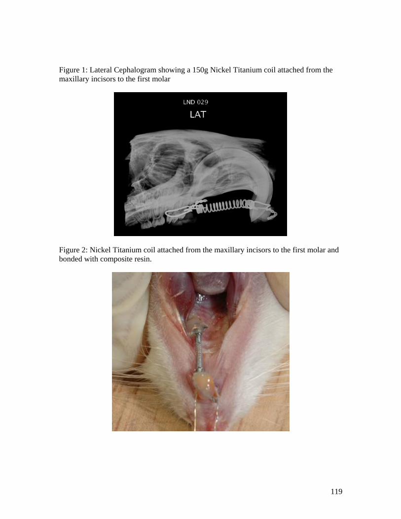
Figure 1: Lateral Cephalogram showing a 150g Nickel Titanium coil attached from the maxillary incisors to the first molar
Figure 2: Nickel Titanium coil attached from the maxillary incisors to the first molar and bonded with composite resin.
119

Figure 3: Experiment Flow Chart
CPP Administration
2 weeks 2 weeks
2 weeks 2 weeksControl Appliance Insertion
Samples Harvested
Samples Harvested No CPP
Experimental Appliance Insertion CPP
Force Application
150cN
120
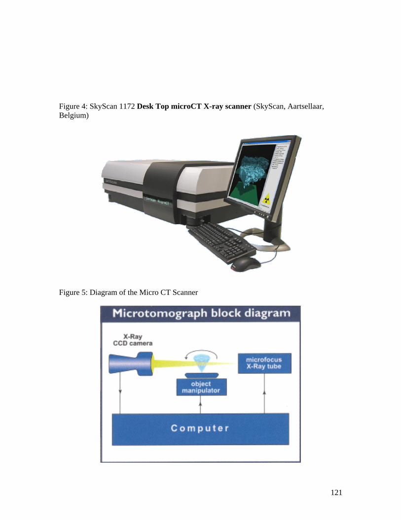
Figure 4: SkyScan 1172 Desk Top microCT X-ray scanner (SkyScan, Aartsellaar, Belgium)
Figure 5: Diagram of the Micro CT Scanner
121

Figure 6: Scanned segment of the maxilla including three maxillary molars. The first maxillary molar is highlighted in yellow.
Figure 7: First maxillary molar and the mesial portion of the second maxillary molar. The thin red line between the contacts of the first and second molars represents the shortest distance between the two surfaces as detected by the software tool.
122

Figure 8: Digital Extraction of Rat Molar
123
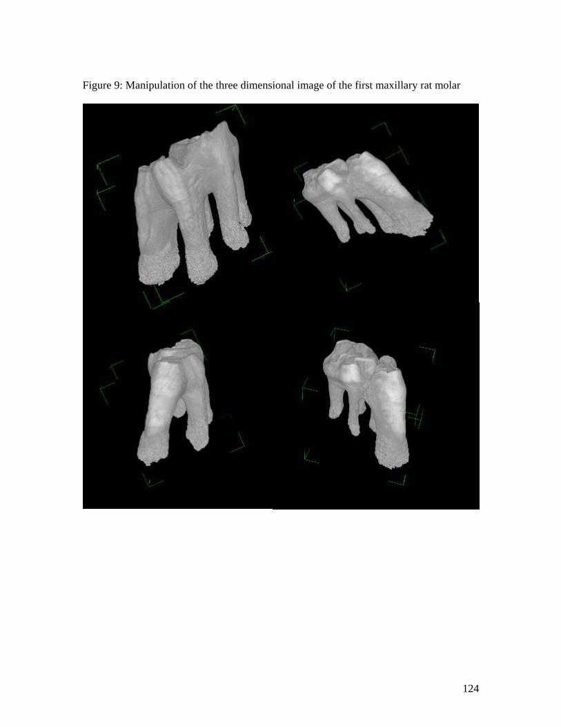
Figure 9: Manipulation of the three dimensional image of the first maxillary rat molar
124

Figure 10: The mesial root of the maxillary first molar. The area defined by the red lines is the segment of the root which was analysed for root resorpiton. The porous apical region can be seen in this view.
Figure 11: Crater isolation method a) Location of the craters in cross section b) Tracing of the crater outline c) Isolated craters. Isolated craters for each axial section are accumulated to achieve
an overall calculation of the total volume of root resorption.
125
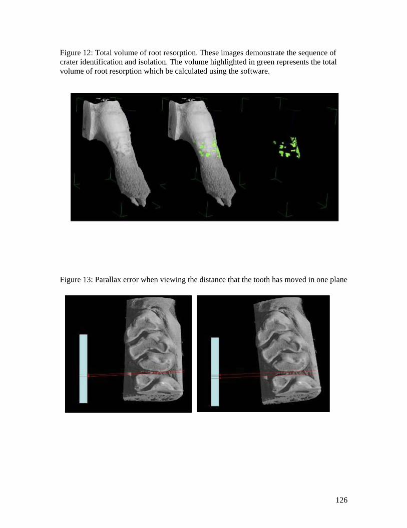
Figure 12: Total volume of root resorption. These images demonstrate the sequence of crater identification and isolation. The volume highlighted in green represents the total volume of root resorption which be calculated using the software.
Figure 13: Parallax error when viewing the distance that the tooth has moved in one plane
126

Figure 14: Tooth movement measured in the axial plane.
Figure 15: Tooth movement measured in the sagittal plane
127

Figure 16: Box-plot of the total volume of root resorption for no dietary supplements and dietary supplements of CPP.
Figure 17: Box-plot for the amount of tooth movement for no dietary supplements and dietary supplements of CPP.
Dietary CPP SupplementsCPPNo CPP
30000
RRvol
25000
20000
15000
10000
5000
0
Dietary CPP Supplements
CPPNo CPP
60.000
TMav 40.000
20.000
0.000
128