copyright © 2015 by the american physiological society
TRANSCRIPT
1
Title 1
Rebamipide suppresses mite-induced asthmatic responses in NC/Nga mice 2
Authors 3
Ikuo Murakami,1,2 Ran Zhang,1 Masayuki Kubo,1 Kenjiro Nagaoka,1 Eri Eguchi,1 Keiki 4 Ogino1 5
6
Affiliations: 7
1Department of Public Health, Okayama University Graduate School of Medicine, 8
Dentistry and Pharmaceutical Sciences, 2-5-1 Shikata-cho, Okayama 700-8558, Japan 9
2Third Institute of New Drug Discovery, Otsuka Pharmaceutical Co., Ltd., 463-10 10
Kagasuno, Kawauchi-cho, Tokushima 771-0192, Japan 11
12
Running Head: 13
Rebamipide suppresses mite-induced asthmatic responses 14
15
Corresponding author: 16
Keiki Ogino, MD, PhD 17
Department of Public Health, Okayama University Graduate School of Medicine, 18
Dentistry and Pharmaceutical Sciences, 2-5-1 Shikata-cho, Okayama 700-8558, Japan 19
Articles in PresS. Am J Physiol Lung Cell Mol Physiol (August 21, 2015). doi:10.1152/ajplung.00194.2015
Copyright © 2015 by the American Physiological Society.

2
Tel: +81-86-235-7184, Fax: + 81-86-226-0715 20
E-mail: [email protected]. 21
22
Abstract: 23
Allergic asthma caused by continuous allergen exposure evokes allergen-specific Th2 24
responses and is characterized by chronic airway inflammation and hyperresponsiveness. 25
A previous report showed that rebamipide improved asthmatic symptoms in an 26
ovalbumin/trypsin mice model. However, it is still unclear how rebamipide exerts its 27
effects in asthma. In this study, rebamipide improved the asthmatic responses induced 28
by mite exposure in NC/Nga mice, revealing the mechanism of this therapeutic effect. 29
Rebamipide suppressed the infiltration of eosinophils into the airways and lung as well 30
as attenuating the production of reactive oxygen species in tissues. In addition to these 31
anti-inflammatory effects, rebamipide inhibited the production of IL-33, a member of 32
the IL-1 family that drives the subsequent production of Th2-associated cytokines. 33
These observations identify the point where rebamipide exerts its suppressive action on 34
asthma and suggest that rebamipide has therapeutic potential in preventing mite-induced 35
asthma. 36
37
3
Keywords: 38
rebamipide, asthma, eosinophil, IL-33, reactive oxygen species 39
4
Introduction 40
Asthma is a chronic respiratory disease that affects about 300 million people worldwide 41
(32). The major characteristics of asthma are airway hyperresponsiveness (AHR), moist 42
cough and variable airflow obstruction on the basis of airway inflammation (36). Such 43
asthmatic symptoms are triggered by factors such as indoor allergens (house dust mites 44
and pet dander), outdoor allergens (pollens, particulate matter and molds), tobacco 45
smoke, exercise and viral respiratory infections (36). These factors evoke airway 46
inflammation by means of the inflammatory mediators and cytokines derived from 47
eosinophils, lymphocytes and airway epithelial cells (3). In addition, when exposed to 48
allergens or non-allergenic substances these cells can produce reactive oxygen species 49
(ROS), which causes tissue damage via lipid peroxidation of cell membranes and 50
nucleotide damage within DNA (8, 18). 51
Rebamipide (2-{4-cholorobenzoylamino-3-[2(1H)-quinolinon-4-yl]} propionic 52
acid) is widely used for mucosal protection, healing of gastric ulcers and treatment of 53
gastritis. It has various biological effects that include up-regulation of growth factors 54
such as epidermal growth factor and hepatocyte growth factor (31, 34); increased 55
production of mucus and prostaglandins (34, 37); inhibition of oxygen radical 56
production by neutrophils stimulated with N-formyl-methionyl-leucyl-phenylalanine 57
5
(fMLP), opsonized zymosan or Helicobacter pylori (20, 27, 39, 41); scavenging of 58
hydroxyl radicals (40) and inhibition of the production of inflammatory cytokines and 59
chemokines (4, 7). These effects of rebamipide suggest the possibility that rebamipide 60
may be useful for the treatment and prevention for asthma. A previous report showed 61
that oral administration of rebamipide improved asthmatic symptoms in an ovalbumin 62
(OVA)/trypsin mice model; however, how rebamipide exerts these effects remains 63
unclear (5). 64
The aim of this study was to examine whether rebamipide improved the 65
asthmatic responses in a model where NC/Nga mice were exposed to mites, and to 66
clarify the molecular mechanisms involved. 67
68
6
Materials and Methods 69
Chemicals and reagents. 70
Rebamipide was manufactured by Otsuka Pharmaceutical Co., Ltd. (Tokushima, Japan). 71
Carboxymethyl cellulose sodium (CMC) was purchased from Wako Pure Chemical 72
Industries, Ltd. (Osaka, Japan). Acetylcholine chloride was purchased from Daiichi 73
Sankyo Pharmaceutical Co., Ltd. (Tokyo, Japan). Gallamine triethiodide was purchased 74
from Sigma-Aldrich Co. (St. Louis, MO). Isoflurane was purchased from Intervet K. K. 75
(Tokyo, Japan). Sodium pentobarbital was purchased from Kyoritsu Seiyaku Co., Ltd. 76
(Tokyo, Japan). 77
78
Animals. 79
Five-week-old male NC/Nga mice were obtained from Charles River Laboratories 80
Japan (Kanagawa, Japan). The mice were maintained under specific pathogen-free 81
conditions in 12/12-hour light/dark cycle and had free access to standard laboratory 82
food and water. They were acclimatized for at least one week before the experiments. 83
The care and handling of animals were in accordance with the Guidelines for the Care 84
and Use of Laboratory Animals at Shikata Campus of Okayama University. This study 85
was approved by the Okayama University Institutional Animal Care and Use 86
7
Committee. 87
88
Intranasal and intratracheal administrations. 89
NC/Nga mice were sensitized to crude mite extract (Dermatophagoides farinae: Df, 90
Cosmo Bio, Tokyo, Japan) based on a previously described protocol (29) as shown in 91
Fig. 1. Briefly, mice were anesthetized with 3% isoflurane and treated with intratracheal 92
2% rebamipide (500 µg in 25 µl 0.1% CMC) for 14 consecutive days (days 0–13). One 93
hour after each treatment, the anesthetized mice received intranasal instillation of Df 94
crude extract (50 µg in 25 µl saline) for six days (days 0–4 and 11). Saline and 0.1% 95
CMC were used as controls, for comparison with the groups given Df or rebamipide. 96
97
Measurement of AHR. 98
On day 14, the degree of bronchoconstriction was measured according to the overflow 99
method (26). Briefly, mice were anesthetized with 50 mg/kg pentbalbital (i.p.) and 100
connected to an artificial ventilator following surgical incision of the trachea. A 101
pulmotor system was constructed with a rodent ventilator (model 55-7058; Harvard 102
Apparatus Canada, QC, Canada), a bronchospasm transducer (model 7020; Ugo Basile, 103
Comerio-Barese, Italy), and a data recorder (model WR300; Graphtec corporation, 104
8
Kanagawa, Japan). Gallamine triethiodide (350 µg/mouse) was immediately 105
administered intravenously to eliminate spontaneous respiration and followed by 106
repeated administration of acetylcholine with stepwise increases in the concentration 107
from 62.5 to 2,000 µg/kg. Dose-response curves were obtained for acetylcholine in 108
anesthetized, mechanically ventilated mice. Bronchoconstriction was expressed as the 109
respiratory overflow volume provoked by acetylcholine as a percentage of the maximal 110
overflow volume (100%) obtained by totally occluding the tracheal cannula (16). 111
112
Bronchoalveolar lavage fluid. 113
Immediately after the assessment of acetylcholine-induced AHR, the lungs were 114
lavaged with 1 ml aliquots of cold Hanks’ balanced salt solution (HBSS) without 115
calcium and magnesium, containing 50 µM EDTA. The collected bronchoalveolar 116
lavage fluid (BALF) was then centrifuged at 200×g for 10 min at 4°C. The supernatant 117
was collected and stored at −80°C for further analysis, and the cell pellet was 118
resuspended in cold HBSS. The number of leukocytes in the BALF was determined 119
using a hematology analyzer (model XT-200i; Sysmex, Hyogo, Japan). Another aliquot 120
was used for cytospin preparations at 500 rpm for 5 min (Cytospin2; Thermo Fisher 121
Scientific, Kanagawa, Japan) and was stained with Diff-Quik for cell count (Sysmex). 122
9
123
Lung specimens. 124
The lung tissue was utilized for extraction of total RNA for RT-PCR and fixed in 10% 125
buffered formalin for morphological examination. The remaining lung tissue was 126
homogenized in a homogenizing buffer (T-PER tissue extraction reagent containing a 127
protease inhibitor cocktail) with a Multi-Beads Shocker (model MB901OT; Yasui Kikai, 128
Osaka, Japan) set to 1,900 rpm and 30 sec, with repetition for two cycles. 129
130
Quantitative PCR analysis. 131
Total RNA was isolated using the RNeasy mini kit (Qiagen, Redwood City, CA), and 132
reverse transcription was performed with the High Capacity RNA-to-cDNA Kit (Life 133
Technologies, Carlsbad, CA) according to the manufacturer’s instructions. Quantitative 134
RT-PCR was carried out using the ABI 7500 Fast Real-Time PCR System and the 135
Taqman Fast Universal PCR Master Mix (Life Technologies). The reaction mixture was 136
prepared with TaqMan Gene Expression Primer and Probe (Life Technologies) 137
according to the manufacturer’s protocol. Primers specific for interleukin-4 (IL-4, Assay 138
ID: Mm00445259_m1), IL-5 (Mm00439646_m1), IL-13 (Mm00434204_m1), 139
interferon gamma (IFNγ, Mm99999071_m1), Eotaxin-1 (Mm00441238_m1), Eotaxin-2 140

10
(Mm00444701_m1), IL-33 (Mm00505403_m1), Glyceraldehyde 3-phosphate 141
dehydrogenase (GAPDH, 4352932E), arginase-1 (Arg1, Mm00475988_m1), Fizz1 142
(Mm00445109_m1), Ym1 (Mm00657889_m1) and nitric oxide synthase 2 (NOS2, 143
Mm00440502_m1) were used. The thermal cycling conditions were at 95°C for 20 sec, 144
followed by 40 cycles of amplification at 95°C for 3 sec and 60°C for 30 sec. The 145
expression of mRNA was standardized to GAPDH mRNA, and the relative expression 146
of each gene was quantified by the ddCt method, expressed as a ratio, related to the 147
mean value for vehicle-treated lungs (17). 148
149
EIA analysis. 150
The concentration of 8-hydroxy-2’-deoxyguanosine (8-OHdG) was determined by a 151
DNA/RNA oxidative stress EIA kit (Cayman Chemical, Ann Arbor, MI) according to 152
the manufacturer’s instructions. The detection limit of this assay was 10.3 pg/ml. 153
154
ELISA analysis. 155
Protein concentrations were determined by the BCA Protein Assay Reagent Kit 156
(Thermo Fisher Scientific Inc., Waltham, MA) using bovine serum albumin standards. 157
The concentrations of eotaxin-1, eotaxin-2 and IL-33 were determined by ELISA kit or 158

11
DuoSet (R&D Systems, Inc., Minneapolis, MN) according to the manufacturer’s 159
instructions and these were adjusted by total protein. The detection limits of the assays 160
for eotaxin-1, eotaxin-2 and IL-33 were 15.6 pg/ml each. 161
162
Histopathological observations. 163
The fixed lung tissues were dehydrated, embedded in paraffin, and sectioned. 164
Hematoxylin and eosin (H&E) staining was used to assess the degree of inflammation 165
(11). Periodic acid-Schiff (PAS) staining was also performed to observe interstitial 166
goblet cell hyperplasia (30). Images of lung tissue sections stained with H&E and PAS 167
were acquired with a BZ-9000 microscope (Keyence, Osaka, Japan) equipped with a 168
40× objective lens. The levels of inflammation in peribronchial areas of the lung were 169
determined according to an ordinal scale of grades 0 to 3, as described elsewhere (33), 170
in a blinded fashion with examination of at least five airway cross-sections per slide. A 171
value of 0 indicated that no inflammation was detectable; a value of 1, occasional 172
cuffing with inflammatory cells; a value of 2, most bronchi were surrounded by a thin 173
layer (1–5 cells thick) of inflammatory cells; and a value of 3, most bronchi were 174
surrounded by a thick layer (more than 5 cells thick) of inflammatory cells. Goblet cell 175
hyperplasia was semiquantitatively scored in a blinded fashion in at least five airway 176
12
cross-sections per slide on a scale of grades 0 to 4 (absence, < 25% of epithelial lining 177
cells, 25–50% of epithelial lining cells, 50–75% of epithelial lining cells, > 75% of 178
epithelial lining cells, respectively) as previously described (21). 179
180
Statistical analysis. 181
All data, unless otherwise noted, are expressed as mean ± SE. One-way analysis of 182
variance (ANOVA) followed by Dunnett’s test was used to evaluate the significance of 183
differences between more than two groups. A mixed model for repeated measures 184
(MMRM) method was used to determine significant differences in the dose-response 185
studies. The threshold for statistical significance was set at P < 0.05. Statistical analyses 186
were performed using SAS software Release 9.3 (SAS Institute, Tokyo, Japan). 187
188
13
Results 189
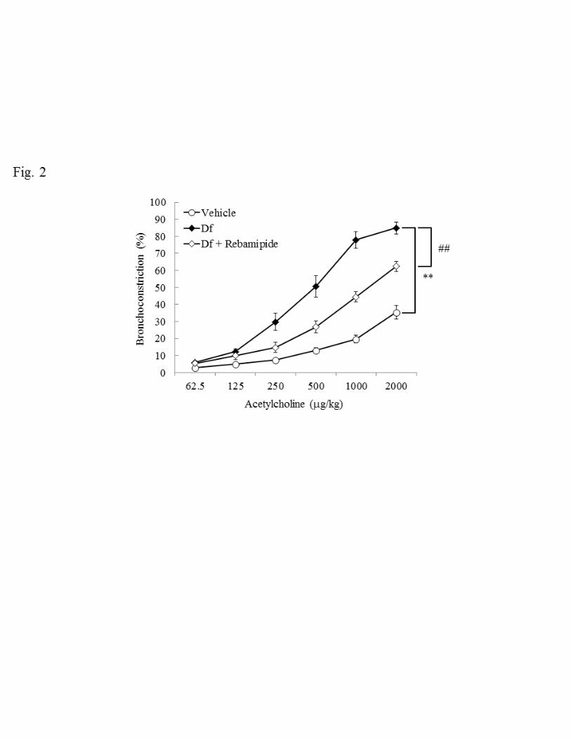
Effect of rebamipide on AHR 190
Mite antigen is one of the major allergic substrates that induce asthma. Here we used 191
Dermatophagoides farinae (Df) for the induction of asthma in NC/Nga mice. To 192
confirm asthmatic features, airway hyperresponsiveness (AHR), which is a basic and 193
reproducible indicator of asthma, was measured. First we examined whether Df 194
treatment changes AHR in response to acetylcholine. In the vehicle group, AHR 195
increased in a dose-dependent manner and such increments in AHR were further 196
enhanced by Df administration (P < 0.01, Fig. 2). We then evaluated the effects of 197
rebamipide on Df-treated mice and found that rebamipide reduced AHR significantly (P 198
< 0.01, Fig. 2). Thus intratracheal administration of rebamipide improved AHR in 199
response to acetylcholine in Df-treated NC/Nga mice. 200
201
Effect of rebamipide on histopathological findings 202
Histopathological changes in lungs of mice treated with Df were examined to estimate 203
the degree of inflammation, using H&E staining. Df treatment increased the 204
inflammation score to about twice that of the vehicle group (0.9 ± 0.0 vs 2.0 ± 0.2, P < 205
0.01, Fig. 3A, B and G) and such Df-induced inflammation was suppressed by 206
14
rebamipide (2.0 ± 0.2 vs 1.4 ± 0.1, P < 0.01, Fig. 3C and G). Since it is known that 207
airway inflammation causes hyperplasia of goblet cells, we attempted to estimate it 208
using PAS staining. Similar to the results of inflammatory scoring, the goblet cell 209
hyperplasia score in the Df-treated group was higher than that in the vehicle group (0.0 210
± 0.0 vs 2.5 ± 0.4, P < 0.01, Fig. 3D, E and H), while the score in the rebamipide group 211
was lower than in the Df-treated group (2.5 ± 0.4 vs 1.6 ± 0.2, P < 0.05, Fig. 3F and H). 212
These results confirmed that rebamipide exhibits anti-inflammatory effects on 213
Df-elicited inflammation in lung. 214
215
Effect of rebamipide on the accumulation of immune cells in BALF 216
To explore the mechanisms of inflammation in the airway and lung, we collected 217
bronchoalveolar lavage fluid (BALF) and analyzed the number of accumulated cells and 218
types in BALF using a hematology analyzer. Df treatment increased total cell number 219
about nine times that in the vehicle group (2.64 ± 0.43 × 10⁴ cells vs 23.58 ± 4.07 × 10⁴ 220
cells) (Fig. 4). We further analyzed BALF to identify the cell types present and found 221
that the numbers of inflammatory cells including eosinophils, macrophages, neutrophils 222
and lymphocytes were notably increased by Df treatment (Fig. 4). In particular, the 223
number of eosinophils in the BALF increased about 4000-fold in comparison with 224
15
vehicle control (0.04 ± 0.04 × 10³ cells vs 157.89 ± 29.82 × 10³ cells), suggesting that 225
Df-induced an inflammatory response mainly mediated by excess numbers of 226
infiltrating eosinophils in the airways and lungs. This increased number of inflammatory 227
cells was reduced for every cell type when rebamipide was administered intratracheally 228
for 14 days, where the attenuation rate in eosinophil number was about 64%. 229
230
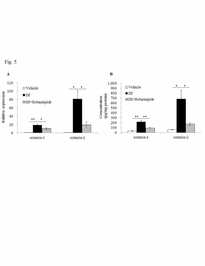
Effect of rebamipide on expression of eotaxins 231
Next we measured the expression of eotaxin-1 and eotaxin-2 in lung tissue to confirm 232
the involvement of eosinophils, as suggested in the results of BALF (Fig. 4). We found 233
that Df challenge increased eotaxin-1 mRNA expression about 20-fold and eotaxin-2 234
mRNA 80-fold compared with those in the vehicle group (Fig. 5A). Elevated protein 235
expression was also detected for both eotaxin-1 and eotaxin-2 (Fig. 5B. eotaxin-1: 36.1 236
± 9.1 vs 219.0 ± 32.8 pg/mg protein, P < 0.01, eotaxin-2: 59.2 ± 6.0 vs 679.5 ± 185.6 237
pg/mg protein, P < 0.01). When rebamipide was administered, augmented mRNA 238
expression induced by Df was reduced about one half and one quarter for eotaxin-1 and 239
eotaxin-2, respectively. In addition, increased protein expression of both eotaxin-1 and 240
eotaxin-2 were also significantly decreased by rebamipide (eotaxin-1: 90.8 ± 12.7 241
pg/mg protein, P < 0.01; eotaxin-2: 174.7 ± 31.5 pg/mg protein, P < 0.05). 242
16
These results demonstrate that main cell infiltrating the lung was the eosinophil, which 243
was also the most abundant cell type in BALF after Df treatment; rebamipide 244
suppressed this infiltration. 245
246
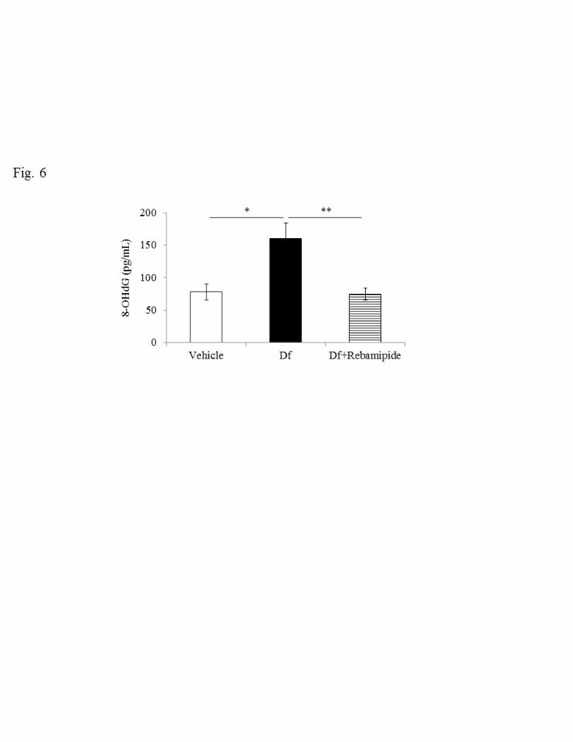
Effect of rebamipide on 8-OHdG in BALF 247
Reactive oxygen species (ROS) are detected extensively in patients with asthma, where 248
they are mainly produced by granulocytes and epithelial cells. Because rebamipide is a 249
direct scavenger of ROS (40) and also inhibits ROS production by activated neutrophils 250
through the competitive inhibition of the formyl peptide receptor (20), we measured 251
8-OHdG in BALF as an indicator of ROS production to examine the suppressive effect 252
of rebamipide in our model. The level of 8-OHdG in BALF increased after Df treatment 253
(78.0 ± 12.6 vs 159.6 ± 24.6 pg/ml BALF, P < 0.05) and this increase in 8-OHdG was 254
significantly reduced to the same level seen with the vehicle control when rebamipide 255
was administered (74.3 ± 9.4, P < 0.01) (Fig. 6). 256
257
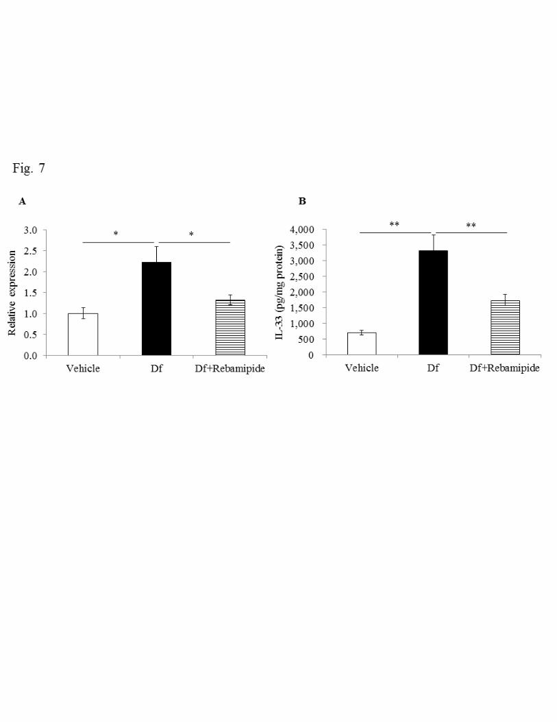
Effect of rebamipide on expression of IL-33 and cytokines 258
It has been revealed that the epithelium-derived cytokine IL-33 plays important roles in 259
asthma in Th2-dependent and independent ways (23), where IL-33 activates effector 260

17
cells such as eosinophils, mast cells and basophils directly and also induces activation 261
of these cells via IL-4, IL-5 and IL-13 produced by Th2 cells (13, 19). We thus 262
examined changes in the expression of IL-33 and Th2 cytokines in lung, before and 263
after Df treatment. 264
We first examined IL-33 expression by measuring mRNA and protein levels. 265
Df treatment increased IL-33 expression significantly compared with the vehicle group 266
(690.1 ± 73.7 vs 3306.5 ± 493.8 pg/mg protein, P < 0.01) (Fig. 7A and B). We then 267
tested the effect of rebamipide on the expression of IL-33. Rebamipide reduced IL-33 268
expression by about half, with respect to both mRNA and protein (1742.1 ± 178.0 269
pg/mg protein, P < 0.01). We next measured the expression of IL-4, IL-5 and IL-13 as a 270
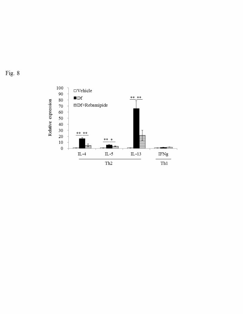
representative Th2 cytokines and IFNγ as a Th1 cytokine, respectively. For all cytokines 271
examined except IFNγ, mRNA expression was elevated, suggesting that Df treatment 272
selectively evokes a Th2 response. These augmented Th2 responses were dramatically 273
suppressed when rebamipide was administered (Fig. 8). 274
275
Effect of Rebamipide on macrophage polarization 276
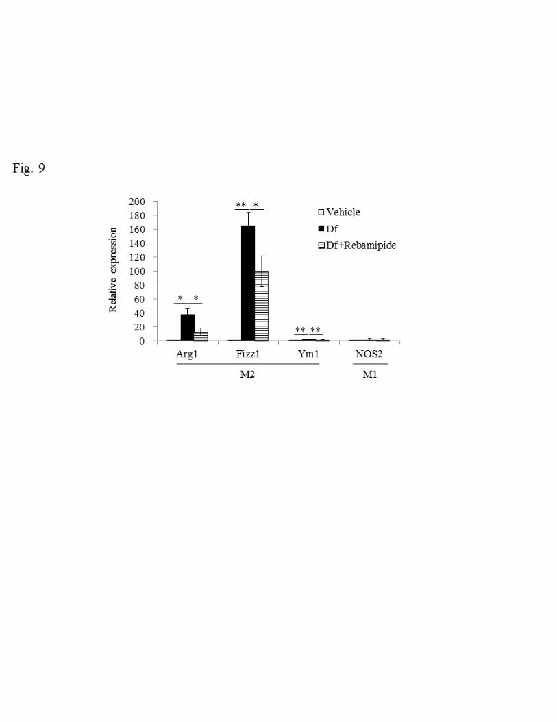
Based on the result of enhanced Th2 cytokine productions shown in Figure 8, we further 277
analyzed macrophage polarization to confirm Th2 response by testing the expression of 278
18
series of polarization marker. Here we utilized arginase-1 (Arg1), found in inflammatory 279
zone-1 (Fizz1) and chitinase-like 3 (Ym1) as a marker for M2 macrophage and nitric 280
oxide synthase 2 (NOS2) was selected for M1 macrophage marker (25, 38). All mRNA 281
levels in M2 macrophage markers increased after Df treatment. On the other hand, M1 282
macrophage maker did not show any changes (Fig. 9). These enhanced expression of 283
M2 macrophage makers were remarkably decreased after rebamipide treatment 284
suggesting the possibility that rebamipide suppress M2 macrophage polarization 285
through the inhibition of production of Th2 cytokines. 286
287

19
Discussion 288
Previous studies reported that rebamipide attenuated the allergic response and 289
respiratory symptoms by improving AHR, reducing leukocyte numbers in BALF and 290
suppressing goblet cell hyperplasia regardless of the different animals, allergens and 291
routes of administration of rebamipide. For example, Lee et al. reported that oral 292
administration of rebamipide reduced the number of infiltrating leukocytes and the 293
amount of TNFα in BALF, and downregulated MUC5AC mucin synthesis in the airway 294
epithelium in a rat model of cigarette smoke-induced mucin production (12). Another 295
group used a different approach, examining the effects of rebamipide on an 296
OVA/trypsin-induced asthmatic mouse model, and found that oral administration of 297
rebamipide decreased the eosinophil number in BALF and improved the respiratory rate, 298
air-flow rate and tidal volume (5). In this study, we used a mite-induced asthmatic 299
model in NC/Nga mice, which resembles human asthma, to examine the therapeutic 300
effects of rebamipide and to clarify the molecular mechanisms involved (26, 29, 30). 301
Our results suggest that rebamipide improves features of asthma in NC/Nga 302
mice through three pathways. First, rebamipide suppressed eosinophil invasion of the 303
airways and lungs (Fig. 4 and 5). It is known that an excess number of infiltrating 304
eosinophils in respiratory tissues is one of the characteristic features of asthma and it is 305
20
strongly associated with the development of AHR. Such chemotaxis of eosinophils 306
towards inflamed tissue may be inhibited by rebamipide directly or indirectly. As 307
previous reports have shown that rebamipide exerts an antagonistic effect on the formyl 308
peptide receptor (FPR) expressed in neutrophils and eosinophils, an essential receptor 309
for the expression of chemotaxis (20, 28), there is a possibility that rebamipide 310
suppresses chemotactic behavior of eosinophils through the inhibition of the FPR. In 311
addition to the direct effects of rebamipide on eosinophils, we found that rebamipide 312
reduced the expression of eotaxin-1 and eotaxin-2 in lung (Fig. 5). The eotaxin family 313
has been identified as chemoattractant for eosinophils, implying that rebamipide may 314
suppress chemotaxis of eosinophils by eliminating the chemoattractant source. Second, 315
rebamipide improved asthmatic symptoms by reducing ROS production by eosinophils 316
and epithelial cells. A marker of oxidative stress formed by ROS is 8-OHdG (10). 317
Several studies demonstrated that 8-OHdG expression was induced and enhanced in the 318
lungs as a result of several types of oxidative stress (1, 24, 35). Similar to previous 319
studies, the 8-OHdG level in BALF was elevated by Df treatment and was reduced 320
significantly by rebamipide administration, suggesting that rebamipide may work as a 321
scavenger of ROS (Fig. 6). Alternatively, rebamipide may reduce ROS produced by 322
activated eosinophils through inhibition of FPR agonist-induced eosinophil activation as 323
21
described above. Finally, rebamipide exhibited a suppressive action on the Th2 response 324
by inhibition of IL-33 production. Previous reports have demonstrated the involvement 325
of IL-33 in asthmatic symptoms (6, 9, 22). The IL-33 level is elevated in asthmatic 326
patients when compared with healthy individuals, and a similar tendency was also 327
observed in mice (6, 9, 22), where IL-33 is released from bronchial epithelial cells in 328
response to allergen exposure and this secreted IL-33 then induces Th2 responses from 329
macrophages, eosinophils and mast cells (19). Direct administration of IL-33 induces 330
eosinophilic inflammation and AHR in mice and administration of neutralizing 331
antibodies against IL-33 and IL-33 receptor ST2 attenuates eosinophilic inflammation 332
and AHR in an OVA-induced airway inflammation model in mice (2, 9, 14, 15). In the 333
present study, rebamipide decreased the expression of IL-33 in lung tissue (Fig. 7) and 334
also attenuated the expression of Df-elicited Th2 cytokines, including IL-4, IL-5 and 335
IL-13 (Fig. 8). Taken together, the inhibition of IL-33 production by rebamipide appears 336
to suppress subsequent Th2 responses. Another approach used to check Th2 response 337
involves examining the polarization of macrophages: macrophage have been classified 338
as M1 (classically activated) or M2 (alternatively activated) macrophages. M1 339
macrophages express inducible nitric oxide synthase (NOS2) and proinflammatory 340
cytokines, and these are essential for protection against infection. Conversely, M2 341
22
macrophages express arginase-1 (Arg1), chitinase-like 3 (Ym1), found in inflammatory 342
zone-1 (Fizz1), and chemokines such as Chemokine (C-C motif) ligand 17 (CCL17), 343
CCL24 (eotaxin-2) and these play important roles in responses to parasite infection, 344
tissue remodeling, angiogenesis and tumor progression (25, 38). Gene expression levels 345
of M2 macrophage markers such as Arg1, Fizz1 and Ym1 were significantly 346
upregulated by Df administration and downregulated by rebamipide treatment, while 347
M1 macrophage marker NOS2 showed no change (Fig. 9). Because both IL-33 and 348
IL-13 can polarize macrophages from M1 to M2 types, it is likely that rebamipide 349
impairs the polarization towards M2 macrophages due to the decreased expression of 350
IL-33 and IL-13. 351
In conclusion, our current study indicates the therapeutic potential of 352
rebamipide in the prevention of mite-induced asthma. Our data demonstrate that 353
rebamipide improves mite-induced asthmatic symptoms through the inhibition of 354
eosinophil infiltration, as well as attenuating oxidative stress and IL-33 production. 355
356
Acknowledgements: 357
Current affiliation of Ran Zhang is Control of Innate Immunity Technology Research 358
Association, 2217-16 Hayashi-cho, Takamatsu, Kagawa 761-0301, Japan. 359
23
360
Disclosures: 361
IM is employed by Otsuka Pharmaceutical Co., Ltd. 362
363
Author contributions: 364
Conception and design of research: IM, KO; Performed experiments: IM, RZ, MK, KN; 365
Analyzed data: IM, EE, KO; Interpreted results of experiments: IM, RZ, MK, KN, EE, 366
KO; Prepared figures: IM; Drafted manuscript: IM; Edited and revised manuscript: MK, 367
KN, EE, KO; Approved final version of manuscript: IM, RZ, MK, KN, EE, KO. 368
369
References: 370
1. Cheng ML, Ho HY, Huang YW, Lu FJ, Chiu DT. Humic acid induces oxidative 371 DNA damage, growth retardation, and apoptosis in human primary fibroblasts. Exp Biol 372 Med 228: 413-423, 2003. 373 2. Coyle AJ, Lloyd C, Tian J, Nguyen T, Erikkson C, Wang L, Ottoson P, Persson 374 P, Delaney T, Lehar S, Lin S, Poisson L, Meisel C, Kamradt T, Bjerke T, Levinson 375 D, Gutierrez-Ramos JC. Crucial role of the interleukin 1 receptor family member 376 T1/ST2 in T helper cell type 2-mediated lung mucosal immune responses. J Exp Med 377 190: 895-902, 1999. 378 3. Fahy JV. Type 2 inflammation in asthma-present in most, absent in many. Nat Rev 379 Immunol 15: 57-65, 2015. 380 4. Fukuda K, Ishida W, Tanaka H, Harada Y, Fukushima A. Inhibition by 381 rebamipide of cytokine-induced or lipopolysaccharide-induced chemokine synthesis in 382 human corneal fibroblasts. Br J Ophthalmol 98: 1751-1755, 2014. 383 5. Gohil P, Thakkar H, Gohil U, Deshpande S. Preliminary studies on the effect of 384
24
rebamipide against the trypsin and egg-albumin induced experimental model of asthma. 385 Acta Pharm 61: 427-433, 2011. 386 6. Kearley J, Buckland KF, Mathie SA, Lloyd CM. Resolution of allergic 387 inflammation and airway hyperreactivity is dependent upon disruption of the 388 T1/ST2-IL-33 pathway. Am J Respir Crit Care Med 179: 772-781, 2009. 389 7. Kim H, Seo JY, Kim KH. Inhibition of lipid peroxidation, NF-kappaB activation 390 and IL-8 production by rebamipide in Helicobacter pylori-stimulated gastric epithelial 391 cells. Dig Dis Sci 45: 621-628, 2000. 392 8. Kirkham P, Rahman I. Oxidative stress in asthma and COPD: antioxidants as a 393 therapeutic strategy. Pharmacol Ther 111: 476-494, 2006. 394 9. Kurowska-Stolarska M, Stolarski B, Kewin P, Murphy G, Corrigan CJ, Ying S, 395 Pitman N, Mirchandani A, Rana B, van Rooijen N, Shepherd M, McSharry C, 396 McInnes IB, Xu D, Liew FY. IL-33 amplifies the polarization of alternatively activated 397 macrophages that contribute to airway inflammation. J Immunol 183: 6469-6477, 2009. 398 10. Kuwano K, Nakashima N, Inoshima I, Hagimoto N, Fujita M, Yoshimi M, 399 Maeyama T, Hamada N, Watanabe K, Hara N. Oxidative stress in lung epithelial 400 cells from patients with idiopathic interstitial pneumonias. Eur Respir J 21: 232-240, 401 2003. 402 11. Lee KS, Lee HK, Hayflick JS, Lee YC, Puri KD. Inhibition of phosphoinositide 403 3-kinase delta attenuates allergic airway inflammation and hyperresponsiveness in 404 murine asthma model. FASEB J 20: 455-465, 2006. 405 12. Lee SY, Kang EJ, Hur GY, Jung KH, Jung HC, Lee SY, Kim JH, Shin C, In 406 KH, Kang KH, Yoo SH, Shim JJ. The inhibitory effects of rebamipide on cigarette 407 smoke-induced airway mucin production. Respir Med 100: 503-511, 2006. 408 13. Liew FY, Pitman NI, McInnes IB. Disease-associated functions of IL-33: the new 409 kid in the IL-1 family. Nat Rev Immunol 10: 103-110, 2010. 410 14. Liu X, Li M, Wu Y, Zhou Y, Zeng L, Huang T. Anti-IL-33 antibody treatment 411 inhibits airway inflammation in a murine model of allergic asthma. Biochem Biophys 412 Res Commun 386: 181-185, 2009. 413 15. Lohning M, Stroehmann A, Coyle AJ, Grogan JL, Lin S, Gutierrez-Ramos JC, 414 Levinson D, Radbruch A, Kamradt T. T1/ST2 is preferentially expressed on murine 415 Th2 cells, independent of interleukin. Proc Natl Acad Sci U S A 95: 6930-6935, 1998. 416 16. Nagai H, Yamaguchi S, Inagaki N, Tsuruoka N, Hitoshi Y, Takatsu K. Effect of 417 anti-IL-5 monoclonal antibody on allergic bronchial eosinophilia and airway 418 hyperresponsiveness in mice. Life Sci 53: PL243-PL247, 1993. 419 17. Nakashima T, Uematsu N, Shibamori M, Sakurai K, Ishida T. Establishment of 420

25
an X-ray irradiation-induced glossitis model in rats: biphasic elevation of 421 proinflammatory cytokines and chemokines. J Pharmacol Exp Ther 347: 660-668, 422 2013. 423 18. Nile SH, Park SW. Optimized methods for in vitro and in vivo anti-inflammatory 424 assays and its applications in herbal and synthetic drug analysis. Mini Rev Med Chem 425 13: 95-100, 2013. 426 19. Oboki K, Ohno T, Kajiwara N, Arae K, Morita H, Ishii A, Nambu A, Abe T, 427 Kiyonari H, Matsumoto K, Sudo K, Okumura K, Saito H, Nakae S. IL-33 is a 428 crucial amplifier of innate rather than acquired immunity. Proc Natl Acad Sci USA 107: 429 18581-18586, 2010. 430 20. Ogino K, Hobara T, Ishiyama H, Yamasaki K, Kobayashi H, Izumi Y, Oka S. 431 Antiulcer mechanism of action of rebamipide, a novel antiulcer compound, on 432 diethyldithiocarbamate-induced antral gastric ulcers in rats. Eur J Pharmacol 212: 9–13, 433 1992. 434 21. Padrid P, Snook S, Finucane T, Shiue P, Cozzi P, Solway J, Leff AR. Persistent 435 airway hyperresponsiveness and histologic alterations after chronic antigen challenge in 436 cats. Am J Respir Crit Care Med 151: 184-193, 1995. 437 22. Préfontaine D, Lajoie-Kadoch S, Foley S, Audusseau S, Olivenstein R, Halayko 438 AJ, Lemière C, Martin JG, Hamid Q. Increased expression of IL-33 in severe 439 asthma: evidence of expression by airway smooth muscle cells. J Immunol 183: 440 5094-5103, 2009. 441 23. Ramadas RA, Ewart SL, Medoff BD, LeVine AM. Interleukin-1–family member 442 9 stimulates chemokine production and neutrophil influx in mouse lungs. Am J Respir 443 Cell Mol Biol 44: 134–145, 2011. 444 24. Sanbongi C, Takano H, Osakabe N, Sasa N, Natsume M, Yanagisawa R, Inoue 445 K, Kato Y, Osawa T, Yoshikawa T. Rosmarinic acid inhibits lung injury induced by 446 diesel exhaust particles. Free Radic Biol Med 228: 1060-1069, 2003. 447 25. Satoh T, Takeuchi O, Vandenbon A, Yasuda K, Tanaka Y, Kumagai Y, 448 Miyake T, Matsushita K, Okazaki T, Saitoh T, Honma K, Matsuyama T, Yui K, 449 Tsujimura T, Standley DM, Nakanishi K, Nakai K, Akira S. The Jmjd3-Irf4 axis 450 regulates M2 macrophage polarization and host responses against helminth infection. 451 Nat Immunol 11: 936-944, 2010. 452 26. Shibamori M, Ogino K, Kambayashi Y, Ishiyama H. Intranasal mite allergen 453 induces allergic asthma-like responses in NC/Nga mice. Life Sci 78: 987-994, 2006. 454 27. Suzuki M, Miura S, Mori M, Kai A, Suzuki H, Fukumura D, Suematsu M, 455 Tsuchiya M. Rebamipide, a novel antiulcer agent, attenuates Helicobacter pylori 456
26
induced gastric mucosal cell injury associated with neutrophil derived oxidants. Gut 35: 457 1375–1378, 1994. 458 28. Svensson L, Redvall E, Björn C, Karlsson J, Bergin AM, Rabiet MJ, Dahlgren 459 C, Wennerås C. House dust mite allergen activates human eosinophils via formyl 460 peptide receptor and formyl peptide receptor-like 1. Eur J Immunol 37: 1966-1977, 461 2007. 462 29. Takahashi N, Ogino K, Takemoto K, Hamanishi S, Wang DH, Takigawa T, 463 Shibamori M, Ishiyama H, Fujikura Y. Direct inhibition of arginase attenuated 464 airway allergic reactions and inflammation in a Dermatophagoides farinae-induced 465 NC/Nga mouse model. Am J Physiol Lung Cell Mol Physiol 299: L17-L24, 2010. 466 30. Takemoto K, Ogino K, Shibamori M, Gondo T, Hitomi Y, Takigawa T, Wang 467 DH, Takaki J, Ichimura H, Fujikura Y, Ishiyama H. Transiently, paralleled 468 upregulation of arginase and nitric oxide synthase and the effect of both enzymes on the 469 pathology of asthma. Am J Physiol Lung Cell Mol Physiol 293: L1419-L1426, 2007. 470 31. Tarnawski AS, Chai J, Pai R, Chiou SK. Rebamipide activates genes encoding 471 angiogenic growth factors and Cox2 and stimulates angiogenesis: a key to its ulcer 472 healing action? Dig Dis Sci 49: 202–209, 2004. 473 32. To T, Stanojevic S, Moores G, Gershon AS, Bateman ED, Cruz AA, Boulet LP. 474 Global asthma prevalence in adults: findings from the cross-sectional world health 475 survey. BMC Public Health 204: 2012. 476 33. Tournoy KG, Kips JC, Schou C, Pauwels RA. Airway eosinophilia is not a 477 requirement for allergen-induced airway hyperresponsiveness. Clin Exp Allergy 30: 478 79-85, 2000. 479 34. Udagawa A, Shiota G, Ichiba M, Murawaki Y. Effect of rebamipide on acetic 480 acid-induced gastric ulcer in rats: involvement of hepatocyte growth factor. Scand J 481 Gastroenterol 38: 141–146, 2003. 482 35. Upadhyay D, kamp DW. Asbestos-induced pulmonary toxicity: role of DNA 483 damage and apoptosis. Exp Biol Med 34: 1060-1069, 2003. 484 36. WHO media centre. WHO Asthma Fact sheet No.307 [Online]. World Health 485 Organization. http://www.who.int/mediacentre/factsheets/fs307/en/ [2013] 486 37. Yamazaki K, Kanbe T, Chijiwa T, Ishiyama H, Morita S. Gastric mucosal 487 protection by OPC-12759, a novel antiulcer compound, in the rat. Eur J Pharmacol 488 142: 23–29, 1987. 489 38. Ye S, Xu H, Jin J, Yang M, Wang C, Yu Y, Cao X. The E3 ubiquitin ligase 490 neuregulin receptor degradation protein 1 (Nrdp1) promotes M2 macrophage 491 polarization by ubiquitinating and activating transcription factor 492
27
CCAAT/enhancer-binding Protein β (C/EBPβ). J Biol Chem 287: 26740-26748, 2012. 493 39. Yoshida N, Yoshikawa T, Iinuma S, Arai M, Takenaka S, Sakamoto K, 494 Miyajima T, Nakamura Y, Yagi N, Naito Y, Mukai F, Kondo M. Rebamipide 495 protects against activation of neutrophils by Helicobacter pylori. Dig Dis Sci 41: 1139–496 1144, 1996. 497 40. Yoshikawa T, Naito Y, Nakamura S, Nishimura S, Kaneko T, Iinuma S, 498 Takahashi S, Kondo M, Yamasaki K. Effect of rebamipide on lipid peroxidation and 499 gastric mucosal injury induced by indometacin in rats. Arzneimittelforschung 43: 1327–500 1330, 1993. 501 41. Yoshikawa T, Naito Y, Tanigawa T, Kondo M. Free radical scavenging activity 502 of the novel anti-ulcer agent rebamipide studied by electron spin resonance. Arzneim 503 Forsch/Drug Res 43: 363–366, 1993. 504
505
Figure Legends: 506
Fig. 1. Schematic representation of the experiment. The vehicle group received 25 µl of 507
saline intranasally on days 0–4 and day 11 and 25 µl of 0.1% CMC intratracheally on 508
days 0–13. The Df group received 50 µg of Dermatophagoides farinae (Df) extract 509
dissolved in 25 µl of saline intranasally on days 0–4 and day 11 and 25 µl of 0.1% CMC 510
intratracheally on days 0–13. The Df plus rebamipide treatment group received 50 µg of 511
Df extract dissolved in 25 µl of saline intranasally on days 0–4 and day 11 and 500 µg 512
of rebamipide dissolved in 25 µl of 0.1% CMC intratracheally. Rebamipide or 513
0.1% CMC was administered one hour before Df or saline administration. 514
515
Fig. 2. Airway hyperresponsiveness (AHR) to acetylcholine. Effect of rebamipide on 516
28
Df-induced increase of AHR in NC/Nga mice. Data were obtained from 5–12 517
mice/group. The bronchoconstriction (%) is expressed as mean ± SE. ** P < 0.01 Df 518
group vs. Vehicle group. ## P < 0.01 Df plus rebamipide group vs. Df group. (MMRM 519
method). 520
521
Fig. 3. Histopathology of H&E-stained lungs or periodic acid-Schiff (PAS)-stained 522
lungs from Vehicle group (A, D), Df group (B, E), and Df plus rebamipide group (C, F). 523
For A, B, C, D, E and F, scale bars are 50 µm and pictures were taken at 40× 524
magnification. Inflammation score (G) and goblet-cell hyperplasia score (H) are 525
expressed as mean ± SE. Data were obtained from 5–12 mice/group. * P < 0.05, ** P < 526
0.01 vs. Df group. 527
528
Fig. 4. Effect of rebamipide on BALF cell numbers. Enlarged view display the results 529
of neutrophil and lymphocyte (Inlet). Data were obtained from 5–12 mice/group. The 530
total cell, eosinophil, macrophage, neutrophil and lymphocyte counts are expressed as 531
mean ± SE. ** P < 0.01 vs. Df group. 532
533
Fig. 5. Effect of rebamipide on Df-induced mRNA and protein expression of eotaxin-1 534
29
and eotaxin-2 in lung tissues (A, B). The expressions of mRNA for eotaxin-1 and 535
eotaxin-2 were standardized to GAPDH mRNA and the relative expression of each gene 536
was quantified by the ddCt method. The mean expression levels in lung tissues exposed 537
to vehicle were normalized to 1. Data were obtained from 5–12 mice/group. 538
Concentrations of eotaxin-1 and eotaxine-2 are expressed as mean ± SE. * P < 0.05, ** 539
P < 0.01 vs. Df group. 540
541
Fig. 6. Effect of rebamipide on Df-induced 8-OHdG production in BALF. Data were 542
obtained from 5–12 mice/group. Concentration of 8-OHdG in BALF are expressed as 543
mean ± SE. * P < 0.05, ** P < 0.01 vs. Df group. 544
545
Fig. 7. Effect of rebamipide on Df-induced mRNA and protein expression of IL-33 in 546
lung tissues (A, B). The expression of mRNA for IL-33 was standardized to GAPDH 547
mRNA and the relative expression of IL-33 was quantified by the ddCt method. The 548
mean expression levels of vehicle lung tissues were normalized to 1. Data were obtained 549
from 5–12 mice/group. Data are expressed as mean ± SE. * P < 0.05, ** P < 0.01 vs. Df 550
group. 551
552
30
Fig. 8. Effect of rebamipide on Df-induced mRNA expression of cytokines in lung 553
tissues. The expression of mRNA for IL-4, IL-5, IL-13 (Th2) and IFNγ (Th1) were 554
standardized to GAPDH mRNA and the relative expression of IL-33 was quantified by 555
the ddCt method. The mean expression levels of lung tissue exposed to vehicle were 556
normalized to 1. Data were obtained from 5–12 mice/group. Data are expressed as mean 557
± SE. ** P < 0.01 vs. Df group. 558
559
Fig. 9. Effect of rebamipide on Df-induced mRNA expression of M2 and M1 560
macrophage markers in lung tissue. The expression of mRNA for Arg1, Fizz1, Ym1 and 561
NOS2 was standardized to GAPDH mRNA and the relative expression of each gene 562
was quantified by the ddCt method. The mean expression levels of lung tissue exposed 563
to vehicle were normalized to 1. Data were obtained from 5–12 mice/group. Data are 564
expressed as mean ± SE. * P < 0.05, ** P < 0.01 vs. Df group. 565
566
Fig. 10. Schematic representation of mechanisms by which rebamipide affects 567
Df-induced asthmatic symptoms. 568