copyright ©2012 by pearson education, inc. all rights reserved. health & physical assessment in...
TRANSCRIPT
Copyright ©2012 by Pearson Education, Inc.All rights reserved.
Health & Physical Assessment in Nursing, Second EditionDonita D’Amico • Colleen Barbarito
Lecture 5Lecture 5
Cardiovascular System
Copyright ©2012 by Pearson Education, Inc.All rights reserved.
Health & Physical Assessment in Nursing, Second EditionDonita D’Amico • Colleen Barbarito
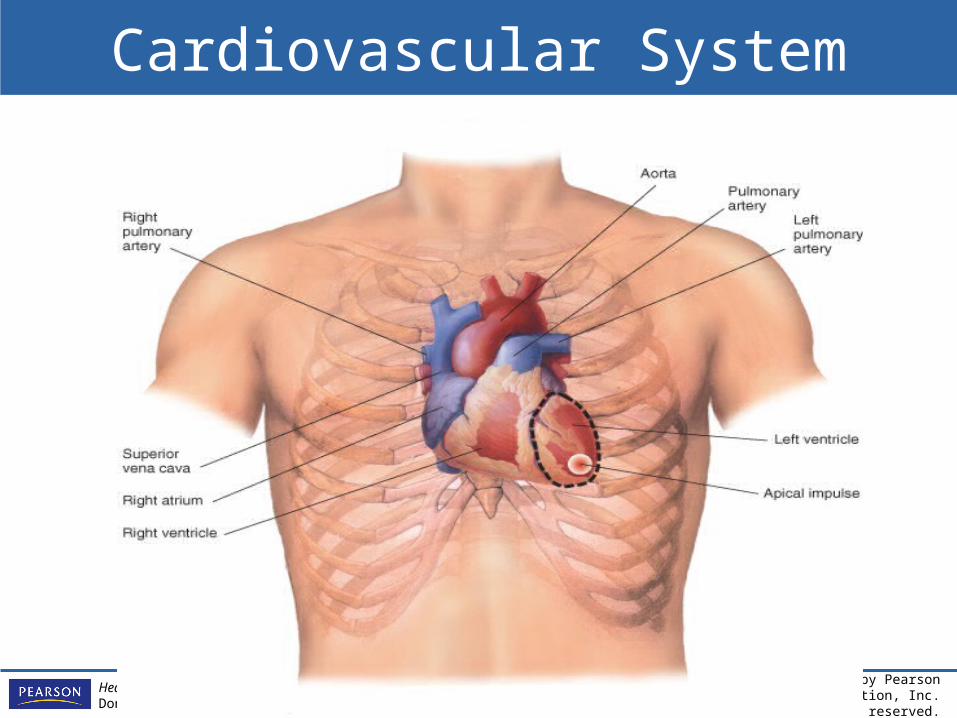
Cardiovascular System
Copyright ©2012 by Pearson Education, Inc.All rights reserved.
Health & Physical Assessment in Nursing, Second EditionDonita D’Amico • Colleen Barbarito
Chambers in the Heart
• Left and right atria• Left and right ventricles

Copyright ©2012 by Pearson Education, Inc.All rights reserved.
Health & Physical Assessment in Nursing, Second EditionDonita D’Amico • Colleen Barbarito
Copyright ©2012 by Pearson Education, Inc.All rights reserved.
Health & Physical Assessment in Nursing, Second EditionDonita D’Amico • Colleen Barbarito
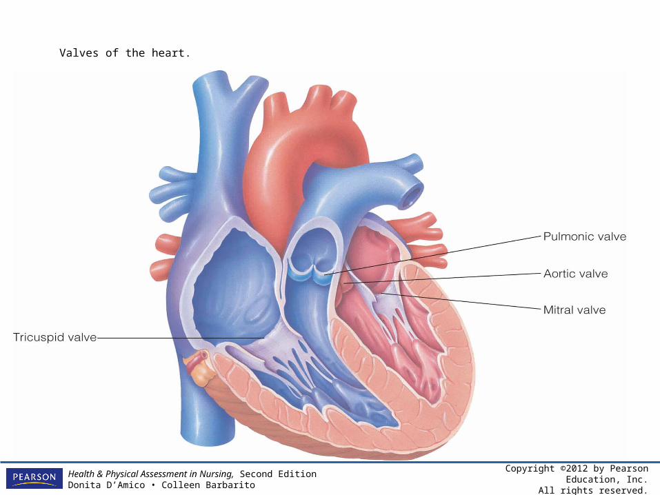
Valves of the heart.
Copyright ©2012 by Pearson Education, Inc.All rights reserved.
Health & Physical Assessment in Nursing, Second EditionDonita D’Amico • Colleen Barbarito
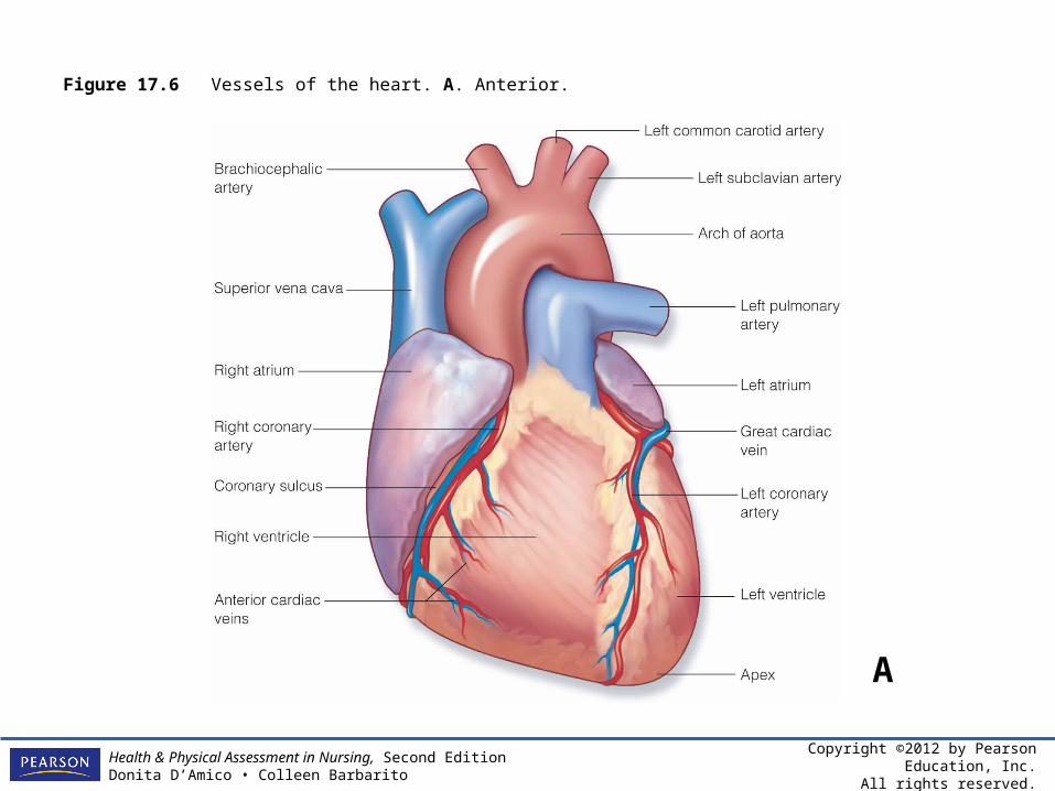
Figure 17.6 Vessels of the heart. A. Anterior.
A
Copyright ©2012 by Pearson Education, Inc.All rights reserved.
Health & Physical Assessment in Nursing, Second EditionDonita D’Amico • Colleen Barbarito
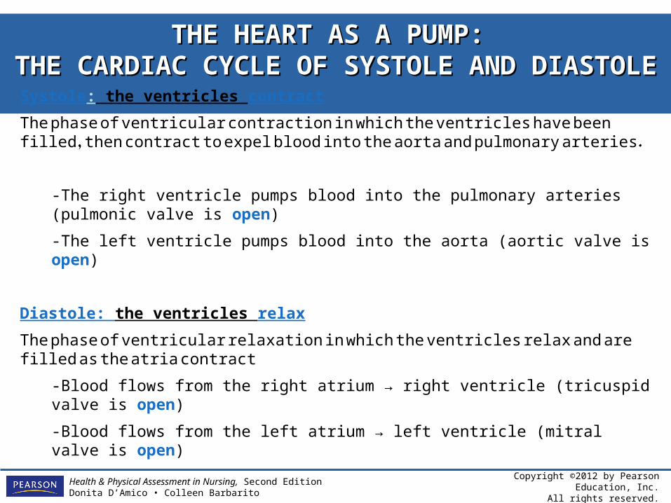
THE HEART AS A PUMP: THE HEART AS A PUMP: THE CARDIAC CYCLE OF SYSTOLE AND DIASTOLETHE CARDIAC CYCLE OF SYSTOLE AND DIASTOLE
Systole: the ventricles contract
The phase of ventricular contraction in which the ventricles have been filled, then contract to expel blood into the aorta and pulmonary arteries.
-The right ventricle pumps blood into the pulmonary arteries (pulmonic valve is open)
-The left ventricle pumps blood into the aorta (aortic valve is open)
Diastole: the ventricles relax
The phase of ventricular relaxation in which the ventricles relax and are filled as the atria contract
-Blood flows from the right atrium → right ventricle (tricuspid valve is open)
-Blood flows from the left atrium → left ventricle (mitral valve is open)
Copyright ©2012 by Pearson Education, Inc.All rights reserved.
Health & Physical Assessment in Nursing, Second EditionDonita D’Amico • Colleen Barbarito
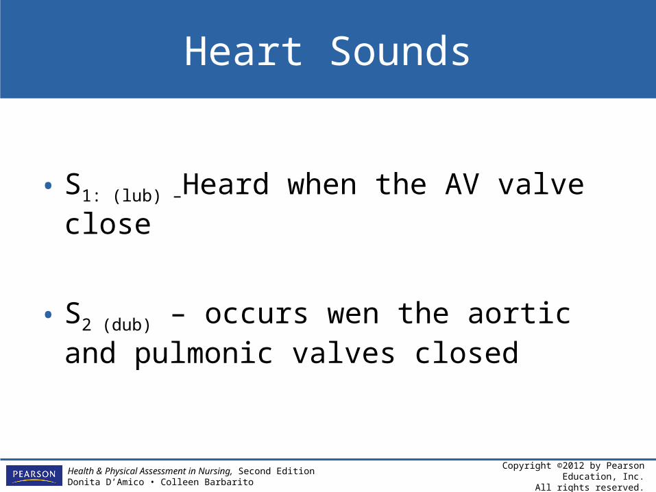
Heart Sounds
• S1: (lub) –Heard when the AV valve close
• S2 (dub) – occurs wen the aortic and pulmonic valves closed
Copyright ©2012 by Pearson Education, Inc.All rights reserved.
Health & Physical Assessment in Nursing, Second EditionDonita D’Amico • Colleen Barbarito
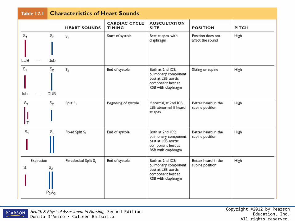
Table 17.1 Characteris
tics of Heart
Sounds
Copyright ©2012 by Pearson Education, Inc.All rights reserved.
Health & Physical Assessment in Nursing, Second EditionDonita D’Amico • Colleen Barbarito
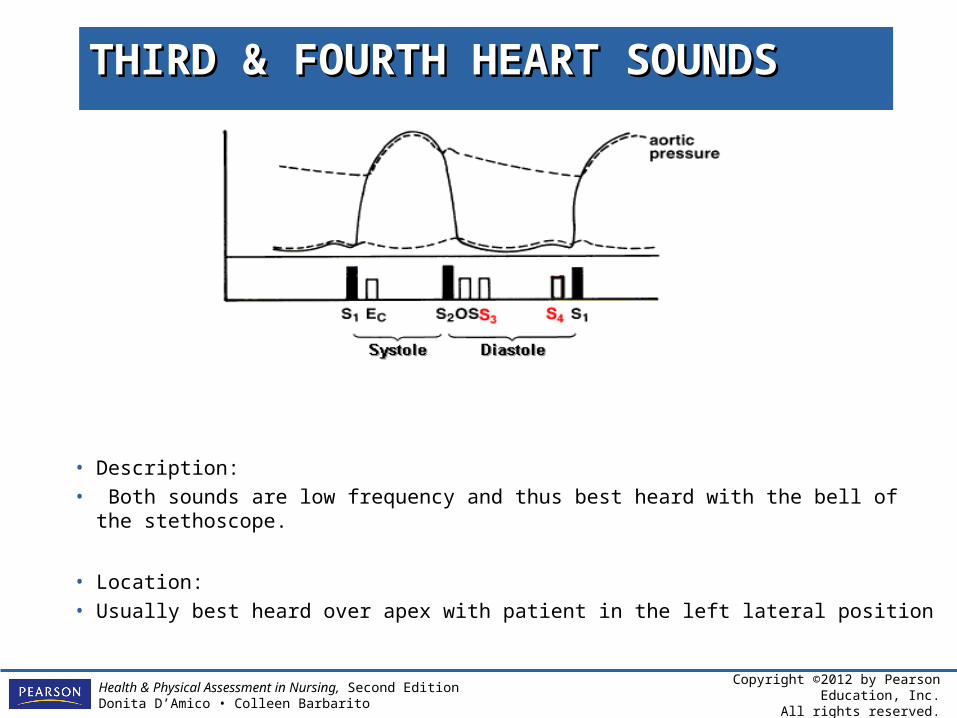
THIRD & FOURTH HEART SOUNDSTHIRD & FOURTH HEART SOUNDS
• Description:
• Both sounds are low frequency and thus best heard with the bell of the stethoscope.
• Location:
• Usually best heard over apex with patient in the left lateral position
Copyright ©2012 by Pearson Education, Inc.All rights reserved.
Health & Physical Assessment in Nursing, Second EditionDonita D’Amico • Colleen Barbarito
THIRD HEART SOUND S3THIRD HEART SOUND S3
• Results from increased atrial pressure leading to increased flow rates, as seen in congestive heart failure, which is the most common cause of a S3.
• May be normal physiological finding in patients less than age 40.
Copyright ©2012 by Pearson Education, Inc.All rights reserved.
Health & Physical Assessment in Nursing, Second EditionDonita D’Amico • Colleen Barbarito
FOURTH HEART SOUND S4FOURTH HEART SOUND S4
Seen in patients with stiffened left ventricles, resulting from conditions such as hypertension, aortic stenosis, ischemic or hypertrophic cardiomyopathy.
Copyright ©2012 by Pearson Education, Inc.All rights reserved.
Health & Physical Assessment in Nursing, Second EditionDonita D’Amico • Colleen Barbarito
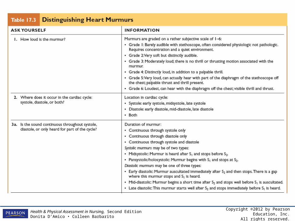
MURMURSMURMURS
• Murmurs are extra heart sounds that are produced as a result of turbulent blood flow that is sufficient to produce audible noise.
• Murmurs may also be the result of various problems, such as narrowing or leaking of valves.
Copyright ©2012 by Pearson Education, Inc.All rights reserved.
Health & Physical Assessment in Nursing, Second EditionDonita D’Amico • Colleen Barbarito
Table 17.3
Distinguishing Heart Murmurs
Copyright ©2012 by Pearson Education, Inc.All rights reserved.
Health & Physical Assessment in Nursing, Second EditionDonita D’Amico • Colleen Barbarito
Cardiac Function
• Stroke volume– Amount of blood that is ejected with each
heartbeat
• Cardiac output– Amount of blood ejected from the left ventricle
over 1 minute
– Cardiac output: stroke volume x heart rate
Copyright ©2012 by Pearson Education, Inc.All rights reserved.
Health & Physical Assessment in Nursing, Second EditionDonita D’Amico • Colleen Barbarito
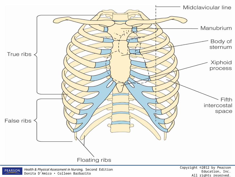
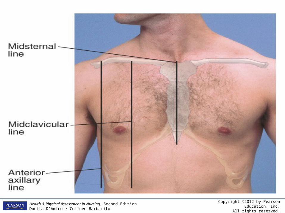
Landmarks for Cardiac Assessment
• Sternum• Clavicles• Ribs• Second through fifth intercostal spaces
Copyright ©2012 by Pearson Education, Inc.All rights reserved.
Health & Physical Assessment in Nursing, Second EditionDonita D’Amico • Colleen Barbarito
Copyright ©2012 by Pearson Education, Inc.All rights reserved.
Health & Physical Assessment in Nursing, Second EditionDonita D’Amico • Colleen Barbarito
Copyright ©2012 by Pearson Education, Inc.All rights reserved.
Health & Physical Assessment in Nursing, Second EditionDonita D’Amico • Colleen Barbarito
Physical Assessment of the Cardiovascular System
• Techniques– Inspection– Palpation– Percussion– Auscultation
Copyright ©2012 by Pearson Education, Inc.All rights reserved.
Health & Physical Assessment in Nursing, Second EditionDonita D’Amico • Colleen Barbarito
Specific Areas of the Cardiovascular Assessment
• Inspection of the face and lips• Inspection of the jugular veins• Inspection of the carotid arteries• Inspection of the hands and fingers• Inspection of the chest and legs
Copyright ©2012 by Pearson Education, Inc.All rights reserved.
Health & Physical Assessment in Nursing, Second EditionDonita D’Amico • Colleen Barbarito
INSPECTION OF THE FACE AND LIPSINSPECTION OF THE FACE AND LIPS
Skin color changes may indicate cardiovascular disease. For example, pallor and cyanosis of lips or extremities are associated with decreased perfusion.
Copyright ©2012 by Pearson Education, Inc.All rights reserved.
Health & Physical Assessment in Nursing, Second EditionDonita D’Amico • Colleen Barbarito
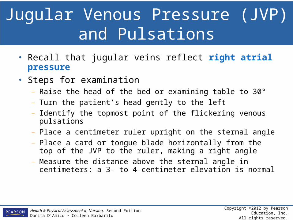
Jugular Venous Pressure (JVP)and Pulsations
• Recall that jugular veins reflect right atrial pressure
• Steps for examination– Raise the head of the bed or examining table to 30°
– Turn the patient’s head gently to the left
– Identify the topmost point of the flickering venous pulsations
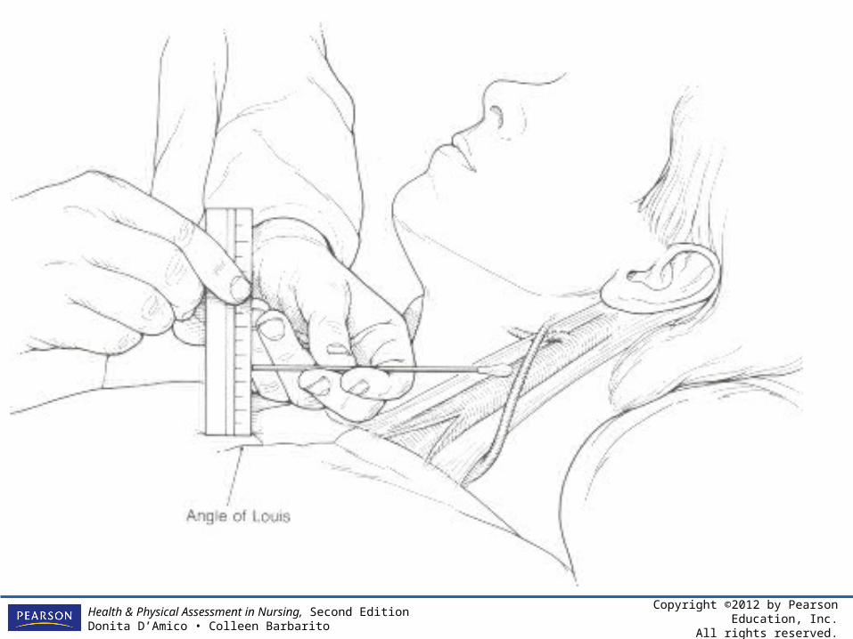
– Place a centimeter ruler upright on the sternal angle
– Place a card or tongue blade horizontally from the top of the JVP to the ruler, making a right angle
– Measure the distance above the sternal angle in centimeters: a 3- to 4-centimeter elevation is normal
Copyright ©2012 by Pearson Education, Inc.All rights reserved.
Health & Physical Assessment in Nursing, Second EditionDonita D’Amico • Colleen Barbarito
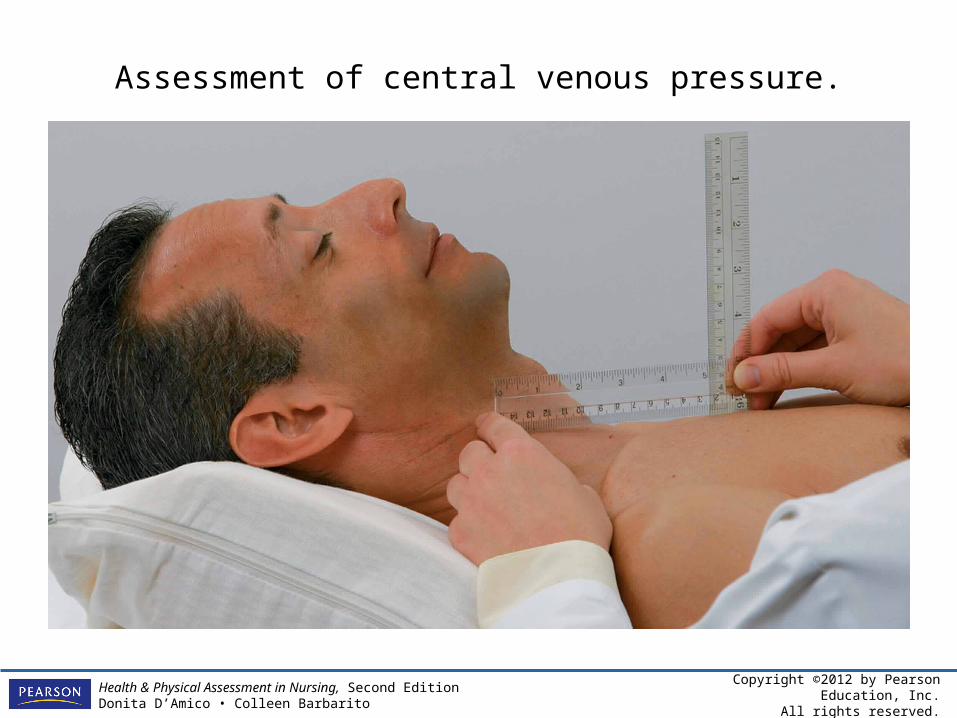
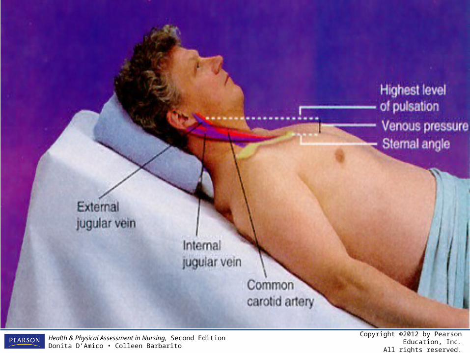
Assessment of central venous pressure.
Copyright ©2012 by Pearson Education, Inc.All rights reserved.
Health & Physical Assessment in Nursing, Second EditionDonita D’Amico • Colleen Barbarito
• Top line – level of the higest visible point of distention
• Bottom line – level of the sternal angle
• Measure: the vertical distance between the sternal angle and the highest level of jugular distention
Copyright ©2012 by Pearson Education, Inc.All rights reserved.
Health & Physical Assessment in Nursing, Second EditionDonita D’Amico • Colleen Barbarito
Copyright ©2012 by Pearson Education, Inc.All rights reserved.
Health & Physical Assessment in Nursing, Second EditionDonita D’Amico • Colleen Barbarito
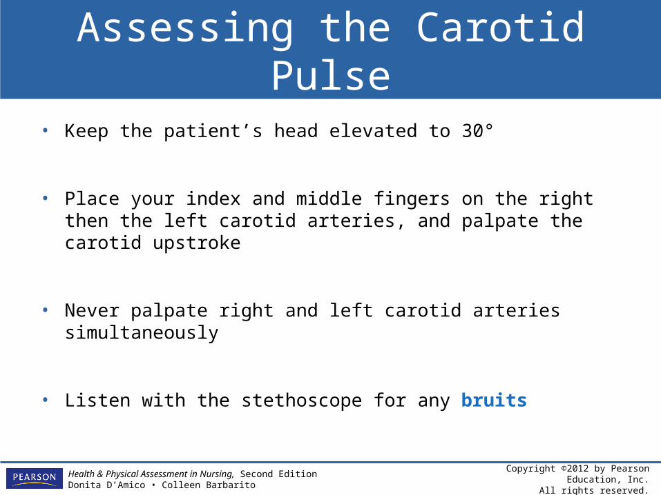
Assessing the Carotid Pulse
• Keep the patient’s head elevated to 30°
• Place your index and middle fingers on the right then the left carotid arteries, and palpate the carotid upstroke
• Never palpate right and left carotid arteries simultaneously
• Listen with the stethoscope for any bruits
Copyright ©2012 by Pearson Education, Inc.All rights reserved.
Health & Physical Assessment in Nursing, Second EditionDonita D’Amico • Colleen Barbarito
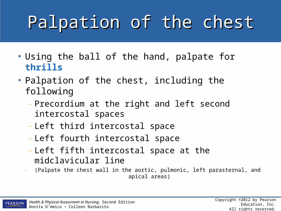
Palpation of the chestPalpation of the chest
• Using the ball of the hand, palpate for thrills• Palpation of the chest, including the following
– Precordium at the right and left second intercostal spaces
– Left third intercostal space– Left fourth intercostal space– Left fifth intercostal space at the midclavicular
line– (Palpate the chest wall in the aortic, pulmonic, left parasternal, and
apical areas)
Copyright ©2012 by Pearson Education, Inc.All rights reserved.
Health & Physical Assessment in Nursing, Second EditionDonita D’Amico • Colleen Barbarito
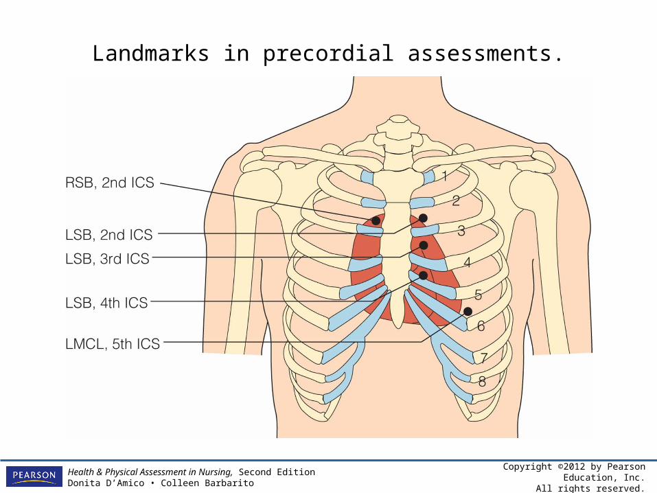
Landmarks in precordial assessments.
Copyright ©2012 by Pearson Education, Inc.All rights reserved.
Health & Physical Assessment in Nursing, Second EditionDonita D’Amico • Colleen Barbarito
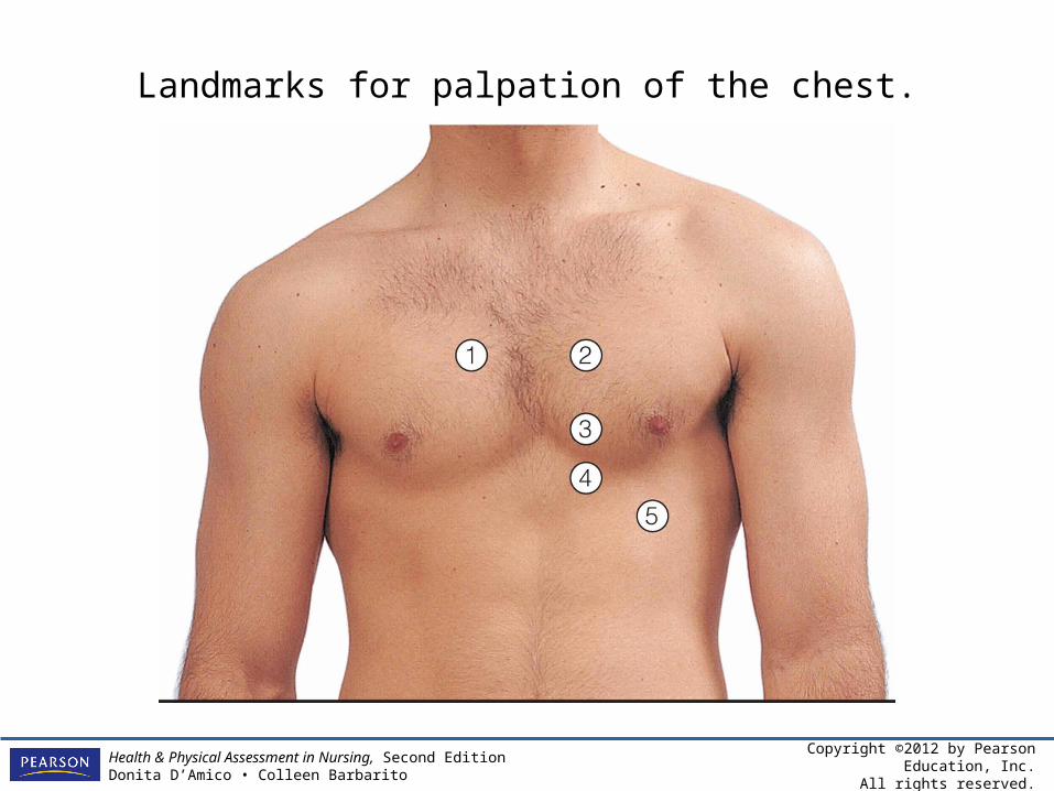
Landmarks for palpation of the chest.
Copyright ©2012 by Pearson Education, Inc.All rights reserved.
Health & Physical Assessment in Nursing, Second EditionDonita D’Amico • Colleen Barbarito
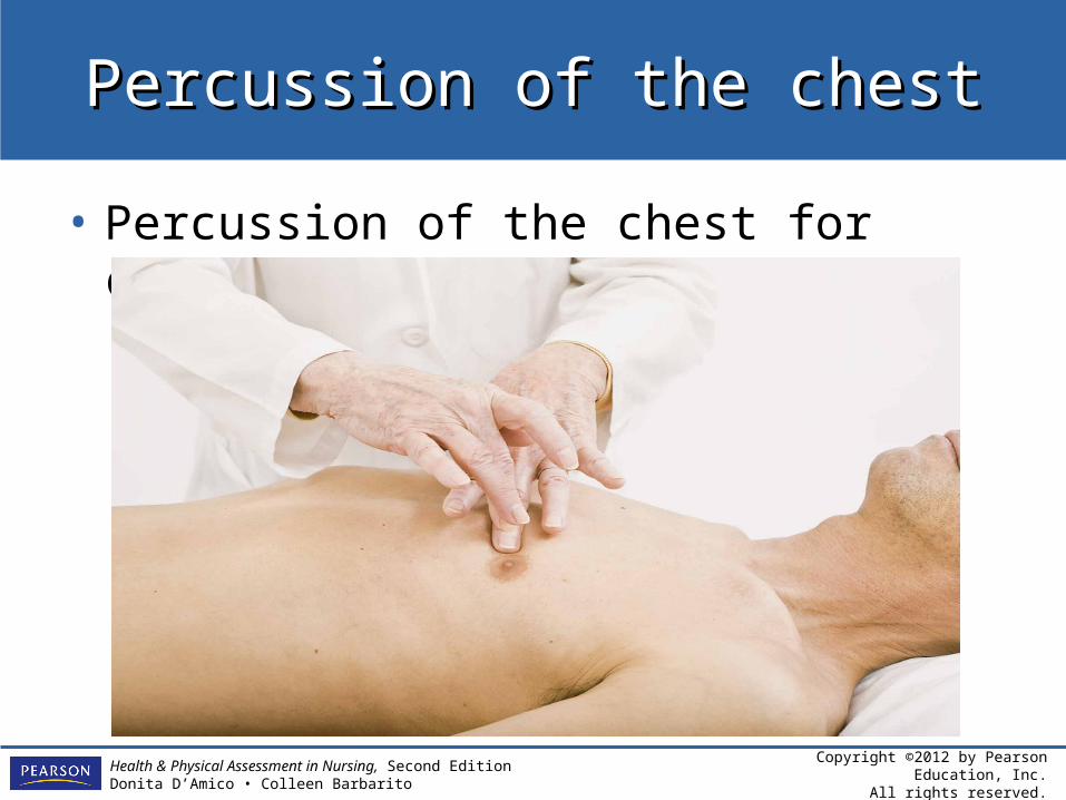
Percussion of the chestPercussion of the chest
• Percussion of the chest for cardiac border
Copyright ©2012 by Pearson Education, Inc.All rights reserved.
Health & Physical Assessment in Nursing, Second EditionDonita D’Amico • Colleen Barbarito
AUSCULTATIONAUSCULTATION
Blood pressure measurement
o Select the proper size cuff
o Position the patient properly
o Make sure there is a brachial pulse
o Apply the cuff correctly
o Assess blood pressure for hypertension
Copyright ©2012 by Pearson Education, Inc.All rights reserved.
Health & Physical Assessment in Nursing, Second EditionDonita D’Amico • Colleen Barbarito
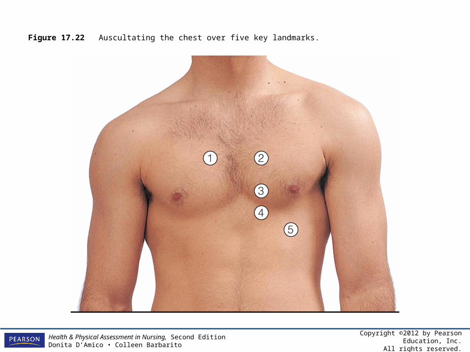
Auscultation of the chestAuscultation of the chest
• Auscultation of the chest using the diaphragm and bell in various positions to include the following locations– Aortic area at the right second intercostal
space–S2 is louder than S1
– Pulmonic area at the left second intercostal space–S2 is louder than S1
– Erb’s point at the left third intercostal space–S1 and S2 are heard equally
Copyright ©2012 by Pearson Education, Inc.All rights reserved.
Health & Physical Assessment in Nursing, Second EditionDonita D’Amico • Colleen Barbarito
Auscultation of the chestAuscultation of the chest
– Tricuspid area at the left fourth intercostal space–S1 is louder than S2
– Apex at the left fifth intercostal space at the midclavicular line–S1 is louder than S2
Copyright ©2012 by Pearson Education, Inc.All rights reserved.
Health & Physical Assessment in Nursing, Second EditionDonita D’Amico • Colleen Barbarito
Figure 17.22 Auscultating the chest over five key landmarks.
Copyright ©2012 by Pearson Education, Inc.All rights reserved.
Health & Physical Assessment in Nursing, Second EditionDonita D’Amico • Colleen Barbarito
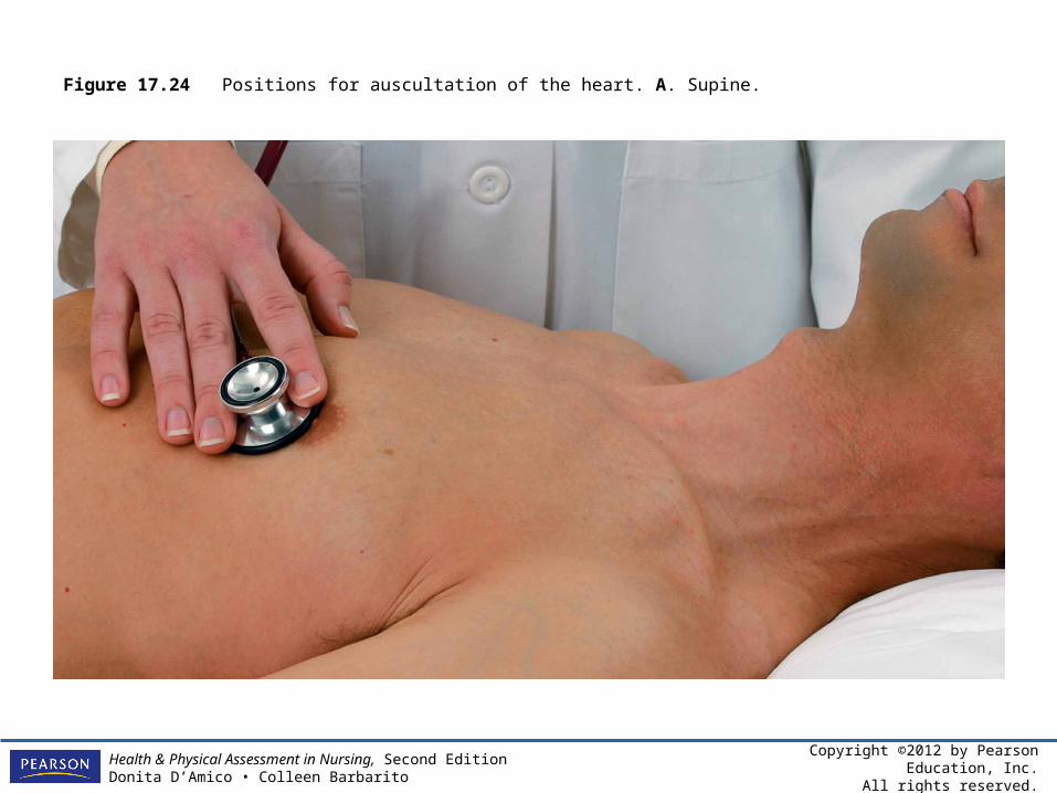
Figure 17.24 Positions for auscultation of the heart. A. Supine.
Copyright ©2012 by Pearson Education, Inc.All rights reserved.
Health & Physical Assessment in Nursing, Second EditionDonita D’Amico • Colleen Barbarito
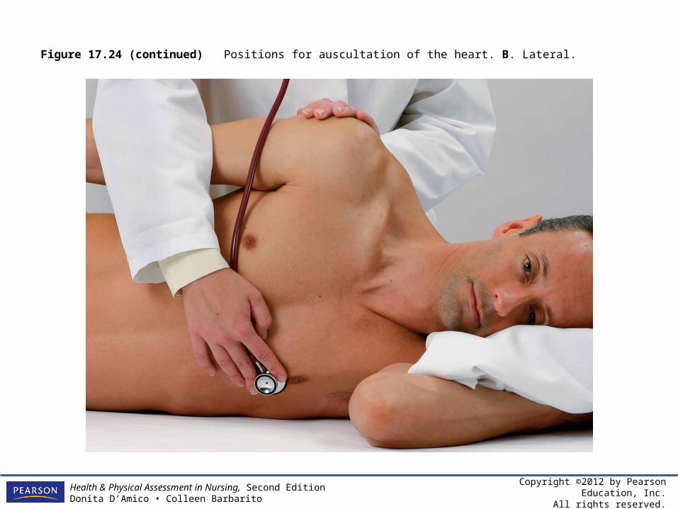
Figure 17.24 (continued) Positions for auscultation of the heart. B. Lateral.
Copyright ©2012 by Pearson Education, Inc.All rights reserved.
Health & Physical Assessment in Nursing, Second EditionDonita D’Amico • Colleen Barbarito
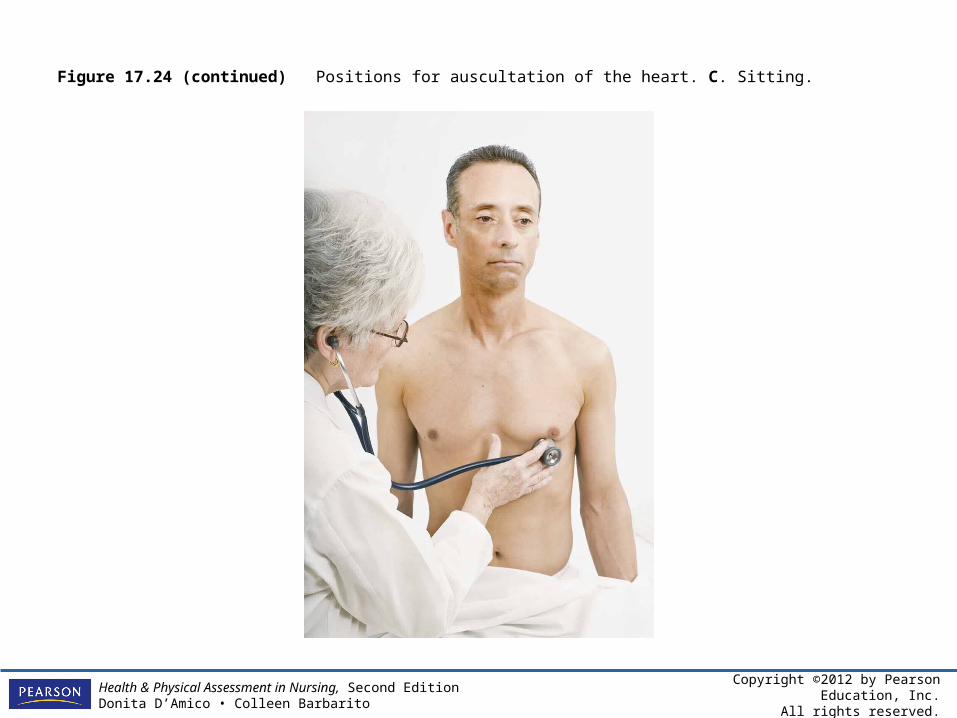
Figure 17.24 (continued) Positions for auscultation of the heart. C. Sitting.
Copyright ©2012 by Pearson Education, Inc.All rights reserved.
Health & Physical Assessment in Nursing, Second EditionDonita D’Amico • Colleen Barbarito
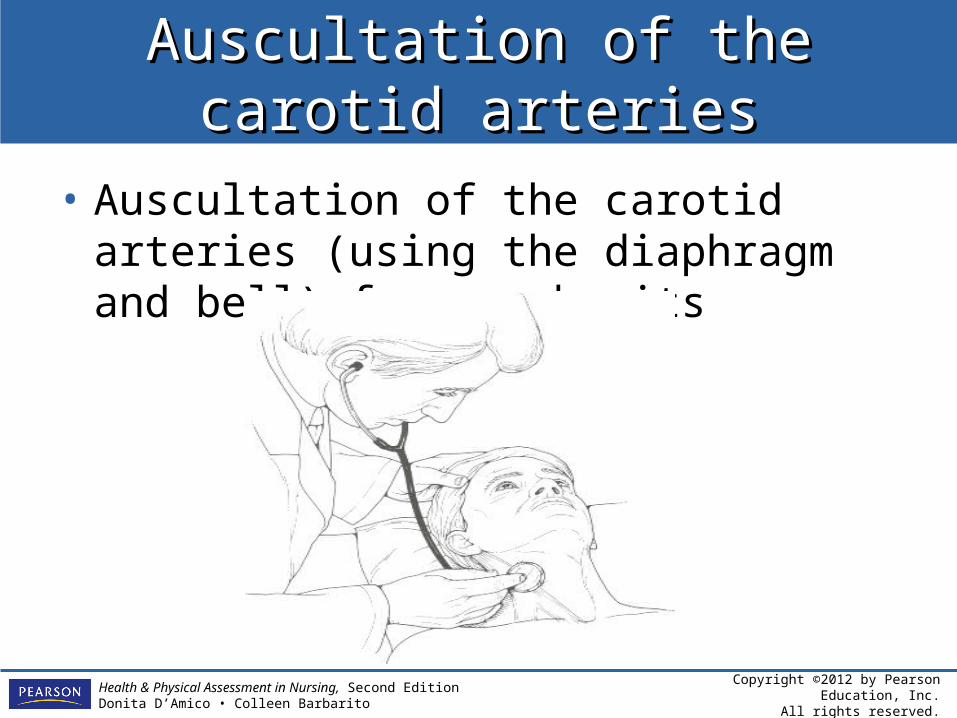
Auscultation of the carotid arteriesAuscultation of the carotid arteries
• Auscultation of the carotid arteries (using the diaphragm and bell) for any bruits