copyright © 2010 pearson education, inc. figure 9.26a arrangement of smooth muscle in the walls of...
Post on 22-Dec-2015
213 views
TRANSCRIPT
Copyright © 2010 Pearson Education, Inc.
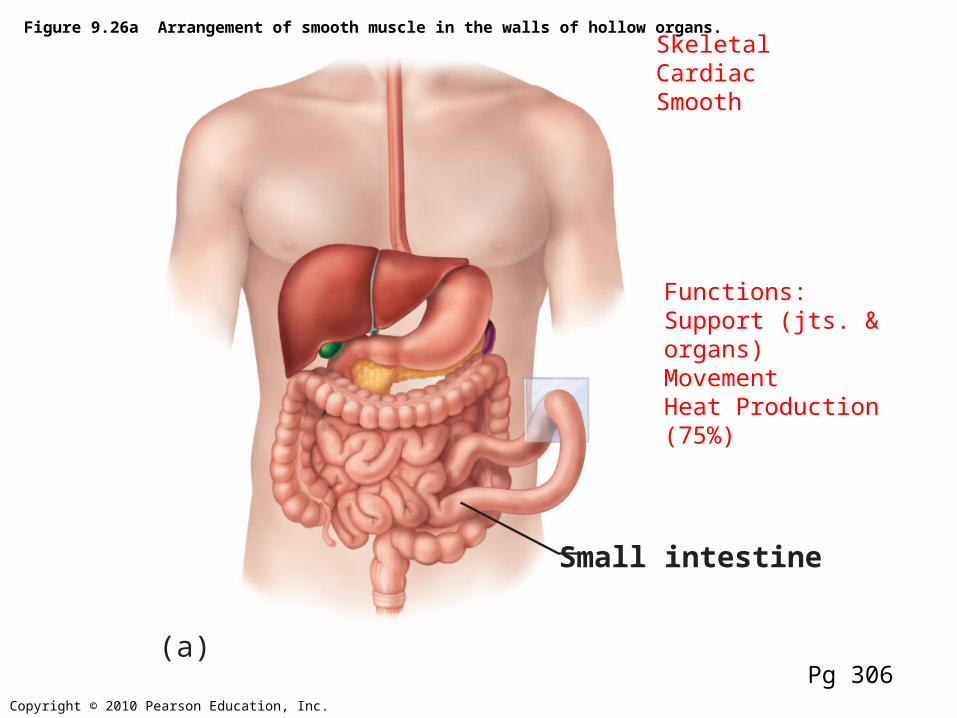
Figure 9.26a Arrangement of smooth muscle in the walls of hollow organs.
Small intestine
(a)Pg 306
SkeletalCardiacSmooth
Functions:Support (jts. & organs)MovementHeat Production (75%)

Copyright © 2010 Pearson Education, Inc.
NucleusLight I bandDark A band
Sarcolemma
Mitochondrion
(b) Diagram of part of a muscle fiber showing the myofibrils. Onemyofibril is extended afrom the cut end of the fiber.
Myofibril
Figure 9.2b Microscopic anatomy of a skeletal muscle fiber.
Pg 280
Muscle fiber = Muscle cell
cytoskeleton
LongDensely packed

Copyright © 2010 Pearson Education, Inc.
Figure 4.10a Muscle tissues.
(a) Skeletal muscle
Description: Long, cylindrical,multinucleate cells; obviousstriations.
Function: Voluntary movement;locomotion; manipulation of theenvironment; facial expression;voluntary control.
Location: In skeletal musclesattached to bones oroccasionally to skin.
Photomicrograph: Skeletal muscle (approx. 460x).Notice the obvious banding pattern and thefact that these large cells are multinucleate.
Nuclei
Striations
Part ofmuscle fiber (cell)
Pg 138
-nicotonic receptors-Myoglobin

Copyright © 2010 Pearson Education, Inc.
Figure 4.10b Muscle tissues.
(b) Cardiac muscle
Description: Branching, short striated, generally uninucleate cells that interdigitate atspecialized junctions (intercalated discs).
Function: As it contracts, it propels blood into the circulation; involuntary control.Location: The walls of the heart.
Photomicrograph: Cardiac muscle (500X);notice the striations, branching of cells, andthe intercalated discs.
Intercalateddiscs
Striations
Nucleus
Pg 139
-Muscarinic & Beta receptors-Lots of myoglobin & good blood supply
Lots of mitochondria

Copyright © 2010 Pearson Education, Inc.
(c) Smooth muscle
Description: Spindle-shapedcells with central nuclei; nostriations; cells arranged closely to form sheets.
Function: Propels substancesor objects (foodstuffs, urine,a baby) along internal passage-ways; involuntary control.
Location: Mostly in the wallsof hollow organs.
Photomicrograph: Sheet of smooth muscle (200x).
Smoothmusclecell
Nuclei
Figure 4.10c Muscle tissues.
Pg 139
-Muscarinic & alpha or beta receptors-No myoglobin

Copyright © 2010 Pearson Education, Inc.
307

Copyright © 2010 Pearson Education, Inc.
Figure 9.5 Relationship of the sarcoplasmic reticulum and T tubules to myofibrils of skeletal muscle.
Myofibril
Myofibrils
Sarcotubular Sys.
Tubules ofthe SR
Sarcolemma
Sarcolemma A.P.
Mitochondria
I band I bandA band
H zone Z discZ disc
Part of a skeletalmuscle fiber (cell)
• T tubule• Sarcoplasmic
Reticulum (Ca)
M line
Pg 284
Myofilaments (sarcomeres)

Copyright © 2010 Pearson Education, Inc.
NucleusLight I bandDark A band
Sarcolemma
Mitochondrion
(b) Diagram of part of a muscle fiber showing the myofibrils. Onemyofibril is extended afrom the cut end of the fiber.
Myofibril
Figure 9.2b Microscopic anatomy of a skeletal muscle fiber.
Pg 280

Copyright © 2010 Pearson Education, Inc.
Figure 9.2c Microscopic anatomy of a skeletal muscle fiber.
I band I bandA bandSarcomere
H zoneThin (actin)filament
Thick (myosin)filament
Z disc Z disc
M line
(c) Small part of one myofibril enlarged to show the myofilamentsresponsible for the banding pattern. Each sarcomere extends fromone Z disc to the next.
Pg 280

Copyright © 2010 Pearson Education, Inc.
Figure 9.3 Composition of thick and thin filaments.
Flexible hinge region
Tail
Tropomyosin Troponin ActinMyosin head
ATP-bindingsite
Heads Active sitesfor myosinattachment
Actinsubunits
Actin-binding sites
Thick filamentEach thick filament consists of manymyosin molecules whose heads protrude at opposite ends of the filament.
Thin filamentA thin filament consists of two strandsof actin subunits twisted into a helix plus two types of regulatory proteins(troponin and tropomyosin).
Thin filamentThick filament
In the center of the sarcomere, the thickfilaments lack myosin heads. Myosin heads are present only in areas of myosin-actin overlap.
Longitudinal section of filamentswithin one sarcomere of a myofibril
Portion of a thick filamentPortion of a thin filament
Myosin molecule Actin subunits
200
Pg 282ATPase

Copyright © 2010 Pearson Education, Inc.
Figure 9.2d Microscopic anatomy of a skeletal muscle fiber.
Z disc Z discM line
Sarcomere
Thin (actin)filament
Thick(myosin)filament
Elastic (titin)filaments
(d) Enlargement of one sarcomere (sectioned lengthwise). Notice the myosin heads on the thick filaments.
pg 280

Copyright © 2010 Pearson Education, Inc.
Figure 9.13 A motor unit consists of a motor neuron and all the muscle fibers it innervates.
Spinal cord
Motor neuroncell body
Muscle
Branching axonto motor unit
Nerve
Motorunit 1
Motorunit 2
Musclefibers
Motor neuronaxon
Axon terminals atneuromuscular junctions
Axons of motor neurons extend from the spinal cord to the muscle.There each axon divides into a number of axon terminals that formneuromuscular junctions with muscle fibers scattered throughoutthe muscle.
Branching axonterminals formneuromuscularjunctions, one permuscle fiber (photo-micrograph 330x).
(b)
(a)
Pg 294
4 100 fibers All or nothing
Polio
Copyright © 2010 Pearson Education, Inc.
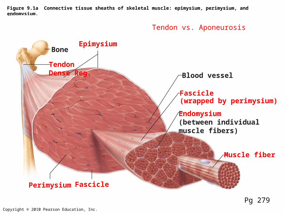
Figure 9.1a Connective tissue sheaths of skeletal muscle: epimysium, perimysium, and endomysium.
Bone
Perimysium
Endomysium(between individualmuscle fibers)
Muscle fiber
Fascicle(wrapped by perimysium)
Epimysium
TendonDense Reg. Blood vessel
Fascicle
Pg 279
Tendon vs. Aponeurosis

Copyright © 2010 Pearson Education, Inc.
Figure 10.6 Lateral view of muscles of the scalp, face, and neck.
Corrugatorsupercilii Orbicularis oculiLevator labiisuperiorisZygomaticusminor and majorBuccinatorRisoriusOrbicularis orisMentalisDepressorlabii inferiorisDepressor anguli orisPlatysma
Galeaaponeurotica
Frontal belly
Occipitalbelly
Temporalis
MasseterSternocleidomastoidTrapezius
Splenius capitis
Epicranius
Pg 331
Copyright © 2010 Pearson Education, Inc.
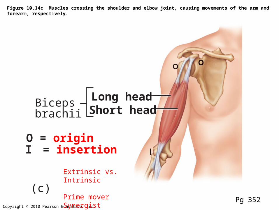
Figure 10.14c Muscles crossing the shoulder and elbow joint, causing movements of the arm and forearm, respectively.
Long headBicepsbrachii Short head
O = origin I = insertion
(c)Pg 352
Extrinsic vs. Intrinsic
Prime moverSynergistAntagonist

Copyright © 2010 Pearson Education, Inc.
Figure 9.8 Events at the Neuromuscular Junction
Nucleus
Actionpotential (AP)
Myelinated axonof motor neuron
Axon terminal ofneuromuscular junction
Sarcolemma ofthe muscle fiber
Ca2+ Ca2+
Axon terminalof motor neuron
Synaptic vesiclecontaining AChMitochondrionSynapticcleft
Junctionalfolds ofsarcolemma
Fusing synaptic vesicles
ACh
Sarcoplasm ofmuscle fiber
Postsynaptic membraneion channel opens;ions pass.
Na+ K+
Ach–
Na+
K+
Degraded ACh
Acetyl-cholinesterase
Postsynaptic membraneion channel closed;ions cannot pass.
1 Action potential arrives ataxon terminal of motor neuron.
2 Voltage-gated Ca2+ channels open and Ca2+ enters the axon terminal.
3 Ca2+ entry causes some synaptic vesicles to release their contents (acetylcholine)by exocytosis.
4 Acetylcholine, aneurotransmitter, diffuses across the synaptic cleft and binds to receptors in the sarcolemma.
5 ACh binding opens ionchannels that allow simultaneous passage of Na+ into the musclefiber and K+ out of the muscle fiber.
6 ACh effects are terminated by its enzymatic breakdown in the synaptic cleft by acetylcholinesterase.
Motor End Plate-1/cell-Middle of cell-Location of receptors
Pg 287


Copyright © 2010 Pearson Education, Inc.
Figure 9.11 Excitation-Contraction Coupling (1 of 4)
Axon terminalof motor neuron
Muscle fiberTriad
One sarcomere
Synaptic cleft
Setting the stage
Sarcolemma
Action potentialis generated
Terminal cisterna of SR ACh
Ca2+
Pg 290
Copyright © 2010 Pearson Education, Inc.
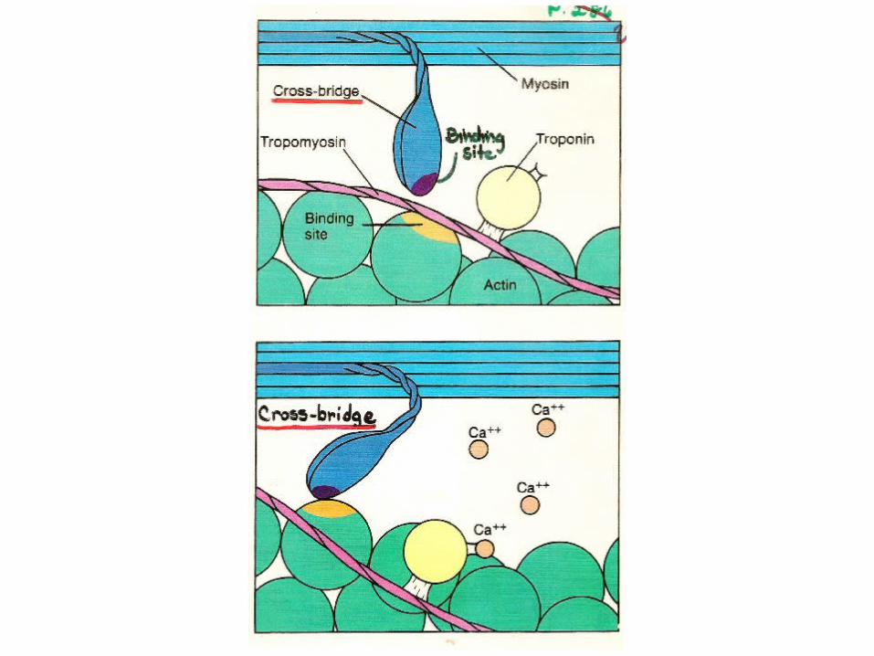
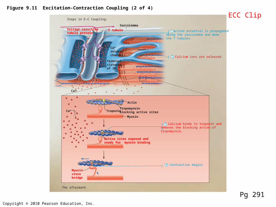
Figure 9.11 Excitation-Contraction Coupling (2 of 4)
Steps in E-C Coupling:
TroponinTropomyosinblocking active sites
Myosin
Actin
Active sites exposed andready for myosin binding
Ca2+
Terminal cisternaof SR
Voltage-sensitivetubule protein
T tubule
Ca2+
releasechannel
Myosincrossbridge
Ca2+
Sarcolemma
Action potential is propagatedalong the sarcolemma and downthe T tubules.
Calcium ions are released.
Calcium binds to troponin andremoves the blocking action oftropomyosin.
Contraction begins
The aftermath
1
2
4
3
Pg 291
ECC Clip

Copyright © 2010 Pearson Education, Inc.
Figure 9.12 Cross Bridge Cycle
Actin
Cross bridge formation.
Cocking of myosin head. The power (working)stroke.
Cross bridgedetachment.
Ca2+
1
2
3
4
Myosinhead
Thickfilament
Thin filament
ADP
Myosin
P i
ADP
P iATPhydrolysis
ADP
P i
ATP
ATP
Pg 292
charged
Rigor mortisCramps
CBC clip

Copyright © 2010 Pearson Education, Inc.
Figure 9.6 Sliding filament model of contraction.
I
Fully relaxed sarcomere of a muscle fiber
Fully contracted sarcomere of a muscle fiber
IA
Z ZH
I IA
Z Z
1
2
Pg 285
Copyright © 2010 Pearson Education, Inc.
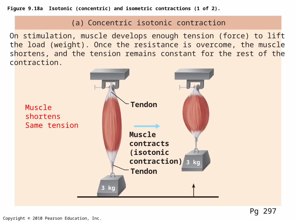
Figure 9.18a Isotonic (concentric) and isometric contractions (1 of 2).
(a) Concentric isotonic contraction
On stimulation, muscle develops enough tension (force) to liftthe load (weight). Once the resistance is overcome, the muscleshortens, and the tension remains constant for the rest of thecontraction.
3 kg
3 kg
Musclecontracts(isotoniccontraction)
Tendon
Tendon
Pg 297
Muscle shortensSame tension

Copyright © 2010 Pearson Education, Inc.
Figure 9.18b Isotonic (concentric) and isometric contractions (1 of 2).
(b) Isometric contraction
Muscle is attached to a weight that exceeds the muscle’s peaktension-developing capabilities. When stimulated, the tensionincreases to the muscle’s peak tension-developing capability,but the muscle does not shorten.
6 kg 6 kg
Musclecontracts(isometriccontraction)
Pg 297
No ShorteningIncreasing TensionMaintains Posture

Copyright © 2010 Pearson Education, Inc.
Figure 9.19 Pathways for regenerating ATP during muscle activity.
Coupled reaction of creatinephosphate (CP) and ADP
Energy source: CP Energy source: glucose Energy source: glucose; pyruvic acid;free fatty acids from adipose tissue;amino acids from protein catabolism
Glycolysis and lactic acid formation
(a) Direct phosphorylation (b) Anaerobic pathway (c) Aerobic pathway
Aerobic cellular respiration
Oxygen use: NoneProducts: 1 ATP per CP, creatineDuration of energy provision:15 seconds
Oxygen use: NoneProducts: 2 ATP per glucose, lactic acidDuration of energy provision:60 seconds, or slightly more
Oxygen use: RequiredProducts: 32 ATP per glucose, CO2, H2ODuration of energy provision: Hours
Creatinekinase
ADPCP
Creatine
Glucose (fromglycogen breakdown ordelivered from blood)
Glucose (fromglycogen breakdown ordelivered from blood)
Glycolysisin cytosol
Pyruvic acid
Releasedto blood
net gain
2
32Lactic acid
O2
O2
O2
O2
H2OCO2
Pyruvic acidFattyacids
Aminoacids
Aerobic respirationin mitochondriaAerobic respirationin mitochondria
ATP
ATP
ATP
net gain perglucose
Pg 299
60-70%
Rapid ATP Production

Copyright © 2010 Pearson Education, Inc.
Figure 9.20 Comparison of energy sources used during short-duration exercise and prolonged-duration exercise.
Short-duration exercise Prolonged-durationexercise
ATP stored inmuscles isused first.
ATP is formedfrom creatinePhosphateand ADP.
Glycogen stored in muscles is brokendown to glucose, which is oxidized togenerate ATP.
ATP is generated bybreakdown of severalnutrient energy fuels byaerobic pathway. Thispathway uses oxygenreleased from myoglobinor delivered in the bloodby hemoglobin. When itends, the oxygen deficit ispaid back.
Pg 300
Lactic Acid Byproduct
70% max muscle activity

Twitch
The contraction of a muscle in response to a brief, single stimulus.
Example: Blinking
Consists of 3 distinct phases

Copyright © 2010 Pearson Education, Inc.
Figure 9.14a The muscle twitch.
Latentperiod
Singlestimulus
Period ofcontraction
Period ofrelaxation
(a) Myogram showing the three phases of an isometric twitch
Pg 295
Refractory period


Increasing # of motor units
No recruitment
Twitches fusing
Piggy-backing twitchesPartial relaxation
Copyright © 2010 Pearson Education, Inc.
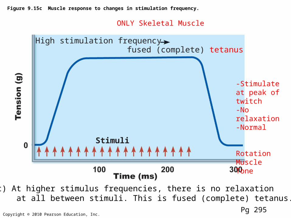
Figure 9.15c Muscle response to changes in stimulation frequency.
Stimuli
High stimulation frequencyfused (complete) tetanus
(c) At higher stimulus frequencies, there is no relaxation at all between stimuli. This is fused (complete) tetanus.
ONLY Skeletal Muscle
-Stimulate at peak of twitch-No relaxation-Normal
RotationMuscle Tone
Pg 295

Copyright © 2010 Pearson Education, Inc.
Sarcomeresgreatly
shortened
Sarcomeres atresting length
Sarcomeres excessivelystretched
170%
Optimal sarcomereoperating length(80%–120% ofresting length)
100%75%
Figure 9.22 Length-tension relationships of sarcomeres in skeletal muscles.
Pg 302
Congestive heart failure
Smooth Muscle-Accommodation-Bladder, Uterus

Temperature
Too cool-Decreases enzyme functioning
Too warm-Decreases enzyme functioning-May even denature enzymes and muscle proteins (107-108 degrees F)-Heat rigor

Fatigue
1. Build up of waste products
2. Decreased energy stores
3. Increased temperature
4. Synaptic fatigue

312

Exercise and Training
Aerobic• Continuous, prolonged• Cellular respiration• Increases mitochondrion• Usage of fat as fuel• Improves endurance• Improves muscle tone
• (red)
AnaerobicBursts of activityCellular respiration &
anaerobic glycolysisIncreases size of muscles
(hypertrophy)Increases strength
(white)

Muscle fiber types
Red Fibers (Slow Oxidative)
• Increased Mitochondrion• Lots of myoglobin• Highly vascular• Slow to fatigue• Use for endurance activities• Slow contraction time
• Efficiency vs. Endurance
White Fibers (Fast Glycolytic)
• Less mitochondrion• Decreased myoglobin• High levels of ATPase• Use for anaerobic activity• Increase in size• Fast contraction time
? Fast Oxidative Glycolytic