copyright © 2009 wolters kluwer health | lippincott williams & wilkins memmler’s the human...
TRANSCRIPT

Copyright © 2009 Wolters Kluwer Health | Lippincott Williams & Wilkins
Memmler’s The Human Body in Health and Disease
11th edition
Memmler’s The Human Body in Health and Disease
11th edition
Chapter 9Chapter 9
The Nervous System: The The Nervous System: The Spinal Cord and Spinal NervesSpinal Cord and Spinal Nerves
Copyright © 2009 Wolters Kluwer Health | Lippincott Williams & Wilkins
Role of the Nervous SystemRole of the Nervous SystemRole of the Nervous SystemRole of the Nervous System
Nervous system coordinates all body systems
•Detects and responds to stimuli
•Brain and spinal cord act as switching centers
•Nerves carry messages to and from centers
Copyright © 2009 Wolters Kluwer Health | Lippincott Williams & Wilkins
Structural DivisionsStructural Divisions
•Central nervous system (CNS)
– Brain
– Spinal cord
•Peripheral nervous system (PNS)
– Cranial nerves
– Spinal nerves
Copyright © 2009 Wolters Kluwer Health | Lippincott Williams & Wilkins
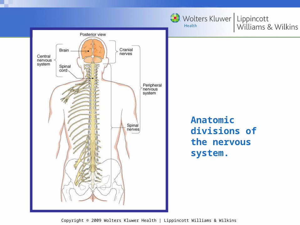
Anatomic divisions of the nervous system.

Copyright © 2009 Wolters Kluwer Health | Lippincott Williams & Wilkins
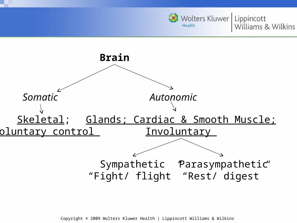
Functional DivisionsFunctional Divisions
Somatic nervous system
•Controlled voluntarily
•Effectors are skeletal muscles
•No further subdivisions
Autonomic (or visceral) nervous system (ANS)
•Controlled involuntarily
•Effectors are smooth muscle, cardiac muscle, and glands
•Subdivided into
– Sympathetic nervous system
– Parasympathetic nervous system
Copyright © 2009 Wolters Kluwer Health | Lippincott Williams & Wilkins
Brain
Somatic Autonomic
Skeletal; Voluntary control
Glands; Cardiac & Smooth Muscle;Involuntary
Sympathetic“Fight/ flight”
Parasympathetic“Rest/ digest”

Copyright © 2009 Wolters Kluwer Health | Lippincott Williams & Wilkins
Checkpoint 9-1: What are the two divisions of the nervous system based on structure?
Checkpoint 9-2: The nervous system can be divided functionally into two divisions based on type of control and effectors. What division is voluntary and controls skeletal muscle, and what division is involuntary and controls involuntary muscles and glands?

Copyright © 2009 Wolters Kluwer Health | Lippincott Williams & Wilkins
Neurons and Their FunctionsNeurons and Their FunctionsNeurons and Their FunctionsNeurons and Their Functions
Neurons
•Functional cells of nervous system
•Highly specialized
•Unique structure
Copyright © 2009 Wolters Kluwer Health | Lippincott Williams & Wilkins
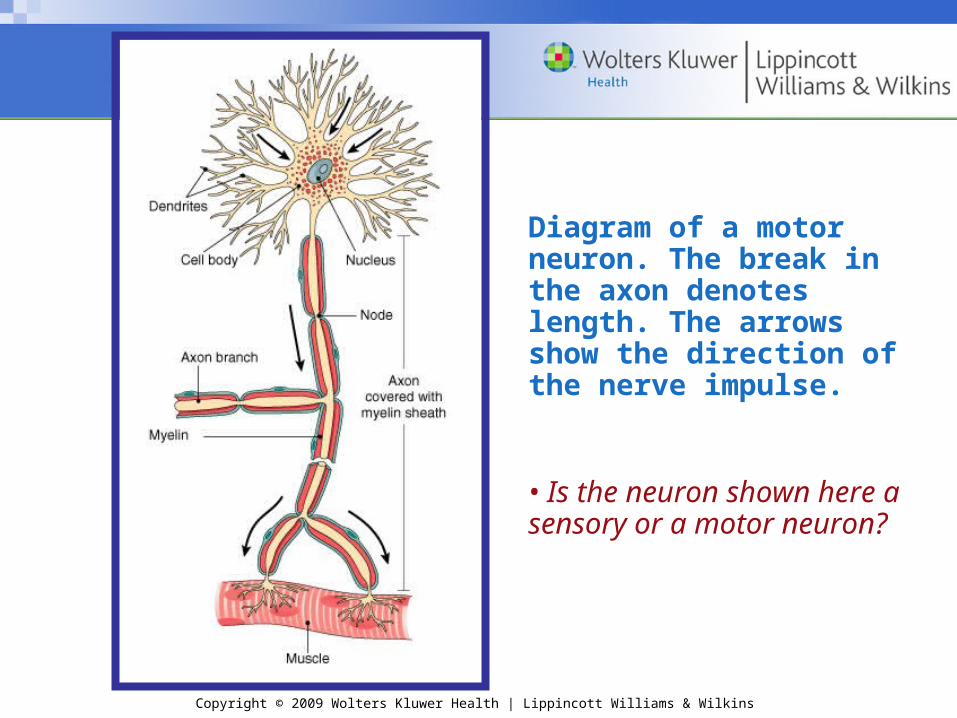
Diagram of a motor neuron. The break in the axon denotes length. The arrows show the direction of the nerve impulse.
• Is the neuron shown here a sensory or a motor neuron?

Copyright © 2009 Wolters Kluwer Health | Lippincott Williams & Wilkins
Structure of a NeuronStructure of a Neuron
Cell body
•Nucleus
•Other organelles
Cell fibers
•Dendrites
•Axons
– Some are protected by myelin sheath
Copyright © 2009 Wolters Kluwer Health | Lippincott Williams & Wilkins
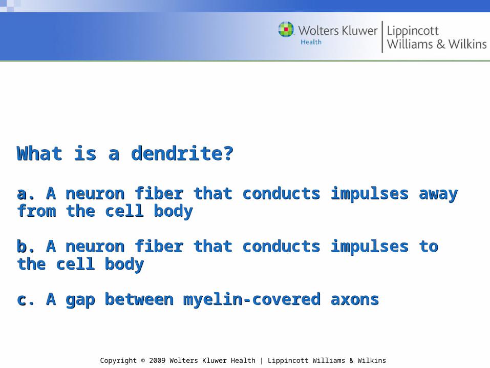
What is a dendrite?
a. a. A neuron fiber that conducts impulses away from the cell body
b. b. A neuron fiber that conducts impulses to the cell body cc. A gap between myelin-covered axons
What is a dendrite?
a. a. A neuron fiber that conducts impulses away from the cell body
b. b. A neuron fiber that conducts impulses to the cell body cc. A gap between myelin-covered axons
Copyright © 2009 Wolters Kluwer Health | Lippincott Williams & Wilkins
Answer:Answer:
b. A neuron fiber that conducts impulses to the cell bodyb. A neuron fiber that conducts impulses to the cell body
Answer:Answer:
b. A neuron fiber that conducts impulses to the cell bodyb. A neuron fiber that conducts impulses to the cell body

Copyright © 2009 Wolters Kluwer Health | Lippincott Williams & Wilkins
Formation of a myelin sheath. (A)Schwann cells wrap around the axon, creating a myelin coating. (B) The outermost layer of the Schwann cell forms the neurilemma.Spaces between the cells are the nodes (of Ranvier).
Copyright © 2009 Wolters Kluwer Health | Lippincott Williams & Wilkins
ACTIVITYACTIVITY
•Make your own myelin covered nerve

Copyright © 2009 Wolters Kluwer Health | Lippincott Williams & Wilkins
Checkpoint 9-3: The neuron, the functional unit of the nervous system, has long fibers extending from the cell body. What is the name of the fiber that carries impulses toward the cell body and what is the name of the fiber that carries impulses away from the cell body?
Checkpoint 9-4: Myelin is a substance that covers and protects some axons. What color describes myelinated fibers, and what color describes unmyelinated tissue of the nervous system?
Copyright © 2009 Wolters Kluwer Health | Lippincott Williams & Wilkins
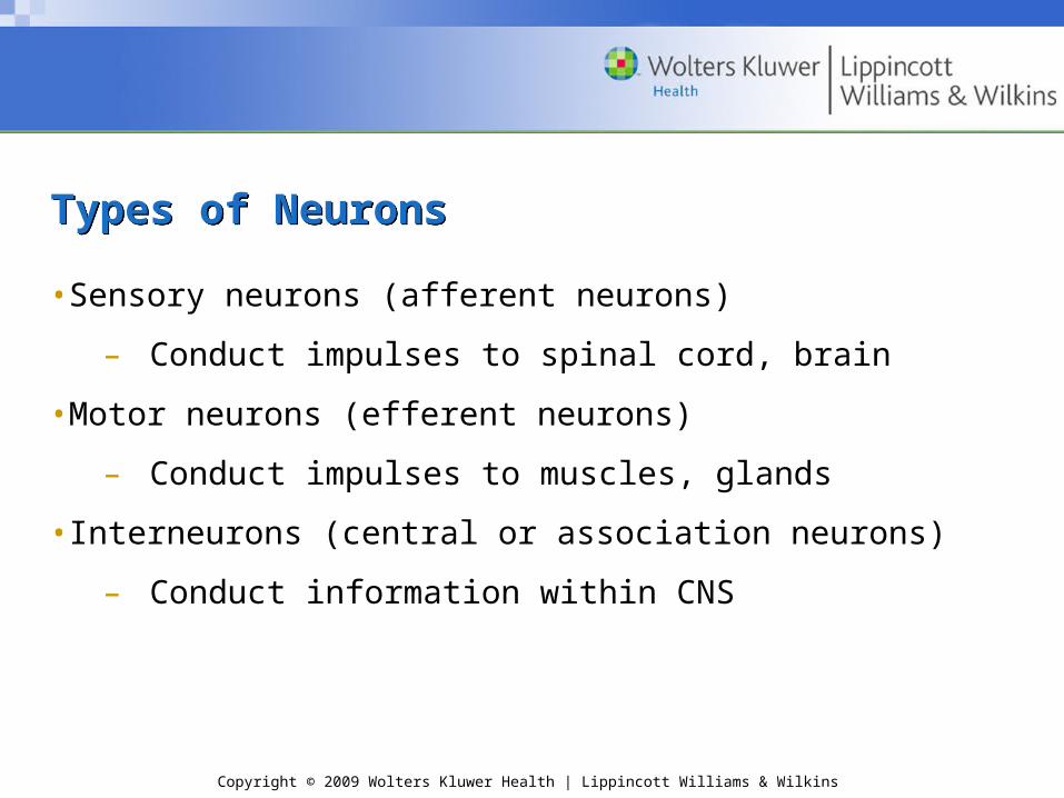
Types of NeuronsTypes of Neurons
•Sensory neurons (afferent neurons)
– Conduct impulses to spinal cord, brain
•Motor neurons (efferent neurons)
– Conduct impulses to muscles, glands
•Interneurons (central or association neurons)
– Conduct information within CNS

Copyright © 2009 Wolters Kluwer Health | Lippincott Williams & Wilkins
Nerves and TractsNerves and Tracts
•Nerve: fiber bundle within PNS
•Tract: fiber bundle within CNS
•Organized into fascicles
•Connective tissue layers
– Endoneurium
– Perineurium
– Epineurium
Copyright © 2009 Wolters Kluwer Health | Lippincott Williams & Wilkins
Checkpoint 9-5: Nerves are bundles of neuron fibers in the PNS. These nerves may be carrying impulses either toward or away from the CNS. What name is given to nerves that convey impulses toward the CNS, and what name is given to nerves that transport away from the CNS?

Copyright © 2009 Wolters Kluwer Health | Lippincott Williams & Wilkins
NeurogliaNeuroglia
Neuroglia (glial cells)
•Protect and nourish nervous tissue
•Support nervous tissue
•Aid in cell repair
•Remove pathogens and impurities
•Regulation composition of fluids around and between cells
Copyright © 2009 Wolters Kluwer Health | Lippincott Williams & Wilkins
Checkpoint 9-6: The nervous system’s nonconducting cells protect, nourish and support the neurons. What are these cells called?
Copyright © 2009 Wolters Kluwer Health | Lippincott Williams & Wilkins
The Nervous System at WorkThe Nervous System at WorkThe Nervous System at WorkThe Nervous System at Work
Electrical impulses sent along neuron fibers and transmitted between cells at junctions

Copyright © 2009 Wolters Kluwer Health | Lippincott Williams & Wilkins
The Nerve ImpulseThe Nerve Impulse
•Plasma membrane carries electrical charge (potential)
•Plasma membrane is polarized (negative charge)
•Membrane potential reverses, generates electrical charge (action potential)
– Resting state
– Depolarization
– Repolarization
• Sodium/potassium (Na+/K+) pump
•Myelin sheath speeds conduction

Copyright © 2009 Wolters Kluwer Health | Lippincott Williams & Wilkins
Question:
Which ions are involved in the action potential?
a. Potassium and calciumb. Sodium and oxygenc. Sodium and potassium
Question:
Which ions are involved in the action potential?
a. Potassium and calciumb. Sodium and oxygenc. Sodium and potassium
Copyright © 2009 Wolters Kluwer Health | Lippincott Williams & Wilkins
Answer:
c. Sodium and potassium
Answer:
c. Sodium and potassium
Copyright © 2009 Wolters Kluwer Health | Lippincott Williams & Wilkins
Checkpoint 9-7: An action potential occurs in two stages. In the first stage, the charge on the membrane reverses, and in the second stage, it returns to the resting state. What are the names of these two stages?
Checkpoint 9-8: What ions are involved in generating an action potential?

Copyright © 2009 Wolters Kluwer Health | Lippincott Williams & Wilkins
The SynapseThe Synapse
Junction point for transmitting nerve impulse
•Axon (presynaptic cell)
•Dendrite (postsynaptic cell)
•Synaptic cleft
•Neurotransmitters
– Epinephrine (adrenaline)
– Norepinephrine (noradrenaline)
– Acetylcholine
•Receptors
Copyright © 2009 Wolters Kluwer Health | Lippincott Williams & Wilkins
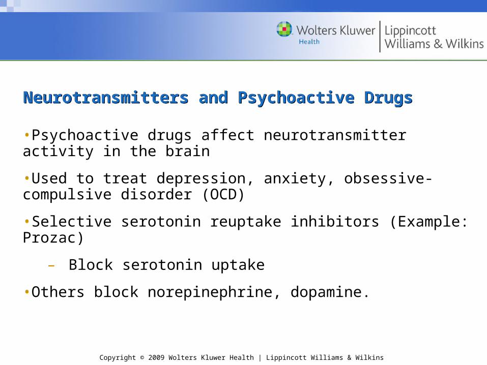
Neurotransmitters and Psychoactive DrugsNeurotransmitters and Psychoactive Drugs
•Psychoactive drugs affect neurotransmitter activity in the brain
•Used to treat depression, anxiety, obsessive-compulsive disorder (OCD)
•Selective serotonin reuptake inhibitors (Example: Prozac)
– Block serotonin uptake
•Others block norepinephrine, dopamine.
Copyright © 2009 Wolters Kluwer Health | Lippincott Williams & Wilkins
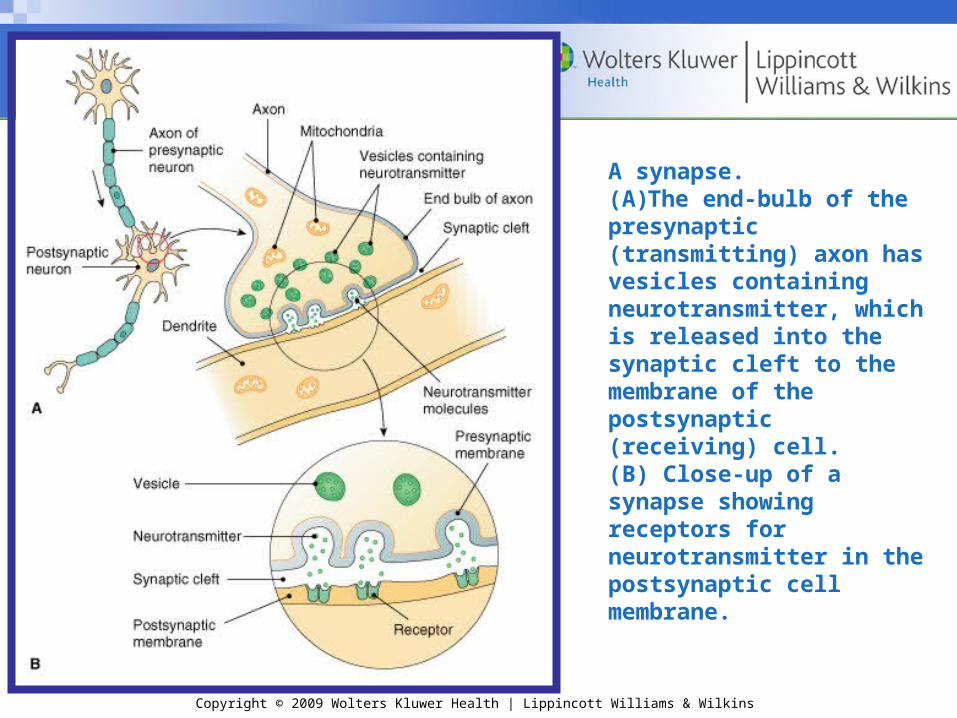
A synapse.(A)The end-bulb of the presynaptic (transmitting) axon has vesicles containing neurotransmitter, which is released into the synaptic cleft to the membrane of the postsynaptic (receiving) cell. (B) Close-up of a synapse showing receptors for neurotransmitter in the postsynaptic cell membrane.
Copyright © 2009 Wolters Kluwer Health | Lippincott Williams & Wilkins
Question:
The point of junction for transmitting a nerve impulse is called what?
a. axonb. synapsec. vesicle
Question:
The point of junction for transmitting a nerve impulse is called what?
a. axonb. synapsec. vesicle

Copyright © 2009 Wolters Kluwer Health | Lippincott Williams & Wilkins
Answer:
b. synapse
Answer:
b. synapse
Copyright © 2009 Wolters Kluwer Health | Lippincott Williams & Wilkins
Checkpoint 9-9: Chemicals are needed to carry information across the synaptic cleft at a synapse. As a group, what are all these chemicals called?
Copyright © 2009 Wolters Kluwer Health | Lippincott Williams & Wilkins
The Spinal CordThe Spinal CordThe Spinal CordThe Spinal Cord
•Links PNS and brain
•Helps coordinate impulses within CNS
•Contained in and protected by vertebrae
Copyright © 2009 Wolters Kluwer Health | Lippincott Williams & Wilkins
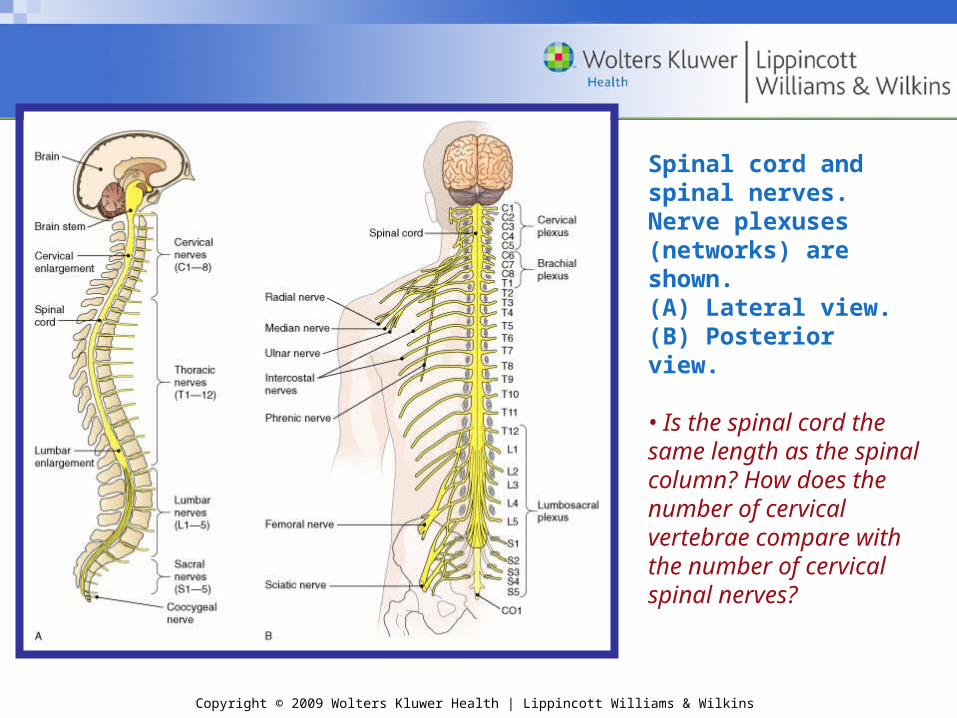
Spinal cord and spinal nerves. Nerve plexuses (networks) are shown. (A) Lateral view. (B) Posterior view.
• Is the spinal cord the same length as the spinal column? How does the number of cervical vertebrae compare with the number of cervical spinal nerves?
Copyright © 2009 Wolters Kluwer Health | Lippincott Williams & Wilkins
Structure of the Spinal CordStructure of the Spinal Cord
•Unmyelinated tissue (gray matter)
– Dorsal horn
– Ventral horn
– Gray commissure
– Central canal
•Myelinated axons (white matter)
– Posterior median sulcus
– Anterior median fissure
– Ascending and descending tracts

Copyright © 2009 Wolters Kluwer Health | Lippincott Williams & Wilkins
Checkpoint 9-10: The spinal cord contains both gray and white matter. How is this tissue arranged in the spinal cord?
Checkpoint 9-11: What is the purpose of the tracts in the white matter of the spinal cord?

Copyright © 2009 Wolters Kluwer Health | Lippincott Williams & Wilkins
The spinal cord. (A) Cross-section of the spinal cord showing the organization of the gray and white matter. The roots of the spinal nerves are also shown. (B) Microscopic view of the spinal cord in cross-section (x5).

Copyright © 2009 Wolters Kluwer Health | Lippincott Williams & Wilkins
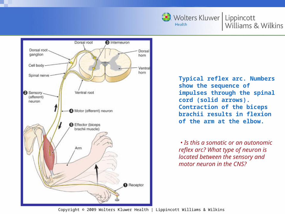
The Reflex ArcThe Reflex Arc
•Receptor detects stimulus
•Sensory neuron transmits impulses to CNS
•CNS coordinates impulses and organizes response
•Motor neuron carries impulses away from CNS
•Effector carries out response
Copyright © 2009 Wolters Kluwer Health | Lippincott Williams & Wilkins
Typical reflex arc. Numbers show the sequence of impulses through the spinal cord (solid arrows). Contraction of the biceps brachii results in flexion of the arm at the elbow.
• Is this a somatic or an autonomic reflex arc? What type of neuron is located between the sensory and motor neuron in the CNS?
Copyright © 2009 Wolters Kluwer Health | Lippincott Williams & Wilkins
Checkpoint 9-12: What name is given to a pathway through the nervous system from a stimulus to an effector?

Copyright © 2009 Wolters Kluwer Health | Lippincott Williams & Wilkins
Reflex ActivitiesReflex Activities
•Simple reflex
– Rapid
– Uncomplicated
– Automatic
•Spinal reflex
– Stretch reflex

Copyright © 2009 Wolters Kluwer Health | Lippincott Williams & Wilkins
Medical Procedures Involving theSpinal CordMedical Procedures Involving theSpinal Cord
•Lumbar puncture (spinal tap)
– Cerebrospinal fluid (CSF) removed for testing
•Drug administration
– Anesthetic (an epidural or spinal anesthesia)
– Pain medication

Copyright © 2009 Wolters Kluwer Health | Lippincott Williams & Wilkins
Diseases and Other Disorders of theSpinal CordDiseases and Other Disorders of theSpinal Cord
•Multiple sclerosis (MS)
•Amyotrophic lateral sclerosis
•Poliomyelitis
•Tumors
•Injuries
– Monoplegia
– Diplegia
– Paraplegia
– Hemiplegia
– Tetraplegia (Quadriplegia)

Copyright © 2009 Wolters Kluwer Health | Lippincott Williams & Wilkins
Question:
In a lumbar tap, what is removed from the body for testing?
a. cerebrospinal fluid b. lymphc. bone marrow
Question:
In a lumbar tap, what is removed from the body for testing?
a. cerebrospinal fluid b. lymphc. bone marrow

Copyright © 2009 Wolters Kluwer Health | Lippincott Williams & Wilkins
Answer:
a. cerebrospinal fluid
Answer:
a. cerebrospinal fluid
Copyright © 2009 Wolters Kluwer Health | Lippincott Williams & Wilkins
The Spinal NervesThe Spinal NervesThe Spinal NervesThe Spinal Nerves
•31 pairs
•Each nerve attached to spinal cord by two roots
– Dorsal root
• Dorsal root ganglion
– Ventral root
•Nerves near end of cord travel together in the cord until each exits from its respective intervertebral foramen
•Mixed nerves

Copyright © 2009 Wolters Kluwer Health | Lippincott Williams & Wilkins
Branches of the Spinal NervesBranches of the Spinal Nerves
•Cervical plexus
– Phrenic nerve
•Brachial plexus
– Radial nerve
•Lumbosacral plexus
– Sciatic nerve
•Dermatomes
Copyright © 2009 Wolters Kluwer Health | Lippincott Williams & Wilkins
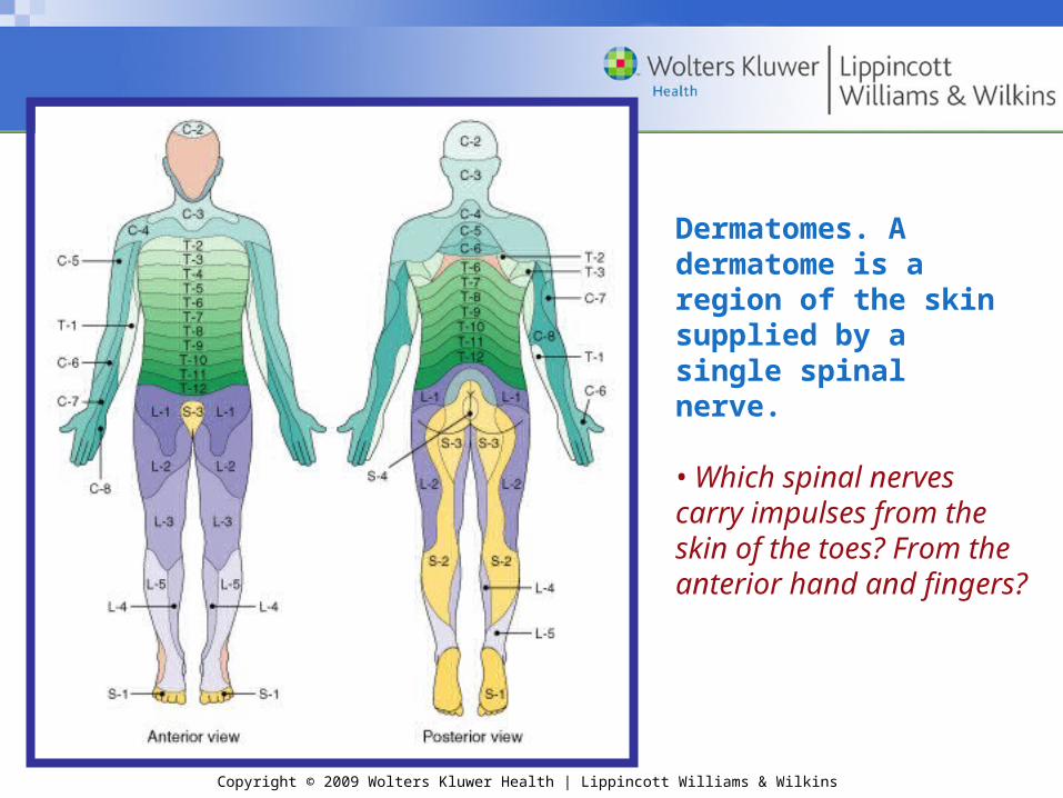
Dermatomes. A dermatome is a region of the skin supplied by a single spinal nerve.
• Which spinal nerves carry impulses from the skin of the toes? From the anterior hand and fingers?

Copyright © 2009 Wolters Kluwer Health | Lippincott Williams & Wilkins
Checkpoint 9-13: How many pairs of spinal nerves are there?
Copyright © 2009 Wolters Kluwer Health | Lippincott Williams & Wilkins
Disorders of the Spinal NervesDisorders of the Spinal Nerves
•Peripheral neuritis
•Sciatica
•Herpes zoster
•Guillain-Barré syndrome

Copyright © 2009 Wolters Kluwer Health | Lippincott Williams & Wilkins
The Autonomic NervousThe Autonomic NervousSystem (ANS)System (ANS)The Autonomic NervousThe Autonomic NervousSystem (ANS)System (ANS)
Regulates the action of glands, smooth muscles of hollow organs and vessels, and heart muscle
•Preganglionic neuron connects spinal cord to ganglion
•Postganglionic neuron connects ganglion to effector
Copyright © 2009 Wolters Kluwer Health | Lippincott Williams & Wilkins
Checkpoint 9-14: How many neurons are there in each motor pathway of the ANS?

Copyright © 2009 Wolters Kluwer Health | Lippincott Williams & Wilkins
Divisions of the Autonomic NervousSystemDivisions of the Autonomic NervousSystem
•Sympathetic nervous system
•Parasympathetic nervous system

Copyright © 2009 Wolters Kluwer Health | Lippincott Williams & Wilkins
Sympathetic nervous systemSympathetic nervous system
•Thoracolumbar area
•Collateral ganglia
– Celiac ganglion
– Superior mesenteric ganglion
– Inferior mesenteric ganglion
•Adrenergic system
•Activated in the four E’s: excitement, emergency, embarassment, exercise

Copyright © 2009 Wolters Kluwer Health | Lippincott Williams & Wilkins
Parasympathetic nervous systemParasympathetic nervous system
•Arise in craniosacral areas
•Terminal ganglia
•Cholinergic system

Copyright © 2009 Wolters Kluwer Health | Lippincott Williams & Wilkins
Question:
Any group of nerve cell bodies located outside the central nervous system is known as what?
a. a ganglionb. a plexusc. a horn
Question:
Any group of nerve cell bodies located outside the central nervous system is known as what?
a. a ganglionb. a plexusc. a horn

Copyright © 2009 Wolters Kluwer Health | Lippincott Williams & Wilkins
Answer:
a. a ganglion
Answer:
a. a ganglion

Copyright © 2009 Wolters Kluwer Health | Lippincott Williams & Wilkins
Autonomic nervous system. The diagram shows only one side of the body for each division.
• Which division of the autonomic nervous system has ganglia closer to the effector organ?

Copyright © 2009 Wolters Kluwer Health | Lippincott Williams & Wilkins
Cellular ReceptorsCellular Receptors
•“Docking sites” on postsynaptic cell membranes
Two types:
•Cholinergic receptors
– Nicotinic (bind nicotine) on skeletal muscle cells
– Muscarinic (bind muscarine, a poison) on effector cells of PNS
•Adrenergic receptors
– Found on receptor cells of sympathetic nervous system
– Bind norepinephrine, epinephrine

Copyright © 2009 Wolters Kluwer Health | Lippincott Williams & Wilkins
Functions of the Autonomic NervousSystemFunctions of the Autonomic NervousSystem
•Sympathetic nervous system
– Fight-or-flight response
•Parasympathetic nervous system
– Returns body to normal
•Systems generally have opposite effects on organ
Copyright © 2009 Wolters Kluwer Health | Lippincott Williams & Wilkins
Checkpoint 9-15: Which division of the ANS stimulates a stress response, and which division reverses the stress response?
Copyright © 2009 Wolters Kluwer Health | Lippincott Williams & Wilkins
Question:
What is the technical name of a “docking site” on a postsynaptic cell membrane?
a. neurotransmitterb. cellular receptorc. dendrite
Question:
What is the technical name of a “docking site” on a postsynaptic cell membrane?
a. neurotransmitterb. cellular receptorc. dendrite

Copyright © 2009 Wolters Kluwer Health | Lippincott Williams & Wilkins
Answer:
b. cellular receptor
Answer:
b. cellular receptor

Copyright © 2009 Wolters Kluwer Health | Lippincott Williams & Wilkins
QUESTIONS?QUESTIONS?