copy number variation of kir genes influences hiv-1...
TRANSCRIPT

Copy Number Variation of KIR Genes Influences HIV-1ControlKimberly Pelak1., Anna C. Need1., Jacques Fellay1,2., Kevin V. Shianna1, Sheng Feng3, Thomas J.
Urban1, Dongliang Ge1, Andrea De Luca4,5, Javier Martinez-Picado6,7, Steven M. Wolinsky8, Jeremy J.
Martinson9, Beth D. Jamieson10, Jay H. Bream11, Maureen P. Martin12, Persephone Borrow13, Norman L.
Letvin14, Andrew J. McMichael15, Barton F. Haynes16, Amalio Telenti17, Mary Carrington10,18, David B.
Goldstein1, Galit Alter18*, on behalf of NIAID Center for HIV/AIDS Vaccine Immunology (CHAVI)
1 Center for Human Genome Variation, Duke University School of Medicine, Durham, North Carolina, United States of America, 2 Global Health Institute, School of Life
Sciences, Ecole Polytechnique Federale de Lausanne, Lausanne, Switzerland, 3 Department of Biostatistics and Bioinformatics, Duke University, Durham, North Carolina,
United States of America, 4 Institute of Clinical Infectious Diseases, Catholic University of the Sacred Heart, Rome, Italy, 5 Division of Infectious Diseases, Siena University
Hospital, Siena, Italy, 6 irsiCaixa Foundation and Hospital Germans Trias i Pujol, Badalona, Spain, 7 Institucio Catalana de Recerca i Estudis Avancats, Barcelona, Spain,
8 Division of Infectious Diseases, Northwestern University Feinberg School of Medicine, Chicago, Illinois, United States of America, 9 Infectious Diseases and Microbiology,
Graduate School of Public Health, University of Pittsburgh, Pittsburgh, Pennsylvania, United States of America, 10 Department of Medicine, David Geffen School of
Medicine, University of California–Los Angeles, Los Angeles, California, United States of America, 11 Department of Molecular Microbiology and Immunology, Johns
Hopkins Bloomberg School of Public Health, Baltimore, Maryland, United States of America, 12 Cancer and Inflammation Program, Laboratory of Experimental
Immunology, SAIC-Frederick, Inc., NCI-Frederick, Frederick, Maryland, United States of America, 13 Nuffield Department of Clinical Medicine, University of Oxford and
Weatherall Institute of Molecular Medicine, John Radcliffe Hospital, Headington, Oxford, United Kingdom, 14 Division of Viral Pathogenesis, Beth Israel Deaconess Medical
Center, Harvard Medical School, Boston, Massachusetts, United States of America, 15 Medical Research Council Human Immunology Unit, Weatherall Institute of
Molecular Medicine, John Radcliffe Hospital, Oxford, United Kingdom, 16 Duke Human Vaccine Institute, Duke University, Durham, North Carolina, United States of
America, 17 Institute of Microbiology, University Hospital Center; and University of Lausanne, Lausanne, Switzerland, 18 Ragon Institute of MGH, MIT and Harvard, Boston,
Massachusetts, United States of America
Abstract
A genome-wide screen for large structural variants showed that a copy number variant (CNV) in the region encoding killercell immunoglobulin-like receptors (KIR) associates with HIV-1 control as measured by plasma viral load at set point inindividuals of European ancestry. This CNV encompasses the KIR3DL1-KIR3DS1 locus, encoding receptors that interact withspecific HLA-Bw4 molecules to regulate the activation of lymphocyte subsets including natural killer (NK) cells. Wequantified the number of copies of KIR3DS1 and KIR3DL1 in a large HIV-1 positive cohort, and showed that an increase inKIR3DS1 count associates with a lower viral set point if its putative ligand is present (p = 0.00028), as does an increase inKIR3DL1 count in the presence of KIR3DS1 and appropriate ligands for both receptors (p = 0.0015). We further providefunctional data that demonstrate that NK cells from individuals with multiple copies of KIR3DL1, in the presence of KIR3DS1and the appropriate ligands, inhibit HIV-1 replication more robustly, and associated with a significant expansion in thefrequency of KIR3DS1+, but not KIR3DL1+, NK cells in their peripheral blood. Our results suggest that the relative amountsof these activating and inhibitory KIR play a role in regulating the peripheral expansion of highly antiviral KIR3DS1+ NK cells,which may determine differences in HIV-1 control following infection.
Citation: Pelak K, Need AC, Fellay J, Shianna KV, Feng S, et al. (2011) Copy Number Variation of KIR Genes Influences HIV-1 Control. PLoS Biol 9(11): e1001208.doi:10.1371/journal.pbio.1001208
Academic Editor: Michael Emerman, Fred Hutchinson Cancer Research Center, United States of America
Received June 20, 2011; Accepted October 20, 2011; Published November 29, 2011
Copyright: � 2011 Pelak et al. This is an open-access article distributed under the terms of the Creative Commons Attribution License, which permitsunrestricted use, distribution, and reproduction in any medium, provided the original author and source are credited.
Funding: This research has been funded in whole or part with federal funds by the Center for HIV/AIDS Vaccine Immunology (‘‘CHAVI’’) under a grant from theNational Institute of Allergy and Infectious Diseases (NIAID), US National Institutes of Health (US NIH), grant number UO1AIO67854. This project has been fundedin whole or in part with federal funds from the National Cancer Institute (NCI), US NIH, under contract number HHSN261200800001E. The content of thispublication does not necessarily reflect the views or policies of the US Department of Health and Human Services, nor does mention of trade names, commercialproducts, or organizations imply endorsement by the US Government. This research was supported in part by the Intramural Research Program of the NIH, NCI,Center for Cancer Research. The MACS is funded by NIAID, with additional supplemental funding from NCI; and the National Heart, Lung, and Blood Institute:UO1-AI-35042, 5-M01-RR-00052 (GCRC), UO1-AI-35043, UO1-AI-37984, UO1-AI-35039, UO1-AI-35040, UO1-AI-37613, and UO1-AI-35041. KP was funded by NIHGenetics Training Grant 5 T32 GM007754-29. AT is supported by the Swiss National Science Foundation. The funders had no role in study design, data collectionand analysis, decision to publish, or preparation of the manuscript.
Competing Interests: The authors have declared that no competing interests exist.
Abbreviations: CNV, copy number variant; HIV-1, human immunodeficiency virus type 1; KIR, killer cell immunoglobulin-like receptors; MHC, majorhistocompatibility complex; NK cells, natural killer cells; SNP, single-nucleotide polymorphism.
* E-mail: [email protected]
. These authors contributed equally to this work.
PLoS Biology | www.plosbiology.org 1 November 2011 | Volume 9 | Issue 11 | e1001208

Introduction
The KIR receptors are expressed mainly on the surface of lym-
phocyte subsets including natural killer (NK) cells and a small subset of
T cells, and they have a unique role in fine-tuning the balance between
self-tolerance and cytotoxicity. KIRs bind to major histocompatibility
complex (MHC) class I ligands on the surface of target cells. The
degree of inhibition and/or activation mediated by interactions
between co-inherited KIR and MHC class I gene products determines
the activation threshold for NK cells (Figure 1) [1].
The KIR region, on chromosome 19q13.4, is highly polymor-
phic in humans [2] and its extensive polymorphism has been
repeatedly associated with the natural history of HIV-1 infection
[3]. The KIR3DL1 and KIR3DS1 genes segregate as allelic variants
at the same locus and both are thought to encode receptors for
molecules that fall within the Bw4 subfamily of HLA-B alleles
(HLA-Bw4). There is also evidence for a single chromosome to
have both KIR3DS1 and KIR3DL1 [4,5].
Both allele groups at this locus have been shown to be involved
in HIV-1 pathogenesis. The activating allele KIR3DS1, in
combination with HLA-Bw4 molecules that have an isoleucine
at position 80 (Bw4-80I), has been associated with lower viral load,
slower decline in CD4+ T cells, and delayed progression to AIDS,
as well as with protection against opportunistic infections [6,7].
In addition, KIR3DS1 has recently been shown to correlate
with strong inhibition of HIV-1 replication [8]. However, some
reports have shown no protective effect associated with the
KIR3DS1+HLA-Bw4-80I genotype [9,10] or show no evidence for
a synergistic effect from the KIR3DS1+HLA-Bw4-80I genotype on
viral load or on CD4+ T cell counts [11]. Recent reports have also
shown that NK cells expressing KIR also directly place pressure of
the virus, driving HIV viral evolution [12].
Similarly, various distinct allelic combinations of the inhibitory
KIR3DL1 receptor and HLA-Bw4 ligands have been associated
with lower HIV-1 viral load and slower progression to AIDS [13].
Two proposed functional explanations may account for the latter
result. The first relates to the education process of NK cells during
development, in which inhibitory receptors must recognize
autologous MHC class I ligands for the NK cell to be functional
upon maturation [14–16], suggesting that ligand engagement by
more highly expressed inhibitory KIR3DL1 allotypes during NK
cell development ultimately may result in stronger NK cell
responses in the event of viral infection when the ligand is missing
or altered [14,17]. The second underlying explanation may relate
to the fact that KIR3DL1 is involved in monitoring the circulation
for normal MHC class I expression; however upon HIV infection,
HIV Nef protein rapidly downregulates MHC class I expression.
Thus, it is equally plausible that higher expression of KIR3DL1
may allow NK cells to recognize reduced MHC class I expression
on infected cells more readily.
KIR receptors are expressed on NK cells in a variegated
manner, where only a fraction of all NK cells express a particular
KIR gene product. Certain KIR receptors are consistently
expressed on a large fraction of NK cells, while others are
expressed on a smaller fraction of NK cells [18]. In individuals
with one copy of KIR3DS1, roughly 20%–50% of NK cells express
the KIR3DS1 receptor [18,19], and in individuals with two copies
of KIR3DS1, 60% or greater of NK cells express the KIR3DS1
receptor [18,19]. To add to the complexity, some KIR3DL1
allotypes have different surface expression levels [20], which have
been shown to have varying impacts on HIV-1 outcomes [13], and
correlate with genealogical groups of KIR3DL1 alleles [21].
The primary outcome studied here is HIV-1 viral load at set
point, which has been shown to be a genetically tractable HIV
outcome [22]. We used a genome-wide screen to identify a copy
number variable region that associated with HIV-1 control, as
measured by plasma viral load at set point, and that encompassed
the KIR3DL1-KIR3DS1 locus. Further dissection of the region and
of the interactions between KIR3DL1, KIR3DS1, and their HLA
ligands demonstrated that the number of gene copies of the
inhibitory KIR3DL1 receptor and activating KIR3DS1 receptor
plays an important role in modulating HIV-1 control, but that this
effect is only detectable after epistatic interactions between HLA
molecules and KIR receptors are taken into account. Further-
more, functional and transcriptional studies on cells derived from
individuals with these particular KIR CNV/HLA combinations
demonstrated a dramatic expansion of KIR3DS1+ NK cells,
which are able to robustly inhibit HIV replication in vitro. Thus,
these data support the genetic association results, suggesting novel
mechanisms of regulation of the antiviral activity of NK cells.
Figure 1. KIR and HLA-B interactions. An NK cell can expressKIR3DL1 alone, KIR3DS1 alone, both receptors, or neither. KIR3DL1 is aninhibitory receptor and has been shown to interact with HLA-Bw4-80Iand with HLA-Bw4-80T. KIR3DS1 is an activating receptor and mayinteract with HLA-Bw4-80I, possibly through an indirect mechanism. AnNK cell can be activated to lyse an infected cell via either the activatingof KIR receptor signaling or the dampening of inhibitory KIR activity.doi:10.1371/journal.pbio.1001208.g001
Author Summary
There is marked intrinsic variation in the extent to whichindividuals are able to control HIV-1. We have identified agenetic copy number variable region (CNV) in humans thatplays a significant role in the control of HIV-1. This CNV islocated in the genomic region that encodes the killer cellimmunoglobulin-like receptors (KIRs) and specificallyaffects the KIR3DS1 and KIR3DL1 genes, encoding twoKIRs that interact with human leukocyte antigen B (HLA-B)ligands. KIRs are expressed on the surface of natural killer(NK) cells, which serve as important players in the innateimmune response, and are involved in the recognition ofinfected and malignant cells through a loss or alteration in‘‘self’’ ligands. We use both genetic association andfunctional evidence to show a strong interaction betweenKIR3DL1 and KIR3DS1, indicating that increasing genecounts for KIR3DL1 confer increasing levels of protectionagainst HIV-1, but only in the presence of at least one copyof KIR3DS1. This effect was associated with a dramaticincrease in the abundance of KIR3DS1+ NK cells in theperipheral blood, and strongly associated with a morerobust capacity of peripheral NK cells to suppress HIV-1replication in vitro. This work provides one of the fewexamples of an association between a relatively commonCNV and a human complex trait.
Copy Number Variation of KIR Genes In HIV Control
PLoS Biology | www.plosbiology.org 2 November 2011 | Volume 9 | Issue 11 | e1001208

Results
CNV Identification in KIR RegionWe investigated the role of large CNVs on HIV-1 control in a
cohort of 2,102 patients of European ancestry from the Euro-
CHAVI Consortium and the Multicenter AIDS Cohort Study
(MACS). Genome-wide single nucleotide polymorphism (SNP)
genotyping was performed using Illumina’s HumanHap550,
Human1M, or Human1MDuo BeadChips, and we used the
PennCNV software [23] to identify large CNVs. For each SNP in
a CNV region, we assigned copy number status (zero, one, two,
three, or four copies) to each sample and carried out a linear
regression analysis on HIV-1 viral load at set point. We examined
duplications and deletions separately using copy number status in
additive genetic models and included as covariates age, sex, and 12
EIGENSTRAT ancestry axes to control for population stratifica-
tion [24]. We tested 5,384 deletions and 3,553 duplications with a
minimum frequency of 0.004, and none of them associated
significantly with viral set point after correction for multiple testing
using straight Bonferroni correction (p threshold = 9.261026 for
deletions and 1.461025 for duplications). However, we note that
this is a conservative correction, because several CNVs can reflect
the same association signal due to the difficulty of distingui-
shing between nearby CNVs when inferring them from the
genotyping data.
We manually inspected all CNVs that showed an association
with set point at p,0.05 (unadjusted) (Table S1). One associated
CNV was located in the KIR region, where both duplications and
deletions associated to variable degrees with HIV-1 control. The
duplications and deletions each occurred in around 3%–5% of the
study population. Many, although not all, of these identified
duplications and deletions covered the KIR3DL1-KIR3DS1 locus,
which has been the subject of intensive study related to control of
HIV-1 [7,13]. Focusing on SNPs included in this copy number
variable region (rs631717, rs649216, rs581623), HIV-1 viral load
at set point was lower for individuals with more copies and higher
for individuals with fewer copies (p = 0.010 for duplications and
p = 0.001 for deletions, as compared to samples that did not show
copy number variability and have two total copies of KIR3DL1
and/or KIR3DS1). If we assign an overall copy number to each
sample based on the PennCNV call for these three SNPs (zero,
one, two, three, or four copies), the CNV in the KIR region
shows an even stronger association with viral load at set point
(p = 361025).
KIR3DL1 and KIR3DS1 Gene CountsTo assess the individual impact of KIR3DS1 and KIR3DL1 on
HIV-1 control and to further investigate the copy number
variability observed in the KIR region, we developed a real-time
PCR assay to quantify the number of copies of each gene (Figure
S1). Individuals without a CNV in the region have a total of two
copies of these genes (one KIR3DL1 or one KIR3DS1 on each
chromosome), whereas in individuals with a deletion or a
duplication their sum corresponds to the copy number state
(e.g., four alleles are measured in individuals with a homozygous
duplication, or zero alleles with a homozygous deletion) (Table
S2). As can be seen in Figure S1, our assay is able to count nearly
all known alleles of KIR3DS1/KIR3DL1 that appear in populations
of European descent. Overall, we found high repeatability of the
assay (Figure S2) and a good correspondence with copy number
assignments called by PennCNV [23] using SNPs in the KIR3DL1-
KIR3DS1 region (Text S1, Table S3).
In the KIR3DL1-KIR3DS1 region, about 3.6% of our samples
had a deletion according to the real-time PCR data, and about 5%
had a duplication. The frequencies of the various genotypes that
show evidence of duplication are listed in Table S4. It is clear that
a single chromosome can have two copies of KIR3DS1 or
KIR3DL1, since respectively 6% and 14.5% of the samples with
duplications had three total copies of either KIR3DS1 or KIR3DL1,
presumably two on one chromosome and one on the opposite
chromosome.
The diversity of genotypes present at this locus and our inability
to discern which genes occur on the same chromosome make it
challenging to determine if this locus is in Hardy-Weinberg
equilibrium. However, previous work has shown that KIR3DL1/
KIR3DS1 are indeed in Hardy-Weinberg equilibrium, in spite of
the existence of the relatively rare haplotypes containing multiple
copies of this gene [25].
Raw Counts of KIR3DS1 and KIR3DL1 Do Not Associatewith Set Point
We first checked to see if the raw number of KIR3DS1 and
KIR3DL1 copies per individual (without accounting for presence of
the cognate HLA-B ligand) associated with HIV-1 set point:
neither raw count associated with viral control (n = 1,736,
p = 0.230 for raw KIR3DS1 count and p = 0.508 for raw KIR3DL1
count, Table 1).
Effective Gene CountThe functionality of a KIR receptor hinges on the presence of
its cognate ligand. The activity of KIR3DS1 and KIR3DL1
therefore depends on the expression of appropriate HLA-Bw4
molecules on the surface of target cells. To take this epistatic
feature into account, we created an ‘‘effective’’ gene count, in
which each copy of KIR3DL1 or KIR3DS1 was counted only when
its specific Bw4 ligand was present. HLA-Bw4-80I and HLA-Bw4-
80T have both been demonstrated to be ligands for KIR3DL1
[26,27]. Although the direct interaction between KIR3DS1 and
HLA-Bw4-80I is less definitive because a physical interaction
between HLA-Bw4-80I and KIR3DS1 has not been demonstrated
[28–30], epidemiological and functional evidence suggests that
under some conditions, such as HIV-1 infection, HLA-Bw4-80I
serves directly or indirectly as a ligand for KIR3DS1 (Table 2) [3].
HLA-Bw6 is not a ligand for either KIR3DS1 or KIR3DL1, and
there has not been evidence to show that any other KIR receptors
interact with HLA-Bw4 molecules. Some HLA-A alleles also carry
the HLA-Bw4-80I motif (16.1% of all HLA-A in dbMHC
Project Anthropology, http://www.ncbi.nlm.nih.gov/gv/mhc/
ihwg.cgi?ID=9&cmd=PRJOV), but for simplicity we restricted
our analyses to HLA-B alleles with Bw4-80I, since it is not clear
that all HLA-A-Bw4-80I molecules serve as ligands for KIR3DL1.
All subsequent analyses used this ‘‘effective’’ gene count.
Table 1. p-Values for association with VL set point.
Alonea (n = 1,736) Alonea (n = 706)
p p
Raw KIR3DS1 count 0.230 0.254
Raw KIR3DL1 count 0.508 0.768
aModel includes age, gender, 12 EIGENSTRAT axes, and either KIR3DL1 orKIR3DS1 raw count.
The first column shows the p values for the association between all sampleswith a raw KIR3DL1 and a raw KIR3DS1 count, and the second column is limitedto just the 706 samples that are used in Table 3.doi:10.1371/journal.pbio.1001208.t001
Copy Number Variation of KIR Genes In HIV Control
PLoS Biology | www.plosbiology.org 3 November 2011 | Volume 9 | Issue 11 | e1001208

In order to determine these ‘‘effective’’ KIR3DL1 and
KIR3DS1 counts, we required that samples had (1) successful
real-time quantitation for both KIR3DL1 and KIR3DS1, (2) HLA-
B data, and (3) available KIR3DL1 allelic subtyping, if at least one
KIR3DL1 was present. A total of 706 samples fit all the criteria.
Allelic subtyping was not included for KIR3DS1 since it shows
little variation [21]. The KIR3DL1 subtyping data were used to
separate the alleles that are expressed on the cell surface
(‘‘KIR3DL1-surface’’), at either high or low levels, from the
special case of KIR3DL1*004, which is not expressed at the cell
surface [13,31].
Effective Counts of KIR3DS1 and KIR3DL1 Associate withSet Point
Each of the effective counts was tested separately. We found
that the effective KIR3DS1 and effective KIR3DL1-surface gene
counts associated with HIV-1 set point (p = 4.261026 and 0.020,
respectively) (Table 3), with an increase in effective gene count
leading to lower viral loads. When KIR3DS1 and KIR3DL1-
surface effective gene counts were considered in the same re-
gression model, they remained separately significant (p = 0.00028
and 0.0085, respectively) (Table 3, Figure 2A–B).
Regardless of KIR3DL1 status, an increase in the effective
count of KIR3DS1 associated with improved viral control
(Table 4, Figure 3A). In contrast, an increase in the effective
count of KIR3DL1 did not show any association in the absence of
KIR3DS1, but did impact HIV-1 set point in the presence of one
or more effective copies of KIR3DS1 (p = 0.0015, Table 4,
Figure 3B).
In the subset of study participants who had two HLA-B alleles
from the Bw6 subfamily (Bw6/Bw6 homozygotes), the raw counts
for KIR3DS1 and KIR3DL1-surface showed no association with
HIV-1 viral load at set point, further supporting the critical nature
of particular KIR-HLA combined genotypes (Table S5).
KIR3DS1 Effective Count Associates with Set Point WhenHLA-B*57, HLA-B*27, and HLA-B*35Px Are Included asCovariates in Model
The interpretation of KIR-HLA epistatic influences on HIV-1
control is complicated by the fact that particular HLA class I alleles
(-A, -B, and -C) have previously been independently implicated in
modulating HIV-1 control, with HLA-B alleles placing the greatest
immune pressure on HIV-1 replication [32]. Specifically, HLA-
B*57 and HLA-B*27 are associated with slower HIV-1 disease
progression, whereas HLA-B*35Px associates with a stronger
susceptibility to developing AIDS rapidly [33–35]. The differential
impact of these HLA alleles has been attributed to differences in
epitope presentation in conserved versus variable regions within
the viral genome. The protective HLA-B*57 and HLA-B*27
generate strong antiviral CD8+ T cell responses that target highly
conserved proteins, where escape mutations place a great impact
on viral fitness. In contrast, HLA-B*35Px restricted CD8+ T cells
tend to target highly variable regions that likely place little pressure
on the virus. However, in addition to CD8+ T cells, NK cells are
also able to interact differentially with these three alleles via
KIR3DL1 and KIR3DS1. HLA-B*57 molecules are a subset of
the HLA-Bw4-80I group (ligand for KIR3DL1 and possibly
KIR3DS1), HLA-B*27 are mainly HLA-Bw4-80T (ligand for
KIR3DL1), and HLA-B*35Px are primarily HLA-Bw6 (except
HLA-B*5301, which is HLA-Bw4-80I) (Table S6). Thus, although
HLA-B*27 and HLA-B*57 may themselves interact with KIR,
these favorable and unfavorable HLA alleles (in terms of HIV-1
control) are present in different proportions within each of the
groups of alleles that are and are not ligands for KIR3DL1 and
KIR3DS1, and could therefore drive an association with viral
control independently of KIR genotypes.
For this reason, to assess the specific effects of KIR, these HLA-B
alleles must be accounted for in the analysis. To do this, we added all
three HLA-B allotypes as covariates to the model considered earlier.
We found that an increase in the effective count of KIR3DS1 still
associated with a decrease in viral load (p = 0.0075), but the effective
count of KIR3DL1-surface did not (p = 0.220) (Table 3).
We note that correction for these controlling alleles could
reduce power and create instabilities in the signal due to
colinearity. Nevertheless, we retain this test as one established
approach [4] for evaluating whether it appears to be a credible
alternative explanation that the apparent signal is due simply to
the contribution of these controlling alleles. We also note that
some fraction of the protection conferred by HLA-B*57 and HLA-
B*27 is due to their interaction with KIR receptors [13].
Table 2. Combinations of KIR3DS1-KIR3DL1 and HLA-Btreated as receptor/ligand pairs in this study.
HLA-Bw4 80I HLA-Bw4 80T HLA-Bw6
KIR3DS1 Y N N
KIR3DL1 Y Y N
doi:10.1371/journal.pbio.1001208.t002
Table 3. p-Values for association with VL set point.
Alone In Combined Model In Combined Model
n p n p n p
Effective KIR3DS1 count 1,429 4.2E-06a 706 2.8E-04b 706 0.0075c
Effective KIR3DL1-surface count 749 0.020a 706 0.0085b 706 0.220c
HLA-B*57 ----- ----- ----- ----- 706 1.4E-07c
HLA-B*27 ----- ----- ----- ----- 706 0.0049c
HLA-B*35Px ----- ----- ----- ----- 706 0.098c
aModel includes age, gender, 12 EIGENSTRAT axes, and the count of one KIR gene.bModel includes age, gender, 12 EIGENSTRAT axes, effective KIR3DS1 count, and effective KIR3DL1-surface count.cModel includes age, gender, 12 EIGENSTRAT axes, effective KIR3DS1 count, effective KIR3DL1-surface count, B*5701, B*27, and B*35Px.doi:10.1371/journal.pbio.1001208.t003
Copy Number Variation of KIR Genes In HIV Control
PLoS Biology | www.plosbiology.org 4 November 2011 | Volume 9 | Issue 11 | e1001208

Higher Effective Counts for Co-Expressed KIR3DL1 andKIR3DS1 Are Associated with the Generation of NK Cellswith a Superior Capacity to Inhibit HIV-1 Replication In Vitro
Previous work examining the role of protective KIR/HLA
genotypes on NK cell functionality showed that NK cells from
healthy HIV-uninfected individuals who expressed KIR3DS1 and
that also expressed HLA-Bw4-80I were associated with a robust
capacity to inhibit HIV-1 replication in vitro, compared to
individuals who expressed KIR3DS1 in the absence of its putative
ligand [8], potentially conferring an enhanced capacity of these
individuals to respond to the virus soon after infection. We were
therefore interested in determining whether individuals with the
observed duplication showed a differential capacity to inhibit viral
replication in vitro, and whether this effect was due to KIR3DS1,
KIR3DL1, or both. To determine whether NK cells generated in
individuals with increased effective counts of KIR3DS1 and
KIR3DL1 showed any variation in NK cell function, we
performed an NK cell viral inhibition assay using fresh blood
collected from HIV-negative individuals with different KIR/HLA
genotype combinations, including several individuals with one
effective copy of KIR3DS1 and two effective copies of KIR3DL1.
We found that NK cells from HIV-negative individuals with one
effective copy of KIR3DS1 and one effective copy of KIR3DL1
inhibited HIV-1 replication more potently than NK cells from
individuals who did not possess at least one effective copy of both
KIR3DL1 and KIR3DS1 (Figure 4, mean inhibition = 42%,
p,0.005). Interestingly, individuals who had one effective copy of
KIR3DS1 and two effective copies of KIR3DL1 exhibited even
more robust NK-cell-mediated inhibition of HIV replication in
vitro than did individuals who had one copy of effective KIR3DS1
and just one copy of effective KIR3DL1 (Figure 4, mean
inhibition = 88%). Individuals who did not have HLA-Bw4-80I
or who did not have both KIR3DL1 and KIR3DS1 showed
markedly less inhibition (mean inhibition,15%). These data
support the association results described in the first part of the
article, with the only discrepancy being that individuals with two
effective copies of KIR3DS1 do show a decrease in viral load at set
point, but do not show an increase in viral inhibition. Overall,
these results demonstrate that, prior to infection, NK cells
generated in the presence of more effective copies of KIR3DS1
and KIR3DL1 have enhanced HIV-1 antiviral activity.
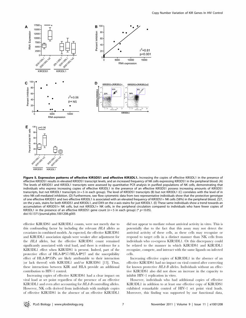
Individuals with Effective Copies of KIR3DS1 and KIR3DL1Show an Elevated Frequency of KIR3DS1+ NK Cells andElevated KIR3DS1 Transcript Levels as the Number ofCopies of KIR3DL1 Increases
Mounting evidence suggests that KIR/HLA compound geno-
types heavily influence the frequency of NK cells expressing a
given KIR receptor [36,37], and it has previously been shown that
KIR3DS1+ NK cells expand in acute HIV infection in the
presence of their putative ligand, HLA-Bw4-80I [38]. This
preferential early expansion of highly antiviral KIR3DS1+ NK
cells could potentially provide enhanced viral control. Given that
increased effective counts of KIR3DL1 in the presence of
KIR3DS1 demonstrated an enhanced capacity to inhibit HIV
replication (Figure 4), we speculated that the increasing doses of
inhibitory KIRs may be associated with unique patterns of
KIR3DL1 and KIR3DS1 expression levels in NK cells. This could
potentially account for their superior antiviral activity.
Using freshly isolated purified NK cells from healthy controls
with distinct KIR/HLA genotypes, we found that increasing
effective counts of KIR3DL1, in the presence of an effective
KIR3DS1, were associated with an elevated number of KIR3DS1
transcripts in purified NK cell populations (Figure 5A, p,0.05).
This suggests that the increasing effective KIR3DL1 counts
potentiate the expression of KIR3DS1 in the circulating NK cell
pool but have little impact on their own expression. More
interestingly, increasing levels of KIR3DS1 RNA transcripts were
strongly associated with the level of HIV-1 inhibition in all
KIR3DS1-carrying individuals expressing the putative ligand
Figure 2. Association between KIR3DS1 and KIR3DL1 effectivecounts and viral load at set point. (A–B) The effective counts ofKIR3DS1 and KIR3DL1-surface associate with viral load (VL) at set point.(C–D) The insets show the association between the raw KIR3DS1 orKIR3DL1-surface count, for the subset of patients from graphs A and Bwhere the effective counts equal zero. An effective count of zero can bedue to the absence of the KIR receptor or due to the absence of theHLA-Bw4 ligand. The raw count does not associate with viral load at setpoint when the effective count for KIR3DS1 or KIR3DL1-surface equalszero. Error bars show one standard deviation.doi:10.1371/journal.pbio.1001208.g002
Table 4. Interaction between KIR3DS1 and KIR3DL1-surface.
Effective KIR3DL1- Surface Count = 0b Effective KIR3DL1- Surface Count = 1b Effective KIR3DL1- Surface Count = 2
nMeanSet Point n
MeanSet Point n
MeanSet Point
Effective KIR3DS1 count = 0a 211 4.28 186 4.16 198 4.12
Effective KIR3DS1 count = 1a 14 4.13 72 3.97 5 3.15
Effective KIR3DS1 count = 2+ 14 3.67 5 3.88 1 2.53
ap = 0.0015. Comparison tests whether KIR3DS1 status influences the effect of KIR3DL1 on viral load.bp = 0.865. Comparison tests whether KIR3DL1 status influences the effect of KIR3DS1 on viral load.doi:10.1371/journal.pbio.1001208.t004
Copy Number Variation of KIR Genes In HIV Control
PLoS Biology | www.plosbiology.org 5 November 2011 | Volume 9 | Issue 11 | e1001208

(r2 = 0.81, p,0.001, Figure 5B), whereas the relative expression of
KIR3DL1 was not associated with NK-cell-mediated inhibition of
HIV infection (Figure 5C). Similarly, in addition to the impact of
increasing KIR3DL1 effective counts on KIR3DS1 transcript
expression, individuals with the protective genotype of an effective
copy of KIR3DS1 and two effective copies of KIR3DL1 showed a
trend towards an expansion of KIR3DS1+ NK cells in the
peripheral circulation, as compared to individuals with a single
effective copy of KIR3DL1 in the presence of an effective copy of
KIR3DS1 (Figure 5D,E). The shift in the whole NK cell
population (Figure 5D) could reflect an increase in the quantity
of KIR3DS1 expressed on the surface of NK cells in the presence
of two copies of KIR3DL1, concomitant with an expansion of the
frequency of KIR3DS1+ NK cells [39]. Individuals with the
protective genotype of an effective copy of KIR3DS1 and two
effective copies of KIR3DL1 also show an increase in the percent
of KIR3DS1+ NK cells, but not an increase in the percent of
KIR3DL1+ NK cells, when compared to individuals with just one
effective copy of KIR3DS1 and one effective copy of KIR3DL1
(Figure 5E). These data suggest that the increasing KIR3DL1
effective gene count, in the presence of an effective KIR3DS1, is
associated with more robust HIV antiviral activity due to a
genotype-driven natural expansion of KIR3DS1+ NK cells in the
peripheral blood prior to infection. This population of KIR3DS1+NK cells may expand even further upon HIV infection, potentially
providing these individuals with an antiviral advantage.
Discussion
We conducted a genome-wide CNV screen that allowed us to
locate a variable region involved in HIV-1 control. The observed
association signal was due to KIR3DL1 and KIR3DS1 copy number
variation encompassed within the region, and to the interaction of
these receptors with their cognate HLA-B ligands. The proportion
of variance explained by the CNV and the proportion of variance
explained by the effective KIR3DL1 and KIR3DS1 counts are
both approximately equal to 0.7% (in models that also include age,
gender, 12 significant EIGENSTRAT axes, HLA-B*57, HLA-
B*27, and HLA-B*35Px). This compares favorably with the
proportion of variance explained by CCR5 (1.7%) and CCR2
(1%) and is substantially lower than that explained by HLA-B*57
(5.8%) [40].
The effective count model that we developed uses one term to
describe the interaction between KIR molecules and their known
or suggested HLA-B ligands, under the assumption that a receptor
is not functional unless its HLA-B ligand is present. The model is
based on the well-established interaction between KIR3DL1 and
HLA-Bw4, and the possible interaction between KIR3DS1 and
HLA-Bw4-80I. Our results actually further support the proposed
epistatic interaction between KIR3DS1 and HLA-Bw4-80I, as can
be seen in the striking difference in viral inhibition exhibited by
KIR3DS1+ cells from KIR3DL1+ individuals who do and do not
have HLA-Bw4-80I.
HLA class I alleles are key players in the adaptive immune
response, having marked differences in their abilities to restrict HIV
through presentation of diverse HIV epitopes to cytotoxic T
lymphocytes (CTL), which will in turn kill the infected cells. But
HLA molecules also interact with KIR to modulate NK cells, thereby
acting within the innate arm of the immune response. In fact, the
three alleles described above also are subsets of the HLA-Bw4-80I (for
HLA-B*57), the HLA-Bw4-80T (for HLA-B*27), and the HLA-Bw6
groups (for HLA-B*35Px). Their impact on HIV control through T-
cell-mediated immunity is therefore also measured in the global
assessment of the effect of the KIR3DS1 and KIR3DL1 effective
counts. We confirmed that our results, showing an association
between a decrease in viral load at set point and an increase in the
Figure 4. NK cell inhibition of HIV-1 replication in vitro. Theinhibitory capacity of NK cells from HIV-negative donors with differentcombinations of KIR3DL1 and/or KIR3DS1 was tested in an NK cellinhibition assay. NK cells derived from individuals who express aKIR3DS1 and two KIR3DL1 exhibit a remarkably robust capacity toinhibit HIV-1 replication in vitro if HLA-Bw4-80I is present (meaninhibition = 88%, n = 5), but not if HLA-Bw4-80I is absent (meaninhibition = 13%, n = 3) (p,0.05). Similarly, NK cells derived fromindividuals who express one KIR3DL1, one KIR3DS1, and HLA-Bw4-80Ialso inhibit HIV-1 replication (mean inhibition = 42%, n = 19) muchbetter than individuals with HLA-Bw4-80I and two KIR3DL1 (meaninhibition = 6%, n = 10), HLA-Bw4-80I and two KIR3DS1 (mean inhibi-tion = 6%, n = 4), or individuals who do not have HLA-Bw4-80I (meaninhibition = 8%, n = 35) (p,0.005) (* p,0.05, ** p,0.005, *** p,0.0005).doi:10.1371/journal.pbio.1001208.g004
Figure 3. Interaction between KIR3DL1 and KIR3DS1. (A) Test ofwhether KIR3DL1 status influences the effect of KIR3DS1 on viral load.(B) Test of whether KIR3DS1 status influences the effect of KIR3DL1 onviral load.doi:10.1371/journal.pbio.1001208.g003
Copy Number Variation of KIR Genes In HIV Control
PLoS Biology | www.plosbiology.org 6 November 2011 | Volume 9 | Issue 11 | e1001208
effective KIR3DS1 and KIR3DL1 counts, were not merely due to
this confounding factor by including the relevant HLA alleles as
covariates in combined models. As expected, the effective KIR3DS1
and KIR3DL1 association signals were weaker after adjustment for
the HLA alleles, but the effective KIR3DS1 count remained
significantly associated with viral load, and there is evidence for a
KIR3DL1 effect when KIR3DS1 is present. Indeed, part of the
protective effect of HLA-B*57/HLA-B*27 and the susceptibility
effect of HLA-B*35Px are likely attributable to their interaction
(or lack thereof) with KIR3DL1 and/or KIR3DS1 [13]. Thus,
these interactions between KIR and HLA provide an additional
contribution to HIV-1 control.
Increasing copies of effective KIR3DS1 had a clear impact on
viral load at set point regardless of the presence of an effective
KIR3DL1 and even after accounting for HLA-B controlling alleles.
However, NK cells derived from individuals with multiple copies
of effective KIR3DS1 in the absence of an effective KIR3DL1
did not appear to mediate robust antiviral activity in vitro. This is
potentially due to the fact that this assay may not detect the
antiviral activity of these cells, as these cells may recognize or
respond to target cells in a distinct manner than NK cells from
individuals who co-express KIR3DL1. Or this discrepancy could
be related to the manner in which KIR3DS1 and KIR3DL1
recognize, compete, and interact with the same ligands on infected
cells.
Increasing effective copies of KIR3DL1 in the absence of an
effective KIR3DS1 had no impact on viral control after correcting
for known protective HLA-B alleles. Individuals without an effec-
tive KIR3DS1 also did not show an increase in the capacity to
inhibit HIV-1 replication in vitro.
However, individuals who had additional copies of effective
KIR3DL1 in addition to at least one effective copy of KIR3DS1
exhibited remarkable control of HIV-1 set point viral loads.
Moreover, this finding was supported by our functional data,
Figure 5. Expression patterns of effective KIR3DS1 and effective KIR3DL1. Increasing the copies of effective KIR3DL1 in the presence ofeffective KIR3DS1 results in elevated KIR3DS1 transcript levels, and an increased frequency of NK cells expressing KIR3DS1 in the peripheral blood. (A)The levels of KIR3DS1 and KIR3DL1 transcripts were assessed by quantitative PCR analysis in purified populations of NK cells, demonstrating thatindividuals who express increasing copies of effective KIR3DL1 in the presence of an effective KIR3DS1 possess increasing amounts of KIR3DS1transcripts, but not KIR3DL1 transcripts (n = 5 in each group). The level of KIR3DS1 transcripts (B) but not KIR3DL1 (C) correlates with the level of invitro NK-cell-mediated inhibition. (D) Furthermore, raw flow cytometric data from two representative individuals show that the protective genotypeof one effective KIR3DS1 and two effective KIR3DL1 is associated with an elevated frequency of KIR3DS1+ NK cells (58%) in the peripheral blood. Z27,on the y-axis, stains for both KIR3DS1 and KIR3DL1, and DX9 on the x-axis stains for just KIR3DL1. (E) These same individuals show a trend towards anaccumulation of KIR3DS1+ NK cells, but not KIR3DL1+ NK cells, in the peripheral circulation compared to individuals who have fewer copies ofKIR3DL1 in the presence of an effective KIR3DS1 gene count (n = 5 in each group) (* p,0.05).doi:10.1371/journal.pbio.1001208.g005
Copy Number Variation of KIR Genes In HIV Control
PLoS Biology | www.plosbiology.org 7 November 2011 | Volume 9 | Issue 11 | e1001208

where individuals with effective KIR3DS1 and KIR3DL1 also
showed an elevated capacity to inhibit HIV replication in vitro.
Interestingly, individuals with two effective copies of KIR3DL1
and an effective copy of KIR3DS1 showed even more inhibition
than individuals with just one copy of each gene. They also
showed a significant elevation in KIR3DS1 transcript levels and in
KIR3DS1+ NK cells expressing this activating KIR receptor as
compared to individuals with just one effective copy of KIR3DS1
and one effective copy of KIR3DL1 (Figures 4, 5A and 5E).
Surprisingly, having additional effective copies of KIR3DL1 also
increased the proportion of KIR3DS1+ NK cells. To our
knowledge, this is the first study to show evidence for a beneficial
interaction between KIR3DS1 and KIR3DL1, and these data
imply that an elevated KIR3DL1 effective count may specifically
provide more robust licensing of KIR3DS1+ NK cells that are
then able to expand and mediate strong antiviral control.
Alternatively, HIV proteins such as Nef [41] and Vpu [42–44]
specifically interfere with the capacity of NK cells to recognize
infected cells via the downregulation of various NK cell receptor
ligands. Thus, it is equally possible that increased expression of
KIR3DL1 may provide a more sensitive measure of reduced
MHC class I expression, potentiating the triggering of other
activating NK cell receptors.
Previous data suggest that individuals with increasing copies of
KIR3DS1 exhibit an expansion of the frequency of KIR3DS1+NK cells in their peripheral circulation [39]. However, such
patterns have not been observed for inhibitory KIR such as
KIR3DL1, perhaps due to the fact that there are many unique
KIR3DL1 alleles, which can be grouped into high, low, and
unexpressed variants. Thus, increasing the dosage of KIR3DL1
may alter NK cell functionality even though it does not necessarily
increase the frequency of KIR3DL+NK cells. Perhaps having
additional copies of KIR3DL1 could contribute to enhanced
licensing, similar to the manner in which additional copies of
HLA-Bw4 enhance bulk NK cell activity [45]. However,
additional work is required to tease out the mechanism underlying
the potential interaction between KIR receptors.
The analyses that we have conducted required several types of
data (genome-wide genotyping, real-time quantitation for
KIR3DL1 and KIR3DS1, HLA-B allelic determination, KIR3DL1
subtyping), which limited our final sample size. However, the
results of our association studies appear clear and biologically
reasonable, and are strongly supported by functional data,
providing a plausible mechanism by which this CNV may impact
HIV-1 control.
KIR receptors are expressed on NK cells in a stochastic manner
and are involved in modulating NK cell functions. The CNV that
we have observed in the KIR region can influence the proportion
of NK cells expressing KIR3DS1, and possibly the overall
expression level of KIR3DS1 on the surface of NK cells. It also
appears to affect the ligand specificity, licensing, or the ability of
the NK cells to recognize virally infected cells, as evidenced by the
differences in inhibition of HIV replication that are seen in
individuals with different genotypes. Interestingly, KIR3DS1+ NK
cells expand aggressively following acute infection in the presence
of HLA-Bw4-80I, potentially allowing these anti-viral cytolytic
effector cells to expand in sufficient numbers to gain effective
control of the incoming virus [38]. However, based on the data
presented here, individuals with increased numbers of effective
copies of KIR3DL1 in the presence of KIR3DS1 may possess an
enlarged pool of KIR3DS1+ NK cells prior to infection, which can
potentially contribute to enhanced anti-viral control immediately
upon transmission, without any proliferative delay to control HIV-
1 replication, if their ligand is present. The fact that an increased
KIR3DS1 effective count appears to impact relatively early
measures of HIV-1 disease control, such as viral load at set point,
reinforces the notion that elevated levels of KIR3DS1+ NK cells in
acute infection may provide the needed effector cells to contain
early viral replication until the HIV-specific CD8+ T cells are able
to respond.
These observations add a new element to what is known about
how genetic variation in the KIR locus modulates the immune
response to HIV-1. It has already been shown that particular KIR
variants interact with their ligands to influence control of HIV-1,
with a strong interaction reported between some KIR3DL1 and
HLA-B*5701 [13]. The novelty of our findings is that the counts of
individual genes in the KIR locus directly influence early aspects of
HIV-1 control, with individuals who have an effective copy of
KIR3DS1, in combination with an effective copy of KIR3DL1,
achieving the highest degree of viral suppression. This effect was
first apparent from our association data and is strongly supported
by functional experiments. In order to assess the possible im-
plications of these findings for vaccine development, it is now a
priority to elucidate the functional basis of how NK cells ex-
pressing sufficient quantities of both KIR3DL1 and KIR3DS1
suppress HIV-1, and in particular whether such suppression
involves elements of adaptive immunity [46], or allow for the
potential of specific recognition of infected cells by KIR3DS1+NK cells that may drive viral evolution [12].
Materials and Methods
Ethics StatementAll samples used in this analysis were de-identified. Samples
were received from collaborators at outside institutions and were
approved under an IRB exemption by the Duke University Health
System Institutional Review Board. The Massachusetts General
Hospital institutional review board approved functional analyses,
and each individual gave written informed consent for participa-
tion in the study.
ParticipantsParticipants were recruited from the Euro-CHAVI Consortium
and from the Multicenter AIDS Cohort Study (MACS). All
samples were consenting according to the IRB guidelines at their
respective sites.
The Euro-CHAVI cohort represents a consortium of eight
European and one Australian Cohorts/Studies that agreed to
participate in the Host Genetic Core initiative of the Center for
HIV-AIDS Vaccine Immunology (CHAVI). CHAVI is a consor-
tium of universities and academic medical centers established by
the National Institute of Allergy and Infectious Diseases, part of
the Global HIV Vaccine Enterprise.
The Multicenter AIDS Cohort Study (MACS) is an ongoing
prospective study of the natural and treated histories of HIV-1
infection in homosexual and bisexual men conducted by sites
located in Baltimore, Chicago, Pittsburgh, and Los Angeles. A
total of 6,973 men have been enrolled. From April 1984
through March 1985, 4,954 men were enrolled; an additional
668 men were enrolled from April 1987 through September
1991. A third enrollment of 1,351 men took place between
October 2001 and August 2003. The 3,427 participants were
HIV-seronegative at study entry and were tested for serocon-
version semiannually by ELISA, with confirmation of positive
tests by Western blotting.
A total of 978 healthy participants from the Boston area were
typed for the KIR CNV. A total of 76 participants were recruited
for this study.
Copy Number Variation of KIR Genes In HIV Control
PLoS Biology | www.plosbiology.org 8 November 2011 | Volume 9 | Issue 11 | e1001208

Set Point DeterminationA seroconverting patient is defined as reaching set point after
acute HIV infection, when at least two consecutive HIV-1 viral load
values, taken at least a month apart, are within a 0.5 log range. All
plasma viral load measurements within the set point range are then
averaged to determine the phenotype [22]. A potential limitation is
that our definition of set point may exclude some rapid progressors
who never maintain a stable stage of infection.
Structural Variants DeterminationTo call CNVs, we used PennCNV [23], a software that applies a
hidden Markov-model-based approach for kilobase-resolution
detection of CNVs from Illumina SNP genotyping data. PennCNV
uses Log R ratio (LRR) and B allele frequency (BAF) measures
automatically computed from the signal intensity files by Bead-
Studio, and we used the standard hg18 PennCNV hidden Markov
model and population frequency of B allele (pfb) files. For data from
the 1MDuo BeadChip, for which there is no standard pfb file, we
used our own data to design a pfb file that would include the SNPs
specific to this genotyping platform. For better handling of low-
quality genotype data, we implemented GC-model signal pre-
processing using standard files from PennCNV. We ran QC analysis
on the samples and removed those with an LRR standard deviation
greater than 0.28, a BAF median outside the range of 0.45 to 0.55, a
BAF drift greater than 0.002, or a waviness factor not between
20.04 and 0.04 (n = 204). Samples were also removed if they were
near threshold for more than one of the QC measurements,
exhibited karyotype abnormalities, or were gross outliers for
number of CNVs called (n = 59, total removals = 256 as some failed
more than 1 QC parameter). CNV calls were restricted to
autosomes. The CNV calls were prepared for regression analysis
by creating separate duplication and deletion files, each containing a
list of the SNPs that were deleted or duplicated, and indicating the
number of copies the participant possessed (zero, one, two for
deletion analysis; two, three, four for duplication analysis). The calls
for each SNP were then run as genotypes in a regression using an
additive genetic model, testing for association with HIV-1 set point.
All samples that were determined to have a deletion or duplication
of the KIR3DS1-KIR3DL1 locus were visually inspected in
BeadStudio to confirm the CNV.
Determination of KIR3DS1 and KIR3DL1 CountKIR3DS1 and KIR3DL1 copy number was measured using a
quantitative real-time PCR assay. Primer sequences were: KIR3DS1
forward primer, 59- CTCGTTGGACAGATCCATGA -39; KIR3DS1
reverse primer, 59- GTCCCTGCAAGGGCAC -39; KIR3DL1
forward primer, 59- GCCTCGTTGGACAGATCCAT-39; KIR3DL1
reverse primer 59- TAGGTCCCTGCAAGGGCAA-39; KIR3DL1-
KIR3DS1 probe, 59-VIC- GGGTCTCCAAGGCCAATTTCTC-
CAT-MGB-39; Beta-globin forward primer, 59- GGCAACCCTA-
AGGTGAAGGC -39; Beta-globin reverse primer, 59-GGTGAGC-
CAGGCCATCACTA-39; Beta-globin probe, 59-6FAM- CATGG-
CAAGAAAGTGCTCGGTGCCT-MGB-39. Primers were pur-
chased from Integrated DNA Technologies (Coralville, IA, USA),
and probes were purchased from Applied Biosystems (Foster City, CA,
USA). Since the sequences for KIR3DL1 and KIR3DS1 are so similar,
we use the same probe for both KIR3DL1 and KIR3DS1. Both reverse
primers were validated in reference [47] as being specific for KIR3DS1
and KIR3DL1, respectively. We designed new forward primers that
create shorter products which are better suited to real-time PCR
analysis. Concentration of DNA samples was determined by
absorbance at 260 nm, and samples were diluted to achieve a
concentration range of 1–20 ng/mL, of which 1 mL was used per
reaction in a total volume of 10 mL per reaction.
Serially diluted DNA from the CEPH lines GM11840 and
GM12752 was used as a standard, with concentrations ranging from
,100 ng/mL to 8 pg/mL. Both lines have one KIR3DL1 and one
KIR3DS1, which was determined by running them against a
standard that did not show copy number variability in the KIR
region and that an external assay determined had both KIR3DL1
and KIR3DS1. Thermal cycling was performed on the Applied
Biosystems 7900 Sequence Detection System and data were
captured using Sequence Detection System software v1.0. Cycling
conditions were: 50uC62 min, 95uC610 min, followed by 40 cycles
of two-step PCR with 15 s at 95uC and a 1 min extension at 60uC.
The threshold DRn was set manually after visual inspection of the
real-time PCR results. The cycle at which the threshold was
achieved (Ct) for each CEPH standard reaction was plotted against
the base 10 log of the input DNA amount, and the line of best fit
through this standard curve was used to estimate the relative input
amount for each gene (KIR3DL1/KIR3DS1 or Beta-globin) in the
unknown samples, which were run in duplicate. The lower limit of
detection of the assay was approximately 16 pg of input DNA.
Samples with estimated DNA input amounts of less than 16 pg, or
for which the coefficient of variation (CV) in duplicate samples
exceeded 0.25, were excluded from analysis. Samples with a CV
greater than 0.1 were manually curated and were excluded if the
results looked irregular for any reason (such as duplicates were not
conclusive or a low DNA concentration). Thirty-nine samples (1.6%)
were excluded due to low DNA concentration. The copy number of
the unknown samples was estimated by the ratio of the KIR3DL1 or
KIR3DS1 amount to the Beta-globin amount. Individual samples were
then assigned a copy number by rounding the KIR/Beta-globin value
to the nearest integer. Of the samples that had a high enough DNA
concentration to use in the real-time assay, we were able to make
98.8% of the KIR3DL1 calls and 98.4% of the KIR3DS1 calls.
KIR3DL1 Allele DesignationsThe KIR3DL1 alleles were characterized according to the
protocol in ref. [47]. A total of 204 of our samples overlapped with
the samples in the VL cohort in ref. [13].
We used the genealogy in ref. [21] to categorize all of the
KIR3DL1 alleles found in our sampled population. KIR3DL1-high:
*001, *00101, *002, *015, *01501, *01502, *008, *009, *020,
*022, *023, *029, *033, *035, *052; KIR3DL1-low: *005, *006,
*007, *019, *028, *053, *054, 3DL1-Lv2, N9; KIR3DL1*004:
*004, *00401, *00402.
KIR3DL1-surface includes all KIR3DL1-high and all KIR3DL1-
low alleles.
Four samples were dropped due to an inconclusive allele type or
because they had a rare KIR3DL1 allele for which the quantity or
presence of any cell surface expression was not known.
We were unable to count the number of KIR3DL1-high,
KIR3DL1-low, and KIR3DL1*004 alleles for six samples where the
KIR3DL1 real-time assay counted three copies of KIR3DL1, since
the allele genotyping assay is not quantitative and thus we could
not discern if one of the alleles was duplicated. These samples were
not included in the analysis.
HLA-B GenotypingHLA-B genotyping was performed by amplification of genomic
DNA with primers that flank exons 2 and 3. PCR products are
cleaned using Ampure (Beckman Coulter). The cleaned products
are sequenced using appropriate nested primers. The sequenced
products are cleaned using CleanSEQ (Beckman Coulter) and
then run on the ABI PRIZM 3730. Sequence analysis is carried
out using Assign (Conexio Genomics).
Copy Number Variation of KIR Genes In HIV Control
PLoS Biology | www.plosbiology.org 9 November 2011 | Volume 9 | Issue 11 | e1001208

Sample SelectionA total of 2,724 individuals with age and gender data were
identified as whites by a principal component analysis of the
genome-wide genotyping data (EIGENSTRAT method) and were
eligible for the study. A total of 126 of these were dropped since
they did not have a set point value. A total of 236 of these were
dropped due to being outliers in the EIGENSTRAT analyses.
This left 2,362 samples, of which 2,102 had Illumina genotype
data and a sufficient PennCNV quality score to be included in the
PennCNV analysis.
Of the 2,362 patients who had a set point value and were not
EIGENSTRAT outliers, 1,751 had KIR3DL1 and KIR3DS1 real-
time results. A total of 771 of these had KIR3DL1 genotype data or
had a KIR3DL1 count of zero. Of these 771 patients, 48 could not
be included because they were missing HLA-B data. Six samples
were not included because the sum of KIR3DL1+KIR3DS1 real-
time counts did not equal the PennCNV call. Six samples were not
included where the KIR3DL1 real-time assay counted three copies
of KIR3DL1 (see previous section). Four samples were dropped
where the KIR3DL1 genotyping assay had two unique alleles and
the real-time assay only counted one KIR3DL1. One sample was
dropped for inconclusive HLA-B results.
Of the samples that were not EIGENSTRAT outliers, 738
samples had complete KIR3DS1 and KIR3DL1 effective calls,
meaning that each had KIR3DL1 and KIR3DS1 real-time counts,
HLA-B data, and KIR3DL1 allele typing when the KIR3DL1 assay
showed the presence of at least one KIR3DL1 allele. Of those with
complete effective calls, 706 had stable HIV-1 viral load set point.
The individuals who were included in the NK cell inhibition
assays were HIV-negative healthy controls from the Boston area.
A total of 76 participants were recruited for these assays, including
eight individuals expressing two copies of KIR3DL1 and one copy
of KIR3DS1. The numbers of included individuals with each
genotype are listed in the legend for Figure 4.
NK Cell Inhibition AssayNK inhibition assays were performed as previously described
[38]. Activated CD4+ T cells were generated from each donor for
4 d in culture with a bispecific antibody to CD3/CD8. The cells
were then infected with a JRCSF (R5) at a multiplicity of infection
of 0.01 for 4 h at 37uC. Cells were then washed twice, and 105
CD4+ T cells were plated in quadruplicate in the presence of
50 U/ml IL-2. NK cells were enriched from whole blood by
negative selection (RosetteSep, Stem Cell technologies) on the
same day as CD4+ T cells were infected. NK cells were then
added at a 10:1 NK:CD4 ratio. Supernatant was collected every
3–4 d for quantification of p24 Gag production by ELISA (p24
ELISA; Perkin Elmer). The percent inhibition was calculated as
the difference between the level of p24 produced in wells
containing medium alone and those also containing NK cells
divided by the total level viral replication in medium alone wells.
KIR3DS1/KIR3DL1+ NK Cell Frequencies andTranscriptional Levels
NK cell populations were isolated from peripheral blood
mononuclear cells (PBMCs) by high-speed cell sorting using a
fluorescence-activated cell sorter (BD FACSAria). For these cell-
sorting experiments, PBMCs were purified from whole blood,
which were then labeled with anti-CD3-phycoerythrin-Cy5.5
(anti-CD3-PE-Cy5.5), anti-CD56-PE-Cy7, anti-CD16-allophyco-
cyanin-Cy7 (anti-CD16-APC-Cy7), anti-CD14-PE-Cy5, anti-
CD19-PE-Cy5, DX9-FITC (KIR3DL1, BD Biosciences), and
z27-PE (Beckman Coulter) antibodies. Gates were set to only
include CD32 CD142 CD192 CD56+/2 CD16+/2 NK cells, and
all CD3+ CD14+ CD19+ cells were excluded. The average purity of
sorted NK cell populations was 97.7% (range, 95.8% to 99.1%).
Sorted NK cells were collected directly in RNA stabilizing buffer
(RLT; Qiagen) and stored at 280uC. RNA was prepared using the
RNeasy kit (Qiagen) and then used to prepare cDNA using the
Superscript III kit (Invitrogen). All sorted events were recorded on
the FACSAria and the frequency of CD32 CD142 CD192
CD56+/2 CD16+/DX9+ (KIR3DL1+ NK cells) and CD32 CD142
CD192 CD56+/2 CD16+/Z27+ DX92 (KIR3DS1+ NK cells) were
analyzed. The level of transcription of all KIRs was measured by
quantitative PCR with SYBR green (Stratagene) as described
previously [37]. To ensure specificity, dissociation curves were
analyzed upon each run. The relative expression of KIR mRNA
was normalized to the expression of glyceraldehyde-3-phosphate
dehydrogenase (GAPDH) in sorted NK cell RNA preparations. The
levels of KIR transcripts were then expressed as 250-cycle number
above threshold.
Statistical AnalysisThe statistical analyses were performed using Stata/IC 10.0 for
Windows. The association with set point was tested by using a linear
regression of the effective gene count after correcting for age,
gender, and ancestry by using the 12 significant EIGENSTRAT
axes. Statistical significance refers to two-sided p values of,0.05.
Accession NumbersThe NCBI (http://www.ncbi.nlm.nih.gov/sites/entrez) acces-
sion numbers for the sequences discussed in this article are: HLA-
B (GeneID 3106; NC_000006.11), KIR3DL1 (GeneID 3811;
NC_000019.9), KIR3DS1 (GeneID 3813).
Supporting Information
Figure S1 KIR3DL1 and KIR3DS1 real-time quantification
assays. Shown is a portion of the exon 4 sequences of all known
alleles of KIR3DL1 (A) and KIR3DS1 (B). Alignment from the
IPD-KIR database (http://www.ebi.ac.uk/ipd/kir/align.html).
The assays for both genes used the same forward primer (red)
and the same probe (orange). The T at position 543 is specific for
KIR3DL1 and KIR3DS1. The A at position 517 is specific to
KIR3DL1, KIR3DS1, and KIR3DL2. KIR3DL2 has multiple
sequence differences in the space where the reverse primers
(green) bind KIR3DL1 and KIR3DS1. KIR3DL1 and KIR3DS1
diverge at position 566 in the reverse primer, where KIR3DL1 has
a T and KIR3DS1 has a G. Images of amplification of B-globin (C),
KIR3DL1 (D), and KIR3DS1 (E) real-time quantification assays.
(DOC)
Figure S2 High repeatability of real-time assay for KIR3DL1
count and KIR3DS1 count. A single plate of samples (n = 94) was
run twice with the KIR real-time assays, as described in the
Materials and Methods section. This figure shows a comparison of
the replicates for KIR3DL1 count (A) and KIR3DS1 count (B).
(TIF)
Table S1 Duplications and deletions that associate with viral
load set point.
(RTF)
Table S2 Raw gene counts from real-time assays.
(RTF)
Table S3 Concordance of real-time results with PennCNV
results.
(RTF)
Copy Number Variation of KIR Genes In HIV Control
PLoS Biology | www.plosbiology.org 10 November 2011 | Volume 9 | Issue 11 | e1001208

Table S4 Genotype frequencies of samples showing a duplica-
tion.
(RTF)
Table S5 p values for association with VL set point if patient is
Bw6/Bw6.
(DOC)
Table S6 Frequency of specified HLA-B alleles among KIR-
related groupings of HLA-B alleles.
(DOC)
Text S1 PennCNV versus real-time comparison.
(RTF)
Acknowledgments
CHAVI is led by Barton Haynes (Duke University, Durham, NC, USA).
Its Host Genetics Core is led by David Goldstein (Duke University,
Durham, NC, USA). The Euro-CHAVI consortium is coordinated by
Amalio Telenti (University of Lausanne, Switzerland), with Sara Colombo
(University of Lausanne, Switzerland) and Bruno Ledergerber (University
of Zurich, Switzerland). Participating Cohorts/Studies (Principal Investi-
gators) are: Swiss HIV Cohort Study, Switzerland (P. Francioli); I.Co.NA
Cohort, Italy (A. d’Arminio Monforte, A. De Luca); San Raffaele del
Monte Tabor Foundation, Milan, Italy (A. Castagna); Royal Perth
Hospital, Perth, Australia (S. Mallal); IrsiCaixa, Barcelona, Spain (J.
Martinez-Picado, J. Dalmau); Guy’s King’s and St. Thomas Hospital,
United Kingdom (P. Easterbrook); Danish Cohort, Denmark (N. Obel);
Modena Cohort, Modena, Italy (A. Cossarizza); Hospital Clinic of
Barcelona, Spain (J.M. Gatell). MACS centers (Principal Investigators)
are located at: The Johns Hopkins Bloomberg School of Public Health
(Joseph Margolick); Howard Brown Health Center and Northwestern
University Medical School (John Phair); University of California, Los
Angeles (Roger Detels); University of Pittsburgh (Charles Rinaldo); and
Data Analysis Center (Lisa Jacobson).
Author Contributions
The author(s) have made the following declarations about their
contributions: Conceived and designed the experiments: KP ACN TJU
DBG GA. Performed the experiments: KP ACN KVS MPM AT GA.
Analyzed the data: KP ACN JF SF DG GA. Contributed reagents/
materials/analysis tools: ADL JMP SMW JJM BDJ JHB GA. Wrote the
paper: KP JF DBG GA. Provided intellectual input: PB NLL AJM BFH
AT MC.
References
1. Bashirova AA, Martin MP, McVicar DW, Carrington M (2006) The killer
immunoglobulin-like receptor gene cluster: tuning the genome for defense. Annu
Rev Genomics Hum Genet 7: 277–300.
2. Uhrberg M, Valiante NM, Shum BP, Shilling HG, Lienert-Weidenbach K, et al.
(1997) Human diversity in killer cell inhibitory receptor genes. Immunity 7: 753–763.
3. Carrington M, Martin MP, van Bergen J (2008) KIR-HLA intercourse in HIV
disease. Trends Microbiol 16: 620–627.
4. Martin MP, Bashirova A, Traherne J, Trowsdale J, Carrington M (2003)
Cutting Edge: expansion of the KIR locus by unequal crossing over. J Immunol
171: 2192–1295.
5. Williams F, Maxwell LD, Halfpenny IA, Meenagh A, Sleator C,
et al. (2003) Multiple copies of KIR 3DL/S1 and KIR 2DL4
genes identified in a number of individuals. Hum Immunol 64:
729–732.
6. Qi Y, Martin MP, Gao X, Jacobson L, Goedert JJ, et al. (2006) KIR/HLA
pleiotropism: protection against both HIV and opportunistic infections. PLoS
Pathog 2: e79. doi:10.1371/journal.ppat.0020079.
7. Martin MP, Gao X, Lee JH, Nelson GW, Detels R, et al. (2002) Epistatic
interaction between KIR3DS1 and HLA-B delays the progression to AIDS. Nat
Genet 31: 429–434.
8. Alter G, Martin MP, Teigen N, Carr WH, Suscovich TJ, et al. (2007)
Differential natural killer cell-mediated inhibition of HIV-1 replication based on
distinct KIR/HLA subtypes. J Exp Med 404: 3027–3036.
9. Long BR, Ndhlovu LC, Oksenberg JR, Lanier LL, Hecht FM, et al. (2008)
Conferral of enhanced natural killer cell function by KIR3DS1 in early human
immunodeficiency virus type 1 infection. J Virol 82: 4785–4792.
10. Gaudieri S, DeSantis D, McKinnon E, Moore C, Nolan D, et al. (2005) Killer
immunoglobulin-like receptors and HLA act both independently and synergis-
tically to modify HIV disease progression. Genes Immun 6: 683–690.
11. Barbour JD, Sriram U, Caillier SJ, Levy JA, Hecht FM, et al. (2007) Synergy or
independence? Deciphering the interaction of HLA Class I and NK cell KIR
alleles in early HIV-1 disease progression. PLoS Pathog 3: e43. doi:10.1371/
journal.ppat.0030043.
12. Alter G, Heckerman D, Schneidewind A, Fadda L, Kadie CM, et al. (2011)
HIV-1 adaptation to NK-cell-mediated immune pressure. Nature 476: 96–100.
13. Martin MP, Qi Y, Gao X, Yamada E, Martin JN, et al. (2007) Innate partnership
of HLA-B and KIR3DL1 subtypes against HIV-1. Nat Genet 39: 733–740.
14. Kim S, Poursine-Laurent J, Truscott SM, Lybarger L, Song YJ, et al. (2005)
Licensing of natural killer cells by host major histocompatibility complex class I
molecules. Nature 436: 709–713.
15. Fernandez NC, Treiner E, Vance RE, Jamieson AM, Lemieux S, et al. (2005) A
subset of natural killer cells achieves self-tolerance without expressing inhibitory
receptors specific for self-MHC molecules. Blood 105: 4416–4423.
16. Anfossi N, Andre P, Guia S, Falk CS, Roetynck S, et al. (2006) Human NK cell
education by inhibitory receptors for MHC class I. Immunity 25: 331–342.
17. Altfeld M, Goulder P (2007) ‘Unleashed’ natural killers hinder HIV. Nat Genet
39: 708–710.
18. Li H, Pascal V, Martin MP, Carrington M, Anderson SK (2008) Genetic control
of variegated KIR gene expression: polymorphisms of the bi-directional
KIR3DL1 promoter are associated with distinct frequencies of gene expression.
PLoS Genet 4: e10000254. doi:10.1371/journal.pgen.1000254.
19. Pascal V, Yamada E, Martin MP, Alter G, Altfeld M, et al. (2007) Detection of
KIR3DS1 on the cell surface of peripheral blood NK cells facilitates
identification of a novel null allele and assessment of KIR3DS1 expression
during HIV-1 infection. J Immunol 179: 1625–1633.
20. Gardiner CM, Guethlein LA, Shilling HG, Pando M, Carr WH, et al. (2001)
Different NK cell surface phenotypes defined by the DX9 antibody are due to
KIR3DL1 gene polymorphism. J Immunol 166: 2992–3001.
21. Norman PJ, Abi-Rached L, Gendzekhadze K, Korbel D, Gleimer M, et al.
(2007) Unusual selection on the KIR3DL1/S1 natural killer cell receptor in
Africans. Nat Genet 39: 1092–1099.
22. Fellay J, Shianna KV, Ge D, Colombo S, Ledergerber B, et al. (2007) A whole-
genome association study of major determinants for host control of HIV-1.
Science 317: 944–947.
23. Wang K, Li M, Hadley D, Liu R, Glessner J, et al. (2007) PennCNV: an
integrated hidden Markov model designed for high-resolution copy number
variation detection in whole-genome SNP genotyping data. Genome Res 17:
1665–1674.
24. Price AL, Patterson NJ, Plenge RM, Weinblatt ME, Shadick NA, et al. (2006)
Principal components analysis corrects for stratification in genome-wide
association studies. Nat Genet 38: 904–909.
25. Single RM, Martin MP, Meyer D, Gao X, Carrington M (2008) Methods for
assessing gene content diversity of KIR with examples from a global set of
populations. Immunogenetics 60: 711–725.
26. Cella M, Longo A, Ferrara GB, Strominger JL, Colonna M (1994) NK3-specific
natural killer cells are selectively inhibited by Bw4-positive HLA alleles with
isoleucine 80. J Exp Med 180: 1235–1242.
27. Gumperz JE, Barber LD, Valiante NM, Percival L, Phillips JH, et al. (1997)
Conserved and variable residues within the Bw4 motif of HLA-B make separable
contributions to recognition by the NKB1 killer cell-inhibitory receptor.
J Immunol 158: 5237–5241.
28. Gillespie GM, Bashirova A, Dong T, McVicar DW, Rowland-Jones SL, et al.
(2007) Lack of KIR3DS1 binding to MHC class I Bw4 tetramers in complex
with CD8+ T cell epitopes. AIDS Res Hum Retroviruses 23: 451–455.
29. Carr WH, Rosen DB, Arase H, Nixon DF, Michaelsson J, et al. (2007)
KIR3DS1, a gene implicated in resistance to progression to AIDS, encodes a
DAP12-associated receptor expressed on NK cells that triggers NK cell
activation. J Immunol 178: 647–651.
30. O’Connor GM, Guinan KJ, Cunningham RT, Middleton D, Parham P, et al.
(2007) Functional polymorphism of the KIR3DL1/S1 receptor on human NK
cells. J Immunol 178: 235–241.
31. Pando MJ, Gardiner CM, Gleimer M, McQueen KL, Parham P (2003) The
protein made from a common allele of KIR3DL1 (3DL1*004) is poorly
expressed at cell surfaces due to substitution at positions 86 in Ig domain 0 and
182 in Ig domain 1. J Immunol 171: 6640–6649.
32. Kiepiela P, Leslie AJ, Honeyborne I, Ramduth D, Thobakgale C, et al. (2004)
Dominant influence of HLA-B in mediating the potential co-evolution of HIV
and HLA. Nature 432: 769–775.
33. Gao X, Bashirova A, Iversen AK, Phair J, Goedert JJ, et al. (2005) AIDS
restriction HLA allotypes target distinct intervals of HIV-1 pathogenesis. Nat
Med 11: 1290–1292.
34. Carrington M, O’Brien SJ (2003) The influence of HLA genotype on AIDS.
Annu Rev Med 54: 535–551.
35. Gao X, Nelson GW, Karacki P, Martin MP, Phair J, et al. (2001) Effect of a
single amino acid change in MHC class I molecules on the rate of progression to
AIDS. N Engl J Med 344: 1668–1675.
Copy Number Variation of KIR Genes In HIV Control
PLoS Biology | www.plosbiology.org 11 November 2011 | Volume 9 | Issue 11 | e1001208

36. Hanke T, Takizawa H, Raulet DH (2001) MHC-dependent shaping of the
inhibitory Ly49 receptor repertoire on NK cells: evidence for a regulatedsequential model. Eur J Immunol 31: 3370–3379.
37. Held W, Dorfman JR, Wu MF, Raulet DH (1996) Major histocompatibility
complex class I-dependent skewing of the natural killer cell Ly49 receptorrepertoire. Eur J Immunol 26: 2286–2292.
38. Alter G, Rihn S, Walter K, Nolting A, Martin M, et al. (2009) HLA class Isubtype-dependent expansion of KIR3DS1+ and KIR3DL1+ NK cells during
acute human immunodeficiency virus type 1 infection. J Virol 83: 6798–6805.
39. Pascal V, Yamada E, Martin MP, Alter G, Altfeld M, et al. (2007) Detection ofKIR3DS1 on the cell surface of peripheral blood NK cells facilitates
identification of a novel null allele and assessment of KIR3DS1 expressionduring HIV-1 infection. J Immunol 179: 1625–1633.
40. Fellay J, Ge D, Shianna KV, Colombo S, Ledergerber B, et al. (2009) Commongenetic variation and the control of HIV-1 in humans. PLoS Genet 5: e1000791.
doi:10.1371/journal.pgen.1000791.
41. Cohen GB, Gandhi RT, Davis DM, Mandelboim O, Chen BK, et al. (1999)The selective downregulation of class I major histocompatibility complex
proteins by HIV-1 protects HIV-infected cells from NK cells. ImmunityJun;10(6): 661–671.
42. Ward J, Davis Z, DeHart J, Zimmerman E, Bosque A, et al. (2009) HIV-1 Vpr
triggers natural killer cell-mediated lysis of infected cells through activation of the
ATR-mediated DNA damage response. PLoS Pathog Oct;5(10): e1000613.
doi:10.1371/journal.ppat.1000613.
43. Richard J, Sindhu S, Pham TN, Belzile JP, Cohen EA (2010) HIV-1 Vpr up-
regulates expression of ligands for the activating NKG2D receptor and promotes
NK cell-mediated killing. Blood Feb 18;115(7): 1354–1363.
44. Shah AH, Sowrirajan B, Davis ZB, Ward JP, Campbell EM, et al. (2010)
Degranulation of natural killer cells following interaction with HIV-1-infected
cells is hindered by downmodulation of NTB-A by Vpu. Cell Host Microbe Nov
18;8(5): 397–409.
45. Kim S, Sunwoo JB, Yang L, Choi T, Song YJ, et al. (2008) HLA alleles
determine differences in human natural killer cell responsiveness and potency.
Proc Natl Acad Sci U S A Feb 26;105(8): 3053–3058.
46. Sun JC, Beilke JN, Lainer LL (2009) Adaptive immune features of natural killer
cells. Nature 457: 557–561.
47. Martin MP, Carrington M (2008) KIR locus polymorphisms: genotyping and
disease association analysis. Methods Mol Biol 415: 49–64.
Copy Number Variation of KIR Genes In HIV Control
PLoS Biology | www.plosbiology.org 12 November 2011 | Volume 9 | Issue 11 | e1001208