copepods from microhabitats in fiji, western samoa, and
DESCRIPTION
Tonga - Micronesica Vol. 19 No. 1-2 Dec., 1983 - By: Yeatman, H.C.TRANSCRIPT

Copepods from Microhabitats in Fiji, Western Samoa, and Tonga
HARRY C. YEATMAN
Department of Biology, University of the South, Sewanee, Tennessee 37375
Abstract- Samples of copepod Crustacea were taken from taro leaf axils, tree holes, crab holes, tin cans, and car tires in conjunction with experiments by collectors on the susceptibility of these species to infection with Coelomomyces fungus, which alternates from copepod to mosquito hosts. To aid collectors and experimenters in identifying copepods from the South Pacific Islands, descriptions and figures are given for each species and subspecies. Included are eleven kinds of cyclopoids: Ha/icyc/ops thermophilus, Ha/icyclops thermophilus spinifer, Ha/icyclops septentrionalis, Bryocyclops fidjiensis, Bryocyclops bogoriensis, Mesocyclops /euckarti, Cryptocyclops bicolor linjanticus, Microcyc/ops microsetus (new species), Tropocyclops confinis, Ectocyclops phaleratus, and Paracyc/ops fimbriatus. Nine harpacticoids are described, including Phyl/ognathopus viguieri, Darcythompsonia inopinata, Tigriopus angulatus, Tisbel/a pulchella, Schizopera tobae, Nitocra lacustris pacificus (new subspecies), Nitocra pseudospinipes (new species), Elaphoidel/a taroi, and Elaphoidella grandidieri. Some of these species are widely distributed over the world. Some are transported in plants and water containers carried by humans.
Introduction
Certain species of copepod Crustacea are obligate alternate hosts for Coelomomyces fungus, which parasitizes and destroys mosquito larvae (Whisler, et a!. 1974, 1975). By determining which species of copepod can be infected with the various species of Coelomomyces, biological control of mosquitoes may be enhanced by the introduction of copepods containing Coelomomyces into mosquito habitats (Toohey, et a!. 1981). For several years, the author identified copepods for the Coelomomyces research of John Couch, Kenan Professor of Botany, Emeritus, University of North Carolina, Chapel Hill. When Marshall Laird, Director of the Research Unit on Vector Pathology, Memorial University of Newfoundland, was seeking someone to identify copepods for Coelomomyces research on several South Pacific islands, Couch suggested the author, who was pleased to undertake the work. Here was an opportunity to identify and record the distribution of the copepods from plant leaf axils, tree holes, tin cans, car tires, crab holes, etc. of Fiji, Western Samoa, and Tonga.
The purpose of this report is to report the species of copepods collected from microhabitats on these islands, to add to the knowledge of distribution of these copepods, to describe new species encountered, and to aid Coelomomyces researchers and collectors in identification of these copepods. Micronesica 19(1 -2): 57- 90. 1983 (December).

58 Micronesica
Materials and Methods
When taking the copepods, collectors recorded the types, exact locations and chemical analyses for each microhabitat. Some copepods from the collections were retained alive for research on infectability with Coelomomyces and some were fixed and preserved in McGregor's fluid (10 cc commercial formalin, 10 cc 5% Borax, 2 cc glycerol, and 80cc distilled water) and placed in small vials to be sent for identification. The author dissected copepods in dilute glycerol ( 40 cc glycerol and 60cc water) and mounted representative specimens in Masters nonresinous mounting medium (CMCP-10, CMCP-9AF, CMCP-9 and CMCP-9AB) and some in glycerine jelly.
All drawings, except the tracing of the electron microscope picture, were done with the aid of a camera Iucida.
Body length measurements were made with a micrometer eyepiece, and caudal setae are not included in this length measurement.
For identification of these copepods, the cyclopoid body is egg-shaped anteriorly and tubular posteriorly, and the first antennae of the female consist of six (Halicyclops) to seventeen segments (usually eleven to seventeen). The harpacticoid body is generally tubular, tapering posteriorly, and the first antennae of the female consist of seven to nine segments (eight segments in most species in this area). Both first antennae of males of both orders are geniculate (modified for grasping females).
Of great importance for identifying the species of cyclopoids are: number of segments of first antennae, proportions and ornamentation of the caudal rami, and shape and ornamentation of the fifth legs. Females are preferable to males (geniculate first antennae)for identification. The shape of the seminal receptacle is useful, if visible.
For identifying female harpacticoid species use: number of body segments, number of segments offirst antennae, number of segments ofpalp of second antenna, proportions and ornamentation of the caudal rami, first legs (perhaps most important), and fifth legs. The shape of the geniculate first antennae and first legs of the male is important.
Species CoUected
The following species of copepods were collected and partially or completely described for identification purposes. In addition one damaged male copepod of the genus Apocyclops was found in a tree hole at Viti Levu, Fiji on 2 May 1979. It is not in condition to be described, but may be Apocyclops borneoensis Lindberg (1954).

Order CYCLOPOIDA Family CYCLOPIDAE Dana
Subfamily HALICYCLOPINAE Kiefer Halicyc/ops thermophilus thermophilus Kiefer
Fig. la-m
59
MATERIAL EXAMINED: Thirty females and thirty males-ten of each sex from Tonga (1976), Western Samoa (1975-1976), and Fiji (1978, 1979, and 1980). These were taken from crab holes and rarely from tires, ponds, and tree holes.
FEMALE: Body length 0.60-0.72 mm for Fiji and 0.53-0.66 mm for Tonga and Western Samoa. Prosome egg-shaped, 5-segmented, opisthosome tubular and 4-segmented. Genital segment generally as broad as long, but slightly longer than broad in some individuals. It bears bluntly-pointed lateral protuberances (Fig. la~). Next to last abdominal segment posterior border bears spinules, medial spinules distinctly longer than lateral. These long spinules specific for all varieties of H. thermophilus. First antenna 6-segmented and second antenna 3-segmented (Fig. Ia and e). Swimming legs biramous and each ramus 3-segmented. Spine formula of terminal segments of exopods of swimming legs is 3, 4, 4, 3 and setal formula is 5, 5, 5, 5 (Fig. lg-j). Fifth leg consists of basal segment bearing an outer seta and fused to last thoracic segment and one distinct segment bearing 3 spines and 1 seta (Fig. lk- 1).
MALE: Body length 0.40-0.54 mm. First anternnae prehensile. Swimming legs like those of female. Fifth leg like that of female, but distinct segment bears 3 spines and 2 setae. Sixth leg consists of 1 spine and 2 setae (Fig. lm).
DISTRIBUTION: H. thermophilus thermophilus has been reported from Java and Madagascar; however, Lindberg's (1952) figure of the species from Madagascar resembles more closely H. thermophilus spinifer.
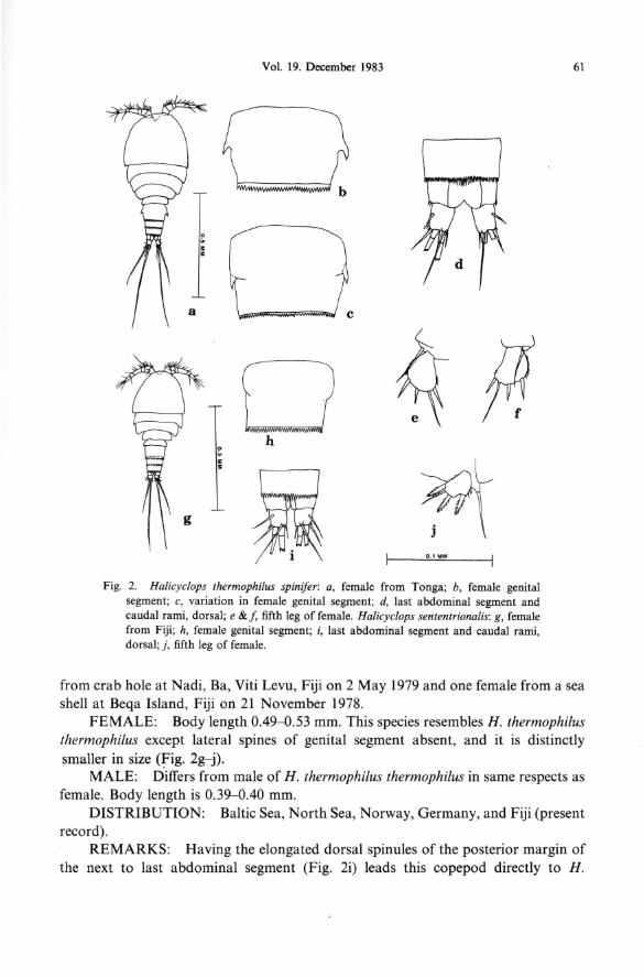
Halicyc/ops thermophilus spinifer Kiefer Fig. 2a- f
MATERIAL EXAMINED: Two females from crab holes, Tonga, 27 August 1976.
FEMALE: Body length 0.58-0.60 mm. This subspecies resembles H. thermophilus thermophilus except lateral spines of genital segment more pronounced, sharper-pointed, and extend further posteriorly (Fig. 2a~). Lindberg's (1952) figures closely resemble the Tonga specimens in these structures.
DISTRIBUTION: India, Iran, Tonga (present record) and probably Madagascar.
REMARKS: Although Lindberg (1957) considered this copepod a distinct species in his key, this form should be retained as a subspecies of H. thermophilus, because there are only minor differences in structures. More specimens and breeding experiments are indicated.


62 Micronesica
thermophilus septentrionalis in Kiefer's (1936) key, thus differing from our specimens of H. neglectus. In Lindberg's (1957) key, using different characters, it goes to H . neglectus septentrionalis. Although showing morphological similarities to both H. thermophilus and H. neglectus, it can be distinguished from these species and should be raised to the rank of distinct species. The presence of this northern copepod on a tropical South Pacific island is unusual, but several species of copepods described from northern, cold waters have later been found in warm southern waters. This is especially true of some harpacticoid copepod species.
Subfamily CYCLOPINAE Dana Bryocyclops fidjiensis Lindberg, 1954
Fig. 3a- g
MATERIAL EXAMINED: Thirty females and twenty males from Fiji (1978, 1979, and 1980), Tonga (1976), and Western Samoa (1975 and 1976) in tree holes, leaf axils of taro and Pandanus, tin cans, bamboo, bottles, and ditches.
FEMALE: Body length is 0.47 to 0.55 mm. Body small and stubby and described by collectors, Karen Toohey and Mark Goettel, as shiny in appearance when alive. Prosome egg-shaped and 5-segmented and opisthosome tubular and 4-segmented (Fig. 3a). Anal operculum conspicuous and triangular (Fig. 3c). Caudal rami short, twice as long as broad. First antennae short, eleven-segmented. Swimming legs biramous and each ramus 2-segmented, except last exopod, which is one-segmented (Fig. 3f). Lindberg (1954) drew the female fourth foot with !segmented endopod, but in all specimens from type locality, endopod shows definite joint to make 2-segmented endopod, as in male fourth leg. Spine formula of terminal segments of exopods is 3, 3, 3, 4 ( 4 spines in the single fourth leg exopod) and setal formula is 5, 5, 5, 4 (Fig. 3d- f). Second and third legs alike in armature. Fifth leg not distinct from fifth body segment and bears 3 setae (Fig. 3g).
MALE: Body length 0.39 to 0.47 mm. First antennae geniculate (Fig. 3b). Fourth leg endopod 2-segmented, as in female endopod (Fig. 3f).
DISTRIBUTION: This species is abundant on Fiji, Tonga, and Western Samoa.
REMARKS: B. fidjiensis differs from the closely related B. anninae (Menzel) in the !-segmented exopod of the fourth leg. B. anninae has a 2-segmented exopod of this leg. The author has a B. anninae female from the leaf axil of Pandanus, The Pali, Oahu, Hawaiian Islands, collected August 1963 by Bassett Maguire, Jr.
Bryocyclops bogoriensis (Menzel) Fig. 4a-j
MATERIAL EXAMINED: Eight females and five males from lvi tree holes (lnocarpus edulis),yavua Dump, Viti Levu, Fiji, 2 May 1979.
FEMALE: Body length 0.40 to 0.50 mm. Like B. fidjiensis, small and stubby,


Vol. 19. December 1983 67
middle of segment. Inner spine may be shorter than terminal seta or longer (Figs. 5f & 6e). Sixth leg reduced to 2 outer spinules and 1 inner seta (Fig. 6f). Seminal receptacle T-shaped, with posterior portion bag-like (Fig. 5g).
DISTRIBUTION: Cosmopolitan- Europe, Asia, Africa, North America, Australia, New Zealand, and islands in the Pacific. This species can tolerate brackish water as shown by its living in brackish crab holes. Wilson (1942) collected several specimens from a lagoon on Penrhyn Island (Solomon Islands) in the tropical Pacific along with nine other species, all typically marine. Lindberg (1954) reported it from Sikaiana Island (Solomon Islands) and Toutouba and Viti Levu (Fiji Islands). Watkins and Belk (1975) reported it from a spring pond, an ephemeral pond, and a lake on Guam.
REMARKS: This species has been successfully infected with a species of Coelomomyces fungus in Africa. It is interesting that this copepod could directly control mosquito larvae by predation and also indirectly by acting as intermediate host for this fungus that destroys mosquito larvae.
Cryptocyclops bico/or linjanticus (Kiefer, 1928a) Fig. 7a- g
MATERIAL EXAMINED: Ten females from tree holes, blocked drains, ponds, rice paddies, and metal drums from Viti Levu, Fiji on 30 November 1978 and 4 and 5 April 1979.
FEMALE: Body length 0.60 to 0. 70 mm. Prosome egg-shaped, 5-segmented, opisthosome tubular and 4-segmented. Caudal rami 3.2 to 3.5 times longer than broad. Inner long terminal seta of caudal ramus 3.5 to 4 times longer than the ramus (Fig. 7b ). This seta less than 3 times longer than ramus in Cryptocyclops bicolor bicolor (Sars). This is the principle difference between these two subspecies. First antenna 11-segmented. Swimming legs biramous, each ramus 2-segmented. Spine formula of terminal segments of leg exopods is 3, 4, 4, 3 and setal formula is 5, 5, 5, 5 (Fig. 7c-f). Fifth leg consists of basal segment bearing an outer seta and not separated by a joint from the fifth body segment and a narrow tubular, distinct segment with a terminal seta (Fig. 7g).
DISTRIBUTION: East Africa, West Africa, Madagascar, Sumatra, Java, India, Iran, Afghanistan, Burma, Cambodia, Philippines, Taiwan, Manchuria, New Hebrides, Fiji. Lindberg (1954) was the first to report it from Viti Levu, Fiji. Recently, the author identified this copepod in material sent by B. F. Gabriel of the University of the Philippines at Los Baiios.
REMARKS: Recently, Microcyclops varicans (Sars) has been sent to me in collections made by B. F. Gabriel in the Philippines. This species differs from C. B. linjanticus by having the longest caudal seta longer than the abdomen (shorter in C. B. linjanticus) and a bag-shaped posterior portion of the female seminal receptacle (short and oval in C. B. linjanticus).


Vol. 19. December 1983
Fig. 8. Microcyc/ops rnicrosetus: a, female from Fiji; b, last abdominal segment and caudal rami, dorsal; c, first antenna of female; d, first antenna of male; e, second antenna; f, mandible, turned; g, first maxilla; h, second maxilla; i, maxilliped; j, first leg; k, second leg; I, third leg; rn, endopod and terminal exopod segment of third leg.
69
about same length as caudal ramus. Innermost terminal seta so slender and tiny that it is not visible under magnification of 600 diameters, light microscopy and was believed to be absent. Using scanning electron microscopy (magnification of 2,000 diameters), Harry Blanton Miller photographed the caudal rami and showed these setae to be present. They are quite hair-like and frequently lie on or close to the base of the longest terminal seta (Fig. 9i). Small size of this seta indicated in specific name of this copepod. First antennae 10-segmented and reach posterior border of third body segment. Second antennae 4-segmented (Fig. 8e). Mandible typical for freshwater cyclopoids, being the biting and chewing form. Two setae very long and quite


Vol. 19. December 1983 71
visible in a whole mount of copepod (Fig. Sf). First and second maxillae and maxillipeds shown in Fig. 8g-i. Swimming legs biramous and each ramus 2-segmented (Figs. 8j-m, 9a, b). Spine formula of terminal segments of the exopods is 3, 4, 4, 3 and setal formula is 5, 5, 5, 5. Setal formula of inner side of the endopods (not counting terminal seta) is 3, 4, 3, 3 (in one individual-3, 4, 4, 3). Basal segment of fifth leg not separated from fifth body segment and bears an outer seta and a narrow tubular segment with a terminal seta (Fig. 9c-f). Seminal receptacle T-shaped, with posterior portion baglike, similar to that structure in Mesocyclops leuckarti (Fig. 9g, h).
MALE: Body length 0.50 to 0.60 mm, usually 0.52 mm. Prosome 5-segmented, opisthosome 5-segmented. Both first antennae geniculate (Fig. 8d).
REMARKS: This new species keys to the Genus Microcyclops. Its distinct segment of the fifth leg is narrow and tubular like that of Microcyclops and Cryptocyclops and not broad as in Apocyclops. The length of the shorter terminal spine of the endopod of the fourth leg exceeds that of Cryptocyclops, and the bag-like posterior portion of the seminal receptacle is more like that of Microcyclops than the very short, posterior portion of seminal receptacle of Cryptocyclops (Dussart, 1969). The tiny innermost terminal caudal seta should not eliminate consideration o,( this copepod in Genus Microcyclops. Within Genus Diacyclops are species in which this seta is very short (D. crassicaudis) and some long (D. bicuspidatus). In spite of being an inhabitant of brackish water and having a slight difference in setal armature in the third leg endopod (three instead of four inner setae of the terminal segment), the species is considered by the author to be within Genus Microcyclops.
Tropocyclops confinis Kiefer Fig. 9j- k
MATERIAL EXAMINED: One female from a stream in mountain highlands, Western Samoa, 13 October 1976.
FEMALE: Body length 0.52 mm. Prosome egg-shaped and 5-segmented, opisthosome tubular and 4-segmented. Caudal rami 2.3 times longer than broad. Tropocyclops prasinus prasinus (Fischer) generally has slightly longer caudal rami (2.5 to 3 times longer than broad). First antenna 12-segmented and reaches beyond second segment of body, with fine hyaline membrane on last three segments. Swimming legs biramous, and each ramus 3-segmented. Spine formula of terminal segments of exopods of swimming legs is 3, 4, 3, 3 (3, 4, 4, 3 for T. prasinus prasinus) and setal formula is 5, 5, 5, 5 (Fig. 91-o ). Fifth leg consists of single segment armed with a slender inner spine, an outer seta, and a terminal seta (Fig. 9p).
DISTRIBUTION: Africa, Madagascar, Java, Flores, Sumatra, India, Iran, Burma, Syria, United States (rare), New Hebrides, Czechoslovakia, Russia.
REMARKS: Although this single individual was taken from a stream and the genus is usually limnetic, the species is available on Western Samoa for distribution to microhabitats. The author has many specimens of the nearly related T. prasinus

72 Micronesica
mexicanus collected from bromeliads in Jamaica by Laessle (1961). It is therefore a potential microhabitat species and included here.
Ectocyclops phaleratus (Koch) Fig. lOa-d
MATERIAL EXAMINED: Two females from ltatoko, Ba, Viti Levu, Fiji, in a blocked drain and a crab hole, 6 June 1979.
FEMALE: Body length 0.80 and 0.75 mm. A stubby species (Fig. lOa). First antenna very short and 10-segmented. Caudal rami very short, about 1.3 to 2 times as long as broad and ornamented with oblique rows of spinules on inner surfaces (Fig. lOb). Swimming legs biramous and each ramus 3-segmented. Spine formula of terminal segments of exopods is 3, 4, 4, 3 and setal formula is 5, 5, 5, 5. Terminal segment of endopod of fourth leg about 1.5 times longer than broad and inner terminal spine twice as long as outer terminal spine (Fig. IOc). Fifth leg not distinct from fifth body segment and armed with two strong spines and an outer seta (Fig. lOd).
DISTRIBUTION: Europe, Africa, Asia, America, Australia, and Fiji (present record).
REMARKS: Although several subspecies of E. pha/eratus and several additional species for the genus have been described, the Fiji specimens are not different from the North American specimens or those available from Europe.
Paracyclops fimbriatus (Fischer) Fig. lOe-i
MATERIAL EXAMINED: Five females from Beqa Island and Viti Levu, Fiji in tree holes and a rubber tire, 21 November 1978 and 15 February 1979.
FEMALE: Body length 0.70 to 0.80 mm. A small species, but more slender in body than Ectocyclops phaleratus (Fig. 1 Oe ). Caudal rami are 3.2 to 4 times as large as broad and bear a short transverse row of spinules just above dorsolateral seta (Fig. 1 Of). North American specimens show more variation in proportions of caudal rami than do those from Fiji. P. fimbriatus poppei (Rehberg), which is common in North America and bears longitudinal dorsal row of spinules on caudal rami, not been found on Fiji, but recorded by Sars (1904) from Hawaii. First antennae very short and 8-segmented (Fig. lOe), not 11-segmented as in Paracyclops affinis (Sars). Swimming legs biramous and 3-segmented. Spine formula of terminal segments of exopods is 3, 4, 4, 3 and setal formula is 5, 5, 5, 5. Terminal segment of endopod of fourth leg about 1.6 to 1.8 times longer than broad and inner terminal spine about twice as long as outer terminal spine (Fig. I Og, h). Fifth leg consists of one broad segment, armed with an inner spine and two outer seta, terminal seta much longer than inner sp~e (Fig. 1 Oi).
DISTRIBUTION: Europe, Asia, Africa, New Zealand, New Guinea, North


74 Micronesica
Order HARPACTICOIDA Family PHYLLOGNATHOPODIDAE Gurney
Phyllognathopus viguieri (Maupus) Fig. lla-1
MATERIAL EXAMINED: Thirty females and fifteen males-ten females and five males from each of the localities-Tonga (1976), Western Samoa (1975, 1976), and Fiji (1978, 1979, 1980). These were taken from tree holes, taro leaf axils, old auto tires, tin cups, plastic containers, bamboo, old boats, and ground pools.
FEMALES: Body length 0.40 to 0.55 mm. A tiny, tubular-bodied copepod with 10 body segments and conspicuous rostrum (Fig. lla). Caudal rami stubbyeither slightly longer than broad or just as long as broad. Considerable variation of shape and armature of these rami, even in the same population in a microhabitat (Fig. llc-e). Long terminal caudal rami may be represented by spike-like projections (Fig. lie). First antennae very short and 8-segmented. Swimming legs biramous and each ramus except endopod of fourth leg (2-segmented) is 3-segmented. Armature of swimming legs of this well-described species is shown in Figure 11 f- i. Fifth leg a projection from sixth body segment and has median indention. Outer portion bears seta and spines, and the inner has 2 large spines (Fig. llj, k).
MALE: Body length 0.35 to 0.55 mm. Body consists of 11 segments (Fig. 11 b). First antennae geniculate and very short. Although variable, fifth leg of these specimens consists of basal segment with an outer seta and inner, strong spine and usually distinct exopod with total of six setae and spines (Fig. 111).
DISTRUBUTION: Cosmopolitan-Europe, Asia, Africa, America, Pacific tropical islands, Caribbean islands, etc. It is easily transported with plants, food, water containers, etc. Lowndes (1931) stated that it could be "found at any time by taking some of the liquid enclosed by the leaves of the Bromeliaceous plants (pineapples) from the Botanic Gardens" (Birmingham, England). He also said he had obtained them from pineapples bought from any food store by adding water to the leaf axils and examining this water after a few weeks. The author has specimens from Guzmania bromeliad leaf axils on Jamaica and Puerto Rico and from Pandanus leaf axils on Oahu, Hawaiian Islands. Watkins and Belk (1975) reported P. veguieri menzeli (Chappuis) from Guam. This subspecies is mainly based on the details of structure of the male fifth legs. The author prefers to avoid subspecific designation for such a variable species.
Family DARCYTHOMPSONIIDAE Lang Darcythompsonia inopinata Smirnov
Fig. 12a- k
MATERIAL EXAMINED: Two females and two males from crab holes on Western Samoa (15 April 1976 and 29 January 1976), and twelve females and five males from crab holes at Culanuku, Viti Levu, Fiji (15 September 1978).


Vol. 19. December 1983 77
fiensis (T. Scott) (also with 7-segmented first antennae) from Ireland, by the absence of angular caudal ramus projections on Darcythompsonia inopinata.
Family HARPACTICIDAE Sars, 1904 Tigriopus angulatus Lang
and Tigriopus californicus (Baker)
Fig. 13a- i, j-n
MATERIAL EXAMINED: Five females and three males of T. angulatus from Otago, New Zealand (February 1976). One female T. angulatus (1976, in vial with Elaphoidella taroi and Bryocyclopsfidjiensis) from Fiji. Six females and three males of T. californicus from Che Ju Island and Pusan, South Korea (6 January and 4 January 1979). Collected from brackish supralittoral pools and a freshwater pool.
DESCRIPTIONS: Because these two species have been so frequently confused in identification and in literature on copepod distribution, the following descriptions are given in the form of comparisons. Where the species agree, no distinctions are made in the descriptions.
FEMALES: Body length forT. angulatus 0.75 to 0.81 mm, forT. californicus 1.04 to 1.16 mm (Fig. 13a, j). Prosome narrowly egg-shaped tapering posteriorly to a tube-shaped opisthosome. Caudal rami stubby, about as long as broad. First antennae short and 9-segmented. Second antenna bears 4-segmented exopodite (Fig. 13c). Bradford (1967) stated that there is a difference in the "proportions of their [the two species] limbs, especially the first leg in both sexes", but the author's specimens show no differences in the first legs except the overall size of the legs (Fig. 13d, k). Bradford (1967) stated that the last segment of the fourth leg exopod of T. angulatus has a total of eight spines and setae instead of seven as in T. californicus. This is true of both sexes of the species and is a notable difference (Fig. 13h, 1). As Bradford pointed out, the fifth legs of the females differ in the two species. ForT. angulatus, the basal expansion extends much longer than the exopod segment, and for T. californicus, this expansion is much shorter than the exopod (Fig. 13f, m).
MALES: Body le~gth for T. angulatus 0. 71 to 0. 73 mm, for T. californicus 1.03 to 1.05 mm. First antennae geniculate (Fig. 13b). Second leg differs from that of female in both species by presence of long outer terminal projection on second segment of endopod (Fig. 13g). Fourth legs like those of females and differ in two species as described for females. Fifth legs of T. californicus show greater separation of 2 basal setae from 3 median setae of exopod than is shown in fifth legs of T. angulatus (Fig. 13i, n).
DISTRIBUTION: Because of confusion of these two species, many of the records of T. californicus from the southwest Pacific are probably T. angulatus. T. californicus has definitely been collected at Laguna Beach, California and Vancouver Island, Canada, and South Korea (present record). T. angulatus has been collected from New Zealand; Macquarie Island; Santiago, Chile; and possibly East Africa. The


Vol. 19. December 1983 79
tide pools. REMARKS: J. S. Pillai, who collected and sent specimens of both species,
stated that in life, these species are orange in color. T. angulatus has been infected with a Coelomomyces fungus that destroys mosquito larvae.
Family TISBIDAE (Stebbing) Lang, 1948 Tisbella pulche/la (Wilson)
Fig. 14a-f
MATERIAL EXAMINED: One female from crab hole at Bau Landing, Fiji (2 August 1978) and one female from ground pool on Yanuca Island, Fiji (5 March 1979).
FEMALE: Body length 1.0 to 1.12 mm. Prosome egg-shaped and 5-segmented, opisthosome tubular and 4-segmented. General body appearance like that of copepods of genus Tisbe. Caudal rami stubby, about as long as broad (Fig. 14b). First antennae slender and 8-segmented (Fig. 14a). Second antennae and mouthparts described by Yeatman (1963), as have swimming legs. As for most harpacticoid copepods, proportions of segments of first legs and their ornamentation is of importance in identification. This leg biramous, its exopod 3-segmented and endopod 2-segmented. This species readily distinguished from Gurney's (1927) Tisbella timsae by length of second endopod segment-as long as first segment in T. timsae and much shorter than first segment in T. pulchella (Fig. 14c, d). Fifth leg has short basal segment bearing an outer seta and two inner setae and a long ( 4 to 5 times longer than broad) exopod segment. This segment bears five terminal and subterminal setae (Fig. 14e). Sixth leg present, and consists of a long inner seta and a short outer seta (Fig. 14f).
DISTRIBUTION: Chappaquiddick Island, Massachusetts; Bermuda Islands; drainage ditch to North Sound, Grand Cayman, BWI (many collected by the author on 26 August 1978); Fiji Islands (present record). The species is probably more widespread in marine and brackish littoral water in the tropics than past collecting would indicate.
REMARKS: The occurrence of this copepod in water far from America and the Caribbean Sea raises again the question of whether it is the same as Gurney's (1927) T. timsae. If Gurney's figures of the first leg and first antenna (?-segmented) are correct and his single specimen from Ismailia, Egypt is not abnormal, T. pulchella is certainly distinct from that species. Collecting specimens from Egypt is necessary to close the case. Nevertheless, the discovery of these specimens of Tisbella on Fiji is exciting for copepod distribution studies.


Vol. 19. December 1983 81
well described by Lang (1948) and no females were collected from microhabitats on Fiji. Brief description of male from Fiji given here to show it is representative of species.
MALE: Body length 0.50 mm. Body small, tubular and 10-segmented (Fig. 14g). Posterior border of anal operculum bears tiny spinules. Caudal rami about as long as broad, and outer corner armed with strong spine (Fig. 14h). Innermost terminal seta very tiny. First segment of endopod of first leg slightly longer than exopod of that leg and slightly more than twice as long as two end segments (combined) of this ramus. Fifth leg consists of basal segment armed with outer seta and two strong, inner spines and an almost round exopod segment with a total of five setae and spines (Fig. 14i). Unlike some species of Schizopera, there is a seta on the first segment of the fourth leg endopod.
DISTRIBUTION: Sumatra, Java, and Fiji (present record). REMARKS: This species is probably not a regular inhabitant of crab holes,
because this type of microhabitat was extensively investigated on the Fiji Islands.
Family AME1RIDAE (Monard) Lang Nitocra lacustris pacijicus n. subsp.
Fig. 15a- p
MATERIAL EXAMINED: Two females and one male from Western Samoa (18 March 1976). U. S. National Museum, No. 204473 (holotype), No. 204475-204478 (paratypes), and No. 204474 (allotype). Four females and one male from Tonga (27 August 1976), and one female from Fiji (17 July 1978)-all taken from crab holes. One female N. lacustris from India.
FEMALE: Body length 0.37 to 0.40 mm. Body tubular, tapering posteriorly. Prosome 5-segmented, opisthosome 4-segmented (Fig. 15a). Rostrum conspicuous. Anal operculum bears small spinules; caudal rami slightly longer than broad. Posterior lappets of last abdominal segment bear long seta and short seta, viewed between caudal rami (Fig. 15b,c). Longer setae exceed caudal rami in length and distinguish this subspecies from typical N. lacustris, which have very short setae in this place (Fig. 15d, e). First antennae short and 8-segmented. Exopod of second antenna is one-segmented (Fig. 15f). Segmentation and armature of swimming legs typical for the species (Fig. 15g, i- k) . Unlike Nitocra lacustris sinoi Marcus et Por, basal segment of first leg endopod is shorter than first two segments of exopod. Third segment of second leg endopod bears three setae. Exopod of fifth leg bears 6 setae and inner expansion of basal segment bears a total of 5 spines and setae (Fig. 15n, o ).
MALE: Body length 0.35 to 0.39 mm. Prosome 5-segmented, opisthosome 5-segmented. Lappet setae of last abdominal segment long, as in female. Inner spine of first leg basal segment modified into blunt hook (Fig. 15h), as typical for species and sex; armature of legs typical.
REMARKS: Except for the elongated setae on lappets of the last body segment, these specimens resemble the well-described, typical N. lacustris.


Vol. 19. December 1983
Family CANTHOCAMPTIDAE (Sars) Monard Elaphoidella taroi Chappuis
Fig. 18a-p
85
MATERIAL EXAMINED: Twenty females and twelve males from taro leaf axils, tree holes, coconut shells, and tin cans at Wailoku, Viti Levu, Fiji (29 August 1979 and 30 September 1978). Three females and two males from taro leaf axils on Western Samoa (7 August 1976).
FEMALE: Body length 0.59 to 0.67 mm. Body tubular and 9-segmented. Small rostrum present at anterior end (Fig. 18a). Anal operculum has row of small spinules at its posterior border. Caudal rami short, taper posteriorly and have dorsal, inner hooklike process (Fig. 18b ). First antennae short and 8-segmented. Exopod of second antenna !-segmented (Fig. 18e). Swimming legs biramous. Exopods and first leg endopod 3-segmented and other leg endopods 2-segmented. Leg segments armed with spines and setae as shown in Figure 18f- i. Fifth leg basal segment bears an outer seta and its long inner expansion bears 4 setae. Exopod short, almost circular and bears 4 setae (Fig. 18n).
MALE: Body length 0.60 to 0.65 mm. Body consists of 10 segments (Fig. 18c). Rostrum and anal operculum like those of female. Caudal rami have dorsal, inner hook smaller than that of female (Fig. 18d). First leg like that of female, but other legs conspicuously different. Second segment of second leg endopod cone-shaped and bears 3 setae instead of 4. Some of spines of third leg exopod enlarged and endopod 3-segmented, middle segment bearing long spine-like projection and end segment with only 2 terminal setae. Outer terminal spine of fourth foot exopod has 2 or 3 long barbs. For comparison of these legs in sexes, Figure 18g-i (female legs) are placed in a row and corresponding male legs (Fig. 18j- l) are placed directly below them. Right and left fifth legs are joined at the inner base and there is an outer seta. Exopod round and bears 3 setae or spines (Fig. 18o, p ).
DISTRIBUTION: Fiji and Western Samoa (present record). REMARKS: This species may readily be distinguished from Elaphoidella
bromeliacola (Chappuis, 1928), which has a total of 4 setae and spines (instead of 3) on the end segment of the fourth leg endopod (Lang, 1948). Karen Toohey and Mark Goettel, who collected the Fiji material, described E. taroi as "mostly a bottomdweller, always gripping onto the substrate." They were able to infect this species with Coelomomyces fungus (Toohey, et al. 1982). It therefore has great potential for use in mosquito control, where it occurs.
Elaphoidella grandidieri (Guerne and Richard) Fig. 19a- j
MATERIAL EXAMINED: Three females from blocked drain at ltatoko, Ba, Viti Levu, Fiji (6 June 1979) and three females from tires on Western Samoa (22 September 1975).

86 Micronesica
Fig. 18. E/aphoidel/a taroi: a, female from Fiji; b, last abdominal segment and caudal rami of female, dorsal; c, male from Fiji; d, left caudal ramus of male, dorsal; e, second antenna;/, first leg of female; g, second leg of female; h, third leg of female; i, fourth leg of female; j , second leg of male; k, third leg of male; /, fourth leg of male; m, terminal exopod segment of fourth leg of male; n, fifth legs of female; o, p, fifth legs of male.


88 Micronesica
terminal, dorsal, sharp projection (Fig. 19b ). This process not as conspicuous as process of caudal rami of E. taroi. First antennae short and 8-segmented (Fig. 19c). Exopod of second antenna !-segmented. Swimming legs biramous. Exopods and first leg endopod 3-segmented. Unlike E. taroi, first leg endopod longer than exopod. Leg segments armed with setae and spines as shown in Fig. I ~e-h. Fig. 19i shows an abnormal fourth leg with no outer spine on second exopod segment. There is no socket to indicate that this spine was lost by breakage. Fifth leg notably different from that of E. taroi. Its basal segment expansion, bearing 4-setae, is short, and the exopod bearing a total of 6 setae and spines, is elongated (Fig. 19j).
DISTRIBUTION: Africa, Madagascar, Ceylon, China, Thailand, Vietnam, Java, Sumatra, Flores, New Guinea, Hawaiian Islands, Fiji, and Western Samoa (last 2 are present record).
Discussion
Some microhabitats such as crab holes may be flooded during high tides or connected with the ocean or with other crab holes by underground tunnels. The freshwater habitats such as leaf axils, tree holes, and some types of containers may have their small flora and fauna shifted by splashing of water during heavy rains or these organisms may be transported by frogs, insects, etc. Of considerable interest is the means of transport from one island to another or even from one continent to another. As pointed out in the "Distribution" section under Phyllognathopus viguieri, species have stages that can survive much drying during transportation of plant leaf axils, such as pineapples. Because pineapples evolved in South America and were taken by Indians and later explorers to other areas, one wonders if Phyllognathopus viguieri evolved in the Americas. This copepod may have evolved in the Hawaiian Islands or other islands and invaded these transplanted pineapples to eventually be shipped to anywhere in the world. Humans, in transporting their boats, plants, drinking water, etc. have played a great part in extending the distribution of many species of copepods. Botanic gardens, having plants from various continents, can be sources of unusual copepods and other fauna and flora.
The ecology of the microhabitats is of interest and importance. In many of the microhabitats, Bryocyclops fidjiensis and Elaphoidella taroi are commonly found together in harmony, and both species are numerous. Apparently, Bryocyclops is not predacious on adult copepods of its species or other species, although it might eat copepod nauplii. Elaphoidella taroi also commonly occurs with Phyllognathopus viguieri. The difference in size between these two harpacticoid species may prevent competition by selection of different-sized foods. In many collections Bryocyclops fidjiensis accompanies these two harpacticoids. Mesocyclops leuckarti, a known predator of other copepods and even mosquito larvae, occurred with Cryptocyclops bicolor linjanticus in a few crab holes, but numbers of each species were low. Halicyclops thermophilus and Halicyclops septentrionalis occurred together in collections from six crab holes. This shows that because these copepods can coexist

Vol. 19. December 1983 89
without interbreeding, they are very likely different species.
ACKNOWLEDGMENTS
It is with pleasure that the author expresses his gratitude to the World Health Organization Collaborating Centre on Biological Control for the identification, ecology, and safety of nontarget organisms, within the Research Unit on Vector Pathology of the Memorial University of Newfoundland, St. John's, Canada. I am particularly grateful to Marshall Laird, Director of RUVP, for making the copepod collections available for my identification and study and for his encouraging and interesting correspondence. I also thank J. S. Pillai, Senior Lecturer in Microbiology, University of Otago, Dunedin, New Zealand, for furnishing his collections of copepods from South Korea and New Zealand (used in comparisons), Tonga, and Western Samoa, and for important literature on the areas involved. I take great pleasure in acknowledging the outstanding work of Karen Toohey and Mark Goettel for their superior collecting of copepods from Fiji, labelling collection data, sorting the collections, and wonderful correspondence. They were recipients of fellowship support from the Canadian International Development Agency. While in Fiji, they established the principal copepod host for a species of Coelomomyces in that area. I wish to thank Barry Engber, graduate student at John Hopkins University, Baltimore, for participating in the Western Samoa collecting with V. S. Pillai, and the late Harry Blanton Miller, former student of the author and later at Vanderbilt University, Nashville, for scanning electron microscopy studies of Microcyclops microsetus n. sp. The Department of Biology, University of the South, Sewanee, furnished typing for the preparation of this report. John Couch of Chapel Hill, North Carolina, involved the author in Coelomomyces research, and for this help I am very grateful.
References Cited
Bradford, J. M. 1967. The genus Tigriopus Norman (Copepoda: Harpacticoida) in New Zealand with a description of a new species. Transactions of the Royal Society of New Zealand, Zoology 10(6): 51- 59.
Chappuis, P. A. 1955. Notes sur les copepodes. 20. Copepodes harpacticoides des iles du Pacifique. Notes Biospeologiques X: 97- 101.
Dussart, B. 1967. Les copepodes des eaux continentals d' Europe Occidentale. Tome I. Calanoides et harpacticoides. N. Boubee and Cie, Paris. 500 p.
---. 1969. Les copepodes des eaux continentales d' Europe Occidentale. Tome II. Cyclopoides, N. Boubee and Cie, Paris. 292 p.
Gurney, R. 1927. Cambridge expedition to the Suez Canal 1924. Report on the Crustacea: Copepoda (littoral and semiparasitic). Transactions of the Zoological Society, part 4: 451-577.
1933. British fresh-water copepoda 3. Ray Society, London. Kiefer, F. 1928a. Beitriige zur copepodenkunde (VIII). Zoologischer Anzeiger 76: 17-18.
1928b. Beitriige zur copepodenkunde (IX). Zoologischer Anzeiger 76(5): 99- 110. 1936. Freilebende siiss-und salzwassercopepoden von der insel Haiti. Mit einer revision der
gattung Halicyclops Norman. Archiv Hydrobiologie 30: 263-317.

90 Micronesica
Kunz, H . 1961. Beitriige zur kenntnis der D 'Arcythompsonidae (Copepoda, Harpacticoida). Zoologischer Anzeiger 167(7 /8): 275-280.
Laessle, A.M. 1961. A micro-limnological study of Jamaican bromeliads. Ecology 42(3): 449-517. Lang, K. 1948. Monographie der harpacticiden. H. Ohlsson, Lund. 2 vols. 1682 p. Lindberg, K. 1952. Cyclopides (Crustaces copepodes) de Madagascar. Troisieme note. Memoires de
L'lnstitut Scientifique de Madagascar, Serie A, VII (1): 62-65. 1954. Cyclopides (Crustaces copepodes) d'iles du Pacifique Sud (Melanesie et Micronesie) et
ed Borneo. Kung!. Fysiografiska Siillskapets I Lund Forhandlingar 24(18): 1-14. 1957. Cyclopides (Crustaces copepodes) de Ia Cote d'Ivoire. Bulletin lnstitut Francaise
Afrique Noire. Serie A, 19: 134-179. Lowndes, A. G. 1931. Some fresh-water entomostraca of the Birmingham District. Annals and
Magazine of Natural History VIII (10): 561-577. Menzel, R. 1925. Cyclopides muscicoles et bromelicoles de Java. Annales de Biologique lacustre XIV. Sars, G . 0 . 1904. Pacific plankton-crustaceen. Zoologischer Jahrbuch Systematik XIX: 641-642. Toohey, M. K., M. S. Goettel, and J. S. Pillai. 1981. A review of the prospects of using biological
control against mosquito vectors of subperiodic filariasis and arboviruses in Polynesia. South Pacific Journal of Natural Science 2: 4-43.
Toohey, Mi. K ., G. Prakash, and M. S. Goettel. 1982. Elaphoidella taroi: the intermediate copepod host in Fiji for the mosquito pathogenic fungus Coelomomyces.Jurna! oflnvertebrate Pathology 40: 378-382.
Watkins, R. L. , and D. Belk. 1975. The Copepoda of Guam. Crustaceana 28(3): 302-304. Whisler, H. C., S. L. Zebold, and J . A. Shemanchuk. 1974. Alternate host for mosquito parasite
Coe/omomyces. Nature 251 : 715-716. 1975. Life history of Coelomomyces psorophorae. Proceedings of the National Academy of
Science 72(2): 693- 696. Wilson, C. B. 1942. The copepods of the plankton gathered during the last cruise of the Carnegie.
Carnegie Institution of Washington Publication 536: 1-237. Yeatman, H . C. 1963. Some redescriptions and new records of littoral copepods for the Woods Hole,
Massachusetts region. Transactions of the American Microscopical Society LXXXII (2): 197-209.