controversial stuff that we fuss a lot over goblet cells ...labmed.ucsf.edu/uploads/518/255_henry...
TRANSCRIPT

1
Henry Moon was one of the giants in academic pathology during my early years. He and my boss, Jim French were cronies going back to WWII
I never met him, but I heard a lot about him, all of it good. Thanks to Linda and her gang for giving me this honor of delivering the Moon Lecture.
Controversial stuff that occurs slightly above,
within or slightly below the gastroesophageal junction, including Barrett’s mucosa:
What role do we pathologists play?
We fuss a lot over goblet cells & cancer
in and around the GE junction,
but do they deserve all the fuss?

2
This is it!Pretty small considering the size of everything around it!
Sometimes the lower esophagus and the GEJ are connected by the same changes, almost as if they are a single entity
Let’s start with 2 cases
#1: Dyspeptic adult woman not responding to medication (PPIs) has upper endoscopy.
The endoscopist saw erythemaat the gastroesophagealjunction. Nothing else.
The erythema was biopsied

3
Biopsies of erythema are among the least informative of all biopsies.
But we won’t discourage the GI people from
biopsying erythemaWe need the business!!Biopsies of erythema account for about 7% of my income
squamous
squamous
squamous
squamous
columnar
columnar
columnarcolumnar
columnar#1
#2
1
2
3 #1 Chronic inflammation!1

4
Plasma cells
Pancreatic acinar cells mixed with cardiac gland mucus cells
2
Pancreatic acinar metaplasia (PAM)
Huge pit cells: pseudogoblet cells3Don’t confuse these with real goblet cells

5
Squamo-columnar junction
No goblet cells!
#2 Finally, way off at the edge of the biopsy
The evil, dreaded goblet cells!!
SummaryEndoscopic erythema at the GE Junction. No endoscopic Barrett’s mucosaSquamous and columnar mucosae
The columnar mucosa has
Inflammation: plasma cells
Goblet cells….and mimics
Pancreatic acinar cells in the cardiac glands
SO?does not have a standard namehas a lot of features, but what do they all mean? I will deal with this.
does not answer the clinical question: what caused dyspepsia?
This is a common biopsy.It is annoying, because it

6
#2: Obese adult white male (the Barrett model)Heartburn for 20 years, recently worse Not responding to PPIs endoscopyGEJ tongues: “cannot tell if this is
an exaggerated Z-line or short segment Barrett’s”
Bx taken of the tonguesPathologist told (not asked)
to R/O Barrett’s
(The true request was to R/I Barrett’s)
Goblet cells
Inflammationlike the first bx
Pseudogoblet cells
SummaryEndoscopic: changes that may be either an exaggerated Z-line (squamocolumnarjunction) or short segment Barrett’s mucosaHistologic:
Columnar mucosa Inflammation Goblet cells…..and mimics
These 2 sets of biopsies around the gastroesophageal junction have
Columnar mucosaImpressive chronic
inflammationGoblet cells
SO?
7
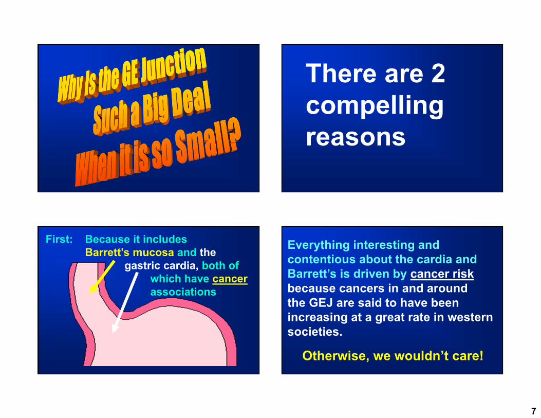
There are 2 compelling reasons
First: Because it includesBarrett’s mucosa and the
gastric cardia, both of which have cancerassociations
Everything interesting and contentious about the cardia and Barrett’s is driven by cancer riskbecause cancers in and around the GEJ are said to have been increasing at a great rate in western societies.
Otherwise, we wouldn’t care!

8
Adenocarcinomas at and around the Gastroesophageal Junction
FundusCardia
Upper Body
Distal Esophagus(Barrett’s)Junctional NOS
Sometimes (often?) we cannot tell where the cancer is arising!
Second: The GE junction affects my standard of living much more than its size suggests it should!
This is it!Disclaimer:
About 10% of my income is derived from specimens taken from the GEJ and nearby.
Our clients, the gastroenterologists actually have to deal with 2 junctions.
9
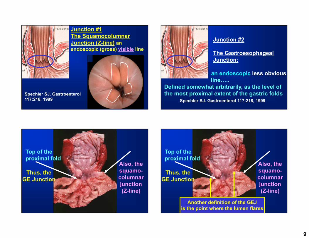
Junction #1The Squamocolumnar Junction (Z-line) an endoscopic (gross) visible line
Spechler SJ. Gastroenterol117:218, 1999
Junction #2
The Gastroesophageal Junction:
an endoscopic less obvious line…..
Defined somewhat arbitrarily, as the level of the most proximal extent of the gastric folds
Spechler SJ. Gastroenterol 117:218, 1999
Top of theproximal fold
Thus, theGE Junction
Also, thesquamo-columnarjunction (Z-line)
Top of theproximal fold
Thus, theGE Junction
Also, thesquamo-columnarjunction (Z-line)
Another definition of the GEJis the point where the lumen flares

10
GEJ(top offolds & point of
flare)
SCJ(Z-line)
Columnar epithelium lined lower esophagus
Clustered mucus glands
Normal Cardiac Mucosa
Pits andglands equal
thickness
The CardiaThere are 2 cardias1. The gross anatomic
structure 2. The microscopic mucosa
Of these, the important one is the microscopic mucosa
If we want to study
the cardia, where
should we find it?

11
The Gross CardiaWhere in the hell is it?
The Gross CardiaWhere in the hell is it?
The AJCC gave it a site code:
C16.0 which includescardia and EG jct.
Their definition of the cardia in 2010:
“The proximal 5 cmof stomach“
Published Definitions of the Cardia: seem to mix gross and microscopic
1. No size. 2. About 1 cm long 3. 1-2 cm long4. Several cm long5. 0.5 to 4 cm long
Published Definitions of the Cardia6. Within 5 cm of EGJ7. 1 cm proximal to 2 cm
distal to the EGJ8. Narrow zone between
esophagus and stomach9. A small ill-defined area,
extending 1-3 cm from the GEJ
(Owens, Hist for Pathol, 2012)

12
Where is the cardia?
Somewhere around
here
Cardiac mucosa
Body mucosa
Squamousmucosa
Cardiac mucosa may be minute. The only way to study it is to biopsy the SCJ
If you want to study the cardia,where do you take biopsies?
Across the normalsquamo-columnar junction
AJCC cardia
Hiatal hernia
Is cardiac mucosa normal?
Studies from U Southern California conclude that cardiac mucosa is abnormal and due to reflux, and that it is the precursor of Barrett’s mucosa(Chandrasoma, et al, AJSP, 2000 to present)
Other studies indicate cardiac mucosa occurs in infants and children, suggesting that it is normal (Zhou, et al, Mod Pathol, 1999, Kilgore,et al, AJG, 2000)
Suggestions that it may be normal in some and abnormal in others

13
It doesn’t matter if cardiac mucosa is normal or abnormal. It exists, so we have to deal with it! Cardiac mucosa is
usually inflamedCardiac mucosa is usually inflamed
CarditisChronic: Plasma cellsActivity: PMNs
Cardiac intense inflammation
Oxyntic very mild inflammation
14
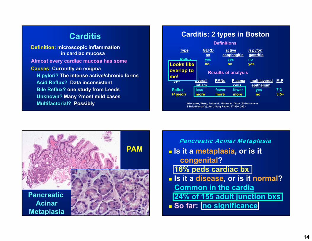
CarditisDefinition: microscopic inflammation
in cardiac mucosaAlmost every cardiac mucosa has someCauses: Currently an enigma
H pylori? The intense active/chronic formsAcid Reflux? Data inconsistentBile Reflux? one study from LeedsUnknown? Many ?most mild casesMultifactorial? Possibly
Carditis: 2 types in Boston
Type GERD active H pylorisx esophagitis gastritis
Reflux yes yes noH pylori no no yes
Type Overall PMNs Plasma multilayered M:Finflam cells epithelium
Reflux less fewer fewer yes 7:3H pylori more more more no 3:5+
Definitions
Results of analysis
Wieczorek, Wang, Antonioli, Glickman, Odze (BI-Deaconess& Brig-Woman's), Am J Surg Pathol, 27:960, 2003
Looks like overlap to me!
Pancreatic Acinar
Metaplasia
PAMPancreatic Acinar Metaplasia
Is it a metaplasia, or is it congenital?
16% peds cardiac bx Is it a disease, or is it normal?
Common in the cardia24% of 155 adult junction bxs
So far: no significance
15
Gobletcells
Gobletcells
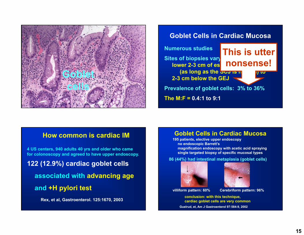
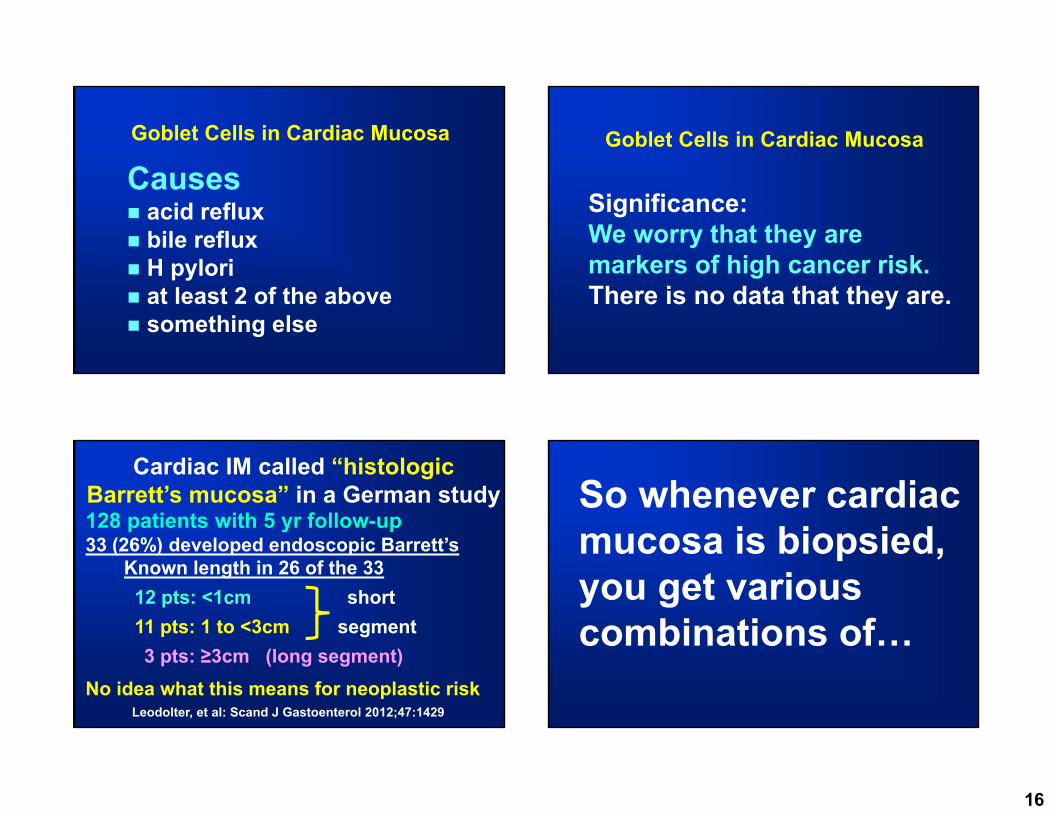
Goblet Cells in Cardiac Mucosa
Numerous studies
Sites of biopsies vary fromlower 2-3 cm of esophagus
(as long as the SCJ is normal) to 2-3 cm below the GEJ
Prevalence of goblet cells: 3% to 36%
The M:F = 0.4:1 to 9:1
This is utternonsense!
How common is cardiac IM
4 US centers, 940 adults 40 yrs and older who came for colonoscopy and agreed to have upper endoscopy.
122 (12.9%) cardiac goblet cells
associated with advancing age
and +H pylori test Rex, et al, Gastroenterol. 125:1670, 2003
Goblet Cells in Cardiac Mucosa195 patients, elective upper endoscopy
no endoscopic Barrett’smagnification endoscopy with acetic acid sprayingsingle targeted biopsy of specific mucosal types
conclusion: with this technique, cardiac goblet cells are very common
villiform pattern: 60% Cerebriform pattern: 96%
Guelrud, et, Am J Gastroenterol 97:584-9, 2002
86 (44%) had intestinal metaplasia (goblet cells)
16
Goblet Cells in Cardiac Mucosa
Causes acid reflux bile reflux H pylori at least 2 of the above something else
Goblet Cells in Cardiac Mucosa
Significance: We worry that they are markers of high cancer risk.There is no data that they are.
Cardiac IM called “histologic Barrett’s mucosa” in a German study128 patients with 5 yr follow-up 33 (26%) developed endoscopic Barrett’s
Known length in 26 of the 3312 pts: <1cm short11 pts: 1 to <3cm segment3 pts: ≥3cm (long segment)
No idea what this means for neoplastic riskLeodolter, et al: Scand J Gastoenterol 2012;47:1429
So whenever cardiac mucosa is biopsied, you get various combinations of…

17
InflammationGoblet
cells
PAM
Histologic features of cardia biopsies in volunteers
226 adults, mean age 45, 61% F, 49% Afr-Am
2 jumbo bx at or within 5mm of the SCJ (some may be too distal)
Cardia, defined simply as presence of mucus glands, found in 191 (85%)
Chronic carditis in 70%
Active carditis in 30%, all definitely or probably H pylori
Goblet cells in 15%; PAM in 13%El-Serag, et al, Scand J Gastroenterol, 42:1158-1166, 2007
#1: Dyspeptic adult woman not responding to medication (PPIs) has upper endoscopy.
The endoscopist saw erythemaat the GE junction. Nothing else.
The erythema was biopsiedLook what we got:
Inflammation
Pancreatic acinar cells
Goblet cells

18
Possible Diagnoseschronic carditis ± PAM ± IM of unknown etiologyorchronic carditis ± PAM ± IM due to _____ ( if you really believe you know) orno significant abnormality (since everyone has some, who cares?)
What do I do every day?
Before deciding, I polled my gastroenterologist colleagues to see what they wanted.
I asked them if they wanted to know if there was carditis, PAM and/or IM, and if so, which item would change their management of the patient.
They said they did not care about any of these items except for IM, which mightaffect management in certain circumstances.
My diagnosis (they want this):
Minute focus of IM at the GEJWhat should be the diagnosis in other institutions or practices?
This depends on what the GI colleagues want to know. The best way to find out is to ask them.
Then tell them what they want.

19
Summary
Cardias are smallCardias are often biopsied, so we see stuffInflammation is almost universal
The cause is unknownGoblet cells are common
The cause is unknownSignificance is minimal if that much
Pancreatic acinar cells are commonThe cause is unknown
Other than for cancer and dysplasia, almost everything else that we say about a cardiain our reports is meaningless!
Now that I have killed cardiac mucosa, what about the other part of this discussion, Barrett’s mucosa? This summarizes our approach to Barrett’s
mucosa, including the definition we use.
Am J Gastroenterol. 2008;103:788-797

20
Barrett’s Esophagus: DefinitionA change in the distal esophageal epithelium of any length that can be recognized as columnar type mucosa at endoscopy and is confirmed to have intestinal metaplasia by biopsy of the tubular esophagus
Wang, Sampliner and the ACG Practice Parameters Committee, Am J Gastro, 103:788, 2008
A change in the distal esophagealepitheliumof any length…..
Barrett’s definition
it is an esophagealdisease, not a GE junction disease!)
…..that can be recognized as columnar type mucosa atendoscopy(it is grossly, i e, endoscopicallyabnormal.)
Barrett’s definition
Tongues of pink mucosa
Barrett’s Esophagus: Definition
…..and is confirmed to have intestinal metaplasia by biopsy of the tubular esophagus.(IM means goblet cells.)

21
Goblet cells in columnar mucosa
A few basal mucous glands
TypicalBarrett’s Mucosa
Goblet cells are irrefutable evidence of metaplasia. This definition also avoids dealing with cardiac mucosain the distal esophagus.
Gastric mucosa with one type of intestinal metaplasia has an increased cancer riskMucosa without IM has no increased cancer risk.
The cancer rationale:
Esophageal mucosa with that same type of intestinal metaplasia has an increasedcancer riskMucosa without IM has no increased cancer risk.
The cancer supposition:

22
2014 Diagnosis of mucosal biopsies at or slightly above the GEJ
Histologic findings DiagnosisNo goblet cells No Barrett’s!!!!!Goblet cells
Tongues above the GEJ Barrett’sZ-line, no tongues Cardiac goblet cellsNot certain if tongues Not certain if Barrett’sNo information Not certain if Barrett’s
Endoscopic
Barrett’sGoblet cells
Alcian blue
H & E
ColumnarBlues
Alcian Blue
H & E
ColumnarBlues
Alcian BlueThese columnar cells with acid mucin are metaplastic cells, but they are not considered to be equal to goblet cells for diagnosis.

23
Barrett’s: other cell types
Paneth cells
Endocrine cells
Barrett’s mucosa is also commonly inflamed.No one seems to care!
Probably they just blame reflux.
How does the mucosa turn fromsquamous to columnar?
Squamous (normal) Columnar (Barrett’s)
Injury
Inflammation
Metaplasia (Barrett’s)
Dysplasia
Carcinoma
We assume that this is refluxate
Mediators > cellular
Why metaplasia? Squamous epithelium heals perfectly well.
We know a lot about the molecular and
genetic changes here
Barrett’s mucosa: theoretical progression

24
How does the mucosa turn fromsquamous to columnar?
gene gene genefactor factor
Barrett’s
Studies using cultures of esophageal squamesor mucosa found that acid and/or bile salts
up-regulate intestinal differentiation factors like CDX2 and CDX1,or up-regulate HB-EGF in lamina
propria fibroblasts that promotes CDX2, or stimulate BMP4 in stromal cells that promotes columnar cell keratins
CK7+ columnar cell keratin in squamous cells above Barrett’s In the laboratory, reflux type
substances induce changes in esophageal squamous cells that might precede intestinal metaplasia. We need to prove that these (or other) factors actually cause this metaplasia in vivo.

25
Columnar metaplasia may be an adaptation by the host to better withstand the chemical (acid and bile) injury.El-Omar and Jankowski, Am J Gastroenterol. 107:1342, 2012
Why do we need columnar mucosa? Barrett’s esophagus: putative precursors
Submucosalgland duct
Cardiacmucosa
Stem cells at squamous base
Barrett’s MucosaMultilayered epithelium
the proposed origin in Boston
Barrett’s mucosa has been separated into two types, based on segment length:Long segment (LSBE): 3 cm or moreShort segment (SSBE): less than 3 cmA less well recognized segment length has been called “ultrashort segment” (USSBE).
The definitions are not uniform. One definition uses less than 1 cm.
26
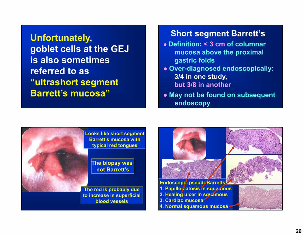
Unfortunately, goblet cells at the GEJ is also sometimes referred to as “ultrashort segment Barrett’s mucosa”
Short segment Barrett’s Definition: < 3 cm of columnar
mucosa above the proximal gastric folds
Over-diagnosed endoscopically:3/4 in one study, but 3/8 in another
May not be found on subsequent endoscopy
Looks like short segmentBarrett’s mucosa with
typical red tongues
The biopsy was The biopsy was not Barrett’s
The red is probably dueto increase in superficial
blood vessels
Endoscopic pseudoBarretts1. Papillomatosis in squamous2. Healing ulcer in squamous3. Cardiac mucosa4. Normal squamous mucosa

27
Barrett’s: squamous metaplasia(Pseudoregression)
Broad stretch
Squamous island
May be stimulated by PPIs: lead to decreasedendoscopic length and hidden stuff
What is hiding below the squamous metaplasia?
The Barrett’s onlyDysplasia
Carcinoma
Barrett’s mucosa is a high-risk cancer precursor, right?
So all this fuss is worthwhile, right?
Or is it?
We need to know 2 things
1. How common is
Barretts?
2. What really is the
cancer risk?

28
3 US studies: Prevalence of Barrett’s in Males Stratified by Age
Author # Age %Barr
Ward 161 65+ 22
Gerson 110 50+ 25
Rex 572 40+ 8
How common is Barrett’s in Sweden?1000 randomly selected people in 2 Swedish places underwent upper endoscopy. Mean age 53.5 yrs, 51% women
16 (only 1.6%) had Barrett’s, 5 long segment
400 had reflux sx: 2.3% had Barrett’s
600 had no reflux sx: 1.2% had Barrett’s
103 had endoscopic esophagitis: 2.6% had Barrett’s
897 had no endoscopic esophagitis: 1.4% had Barrett’s
Alcohol and smoking were independent risk factors
Ronkainen J, et al. Gastroenterol 129:1825, 2005
There seems to be a lot of Barrett’s mucosa in the USA in older men.
The Swedes have very little, but we don’t live there!
What really is the cancer risk?
Author Date Location #pts Cancer incidence
Spechler 2011 USA N/A 0.5%/yr estimate
Wani 2011 USA 1204 0.27%/yr
Bhat 2011 No Ire 8522 0.22%/yr****
Hvid-Jen 2012 Denmark 11028 0.12%/yr
***included both IM and non-IM, CA esoph and cardia
29
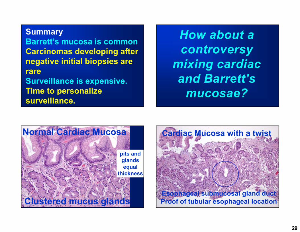
SummaryBarrett’s mucosa is commonCarcinomas developing after negative initial biopsies are rareSurveillance is expensive.Time to personalize surveillance.
Clustered mucus glands
Normal Cardiac Mucosa
pits andglands equal
thickness
Cardiac Mucosa with a twist
Esophageal submucosal gland ductProof of tubular esophageal location

30
Cardiac Mucosa in the tubular esophagus
Columnar lined lower esophagus
Gastric oxyntic mucosa
Submucosalglands, also proof of esophageal location
2006 British Society of Gastroenterology guidelines for the diagnosis and management of Barrett’s oesophagus (BO)BO is defined as an endoscopicallyapparent area above the OGJ that is suggestive of Barrett’s which is supported by the finding of columnar lined oesophagus on histology. …..IM…is not a requirement for diagnosis. (because sampling may miss IM)
Playford. Gut 55:442-3, 2006
….They suggest that IM not be required for the definition of BO…..

31
If these came from mucosae that looked like endoscopic Barrett’s
Then these would be Barrett’s in the UK
#2: Obese adult maleHeartburn for 20 years, recently worse Not responding to PPIs endoscopyGEJ tongues: “cannot tell if this is
an exaggerated Z-line or short segment Barrett’s”
Bx taken of the tonguesPathologist told (not asked)
to R/O Barrett’s
(The true request was to R/I Barrett’s)
Goblet cells
Chronic inflammation
Diagnosis:Cardio-esophageal junction, biopsy:
Columnar mucosa with goblet cellsMaybe add: either in the cardia or in short segment Barrett’s mucosa.
Comment:
If you can’t tell it is Barrett’s, neither can I!(with a reference to Wang and Sampliner or Spechler or Fitzgerald, if that seems necessary)
Even in the UK this is not Barrett’s because of the endoscopic uncertainty

32
Some people in the US and in a few other places want us to adopt the British definition for Barrett’s that doesn’t require goblet cells.
They have some data to support this
3 studies: cardiac mucosa without IM in the distal esophagus had CDX2, an intestinal differentiation marker, in some, but not all cases.
Phillips, et al, Am J Surg Pathol, 27:1442, 2003Groisman, et al, Mod Pathol, 17:1282, 2004 Shi, et al, Am J Clin Pathol, 129:571, 2008
A study of endoscopically confirmed columnar epithelium in the distal esophagus by image analysis:mucosa with IM and without (cardiac type) had similar DNA content changes.
Stomach No IM IM Liu, et al. Am J Gastroenterol 104:816, 2009

33
One study from Germany: 70% of 141 small (>2 cm) distal esophageal cancers treated by EMR were surrounded by cardiac mucosa, not mucosa with goblet cells.No IM anywhere in over half of the EMR specimens
Conclusion: no support for the view that Barrett adenocarcinoma is nearly always accompanied and preceded by IM.
Takubo, et al. Hum Pathol. 40:65, 2009
In contrast, Another study from U of Southern California of esophageal, EGJ and cardiac carcinomas:residual IM was found next to
52% of 33 tumors >4cm76% of 36 tumors <4cm
100% of 8 tumors ≤1cm92% of 26 tumors confined to the wallResidual IM was related to tumor size.
Chandrasoma, et al. Dis of the Esophagus. 20:36, 2007
Problems with these data: they are all retrospective
We want to know if non-IM mucosa needs surveillance. Specifically, does it have the same cancer risk as does IM mucosa AGA Institute Medical Position Panel
Spechler, et al. Gastroenterol 140:1084, 2011
The latest word from the US folks

34
Definition of Barrett’s Esophagus
“the condition in which any extent of metaplasticcolumnar epithelium that predisposes to cancerdevelopment replaces the stratified squamous epithelium……..
Definition of Barrett’s Esophagus
Presently, intestinal metaplasia is required for the diagnosis….. because intestinal metaplasia is the only type of esophageal columnar epithelium that clearly predisposes to malignancy…
“Although cardia-type epithelium might be a risk factor for malignancy, the magnitude of that risk remains unclear.”
“Based on this lack of data, it is justified not to perform endoscopic surveillance for patients solely with cardia-type epithelium…”
If these came from mucosae that looked like endoscopic Barrett’s
Then these would be Barrett’s in the UK as of 2006

35
The new BSO Barrett’s guidelines
Gut, 2014;63:7-42
BO: any portion of the normal distal squamous epithelial lining that has been replaced by metaplastic columnar epithelium, which is clearly visible endoscopically (≥1 cm) …. and is confirmed microscopically from biopsies…..
British Society of Gastroenterology guidelines 2014
Fitzgerald, et al. Gut. 2014 63:7-42
Old: Has both endo and histo requirementsNew: A minimum length is now defined.
If these came from mucosae that looked like endoscopic Barrett’s, ≥1 cm
then these would be still Barrett’s in the UK as of 2014
….The BSG suggests that IM not be required for the definition of BO, but it (the lack of IM) should be taken into account when deciding on the clinical management……

36
…..even though the insistence of the identification of IM to define or confirm a diagnosis of Barrett’s oesophagus is problematic, it is recognised that the inclusion of gastric-type mucosa in short tongues of columnar-lined oesophagus is of less clinical importance in terms of the likelihood of malignant transformation and has the potential to greatly influence the frequency of diagnosis of Barrett’s oesophagus at index endoscopy and the number of patients entering into follow-up and surveillance programmes.
Long discussion by the BSG summarized in the next slide
…..non-IM columnar mucosa has little cancer risk, and inclusion of it in the BO diagnosis will greatly increase the number of people on surveillance who don’t need it.
Decreasing the requirement for goblet cells would increase the diagnosis of BE by 147%.
Among patients with short columnar segments, 12% had goblet cells on subsequent endoscopy, so most of the columnar mucosa might represent proximal stomach.
No patient without goblet cells developed carcinoma.
Decreasing the requirement for goblet cells would cause many patients to be inaccurately labeled as BE.
U of Chicago study 2012: Westerhoff, et al. Clin Gastroenterol Hepatol 2012;10:1232–1236
Decreasing the requirement for goblet cells would increase the diagnosis of BE by 147%.
Among patients with short columnar segments, 12% had goblet cells on subsequent endoscopy, so most of the columnar mucosa might represent proximal stomach.
No patient without goblet cells developed carcinoma.
Decreasing the requirement for goblet cells would cause many patients to be inaccurately labeled as BE.
U of Chicago study 2012: Westerhoff, et al. Clin Gastroenterol Hepatol 2012;10:1232–1236 Sounds like a
waste of time, resouces and money to include these people!

37
What happens to people with non-IM columnar lined lower esophagus (CLE) over time? There is limited long-term follow-up data
U of Chicago study: 12% of CLE patients without IM developed goblet cells on F-U exam within 5.8 years.Westerhoff, et al. Clin Gastroenterol Hepatol 2012;10:1232–1236
Houston VA study: 29% of CLE patients without IM developed goblet cells on F-U exam within 2 yearsKhandwalla, et al. Am J Gastroenterol 2014;109:178-182
Does non-IM CLE have a cancer risk? There is very little data. U of Chicago study, 2012: No patient without IM developed carcinoma, over a mean F-U of 5.8 years. This is a small series, and 5.8 years is not long enough.
Northern Ireland study, 20118,522 Barrett's pts, mean 7 years FU
Incidence/yr of esoph/cardia AdCA
With IM 0.38%
Without IM 0.07%. Bhat, et al. JNCI, 2011;103:1049–1057
38
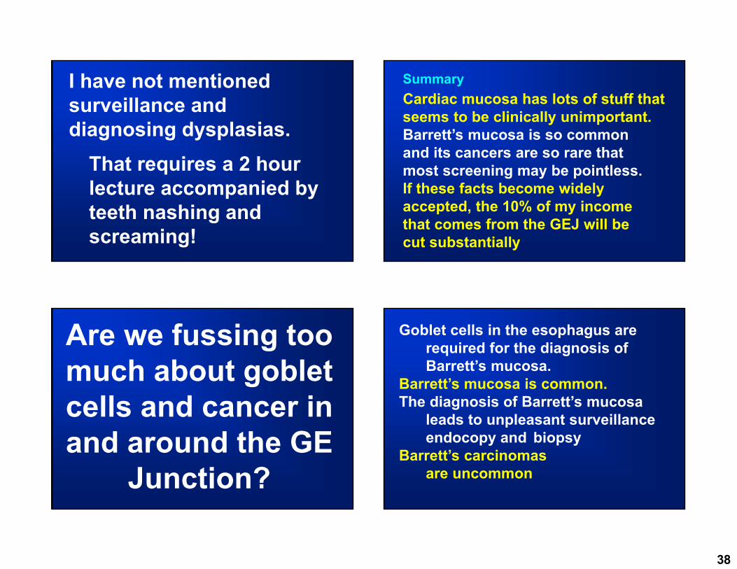
I have not mentioned surveillance and diagnosing dysplasias.
That requires a 2 hour lecture accompanied by teeth nashing and screaming!
Cardiac mucosa has lots of stuff that seems to be clinically unimportant.Barrett’s mucosa is so common and its cancers are so rare that most screening may be pointless.If these facts become widely accepted, the 10% of my income that comes from the GEJ will be cut substantially
Summary
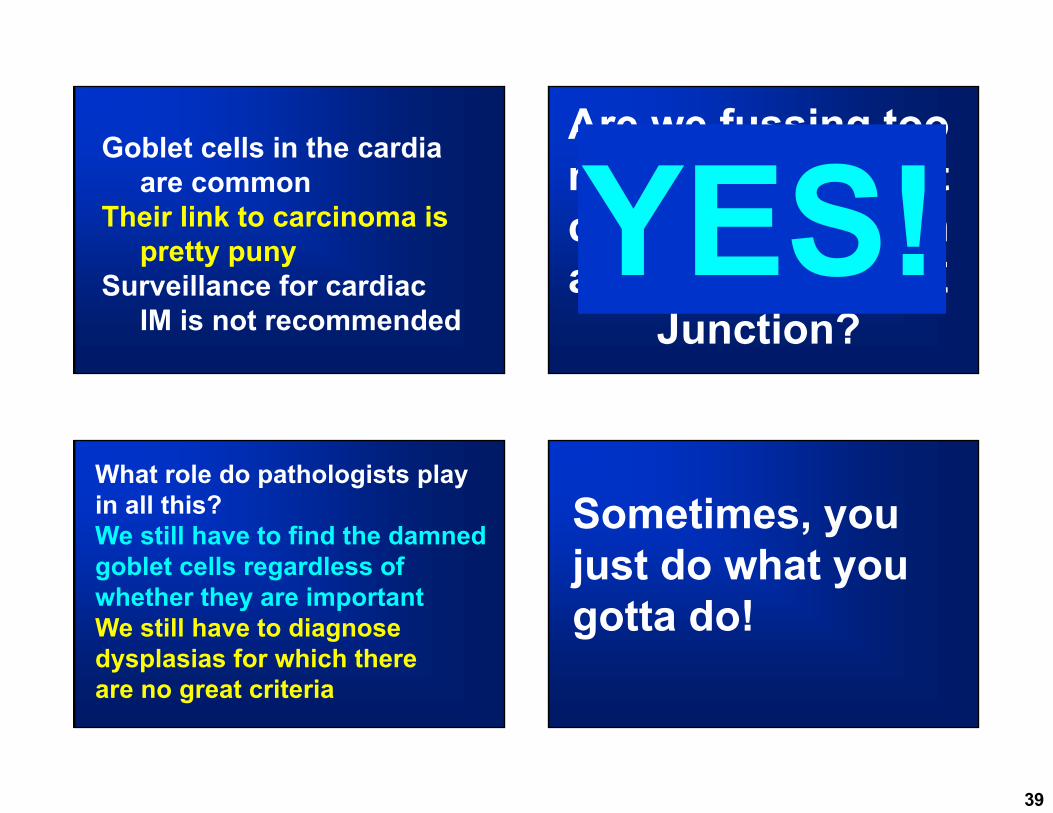
Are we fussing too much about goblet cells and cancer in and around the GE
Junction?
Goblet cells in the esophagus are required for the diagnosis of Barrett’s mucosa.
Barrett’s mucosa is common.The diagnosis of Barrett’s mucosa
leads to unpleasant surveillance endocopy and biopsy
Barrett’s carcinomas are uncommon
39
Goblet cells in the cardiaare common
Their link to carcinoma is pretty puny
Surveillance for cardiac IM is not recommended
Are we fussing too much about goblet cells and cancer in and around the GE
Junction?
What role do pathologists play in all this?We still have to find the damned goblet cells regardless of whether they are importantWe still have to diagnose dysplasias for which there are no great criteria
Sometimes, you just do what you gotta do!

40
It takes
To be a GI pathologist