controlling the luminescence of carboxyl-functionalized cdse/zns core–shell quantum dots in...
TRANSCRIPT

Controlling the Luminescence of Carboxyl-Functionalized CdSe/ZnSCore−Shell Quantum Dots in Solution by Binding with GoldNanorodsMonica Focsan,† Ana M. Gabudean,† Adriana Vulpoi,‡ and Simion Astilean*,†
†Nanobiophotonics and Laser Microspectroscopy Center, and ‡Nanostructured Materials and Bio-Nano-Interfaces Center,Interdisciplinary Research Institute in Bio-Nano-Sciences and Faculty of Physics, Babes-Bolyai University, 1 M. Kogalniceanu,400084, Cluj-Napoca, Romania
*S Supporting Information
ABSTRACT: Plasmonic nanostructures offer promisingroutes toward artificial control of the photoluminescenceproperties of various emitters. Here, we investigated thephotoluminescence of carboxyl-functionalized CdSe/ZnScore−shell quantum dots (c-QDs) localized near goldnanorods (AuNRs) as a function of c-QDs−AuNRs distanceusing the cetyltrimethylammonium bromide (CTAB) surfac-tant and Bovine Serum Albumin (BSA) protein layers overcoating metal surface as spacer. The direct binding ofnegatively charged c-QDs to positively charged CTAB (3−4nm thickness) caused close contact with the metal, resulting inan efficient metal-induced energy transfer (quenching). Wefound that quenching is modulated by the degree of spectraloverlap between the photoluminescence band of c-QDs (620nm) and longitudinal localized surface plasmon resonance (LSPR) of AuNRs (637 and 733 nm). Deposition of BSA layer overCTAB coated-AuNRs and subsequent decoration with c-QDs yielded an increase in photoluminescence signal when exciting inresonance with the transverse LSPR of AuNRs. On the basis of experimental studies using steady-state and time-resolvedfluorescence measurements as well as finite-difference time-domain calculations, we report over 70% quenching efficiency for allinvestigated AuNRs along with a 4.6-fold in photoluminescence enhancement relative to free c-QDs (39-fold enhancementrelative to c-QDs loaded AuNRs) after BSA deposition.
■ INTRODUCTIONIn the field of nanomaterials research, a key goal is to integratewithin the same nanosystem multiple functionalities in view ofbiosensing and bioimaging applications.1 Resonant couplingbetween luminescent semiconductor nanoparticles (quantumdots, QDs) and plasmonic metallic nanoparticles can generatenew remarkable optical effects, extending thus the applicationsfield of as-designed nanometer-scale hybrid structures. Becauseof their broad excitation spectra, size-tunable photolumines-cence emission spectra, and superior photostability againstphotobleaching, QDs are very appealing in practical biologicalapplications, especially for multiplexed labeling or multipleimmunoassays, as an alternative to ionic and molecularfluorophores.2 On the other hand, due to their unique opticalproperties related to their localized surface plasmon resonance(LSPR), gold nanoparticles (AuNPs) act as powerful nanoscaleoptical antennas,3 as they are able to significantly enhance lightabsorption or alter the radiative and nonradiative decay rates ofnearby located dipoles.4 In particular, luminescence enhance-ment occurs when the dominant relaxation pathway is radiativedecay, and vice versa, the luminescence is quenched when thenonradiative decay represents the dominant mechanism. For
instance, QDs were successfully exploited for metal-enhancedfluorescence (MEF),5,6 as well as for fluorescence resonanceenergy transfer.1,7
Furthermore, it has been already demonstrated that severalfactors influence the plasmon−exciton interaction such asdistance between QDs and metal surface, the excitationwavelength, the polarization of excitation, the size of QDs,the geometry of nanoparticles, and the spectral overlap betweenthe luminescence of QDs and the LSPR band of metalnanoparticles.8−10 The interplay between these factorsdetermines the magnitude of the luminescence enhancementor quenching of QDs. In fact, to control these above-mentionedparameters, a number of experimental methods have beendeveloped, including layer-by-layer (LBL) polyelectrolytedeposition technique,11 the utilization of hybrid metal@silica@QDs structures,12 or the utilization of biomolecules(e.g., DNA molecule, streptavidin, biotin) to adjust theinterparticle distance.13−15 However, despite several exper-
Received: February 5, 2014Revised: October 1, 2014
Article
pubs.acs.org/JPCC
© XXXX American Chemical Society A dx.doi.org/10.1021/jp501281v | J. Phys. Chem. C XXXX, XXX, XXX−XXX

imental and theoretical studies performed on solid substrates,MEF of QDs in aqueous solution has received less attention,especially using gold nanorods (AuNRs) as sensitiveamplification platform. This interest is essential for a clearunderstanding of the plasmon−exciton interaction in suchQDs@AuNPs hybrid assemblies in aqueous solution, openingthus the door to single molecule detection in biosciences16 aswell as photonic plasmonic devices and solar energy harvest-ing.17,18
Among various shapes of AuNPs, the nanorods areparticularly attractive due to the fine-tunability of their plasmonresonances across a wide spectral region, offering thus excellentcontrollable overlap with the emission of QDs, a key aspect forfurther multiplexed biodetection applications. While goldnanospheres present a well-defined plasmonic band in thevisible spectral region, AuNRs represent a unique class ofmetallic nanostructures with two surface plasmonic resonancesin the visible/near-infrared spectral range associated withoscillations of free electrons along the transverse andlongitudinal dimensions of the nanoparticles.19 Furthermore,due to their strong scattering and local-field enhancementsassociated with the LSPR excitation at the surface of thenanoparticle, AuNRs are an excellent candidate for theinvestigation of the luminescence enhancement mechanism.3
In this Article, we study the modification of carboxyl-functionalized CdSe/ZnS QDs (c-QDs) luminescence at 620nm in close vicinity of AuNRs in aqueous solution, as afunction of both the spectral overlap between the luminescenceof c-QDs and LSPR band of nanoparticles and the interparticledistance. In particular, two types of AuNRs were synthesizedutilizing the seed-mediated growth method to exhibitlongitudinal LSPR bands at 637 nm (AuNRs-637) and 733nm (AuNRs-733), respectively. First, we found that the directinteraction between the negatively charged c-QDs with thepositively charged cetyltrimethylammonium bromide (CTAB)bilayer that covers AuNRs surface induces a strongluminescence quenching of c-QDs with different efficiencyrate, ranging from 78% for AuNRs-733 to 91% for AuNPs-637,and a significant shortening of the exciton lifetime. The highestquenching efficiency obtained for AuNPs-637 (herein 91%) isdue to better spectral overlap of the longitudinal LSPR bandwith the luminescence band of c-QDs. Subsequently, with theaim to increase the spacer thickness, which will directly preventthe quenching mechanism, the surface of CTAB-coatedAuNRs-637 was first coated with Bovine Serum Albumin(BSA) protein and then decorated with c-QDs. BSA, a largemultidomain protein in plasma with an important role in
physiological functions,20 was selected in this study consideringits ability to easily attach to the surface of AuNRs throughelectrostatic interaction with the positively CTAB bilayer.21−23
The experimental optical data obtained on the attachment of c-QDs and BSA molecules to the CTAB coated-AuNRs-637surface were confirmed by simulations based on finite-difference time-domain (FDTD). As a consequence of theincreased spacer thickness, the plasmonic enhancement effectbecame dominant in our hybrid system made of AuNR-637,BSA, and c-QDs, leading to 4.6-fold luminescence enhance-ment relative to free c-QDs and 39-fold luminescenceenhancement relative to the corresponding case of c-QDs@AuNR system, with only slight shortening of the excitonlifetime.
■ EXPERIMENTAL METHODS
Reagents. Tetrachloroauric (III) acid, cetyltrimethylammo-nium bromide (CTAB), and ascorbic acid were purchased fromAldrich. Sodium borohydride (NaBH4, 99%) and silver nitrate(AgNO3) were obtained from Merck. Bovine Serum Albumin(BSA, 66 kDa) was purchased from Aldrich. Commercial core/shell CdSe/ZnS quantum dots (c-QDs) with a polyethyleneglycol (PEG) surface coating coupled to carboxyl terminalgroups were purchased from Evident Technologies Inc. (T2-MPEviTags). The concentration of c-QDs was 12 nmol/mL.All chemicals were used as received.
Samples Preparation. AuNRs of two different aspectratios were synthesized utilizing the seed-mediated growthmethod.19 Gold seeds were first prepared by mixing 1.25 mL of5 mM HAuCl4, 1.25 mL of 0.2 M CTAB, and 0.9 mL of freshlyprepared ice cold 1 mM NaBH4 solution under continuousstirring, resulting in a brownish yellow solution. The growthsolution was obtained by mixing 5 mL of 1 mM HAuCl4 with 5mL of 0.2 M CTAB and various amounts of 4 mM AgNO3.0.07 mL of 0.0788 M ascorbic acid was then added while gentlystirring. Ascorbic acid acts as a mild reducing agent and changesthe color of growth solution from dark yellow to colorless.Finally, 0.012 mL of seed solution was added into the as-prepared solution to start the growth of nanorods. The color ofthe solution gradually changed in the first 15−20 min beforefinally stabilizing. The obtained AuNRs were collected twice bycentrifugation to remove the excess CTAB surfactant and thendispersed in ultrapure water. Considering their longitudinalLSPR peak values at 637 and 733 nm, the as-prepared AuNRsin aqueous solution will be further referred to as AuNRs-637and AuNRs-733, respectively (see Figure 1, spectra a,b).
Figure 1. (a) Spectral overlap between the normalized luminescence of c-QDs (dot−dashed spectrum) and the extinction spectra of synthesizedAuNRs-637 (spectrum a) and AuNRs-733 (spectrum b), respectively; (b) representative TEM images of the synthesized AuNRs.
The Journal of Physical Chemistry C Article
dx.doi.org/10.1021/jp501281v | J. Phys. Chem. C XXXX, XXX, XXX−XXXB
The c-QDs are negatively charged (−23.8 mV, zeta-deviation= 0.3 at pH 7.6) in the PBS solution because of the carboxyl-functionalized groups surrounding the surface of the QDs,while the as-prepared AuNRs are positively charged due to theCTAB bilayer adsorbed on the surface of the nanorods (+ 40mV, zeta-deviation = 8). Therefore, when 2 nM c-QDs inphosphoric buffered saline (PBS, pH 7.6) are mixed withdifferent concentrations of the CTAB-coated AuNRs-637(stock concentration 1.47 × 10⁻⁹ M, as calculated from theabsorbance of longitudinal plasmon resonance for a molarextinction coefficient of ε = 3.13 × 10⁹ M−1 cm−124) andCTAB-coated AuNRs-733 (1.66 × 10⁻⁹ M, as calculated fromthe absorbance of longitudinal plasmon resonance for a molarextinction coefficient of ε = 3.6 × 10⁹ M−1 cm−124), the c-QDsinteracted with AuNRs due to electrostatic interaction.The attachment of BSA protein to CTAB-coated AuNRs-637
and the subsequent decoration with c-QD was performed intwo steps at pH ≈ 7.5. First, the albumin solution was freshlyprepared at room temperature by dissolving 1 mg of the solidBSA in 1 mL of ultrapure water at pH 4.7. Next, BSA@AuNRs-637 conjugates were prepared through electrostatic attractiveinteractions.21 BSA@AuNRs-637 system was centrifuged at8000 rpm for 10 min to remove the excess protein in thesolution. Second, the c-QDs decorated BSA@AuNRs-637 wereobtained by mixing the c-QDs (0.5 mL) and the BSA@AuNRs-637 conjugates (1 mL) for more than 3 h. The final solutionwas centrifuged to remove the unadsorbed c-QDs, and theobtained c-QDs decorated BSA@AuNRs-637 were redispersedin PBS.Experimental Measurements. The UV−vis−NIR absorp-
tion spectra were measured at room temperature using a JascoV-670 UV−vis−NIR spectrophotometer with a bandwidth of 2and 1 nm spectral resolution.Morphology and mean size of AuNRs were determined by
JEOL 100 U type transmission electron microscopy (TEM)operated at 100 kV accelerating voltage in which samples weredeposited on carbon-coated copper grids and dried at roomtemperature. The decoration of BSA@AuNRs-637 conjugateswith c-QDs was further confirmed by TEM images using a
Tecnai F20 field emission operated at 200 kV acceleratingvoltage and equipped with an Eagle 4 k CCD camera.The ζ-potential of the colloidal solutions was measured using
a particle analyzer (Nano ZS90 Zetasizer, Malvern Instru-ments) equipped with a He−Ne laser (633 nm, 5 mW) andusing a measurement angle of 90°. Each sample was measuredthree times, and the mean value was reported.Luminescence spectra were collected at room temperature
using a Jasco LP-6500 spectrofluorimeter equipped with axenon lamp as a light source. Luminescence quenchingexperiments were carried out in aqueous solution using anexcitation wavelength fixed at 375 nm. Luminescence spectrawere recorded in the wavelength range of 560−680 nm.Luminescence quenching was analyzed with values ofintegrated intensities computed over full spectral range ofluminescence bands for both free c-QDs and c-QDs in thepresence of increasing AuNRs concentration. For MEFexperiments, we used the 510 nm excitation wavelength withbandwidths of 1 nm in excitation and 3 nm in emission.Luminescence measurements were also performed to evaluatethe long-term stability of c-QDs in PBS solution.Fluorescence lifetime measurements were performed on a
PicoQuant MicroTime 200 time-resolved confocal fluorescencemicroscope system (Picoquant GmbH, Germany) based on aninverted microscope (IX 71, Olympus) equipped with aUPLSAPO 60×/NA = 1.2 water immersion objective. Theexcitation beam was provided by 0.55 μW picosecond diodelaser heads (LDH-D-C-375 and LDH-D-C-510, PicoQuant)operating at 375 and 510 nm and pulsed at 20 MHz repetitionrate. The samples were dropped on microscope cover glassesand analyzed in solution. The signal collected through theobjective was spatially and spectrally filtered by a 50 μmpinhole and long pass emission filters (HQ405LP andHQ519LP, Chroma, Brattleboro, U.S.), respectively, beforebeing focused on a photon counting detector module (PDM-series) Single Photon Avalanche Diode (SPAD) from MicroPhoton Devices (MPD). The detector signals were processedby the PicoHarp 300 Time-Correlated Single Photon Counting(TCSPC) data acquisition unit, from PicoQuant. Time-
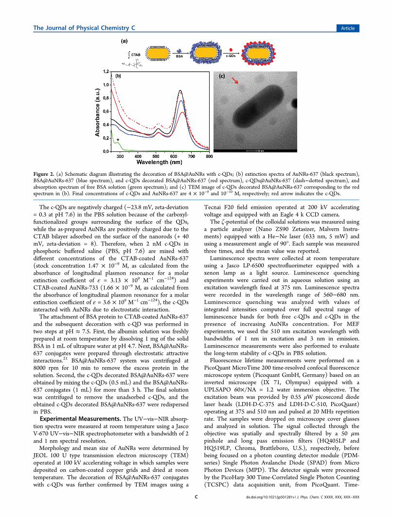
Figure 2. (a) Schematic diagram illustrating the decoration of BSA@AuNRs with c-QDs; (b) extinction spectra of AuNRs-637 (black spectrum),BSA@AuNRs-637 (blue spectrum), and c-QDs decorated BSA@AuNRs-637 (red spectrum), c-QDs@AuNRs-637 (dash−dotted spectrum), andabsorption spectrum of free BSA solution (green spectrum); and (c) TEM image of c-QDs decorated BSA@AuNRs-637 corresponding to the redspectrum in (b). Final concentrations of c-QDs and AuNRs-637 are 4 × 10⁻⁹ and 10−10 M, respectively; red arrow indicates the c-QDs.
The Journal of Physical Chemistry C Article
dx.doi.org/10.1021/jp501281v | J. Phys. Chem. C XXXX, XXX, XXX−XXXC
resolved fluorescence decay curves were recorded and analyzedusing the SymPhoTime software (version 1.6) provided byPicoQuant. The fluorescence lifetimes were obtained throughnonlinear iterative deconvolution algorithm. The instrumentresponse function (IRF) was recorded from the laser light backscattered from plain cover glass working in similar experimentalconditions (see Supporting Information Figure S1 for IRF oflaser 510 nm). The quality of the fits was judged by analyzingthe chi-square (χ2) values and the distribution of the residuals.
■ RESULTS AND DISCUSSIONConjugation of c-QDs to AuNRs. Figure 1a illustrates the
spectral relationship between the normalized extinction spectraof two as-synthesized AuNRs (solid spectra) and theluminescence spectrum of c-QDs (dot−dashed spectrum).The almost unmodified spectral position of transverse LSPRband at approximately 514 nm and the longitudinal LSPRbands at 637 and 733 nm (see Figure 1a, spectra a,b)correspond to AuNRs with aspect ratios of ∼2.1 (length/width:47 ± 4/22 ± 2 nm) and ∼3.2 (length/width: 38 ± 3/12 ± 1nm), respectively, obtained from analysis of TEM measure-ments (see Figure 1b for representative images).Note here that the luminescence emission band of the c-QDs
(620 nm) exhibits good spectral overlap with the longitudinalLSPR band of the AuNRs-637 and almost no overlap with thelongitudinal LSPR band of AuNRs-733. First, we wereinterested in examining the luminescence quenching of c-QDs when they are directly conjugated to CTAB-coatedAuNRs with two distinct longitudinal LSPR bands (i.e., 637 and733 nm) and to investigate the involved quenching mechanism.Second, to control the distance between the c-QDs and theAuNRs surface and avoid quenching, a layer of BSA protein wasdeposited onto the surface of CTAB-coated AuNRs beforeattaching c-QDs as shown schematically in Figure 2a andproved by TEM analysis in Figure 2c.The changes of the LSPR spectrum of AuNRs-637 after
conjugation with c-QDs and BSA protein were firstinvestigated. The synthesized AuNRs-637 exhibit in solutiona weak transverse LSPR band at 514 nm and a stronglongitudinal LSPR band at 637 nm (see Figure 2b, blackspectrum). The conjugation of CTAB-coated AuNRs with c-QDs (denoted as c-QDs@AuNPs-637) and BSA protein(denoted as BSA@AuNRs-637), respectively, is clearly provenby the modification induced in the longitudinal and transverseLSPR bands, the red-shifts of 3 and 1 nm in the first case(Figure 2b, dash−dotted spectrum) and 4 and 1 nm,respectively, in the second case (Figure 2b, blue spectrum).Such spectral modifications are commonly observed in the caseof LSPR sensors25 and can be assigned to the increase of thelocal refractive index relative to water. Thus, LSPR results provethe direct attachment of negatively charged c-QDs over theentire surface of positively charged CTAB bilayer wrappedaround AuNRs-637 through electrostatic interaction. Addition-ally, the adsorption of BSA molecules onto the surface ofCTAB-coated AuNRs-637 is also confirmed by the sensitiveblue-shift of the characteristic absorption band of free BSA insolution from 285 nm, marked with an asterisk in Figure 2b,green spectrum, due to π → π* transition of the aromaticamino acids residues,26 to 276 nm. This is clearly indicating theformation of bioconjugates (see Figure 2b, blue spectrum). Theadsorption of albumin on the surface of AuNRs-637 could leadto a partial unfolding of protein, causing conformationalchanges.27
Subsequently, more noticeable spectral modifications occurwhen negatively c-QDs interact with BSA@AuNRs-637 system.Figure 2b, red spectrum, depicts the extinction spectrum ofBSA@AuNRs-637 after conjugation with c-QDs. Specifically,red-shifts of the longitudinal and transverse LSPR bands with 7and 1 nm, respectively, were clearly identified, along with theirbroadening, indicating the presence of the c-QDs “corona” overthe entire surface of the BSA@AuNRs-637 system. Further-more, the presence of the c-QDs “corona” over the surface ofthe BSA@AuNRs system was clearly confirmed by TEM (see inFigure 2c; additional TEM images in Supporting InformationFigure S2). The LSPR measurements were simulated throughFDTD calculations on a single CTAB-coated AuNR, plainBSA-capped AuNR and decorated with c-QDs, using acommercially available FDTD solution software package fromLumerical Solutions, Inc. (Vancouver, Canada).28 We consid-ered a total field scattered field (TFSF) source with a wavevector k normal to the plan comprising the longitudinal axis ofthe nanorod and the incident electromagnetic field E0 polarizedalong the longitudinal axis. The computation volume wasdivided into two different regions, one including both incidentand scattered fields (total field) while the other including onlythe scattered field. The extinction spectra were calculated usinga 0.8 nm grid size. The nanoparticle was treated as asemispherical end-capped cylinder with 47 nm length and 22nm width, according to the average size obtained from TEMmeasurements and the position of the plasmon resonancesband at 637 nm. The structure was placed in an aqueoussurrounding media (n = 1.33). The dielectric dispersion of goldwas determined by best fitting the experimental data reportedin the literature.29 Considering that the CTAB layer is moredensely packed on the lateral facets of AuNR as compared to itsends, the coating CTAB shell was modeled as a 4 nm layer witha refractive index of 1.43530 wrapped around the cylinder, whilefor less densely packed layer from the ends we considered aneffective refractive index of 1.363 obtained for a combination of30% CTAB and 70% water. Furthermore, the compact layer ofBSA covering the AuNR surface was modeled as a 4 nmsemispherical shell with a refractive index of 1.445,31 under thehypothesis that the whole gold surface is covered by BSAmolecules. Consequently, the longitudinal LSPR band was red-shifted by 4 nm, in good agreement with the experimentalresults (see Supporting Information Figure S3, red spectrum).Finally, we attempted on simulating the decoration of BSA-coated CTAB-AuNR with c-QDs. We point out that the c-QDsused in our experiments consist of CdSe/ZnS core−shell dotsof approximately 3 nm radius32 functionalized with sparsecarboxylated polyethylene glycol chains. Considering the PEG-functionalized c-QDs as rigid spheres with radius of 6 nm,33 thenumber of c-QDs bound to one BSA@CTAB-AuNR of 7500nm2 total area, as obtained for a 47 nm length and 22 nm widthAuNR covered with 4 nm layers of CTAB and BSA, would bearound 50 for a full coverage with closely packing spheres.However, in reality the loading capacity of BSA@CTAB-AuNRis much lower as proved by the TEM picture in Figure 2c wherec-QDs are separated at distance above 6 nm, meaning less than50 c-QDs per particle. Such corona of c-QDs can be modeledin the first approximation as a continuous layer of dielectric of 6nm thickness with an effective refractive index of about 1.41(volume weighted value calculated from the refractive indices ofCdSe/ZnS core−shell dots and PEG layer, which are 2.433 and1.465, respectively, under the supposition of ∼33% coverageinferred from TEM analysis). As a consequence, we have
The Journal of Physical Chemistry C Article
dx.doi.org/10.1021/jp501281v | J. Phys. Chem. C XXXX, XXX, XXX−XXXD

obtained an additional red-shift of 8 nm (see SupportingInformation Figure S3, blue spectrum), rather close to theexperimental value (see Supporting Information Figure S3).Luminescence Quenching of c-QDs by AuNRs. The
effect of conjugation of c-QDs to as-synthesized nanoparticles(i.e., AuNRs-637 and AuNRs-733) on the luminescenceproperties was systematically investigated by steady-stateluminescence spectroscopy. It is worth mentioning that theluminescence spectrum of c-QDs was measured by using 375nm as excitation wavelength, excluding in this case theexcitation of metallic nanoparticles via its surface plasmonresonance, which occurs above 510 nm for AuNRs. Indeed,when a certain amount of c-QDs solution was mixed withCTAB-coated AuNRs solution, the luminescence intensitydecreased rapidly together with a slight blue-shift of theluminescence maximum, as compared to reference sample of c-QDs in the absence of the AuNRs (see Figure 3). A controlexperiment has been carried out to evaluate the long-termstability of free c-QDs in PBS solution for up to 20 days andfound no sign of luminescence quenching. This suggests thatthe c-QDs are not vulnerable to aggregation in theconcentration range of 1−2 nM in PBS solution (seeSupporting Information Figure S4). We then noticedinteresting behavior of c-QDs luminescence when mixed withthe CTAB molecules in water. For instance, at high CTABconcentration (10−4 M), c-QDs exhibit increased luminescencerelative to low concentration and free c-QDs in pure water,which can be explained by the fact that CTAB molecules at
high concentration form colloidal micelles, which in turn canengage the anionic c-QDs molecules and reduce the overall rateof nonradiative process. On the other hand, at low CTABconcentration, the luminescence decreases with respect to freec-QDs as in this case for c-QDs strongly interacting withcationic CTAB molecules the nonradiative deactivationincreases (data not shown here).34 It is therefore expectedthat similar quenching effect should contribute to overallquenching when c-QDs molecules bound to AuNRs via CTABlayer coating.More precisely, Figure 3 shows the evolution of lumines-
cence as result of mixing the same amount (2 nM) of c-QDswith increasing amounts of AuNRs (0.32 × 10⁻¹⁰ M; 0.6 ×10⁻¹⁰ M; 0.9 × 10⁻¹⁰ M; 1.2 × 10⁻¹⁰; 1.48 × 10⁻¹⁰ M; 1.74 ×10⁻¹⁰ M; and 2 × 10⁻¹⁰ M), which provide 63 to 10 c-QDs perone metallic nanoparticle. Given the number of PEG-functionalized c-QD per metallic nanoparticle for full coverage(34:1, as calculated for a AuNR of 47 nm length and 22 nmwidth, covered with a 4 nm CTAB layer, and 6 nm radius PEG-functionalized c-QDs) and relatively large QDs−QDs distance(see Figure 2c), the interaction between loaded and free QDscan be considered negligible and not affecting the evolution ofluminescence presented in Figure 3.In our case, the variation of the quenching efficiency
presented in Figure 3 originates from the degree of spectraloverlap between the longitudinal LSPR band of the AuNRs andthe luminescence band of the c-QDs. Experimentally, the
Figure 3. Evolution of luminescence spectra of c-QDs (2 nM) in the presence of increasing concentrations of (a) AuNRs-637 and (b) AuNRs-733,respectively. The nominal AuNRs concentrations from top to bottom are (a) 0, (b) 0.32 × 10⁻¹⁰ M, (c) 0.6 × 10⁻¹⁰ M, (d) 0.9 × 10⁻¹⁰ M, (e) 1.2 ×10⁻¹⁰ M, (f) 1.48 × 10⁻¹⁰ M, (g) 1.74 × 10⁻¹⁰ M, and (h) 2 × 10⁻¹⁰ M; λexcitation = 375 nm.
Figure 4. (a) Comparison of quenching efficiencies of AuNRs-637 and AuNRs-733; and (b) Stern−Volmer plots showing the luminescencequenching efficiencies of AuNRs-637 and AuNRs-733.
The Journal of Physical Chemistry C Article
dx.doi.org/10.1021/jp501281v | J. Phys. Chem. C XXXX, XXX, XXX−XXXE

quenching efficiencies (QE) can be determined from thesteady-state luminescence data:35
= −QEII
1 i
0 (1)
where Ii is the integrated luminescence intensity of the donor inthe presence of acceptor and I0 is the integrated luminescenceintensity of the donor alone.As shown in Figure 4a, the direct interaction of negatively
charged c-QDs with the positively charged AuNRs induces astrong quenching of c-QDs luminescence with differentefficiency rate, ranging from 78% for AuNRs-733 to 91% forAuNRs-637, as the concentration of nanoparticles reaches 2 ×10⁻¹⁰ M.Furthermore, the difference in luminescence quenching
efficiency is also clearly shown by linear Stern−Volmer (SV)plots, as indicated in Figure 3b, and quantified by the followingequation:35
= +FF
K q1 [ ]i
0sv
(2)
where F0 and Fi are the luminescence intensities in the absenceand in the presence of different concentration of quencher; Ksvis the SV quenching constant; and q is the concentration of thequencher, respectively. The calculated values of Ksv from SVplots are presented in Table 1.
In general, the rate of energy transfer is strongly dependenton several factors, such as the distance (d) of separationbetween the donor and proximal and the extent of spectraloverlap.35 For example, Forster resonance energy transfer(FRET), ascribed to 1/d6 dependence, is currently explored asa crucial tool for investigating different biological phenomenainvolving energy transfer. Li et al. reported that the energy
transfer efficiency of CdSe/ZnS QDs followed the FRETmechanism and that the highest quenching efficiency value wasobtained for 80 nm sized acceptor gold nanospheres, due toincreased spectral overlap of the plasmonic band with theluminescence band of QDs.36
Surface energy transfer (SET) model has been recentlyreported to investigate the quenching effects of QDs or dyemolecules.37−39 Reineck et al. determined, both theoreticallyand experimentally, the distance and wavelength dependence ofthe electromagnetic coupling between Au@SiO2 core−shellnanoparticles and nearby fluorophores.37 Strouse’s groupdemonstrated that SET is able to measure intermoleculardistances up to 50 nm, depending on the size of the metallicnanoparticles involved.40,41 For SET model, the quenching isinversely proportional to the fourth power of the distancebetween particles in analogy to the six power distancedependence that governs FRET formalism. More recently,Samanta et al. demonstrated that the quenching of QDsfluorescence is due to an increase in the nonradiative decay ratethat is induced by the presence of 30 nm AuNPs. They foundthat, unlike FRET, the energy transfer is inversely proportionalto the 2.7th power of the distance between particles.42
Herein, the observed quenching efficiency is attributed to acombination of factors such as (i) the large extinctioncoefficient of AuNRs, (ii) the overlap between theluminescence band of the c-QDs and the longitudinal LSPRband of AuNRs, and (iii) the close vicinity between c-QDs andCTAB-coated AuNRs surface. Additionally, we assume that thequenching of c-QDs induced by CTAB might also contribute tooverall quenching of c-QDs molecules bound to AuNRs viaCTAB layer coating. It should be mentioned that, in general,the energy transfer is most efficient when the distance betweendonors and acceptors is in the 20−60 Å range.36 This conditionis also fulfilled in our system taking into consideration that theCTAB bilayer covering the AuNRs surface plays the role ofspacer by fixing the c-QDs at about 4 nm far from the AuNRssurface. Similar quenching effects of dye molecules on goldnanoparticles have been previously reported by Reineck et al.when the distance is less than 5 nm,37 which is in agreementwith our observations.
Luminescence Enhancement of c-QDs by AuNRs. Theresonant excitation of the surface plasmons of the metallic
Table 1. Calculated Stern Volmer quenching constants (Ksv)
AuNRs Ksv (M−1) SDa Rb
AuNRs-637 3.99 × 10¹⁰ 0.326 0.992AuNRs-733 1.55 × 10¹⁰ 0.057 0.998
aSD is the standard deviation of the Ksv values.bR is the correlation
coefficient.
Figure 5. (a) Luminescence spectra of c-QDs (dashed spectrum), c-QDs@BSA system (dotted spectrum), and c-QDs decorated BSA@AuNRs-637(red spectrum). λexcitation = 510 nm. (b) Normalized fluorescence lifetime decay curves of c-QDs (i), c-QDs decorated BSA@AuNRs-637 (ii), and c-QDs@AuNRs-637 (iii) obtained at λexcitation = 375 nm and λexcitation = 510 nm, respectively. Inset: Comparison between the position of the excitationwavelength and the optical response of AuNRs-637 and c-QDs.
The Journal of Physical Chemistry C Article
dx.doi.org/10.1021/jp501281v | J. Phys. Chem. C XXXX, XXX, XXX−XXXF

nanoparticles leads to the enhancement of the electromagneticfield at the nanoparticles surface, which can subsequentlyinfluence the luminescence rates of the molecules situated intheir close proximity. We previously demonstrated that when c-QDs are directly adsorbed onto the CTAB-coated AuNRsurface, the luminescence is quenched. The degree ofquenching is depending on the spectral overlap between thelongitudinal LSPR band of AuNRs and luminescence spectrumof c-QDs. However, it is known that at a distance of a fewnanometers from the AuNR surface,35 the luminescence of theQDs can be strongly enhanced by the means of severalmechanisms, as the increase of excitation rate in the enhancedlocal electromagnetic field, increase of radiative lifetime of QDsby plasmon−exciton coupling in the near field, as well as byinteraction of free space photons by scattering processes.We note that the intensity of the c-QDs luminescence,
located at 620 nm (Figure 5a, dashed spectrum), decreaseswhile the band maximum blue-shifts with 5 nm after interactionof c-QDs with albumin (Figure 5a, dotted spectrum). This shiftcan be attributed to a change of the electronic structure of thec-QDs coated with protein.43 Finally, when BSA protein acts asspacer layer to prevent the quenching mechanism, our c-QDsdecorated BSA@AuNRs-637 system exhibits up to a 4.6-foldluminescence enhancement as compared to free c-QDs (Figure5a, red spectrum) and a 39-fold luminescence enhancementrelative to the corresponding case of c-QDs@AuNR system,under the excitation with the 510 nm wavelength. Theseenhanced values of the luminescence are attributed to the effectof local field excitation due to surface plasmon resonance. It isworth mentioning that all luminescence spectra were collectedunder identical excitation and detection conditions. On thecontrary, under 375 nm excitation, we obtained 1.1-foldluminescence enhancement, as Supporting Information FigureS6 shows. This amplification factor is well correlated with theFDTD evaluation of the local field exhibited by AuNRs-637 atapproximately 7−8 nm from the metal surface at 375 nm,where c-QDs are located, revealing an approximately 2.5-timessmaller field at 375 nm as compared to 510 nm (SupportingInformation Figure S5).To get a better insight into the mechanisms involved in the
enhancement of the luminescence of c-QDs when conjugatedto BSA@AuNRs-637 and to confirm our previous findings, weinvestigated the changes in c-QDs excited-state lifetime andperformed fluorescence lifetime measurements on free c-QDsand c-QDs conjugated to AuNRs-637 in the absence andpresence of BSA. The decay curves, obtained for the threesystems at the excitation of 375 and 510 nm, show twoexponential components after deconvolution of the instrumentresponse function (see Figure 5b). The decay fitting parametersare listed in Table 2.
The fluorescence lifetime of free c-QDs shows abiexponential behavior having an average value of 10.3 ns ascalculated according to the formula:
ττ
⟨ ⟩ =∑ *∑
AA
i i
i (3)
where ⟨τ⟩ is the average fluorescence lifetime and τi and Airepresent the decay time and amplitude, respectively, of eachcomponent (see Table 2). The measured lifetime correspondswell to the previously reported value.44 At 510 nm excitation, inthe presence of AuNRs-637, the average fluorescence lifetime ofc-QDs strongly decreases to 5.7 ns as compared to 10.3 nscorresponding to the free c-QDs. The shortening of the lifetimeof c-QDs confirms the efficient energy transfer45 in our donor−acceptor system, in accordance with the previous resultsobtained on the quenching of the luminescence intensity fromour steady-state fluorescence measurements. Specifically, thesignificant overlap between the emission of the c-QDs and theplasmon resonance of AuNRs-637 (see inset of Figure 5b)favors a resonance energy transfer from c-QDs to AuNRs-637.Additionally, the <5 nm layer of CTAB30 stabilizing theAuNRs-637 promotes a strong interaction between c-QDs andthe metallic surface, thus supporting the quenching of c-QDsfluorescence by AuNRs-637 via nonradiative energy transfer. Aspreviously demonstrated, the larger is the overlapping betweenthe longitudinal LSPR and the emission bands, the greater isthe quenching. Thus, the significant decrease of the excited-state lifetime is well correlated with the results obtained fromsteady-state fluorescence measurements. On the contrary,under excitation at 375 nm, out of the plasmon resonances ofAuNRs we obtain a value of 7 ns.Furthermore, we have focused on the c-QDs decorated
BSA@AuNRs-637 system with enhanced fluorescence proper-ties, and we have investigated the excited-state lifetime underthe same excitation at 510 nm. As illustrated in Figure 5b, curveii, the decay curve displays a slight decrease as compared tobare c-QDs, the average lifetime being calculated at 8.3 ns. Inthis case, the presence of a BSA shell around AuNRs-637surface helps in preventing the process of energy transfer fromc-QDs to Au surface, which was already mentioned as being thedominating quenching effect. A similar behavior has beenreported in an early paper of Medintz et al., who developed aFRET-based prototype QDs sensor assembly.46 They haveshown that overcoming the FRET mechanism between theQDs as donor and a fusion protein as acceptor results in asignificant recovery of the donor lifetime. Additionally, weassume a competition between the decrease of the nonradiativedecay rate due to a possible intercalation of c-QDs in the BSAlayer and the increase of the radiative decay rate as aconsequence of the matching of plasmon band with theluminescence of c-QDs.
Table 2. Decay Parameters of Free and Conjugated c-QDs
excitation (nm) samplea τ1 [ns]b A1 [%] τ2 [ns] A2 [%] ⟨τ⟩ [ns] χ2c
375 A 15.2 ± 0.1 60.5 ± 2 2.74 ± 0.07 39.5 ± 1.5 10.3 1.2B 13.35 ± 0.15 44 ± 1 2.1 ± 0.1 56 ± 1 7 1.5C 14 ± 0.1 52.5 ± 1.5 2.6 ± 0.03 47.5 ± 1.2 8.6 1.2
510 A 15.3 ± 0.3 60.5 ± 1.5 2.71 ± 0.08 39.5 ± 1.5 10.3 1.15B 11.35 ± 0.4 42 ± 1 1.57 ± 0.04 58 ± 1.5 5.7 1.2C 13.5 ± 0.3 53 ± 2.5 2.5 ± 0.06 47 ± 2 8.3 1.11
aA = c-QDs; B = c-QDs@AuNRs-637; C = c-QDs@BSA@AuNRs-637. bτn and An are lifetime and amplitude of the nth component; ⟨τ⟩ is theamplitude-weighted average lifetime. cχ2 indicates the goodness of the fit (1 corresponds to a perfect fit).
The Journal of Physical Chemistry C Article
dx.doi.org/10.1021/jp501281v | J. Phys. Chem. C XXXX, XXX, XXX−XXXG
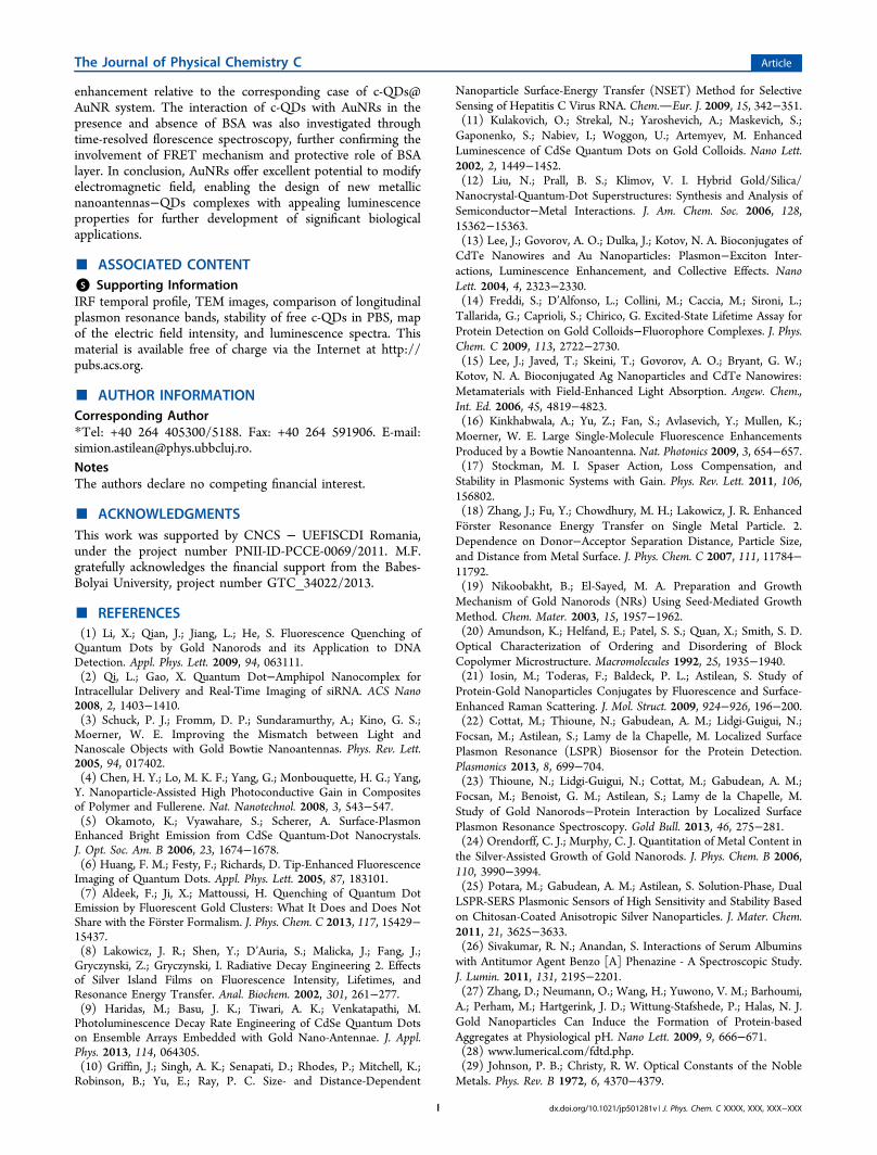
Subsequently, we were interested in investigating the energytransfer between c-QDs and AuNRs-637 in the absence andpresence of BSA because it is known that the nonradiativeenergy transfer between two dipoles is related to the donor−acceptor (here c-QDs−AuNR) separation, as we havepreviously discussed. The general formula known to describethis dependence is47
=+
Ed d1
1 ( / )p0 (4)
where E is the nonradiative energy transfer, d is the separationbetween the donor and the acceptor, d0 is the separation atwhich the energy transfer efficiency is 50%, and p is dependenton the mechanism of energy transfer from the donor to theacceptor. In FRET mechanism, p = 6, while d0FRET can bedescribed as48
λ= Φ −d k n J0.211[ ( )]0FRET2
04 1/6
(5)
where k2 describes the relative orientation in space of thetransition dipoles of the donor and the acceptor and is taken tobe 2/3 assuming rapid orientational averaging of the donorwithin the lifetime of its excited state, Φ0 is the quantum yieldof donor fluorescence without acceptor (here 0.77 for 620 nmemitting c-QDs), n is the refractive index of the medium (here1.426, corresponding to the volume weighted value of CTABand BSA layers employed in the FDTD simulations), and J(λ)is the overlap integral between the normalized donor emissionand the acceptor extinction coefficient, expressed as follows:
∫λ λ ε λ λ= λ∞
J F( ) ( ) ( ) d0
D A4
(6)
where FD(λ) is the fluorescence intensity of the donor in theabsence of acceptor (the integral of the spectrum equals one),and εA(λ) is the wavelength-dependent molar extinctioncoefficient of the acceptor. For the SET mechanism, p = 4,and d0SET can be obtained with49
ω ω=
Φ‐
‐
⎡⎣⎢⎢
⎤⎦⎥⎥d
kcn
0.2251
0SETc QDs
c QDs2
F F
31/4
(7)
where c is the speed of light in a vacuum, n is the refractiveindex of the medium (1.426), Φc‑QDs is the fluorescencequantum yield of donor, here c-QDs, ωc‑QDs is the angularfrequency for the donor (3.04 × 10¹⁵ s−1 for 620 nm emitting c-QDs), while ωF and kF represent the angular frequency of themetal (8.4 × 10¹⁵ s−1, bulk gold) and the Fermi wave vector ofmetal (1.2 × 108 cm−1, bulk gold), respectively.To understand the origin of the energy transfer between c-
QDs and AuNRs, we have plotted the theoretical energytransfer efficiency curves based on FRET and SET mechanisms(see Figure 6) along with the FDTD simulated quenchingefficiencies at different separation distances between c-QDs andthe metal surface of AuNRs with LSPR at 637 nm, respectively.In FDTD simulations, we considered the emitting c-QD as
an oscillating dipole radiating in the interval 530−690 nm,placed at different distances from a 47 × 22 nm AuNRexhibiting longitudinal LSPR at 637 nm (AuNR-637), along itsshort axis and polarized along its longitudinal axis. Thenonradiative rates were calculated for distances d between 2and 18 nm and normalized with respect to the free space case.The value of the overlap integral in our case was found to be3.2 × 1020 M−1 cm−1 nm4, while for d0FRET and d0SET we found
4 and 7.7 nm, respectively. To elucidate the quenchingmechanism in our system, we have investigated the positionof the experimental quenching efficiencies obtained for c-QDsconjugated to AuNRs in the presence and the absence of BSArelative to the theoretical curves of the quenching efficiencybased on the FRET and SET models (see Figure 6). We firstcalculated the quenching efficiencies (QE) of c-QDs coupled toAuNRs-637 and BSA@AuNRs-637 according to the formula:
ττ
= −QE 10 (8)
where τ0 represents the lifetime of free c-QDs (10.3 ns) and τrepresents the lifetime of c-QDs@AuNR-637 (5.7 ns) and c-QDs@BSA@AuNRs-637 (8.3 ns), as presented in Table 2.Figure 6 demonstrates that the FRET model provides a
better fit to the experimental data, this result being expectedconsidering the spectral overlap between the luminescence of c-QDs and the longitudinal LSPR band of AuNRs-637.
■ CONCLUSIONSIn summary, the quenching mechanism between c-QDs andAuNRs of different aspect ratios in aqueous solution was firstinvestigated. We found that the luminescence quenchingefficiency depends on the spectral overlap between theluminescence of c-QDs and the longitudinal LSPR band.Thus, the highest quenching efficiency was obtained for theAuNRs with the longitudinal LSPR located at 637 nm, bestoverlapping the luminescence band of c-QDs. Additionally, theshortening of the excited-state lifetime of c-QDs in the presenceof AuNPs-637 sustains the hypothesis of the energy transfer.We have demonstrated that the quenching of the luminescenceof c-QDs can be successfully avoided by coating the AuNRs-637 with BSA protein prior to decoration with c-QDs byexcitation of metallic nanoparticles via its surface plasmonresonance, which occurs above 510 nm for AuNRs. Theexperimental LSPR response of AuNRs in the presence of BSAand c-QDs molecules was confirmed at the theoretical level byFDTD calculations. By playing the role of both linker andspacer for keeping the c-QDs at a favorable distance from theAuNRs, BSA triggers a 4.6-fold enhancement of theluminescence of free c-QDs and a 39-fold luminescence
Figure 6. Simulated data points of the quenching efficiency at differentseparation distances between c-QDs and AuNR-637 (■) metalsurface, along with the experimental data points obtained for c-QDsconjugated to AuNRs-637 in the presence and absence of BSA (red●) and theoretical curves of the quenching efficiency based on theFRET (red curve) and SET (black curve) models.
The Journal of Physical Chemistry C Article
dx.doi.org/10.1021/jp501281v | J. Phys. Chem. C XXXX, XXX, XXX−XXXH
enhancement relative to the corresponding case of c-QDs@AuNR system. The interaction of c-QDs with AuNRs in thepresence and absence of BSA was also investigated throughtime-resolved florescence spectroscopy, further confirming theinvolvement of FRET mechanism and protective role of BSAlayer. In conclusion, AuNRs offer excellent potential to modifyelectromagnetic field, enabling the design of new metallicnanoantennas−QDs complexes with appealing luminescenceproperties for further development of significant biologicalapplications.
■ ASSOCIATED CONTENT*S Supporting InformationIRF temporal profile, TEM images, comparison of longitudinalplasmon resonance bands, stability of free c-QDs in PBS, mapof the electric field intensity, and luminescence spectra. Thismaterial is available free of charge via the Internet at http://pubs.acs.org.
■ AUTHOR INFORMATIONCorresponding Author*Tel: +40 264 405300/5188. Fax: +40 264 591906. E-mail:[email protected] authors declare no competing financial interest.
■ ACKNOWLEDGMENTSThis work was supported by CNCS − UEFISCDI Romania,under the project number PNII-ID-PCCE-0069/2011. M.F.gratefully acknowledges the financial support from the Babes-Bolyai University, project number GTC_34022/2013.
■ REFERENCES(1) Li, X.; Qian, J.; Jiang, L.; He, S. Fluorescence Quenching ofQuantum Dots by Gold Nanorods and its Application to DNADetection. Appl. Phys. Lett. 2009, 94, 063111.(2) Qi, L.; Gao, X. Quantum Dot−Amphipol Nanocomplex forIntracellular Delivery and Real-Time Imaging of siRNA. ACS Nano2008, 2, 1403−1410.(3) Schuck, P. J.; Fromm, D. P.; Sundaramurthy, A.; Kino, G. S.;Moerner, W. E. Improving the Mismatch between Light andNanoscale Objects with Gold Bowtie Nanoantennas. Phys. Rev. Lett.2005, 94, 017402.(4) Chen, H. Y.; Lo, M. K. F.; Yang, G.; Monbouquette, H. G.; Yang,Y. Nanoparticle-Assisted High Photoconductive Gain in Compositesof Polymer and Fullerene. Nat. Nanotechnol. 2008, 3, 543−547.(5) Okamoto, K.; Vyawahare, S.; Scherer, A. Surface-PlasmonEnhanced Bright Emission from CdSe Quantum-Dot Nanocrystals.J. Opt. Soc. Am. B 2006, 23, 1674−1678.(6) Huang, F. M.; Festy, F.; Richards, D. Tip-Enhanced FluorescenceImaging of Quantum Dots. Appl. Phys. Lett. 2005, 87, 183101.(7) Aldeek, F.; Ji, X.; Mattoussi, H. Quenching of Quantum DotEmission by Fluorescent Gold Clusters: What It Does and Does NotShare with the Forster Formalism. J. Phys. Chem. C 2013, 117, 15429−15437.(8) Lakowicz, J. R.; Shen, Y.; D’Auria, S.; Malicka, J.; Fang, J.;Gryczynski, Z.; Gryczynski, I. Radiative Decay Engineering 2. Effectsof Silver Island Films on Fluorescence Intensity, Lifetimes, andResonance Energy Transfer. Anal. Biochem. 2002, 301, 261−277.(9) Haridas, M.; Basu, J. K.; Tiwari, A. K.; Venkatapathi, M.Photoluminescence Decay Rate Engineering of CdSe Quantum Dotson Ensemble Arrays Embedded with Gold Nano-Antennae. J. Appl.Phys. 2013, 114, 064305.(10) Griffin, J.; Singh, A. K.; Senapati, D.; Rhodes, P.; Mitchell, K.;Robinson, B.; Yu, E.; Ray, P. C. Size- and Distance-Dependent
Nanoparticle Surface-Energy Transfer (NSET) Method for SelectiveSensing of Hepatitis C Virus RNA. Chem.Eur. J. 2009, 15, 342−351.(11) Kulakovich, O.; Strekal, N.; Yaroshevich, A.; Maskevich, S.;Gaponenko, S.; Nabiev, I.; Woggon, U.; Artemyev, M. EnhancedLuminescence of CdSe Quantum Dots on Gold Colloids. Nano Lett.2002, 2, 1449−1452.(12) Liu, N.; Prall, B. S.; Klimov, V. I. Hybrid Gold/Silica/Nanocrystal-Quantum-Dot Superstructures: Synthesis and Analysis ofSemiconductor−Metal Interactions. J. Am. Chem. Soc. 2006, 128,15362−15363.(13) Lee, J.; Govorov, A. O.; Dulka, J.; Kotov, N. A. Bioconjugates ofCdTe Nanowires and Au Nanoparticles: Plasmon−Exciton Inter-actions, Luminescence Enhancement, and Collective Effects. NanoLett. 2004, 4, 2323−2330.(14) Freddi, S.; D’Alfonso, L.; Collini, M.; Caccia, M.; Sironi, L.;Tallarida, G.; Caprioli, S.; Chirico, G. Excited-State Lifetime Assay forProtein Detection on Gold Colloids−Fluorophore Complexes. J. Phys.Chem. C 2009, 113, 2722−2730.(15) Lee, J.; Javed, T.; Skeini, T.; Govorov, A. O.; Bryant, G. W.;Kotov, N. A. Bioconjugated Ag Nanoparticles and CdTe Nanowires:Metamaterials with Field-Enhanced Light Absorption. Angew. Chem.,Int. Ed. 2006, 45, 4819−4823.(16) Kinkhabwala, A.; Yu, Z.; Fan, S.; Avlasevich, Y.; Mullen, K.;Moerner, W. E. Large Single-Molecule Fluorescence EnhancementsProduced by a Bowtie Nanoantenna. Nat. Photonics 2009, 3, 654−657.(17) Stockman, M. I. Spaser Action, Loss Compensation, andStability in Plasmonic Systems with Gain. Phys. Rev. Lett. 2011, 106,156802.(18) Zhang, J.; Fu, Y.; Chowdhury, M. H.; Lakowicz, J. R. EnhancedForster Resonance Energy Transfer on Single Metal Particle. 2.Dependence on Donor−Acceptor Separation Distance, Particle Size,and Distance from Metal Surface. J. Phys. Chem. C 2007, 111, 11784−11792.(19) Nikoobakht, B.; El-Sayed, M. A. Preparation and GrowthMechanism of Gold Nanorods (NRs) Using Seed-Mediated GrowthMethod. Chem. Mater. 2003, 15, 1957−1962.(20) Amundson, K.; Helfand, E.; Patel, S. S.; Quan, X.; Smith, S. D.Optical Characterization of Ordering and Disordering of BlockCopolymer Microstructure. Macromolecules 1992, 25, 1935−1940.(21) Iosin, M.; Toderas, F.; Baldeck, P. L.; Astilean, S. Study ofProtein-Gold Nanoparticles Conjugates by Fluorescence and Surface-Enhanced Raman Scattering. J. Mol. Struct. 2009, 924−926, 196−200.(22) Cottat, M.; Thioune, N.; Gabudean, A. M.; Lidgi-Guigui, N.;Focsan, M.; Astilean, S.; Lamy de la Chapelle, M. Localized SurfacePlasmon Resonance (LSPR) Biosensor for the Protein Detection.Plasmonics 2013, 8, 699−704.(23) Thioune, N.; Lidgi-Guigui, N.; Cottat, M.; Gabudean, A. M.;Focsan, M.; Benoist, G. M.; Astilean, S.; Lamy de la Chapelle, M.Study of Gold Nanorods−Protein Interaction by Localized SurfacePlasmon Resonance Spectroscopy. Gold Bull. 2013, 46, 275−281.(24) Orendorff, C. J.; Murphy, C. J. Quantitation of Metal Content inthe Silver-Assisted Growth of Gold Nanorods. J. Phys. Chem. B 2006,110, 3990−3994.(25) Potara, M.; Gabudean, A. M.; Astilean, S. Solution-Phase, DualLSPR-SERS Plasmonic Sensors of High Sensitivity and Stability Basedon Chitosan-Coated Anisotropic Silver Nanoparticles. J. Mater. Chem.2011, 21, 3625−3633.(26) Sivakumar, R. N.; Anandan, S. Interactions of Serum Albuminswith Antitumor Agent Benzo [A] Phenazine - A Spectroscopic Study.J. Lumin. 2011, 131, 2195−2201.(27) Zhang, D.; Neumann, O.; Wang, H.; Yuwono, V. M.; Barhoumi,A.; Perham, M.; Hartgerink, J. D.; Wittung-Stafshede, P.; Halas, N. J.Gold Nanoparticles Can Induce the Formation of Protein-basedAggregates at Physiological pH. Nano Lett. 2009, 9, 666−671.(28) www.lumerical.com/fdtd.php.(29) Johnson, P. B.; Christy, R. W. Optical Constants of the NobleMetals. Phys. Rev. B 1972, 6, 4370−4379.
The Journal of Physical Chemistry C Article
dx.doi.org/10.1021/jp501281v | J. Phys. Chem. C XXXX, XXX, XXX−XXXI

(30) Yu, C.; Varghese, L.; Irudayaraj, J. Surface Modification ofCetyltrimethylammonium Bromide-Capped Gold Nanorods to MakeMolecular Probes. Langmuir 2007, 23, 9114−9119.(31) Tsargorodskaya, A.; Nabok, A. V.; Ray, A. K. EllipsometricStudy of The Adsorption Of Bovine Serum Albumin into PorousSilicon. Nanotechnology 2004, 15, 703.(32) Pelossof, G.; Tel-Vered, R.; Liu, X.; Willer, I. SwitchableMechanical DNA “Arms” Operating on Nucleic Acid ScaffoldsAssociated with Electrodes or Semiconductor Quantum Dots.Nanoscale 2013, 5, 8977−8981.(33) Kelf, T. A.; Sreenivasan, V. K. A.; Sun, J.; Kim, E. J.; Goldys, E.M.; Zvyagin, A. V. Non-Specific Cellular Uptake of Surface-Functionalized Quantum Dots. Nanotechnology 2010, 21, 285105.(34) Bilski, P.; Dabestani, R.; Chignell, C. F. Influence of CationicSurfactant on the Photoprocesses of Eosine and Rose Bengal inAqueous Solution. J. Phys. Chem. 1991, 95, 5784−5791.(35) Lakowicz, J. R. Principles of Fluorescence Spectroscopy; KluwerAcademic/Plenum Publishers: New York, 1999.(36) Li, M.; Cushing, S. K.; Wang, Q.; Shi, X.; Hornak, L. A.; Hong,Z.; Wu, N. Size-Dependent Energy Transfer between CdSe/ZnSQuantum Dots and Gold Nanoparticles. J. Phys. Chem. Lett. 2011, 2,2125−2129.(37) Reineck, P.; Gomez, D.; Ng, S. H.; Karg, M.; Bell, T.; Mulvaney,P.; Bach, U. Distance and Wavelength Dependent Quenching ofMolecular Fluorescence by Au@SiO2 Core−Shell Nanoparticles. ACSNano 2013, 7, 6636−6648.(38) Acuna, G. P.; Bucher, M.; Stein, I. H.; Steinhauer, C.; Kuzyk, A.;Holzmeister, P.; Schreiber, R.; Moroz, A.; Stefani, F. D.; Liedl, T.; et al.Distance Dependence of Single-Fluorophore Quenching by GoldNanoparticles Studied on DNA Origami. ACS Nano 2012, 6, 3189−3195.(39) Griffin, J.; Singh, A. K.; Senapati, D.; Rhodes, P.; Mitchell, K.;Robinson, B.; Yu, E.; Ray, P. C. Size- and Distance-DependentNanoparticle Surface-Energy Transfer (NSET) Method for SelectiveSensing of Hepatitis C Virus RNA. Chem.Eur. J. 2009, 15, 342−351.(40) Breshike, C. J.; Riskowski, R. A.; Strouse, G. F. Leaving ForsterResonance Energy Transfer Behind: Nanometal Surface EnergyTransfer Predicts the Size-Enhanced Energy Coupling between aMetal Nanoparticle and an Emitting Dipole. J. Phys. Chem. C 2013,117, 23942−23949.(41) Jennings, T. L.; Singh, M. P.; Strouse, G. F. Fluorescent LifetimeQuenching near d =1.5 nm Gold Nanoparticles: Probing NSETValidity. J. Am. Chem. Soc. 2006, 128, 5462−5467.(42) Anirban, S.; Yadong, Z.; Shengli, Z.; Han, Y.; Yan, L.Fluorescence Quenching of Quantum Dots by Gold Nanoparticles:A Potential Long Range Spectroscopic Ruler. Nano Lett. 2014, 14,5052−5057.(43) Dif, A. l.; Henry, E.; Artzner, F.; Baudy-Floch, M. l.; Schmutz,M.; Dahan, M.; Marchi-Artzner, V. R. Interaction between Water-Soluble Peptidic CdSe/ZnS Nanocrystals and Membranes: Formationof Hybrid Vesicles and Condensed Lamellar Phases. J. Am. Chem. Soc.2008, 130, 8289−8296.(44) van Sark, W. G. J. H. M.; Frederix, P. L. T. M.; Bol, A. A.;Gerritsen, H. C.; Meijerink, A. Blueing, Bleaching, and Blinking ofSingle CdSe/ZnS Quantum Dots. ChemPhysChem 2002, 3, 871−879.(45) Shivkumar, M. A.; Inamdar, L. S.; Rabinal, M. H. K.; Mulimani,B. G.; Rao, G. M. A; Inamdar, S. R. FRET from CdSe/ZnS Core-ShellQuantum Dots to Fluorescein 27 Dye. Open J. Phys. Chem. 2013, 3,40−48.(46) Medintz, I. L.; Clapp, A. R.; Mattoussi, H.; Goldman, E. R.;Fisher, B.; Mauro, J. M. Self-Assembled Nanoscale Biosensors Basedon Quantum Dot FRET Donors. Nat. Mater. 2003, 2, 630−638.(47) Singh, M. P.; Strouse, G. F. Involvement of the LSPR SpectralOverlap for Energy Transfer Between a Dye and Au Nanoparticle. J.Am. Chem. Soc. 2010, 132, 9383−9391.(48) Lakowicz, J. R. Principles of Fluorescence Spectroscopy, 3rd ed.;Springer Academic: New York, 2006.(49) Zhang, X.; Marocico, C. A.; Lunz, M.; Gerard, V. A.; Gunko, Y.K.; Lesnyak, V.; Gaponik, N.; Susha, A. S.; Rogach, A. L.; Bradley, A. L.
Wavelength, Concentration, and Distance Dependence of Non-radiative Energy Transfer to a Plane of Gold Nanoparticles. ACSNano 2012, 10, 9283−9290.
The Journal of Physical Chemistry C Article
dx.doi.org/10.1021/jp501281v | J. Phys. Chem. C XXXX, XXX, XXX−XXXJ