controlled differentiation of human and mouse · pdf filecontrolled differentiation of human...
TRANSCRIPT

Controlled Differentiation of Human and Mouse Embryonic Stem Cells to Kidney Precursors
by
Manpreet Sambi
A thesis submitted in conformity with the requirements for the degree of Master of Science
Physiology University of Toronto
© Copyright by Manpreet Sambi 2013

ii
Controlled Differentiation of Human and Mouse Embryonic Stem
Cells to Kidney Precursors
Manpreet Sambi
Master of Science
Physiology University of Toronto
2013
Abstract
Every year, patients require transplants or undergo dialysis to treat kidney disease; therefore, it is
increasingly important to develop new therapies to treat kidney disease. My project focuses on
understanding kidney development. The long-term goal is to use embryonic stem cell
differentiation with co-culture on decellularized kidney matrices to determine if different types
of kidney cells can be derived from embryonic stem cells. I have developed a kidney precursor
differentiation protocol and determined that a decellularized kidney matrix possesses
biomolecules to direct differentiation. The co-culture system I have developed will allow us to
study the interaction between kidney cells and the extracellular matrix. This should help us to
understand kidney development and aid in the development of cell based therapies for the
treatment of kidney disease. The ultimate goal is to generate a three dimensional culture system
using stem cells to assess the potential of candidate cells for kidney therapy.

iii
Acknowledgments
I would first like to thank my supervisor, Dr. Ian Rogers. Thank you for giving me this amazing
opportunity to work alongside you, for guiding me, for giving me a chance to realize my
potential and for being patient with me. It is one experience that I will always remember as an
important part of my life.
I would also like to thank my committee members, Dr. Andras Nagy and Dr. Susan Quaggin. I
know you are incredibly busy, so thank you for sitting through my committee meetings and for
always having a helpful suggestion at every meeting. I learned so much in such a short time.
To my colleagues and friends, Jennifer Whiteley, Mira Li and of course, Theresa Chow, all I can
say is that you guys made my trek downtown worth it. I would like to thank you all for listening
to my weekly rants about the new series I was watching and for jumping on my bandwagon and
for all your advice, both academic and otherwise. I wish you only success in your futures.
I would like to thank my friends and my other halves GG, SK and OC. All three of you have
shown me what it means to be fortunate. You’ve always been in my corner cheering me on,
you’ve put me in my place when I needed it, reminded me of my strength when it faltered and
made me laugh even when things were bleak. I could not ask for a greater blessing than you.
And last but never least, I would like to thank my siblings and my parents. Mom and dad, both of
you have shown more faith and hope in me than anyone else. You’ve never given up on me and
your sacrifices and strength have always made me remember what it means to be your daughter.
It is a privilege to have you as parents. I love you both.

iv
Table of Contents
Acknowledgments .......................................................................................................................... iii
Table of Contents ........................................................................................................................... iv
List of Figures ................................................................................................................................ ix
List of Abbreviations ..................................................................................................................... xi
Chapter 1 Introduction .................................................................................................................... 1
1.1 Stem Cells ........................................................................................................................... 1
1.1.1 Embryonic and Adult Stem Cells ........................................................................... 1
1.1.2 Origin of Mouse and Human ES Cells: .................................................................. 2
1.2 The Mammalian Embryo: Growth and Development after Implantation ........................... 3
1.2.1 The Endoderm Germ Layer .................................................................................... 4
1.2.2 The Mesoderm Germ Layer: ................................................................................... 4
1.2.3 The Ectoderm Germ Layer ..................................................................................... 6
1.3 Important Growth Factors During Development ................................................................ 7
1.3.1 Activin ..................................................................................................................... 7
1.3.2 Bone Morphogenic Protein ..................................................................................... 8
1.3.3 Fibroblast Growth Factor ........................................................................................ 9
1.4 Current Status of Kidney Progenitor Differentiation Studies ........................................... 10
1.5 The ECM and its Importance to Cell Growth and Development ...................................... 12
1.5.1 Basement Membrane Components and Composition: .......................................... 13
1.5.1.1 Collagen Type IV: .................................................................................. 14
1.5.1.2 Laminins: ................................................................................................ 15
1.5.1.3 Entactin/Nidogen .................................................................................... 15
1.5.1.4 Heparan Sulfate Proteoglycans ............................................................... 16
1.5.2 Functional Role of the ECM ................................................................................. 16

v
1.6 The ECM of the Kidney and its Role in Kidney Development ........................................ 17
1.6.1 Basement Membrane and Early Kidney Development ......................................... 17
1.7 Decellularization ............................................................................................................... 20
1.7.1 Decellularization Agents and Methods of Decellularization and Optimizing for Various Tissues ............................................................................................... 20
1.7.2 Cell Growth And Development on Decellularized Kidney Scaffolds .................. 22
1.8 Goals of this Study ............................................................................................................ 24
Chapter 2 ....................................................................................................................................... 26
2.1 Introduction ....................................................................................................................... 26
2.2 Materials and Methods ...................................................................................................... 29
2.2.1 Reagents, Growth Factors, Media and Dilution Buffers ....................................... 29
2.2.2 Mouse ES media ................................................................................................... 30
2.2.3 Growing Mouse ES cells ...................................................................................... 30
2.2.4 Passaging Mouse ES cells ..................................................................................... 30
2.2.5 A30 Base Medium ................................................................................................ 31
2.2.6 A30 Differentiation Medium ................................................................................ 31
2.2.7 Intermediate Mesoderm Base Medium ................................................................. 31
2.2.8 Intermediate Mesoderm Differentiation Medium ................................................. 31
2.2.9 Human ES media .................................................................................................. 32
2.2.10 Growing Human ES Cells ..................................................................................... 32
2.2.11 Passaging Human ES Cells ................................................................................... 32
2.2.12 Preliminary Metanephric Mesenchyme Differentiation Media ............................ 33
2.2.13 Differentiation Protocol for Mouse ES cells ......................................................... 33
2.2.14 Immunocytochemistry .......................................................................................... 34
2.2.15 Performing Cell Count .......................................................................................... 35
2.2.16 Microscopy ........................................................................................................... 35

vi
2.2.17 RT-PCR ................................................................................................................. 35
2.3 Results ............................................................................................................................... 36
2.3.1 Differentiating CA-1 Human ES Cell Line to Mesoderm .................................... 37
2.3.2 Determining the most Efficient Substrate for Inducing Mesoderm ...................... 38
2.3.3 Determining the most Efficient Human ES Cell Line to Induce Mesoderm ........ 39
2.3.4 Determining a Method to Increase Mesoderm Efficiency using LY294002 with the CA-1 cell line .......................................................................................... 39
2.3.5 Using a BRACHYURY reporter and the B6-eGFP cell line to establish a mesoderm induction protocol and to compare mesoderm efficiency of both cell lines ................................................................................................................ 41
2.3.6 Using a BRACHYURY Reporter and the B6-eGFP Cell Line to Establish an Intermediate Mesoderm Induction Protocol and to Compare Mesoderm Efficiency of Both Cell Lines ............................................................................... 43
2.3.7 Determining the Cause of High Cell Death and Optimizing for Greater Cell Survival ................................................................................................................. 44
2.3.8 Determining the Most Efficient Confluency and Condition for Differentiation .. 45
2.3.9 Testing Retinoic Acid Concentrations for PAX-2 Induction ................................ 46
2.3.10 Using Inhibitors to Reduce OCT-4 Expression and Increase PAX-2 Induction .. 47
2.3.11 Increasing PAX-2 Induction Through Modifications to IM medium ................... 48
2.3.12 Testing the Ability of Intermediate Mesoderm Positive Cells to Continue to Differentiate without the Addition of Growth Factors on Gelatin Coated Plates and Cell Culture Inserts ........................................................................................ 51
2.3.13 Determining Time Required in Media for Optimal Differentiation on Gelatin and Cell Culture Inserts ........................................................................................ 52
2.3.14 Varying Concentrations and Combinations of LIF, GDNF and FGF2 in Mouse ES media to Induce Metanephric Mesenchyme and Ureteric Bud Differentiation ....................................................................................................... 54
2.3.15 Inducing SIX-2 Expression with FGF2 in a Medium with Less Serum ............... 55
2.3.16 Returning to our CA-1 cell line and Applying Established A30 and IM media to Human ES Cells ................................................................................................ 57
2.4 Discussion ......................................................................................................................... 57

vii
2.5 Chapter 2 Figures .............................................................................................................. 63
Chapter 3 ....................................................................................................................................... 89
3.1 Introduction ....................................................................................................................... 89
3.2 Materials and Methods ...................................................................................................... 92
3.2.1 Reagents, Growth Factors, Media and Dilution Buffers ....................................... 92
3.2.2 Mouse ES media ................................................................................................... 92
3.2.3 Growing Mouse ES cells ...................................................................................... 92
3.2.4 Passaging Mouse ES cells: .................................................................................... 92
3.2.5 A30 Base Medium ................................................................................................ 92
3.2.6 A30 Differentiation Medium: ............................................................................... 92
3.2.7 Intermediate Mesoderm Base Medium: ................................................................ 93
3.2.8 Intermediate Mesoderm Differentiation Medium: ................................................ 93
3.2.9 Preliminary Metanephric Mesenchyme Differentiation Media: ........................... 93
3.2.10 Differentiation Protocol for Mouse ES cells: ........................................................ 93
3.2.11 Decellularizing Neonatal and Adult Kidneys ....................................................... 93
3.2.12 Recellularizing Adult and Neonatal Kidneys ....................................................... 94
3.2.13 Processing Kidney Tissues: .................................................................................. 94
3.2.14 Immunohistochemistry: ........................................................................................ 95
3.2.15 Immunocytochemistry: ......................................................................................... 95
3.2.16 Microscopy ........................................................................................................... 96
3.3 Results ............................................................................................................................... 96
3.3.1 Dissociating Mesoderm and Intermediate Mesoderm Differentiated BRY-GFP ES Cells ................................................................................................................. 97
3.3.2 Reseeding a Decellularized Adult and Day 8 Neonatal Kidney Matrix with ES Derived Intermediate Mesoderm Cells ................................................................. 98
3.3.3 Differentiation of Mouse ES cells to Intermediate Mesoderm using a Combination of Acellular Kidney and Specific Growth Factors ........................ 100

viii
3.3.4 The Role of the Acellular Kidney Matrix in Directing Differentiation .............. 101
3.3.5 Determining Whether a Triton Wash Following Decellularization with SDS Improves Differentiation on Adult Kidney Sections .......................................... 103
3.3.6 Future Directions on Further ECM Studies ........................................................ 104
3.4 Discussion ....................................................................................................................... 105
3.5 Chapter 3 Figures ............................................................................................................ 110
Chapter 4 ..................................................................................................................................... 116
4.1 Summary and Conclusions ............................................................................................. 116
4.2 Bridging Differentiation and ECM Experiments ............................................................ 118
4.3 Using Alternative Substrates and Cell Lines .................................................................. 119
4.4 What We Have Learned from the Literature .................................................................. 120
4.5 Can we Differentiate ES cells to Ureteric Bud and Metanephric Mesenchyme? ........... 124
4.6 Can we Further Optimize Reseeding Experiments and Use Younger Matrices? ........... 125
4.7 Final Remarks ................................................................................................................. 125
Bibliography ............................................................................................................................... 127

ix
List of Figures
Figure 2.1: CA-1 human ES cells expressing BRACHYURY (20x) ........................................... 63
Figure 2.2 : CA-1 human ES cells expressing BRACHYURY (20x) .......................................... 64
Figure 2.3: CA-2 human ES cells double stained for BRACHYURY and HNF3β (20x) ........... 65
Figure 2.4: CA-1 human ES cells stained for OCT-4 (a,b) in the absence of LY294002 and (c,d) in the presence of LY294002 (5x) ................................................................................... 66
Figure 2.5: B6 EGFP and BRY-GFP mouse ES cells stained positive for BRACHYURY (5x) .................................................................................................................................................. 67
Figure 2.6: B6 EGFP mouse ES cells stained positive for PAX-2 (b) (20x) ................................ 68
Figure 2.7. B6 EGFP and BRY-GFP mouse ES cells stained positive for PAX-2 in 3 different conditions at a density of 40,000 cells/well (20x) .................................................................... 69
Figure 2.8: BRY-GFP ES cells in new basal media with 100nM RA concentrations yielding most PAX-2 induction.............................................................................................................. 71
Figure 2.9: Diminished expression of OCT-4 in PAX-2 positive regions .................................... 73
Figure 2.10: Addition of inhibitors did not reduce OCT-4 expression or increase PAX-2 .......... 74
Figure 2.11: Cells expression PAX-2 at lower serum concentrations with addition of ROCK inhibitor .................................................................................................................................... 75
Figure 2.12: All living cells expressing PAX-2 at 4% serum concentrations with addition of ROCK inhibitor in monolayers ................................................................................................ 76
Figure 2.13: When grown in mouse ES media for 3.5 days after intermediate mesoderm induction, 20% of cells grown on gelatin coated plates were expressing CYTOKERATIN with some organization occurring ............................................................................................ 77
Figure 2.14: Mouse ES cells grown on gelatin plate for 2 days in A30 differentiation medium, 8 days in IM differentiation medium expressed PAX-2, SIX-2 and CYTOKERATIN with differential expression patterns on both substrates ............................. 78
Figure 2.15: Mouse ES cells grown in IM differentiation medium 10 days express SIX-2, PAX-2 and CYTOKERATIN more efficiently on cell culture inserts. ................................... 79
Figure 2.16: Mouse ES cells grown on gelatin plate for 2 days in A30 differentiation medium, 2 days in IM differentiation medium and 6 days in mouse ES media no LIF expressed more kidney markers on filters when compared with gelatin coated plates ........... 80

x
Figure 2.17: Mouse ES cells grown on gelatin plate for 2 days in A30 differentiation medium, 8 days in IM differentiation medium with better growth and differentiation on inserts with cell volumes at 400μL and 500μL ........................................................................ 81
Figure 2.18: Mouse ES cells grown gelatin plate for 2 days A30 differentiation medium, 2 days in IM differentiation media, 3.5 days in mouse ES media 125ng/mL GDNF and LIF 1000U/mL express CYTOKERATIN and may also be forming organized structures ............ 82
Figure 2.19: Mouse ES cells grown in A30 and IM differentiation media, 3.5 days in mouse ES Media 125ng/mL FGF2 express SIX-2 and CYTOKERATIN .......................................... 83
Figure 2.20: Mouse ES cells in A30 and IM differentiation media, 3.5 days in mouse ES media 125ng/mL FGF2 , GDNF, LIF 1000U/mL showed expression of SIX-2 and CYTOKERATIN ..................................................................................................................... 84
Figure 2.21: Mouse ES cells grown in A30 for 2 days and IM differentiation media for 2 days and 3.5 days in new media with 100ng/mL FGF2 were expressing SIX-2 and CYTOKERATIN with some double staining. ......................................................................... 85
Figure 2.22: Mesoderm (Meso), intermediate mesoderm (IM) and metanephric mesenchyme (MM) differentiated cells express Gsc, Eya-1, Cited1 and Wt-1.. ........................................... 86
Figure 2.23: CA-1 human ES cells Activin grown for two days in A30 differentiation medium express PAX-2, BRACHYURY and CYTOKERATIN ............................................ 87
Figure 2.24: CA-1 human ES cells grown for 2 days in A30 media and IM media for two days with cells expressing PAX-2 and CYTOKERATIN ....................................................... 88
Figure 3.1: Transverse kidney section decellularization set up .................................................. 110
Figure 3.2: Mesoderm differentiated mouse ES cells grown in decellularized kidneys for 8 days (20x) showed some DAPI staining ................................................................................ 111
Figure 3.3: Mouse ES cells on decellularized kidney sections for 10 to 14 days in mouse ES media no LIF expressed β-CATENIN, WT-1, CYTOKERATIN ......................................... 112
Figure 3.4: Mouse ES cells grown on decellularized day 8 neonatal kidney sections for 10 days in mouse ES media no LIF and stained positive for CYTOKERATIN and β-CATENIN .......................................................................................................................... 113
Figure 3.5: Mouse ES cells grown on decellularized kidney sections for 4 days in mouse ES media no LIF express PAX-2 and OCT-4 but no SOX-17 or HNF3β and on plates for 4 days in mouse ES media no LIF as a control express SOX-17 and HNF3β .......................... 114
Figure 3.6: Mouse ES cells grown in decellularized triton X-100 washed kidney sections grown on filters for 10 days in mouse ES media expressing OCT-4, PAX-2 and CYTOKERATIN (10x) .......................................................................................................... 115

xi
List of Abbreviations
A30 Activin 30 ActR Activin receptor B6 Black 6 BMP Bone morphogenic protein Bra BRACHYURY BRY BRACHYURY BSA Bovine serum albumin or Albumins from bovine serum Ca+2 Calcium Ca-1 Canada-1 Ca-2 Canada-2 CITED1 Cbp/p300-interacting transactivator 1 c-Myc Cellular myelocytomatosis oncogene CO2 Carbon Dioxide Dabco 1,4-diazabicyclo[2.2.2]octane DAPI 4’,6’-diamidino-2-phenylindole DMEM/F12 Dulbecco’s modified eagle medium/F12 medium DMSO Dimethyl Sulfoxide ECM Extracellular matrix EDTA Ethylenediaminetetraacetic acid ES Embryonic stem EYA Eyes Absent Homolog FBS Fetal bovine serum FGF Fibroblast growth factor GAG Glycosaminoglycans GDNF Glial cell-derived neurotrophic growth factor GFP Green fluorescent protein Gsc GSC HBSS Hank’s Balanced Salt Solution HNF3β Hepatocyte nuclear factor 3 Beta IgG Immunoglobulin G IM Intermediate mesoderm KO/DMEM Knock Out Dulbecco’s modified eagle medium LIF Leukemia inhibitory factor Lim-1 LIM homeobox-1 LY294002 2-(4-Morpholinyl)-8-phenyl-4 H-1-benzopyran-4-one MEF Mouse embryonic fibroblast Mg+ Magnesium MM Metanephric mesenchyme NK Natural Killer NME2 Non-metastatic cells 2 protein OCT-4 Octamer-4 PAX Paired box PBS Phosphate buffered saline PCR Polymerase chain reaction PI3K Phosphatidylinositol 3-kinase
xii
RA Retinoic acid ret Receptor tyrosine kinase ROCK Rho-associated protein kinase rpm Revolutions per minute RT Reverse transcription SDS Sodium dodecyl sulfate SIX Sine Oculis SOX-17 Sex determining region Y-box 17 TAE Tris-acetate-EDTA TBE Tris/Borate/EDTA TGFβ Transforming growth factor beta UB Ureteric bud UV Ultraviolet WNT Wingless WT-1 Wilms’ Tumor protein Y27632 (1R,4r)-4-((R)-1-aminoethyl)-N-(pyridin-4-yl)cyclohexanecarboxamide

1
Chapter 1 Introduction
1.1 Stem Cells
Since the isolation and characterization of human and mouse ES cells, stem cell research has
focused on understanding the biology of stem cells and establishing protocols for directed
differentiation of stem cells. These cells can essentially become any cell in the body once we
provide similar temporal, environmental and growth factor concentration gradient conditions that
are normally present in a developing embryo. Important for proper differentiation are the
interactions between cells and the interactions between cells and the extracellular matrix (ECM).
In my thesis I am exploring many of these different interactions using human and mouse
embryonic stem cell (ES cells) lines to design a directed differentiation protocol and
decellularized kidneys to study a co-culture system with these cells. Identifying the exact
conditions that are required to direct differentiation to kidney precursors, however, continues to
be a challenge.
1.1.1 Embryonic and Adult Stem Cells
Stem cells can be classed into two categories: embryonic stem cells (ES cells) and adult stem
cells. During embryogenesis, the fertilized egg develops into a blastocyst which contains a mass
of cells called the inner cell mass (ICM) surrounded by the trophectoderm. The inner cell mass
is important because these early stage embryonic cells express genes that are associated with
pluripotency and self-renewal and therefore give rise to stem cells. Hence stem cells are defined
based on their ability to self-renew as well as their ability to differentiate into various cell types.
Stem cells can be classed into five different categories based on their ability to differentiate.

2
Totipotent stem cells can differentiate to both embryonic and extraembryonic cell types
(Mitalipov and Wolf 2009). Pluripotent stem cells are derived from the inner cell mass and can
give rise to any cell type from one of the three germ layers which will be later discussed
(Mitalipov and Wolf 2009). Multipotent stem cells produce cells that are of a similar lineage, for
example hematopoietic cells gives rise to all blood cells types (Gunsilius, Gastl et al. 2001).
Oligopotent stem cells are able to differentiate to a few cell types. These cells are unique because
they are considered the “progenitor” population of a particular cell type that can differentiate
based on the location of the progenitor cell and the cues it receives. For example, hematopoietic
stem cells give rise to lymphoid and myeloid progenitors. The lymphoid progenitors give rise to
T, B and Natural Killer (NK) cells while the myeloid will produce macrophages,
megakaryocytes and erythrocytes for example (Hong, Shin et al. 2004). Lastly, unipotent stem
cells maintain the self-renewal property; however, they are only able to produce their own cell
type. Once isolated these are the cells that are expanded and cultured in conditions that allow
them to maintain their undifferentiated state.
1.1.2 Origin of Mouse and Human ES Cells:
One of the biggest differences between human and mouse ES cells is the point in development
when they are isolated. Mouse ES cells are isolated from the ICM of the blastocyst and are
considered true ES cells, human ES cells, on the other hand, are harvested from discarded
embryos whose ICM has lost its “tight even” appearance or very few cells of the inner cell mass
remain and are therefore at a slightly later stage of development when compared with mouse ES
cells (Mitalipova, Calhoun et al. 2003; Stojkovic, Lako et al. 2004). Further differences can be
seen in culturing methods of these cells. Mouse ES cells are generally cultured in the presence of
leukemia inhibitory factor (LIF) which allow the cells to maintain their undifferentiated state,

3
this is important because in the developing embryo, LIF is normally produced by the
trophectoderm and allows inner cell mass cells to remain undifferentiated, therefore, in culture, it
must be supplemented exogenously (Pauklin, Pedersen et al. 2011). In contrast, human ES cells
are cultured in the presence of fibroblast growth factor 2 (FGF2) which allow the cells to retain
their stemness (Stojkovic, Lako et al. 2004). Regardless of these differences, both of these cell
types have the potential to give rise to any cells or tissue derived from one of the three germ
layers formed during gastrulation: endoderm, ectoderm and mesoderm.
1.2 The Mammalian Embryo: Growth and Development after
Implantation
Most integral to directed differentiation of stem cells to a specific cell fate is a thorough
understanding of embryology and development. The process of development of the endoderm,
ectoderm and mesoderm layers involves regulated secretion of growth factors and other
molecules that induce the patterning and development of cells of varying lineages. This process
also involves cell-cell interactions and signaling through receptor interactions (Kessler and
Melton 1994). In addition, each of these primary layers branch off into various subdivisions to
generate progenitor and precursor cells of virtually every organ. The terms “progenitor” and
“precursor” have alternating definitions depending on the tissue of origin (Tajbakhsh 2009). But
for our purposes, the two terms will be used interchangeably and they will be defined as follows:
progenitor and precursor cells are those that arise from stem cells and represent a population of
cells that will give rise to cells of a specific cell lineage (Tajbakhsh 2009). One of the most
important and most studied aspects of development is the mode by which interactions between

4
and within the germ layers allow for cell fate determination also known as induction1 (Harland
1988) (Kessler and Melton 1994).
1.2.1 The Endoderm Germ Layer
The endoderm is one of three germ layers and gives rise to a number of organs including the
digestive tract, pancreas, liver and lungs (Zorn and Wells 2009). The endoderm has been
documented to interact with the mesoderm germ layer and studies have shown that both the
endoderm and mesoderm may be derivatives of a precursor population of cells known as the
mesendoderm (Zorn and Wells 2009). After gastrulation, the endoderm forms a “primitive gut
tube” which is patterned and further develops into the foregut, midgut and hindgut (Zorn and
Wells 2009). Growth and development of endoderm is modulated by interactions with the
mesoderm and ectoderm germ layers which secrete molecules and growth factors that regulate
endoderm development and patterning. A number of growth factors have been implicated in
endoderm development, some of which are secreted by surrounded tissues including FGF, bone
morphogenic protein (BMP), Wnt (Wingless) and retinoic acid (RA) (Zorn and Wells 2009).
These growth factors are also important in mesoderm and ectoderm development, however, the
combinations and concentrations of these growth factors are what determine which germ layer
will be favored and develop.
1.2.2 The Mesoderm Germ Layer:
The mesoderm is very complex and produces organs with specialized functions different from
that of the ectoderm and endoderm (Papaioannou 2004). This layer gives rise to many organs of
1 Induction is the process by which a signal from one tissue elicits a response from a different tissue Papaioannou, A. (2004). Early Embryonic Mesoderm Development Elsevier Academic Press

5
the body including the urogenital system, the heart and muscle tissue (Kessler and Melton 1994).
It is formed during gastrulation by the migration of cells between the endoderm and the ectoderm
and develops based on cues it receives from these and surrounding tissues. As the mesoderm
cells continue to migrate between the ectoderm and the endoderm, the primitive streak is formed.
The mesoderm germ layer will then differentiate into three major subdivisions: the paraxial
mesoderm, intermediate mesoderm and lateral plate mesoderm (Kessler and Melton 1994).
Paraxial mesoderm forms somites2 which further differentiate into muscle and form the
vertebrae of the axial skeleton (Gilbert 2000). The lateral plate mesoderm is involved in limb
development, forming connective tissue of the limbs as well as heart and blood formation
(Papaioannou 2004). Lastly, the intermediate mesoderm is specialized to form kidneys and
gonads (Papaioannou 2004). The most complex aspect of mesoderm development is the need for
interaction between all three mesoderm subtypes as well as communication between the other
developing germ layers and derivatives in order to allow for induction (Harland 1988). This
communication and signaling occurs through the secretion of growth factors once activated
genes begin transcription (Gilbert 2000). There are four major groups of growth factors which
are specifically involved in mesoderm differentiation including members of the FGF family,
transforming growth factor β (TGFβ) superfamily and the Wnt family (Gilbert 2000). However,
as previously mentioned, these growth factors are also important in ectoderm and endoderm
development. Mimicking these communications in vitro presents a challenge in establishing a
method to direct differentiation of ES cells into mesoderm and its kidney derivatives.
2 Somites are “segmental blocks of tissue” Kessler, D. S. and D. A. Melton (1994). "Vertebrate embryonic induction: mesodermal and neural patterning." Science 266(5185): 596-604.

6
1.2.3 The Ectoderm Germ Layer
The ectoderm germ layer gives rise to the central nervous system and epidermis. It is the outer
most layer of the embryo and is divided into two parts: surface ectoderm and neuroectoderm.
The surface ectoderm gives rise to the epidermis as well as the teeth, hair and nails. The
neuroectoderm undergoes more complex patterning and development events that give rise to the
central nervous system and sensory organs. Specification of these organs and tissue types is
determined by the secretion of various growth factors. The neuroectoderm, for example,
develops into the nervous system through signaling from BMPs including noggin, which binds to
BMPs (Carlson 2009). The neuroectoderm then undergoes a series of transformations that occur
under the control of protein signaling, such as noggin, allowing it to develop into the neural plate
followed by the neural groove and finally into the neural tube. The neural tube forms three
distinct regions of the brain, namely the forebrain, midbrain and hindbrain. Other important
growth factors implicated in ectoderm growth and development included FGF and members of
the TGFβ superfamily (Tripathi, Tripathi et al. 1991). However, as previously stated, while these
growth factors are not unique to the development of ectoderm alone, their concentration gradient
and coupling with other growth factors is unique to ectoderm development. For example, when a
morphogen is secreted, a concentration gradient is established in the embryo. Therefore, cells
that are closer to the source of morphogen secretion, such as Activin A, are exposed to a high
concentration of Activin A causing these cells to differentiate to one cell type (i.e. ectoderm);
while cells that are further away from the secretion site are exposed to a lower concentration of
Activin A will differentiate to another cell type (i.e. mesoderm). Therefore, while both types of
cells differentiate based on cues from the same morphogen, the concentration gradient
determines the cell type.

7
1.3 Important Growth Factors During Development
The transforming growth factor β superfamily is made up of structurally similar proteins that are
involved in a number of important cell functions including differentiation (Carlson 2009).
Literature has shown that two important members of the TGFβ superfamily are extremely
important during embryogenesis: Activin and BMPs. These growth factors are important in
patterning and induction of various tissue types, specifically differentiation of mesoderm to
kidney derivatives.
1.3.1 Activin
Activin A is released throughout organogenesis, however, its tissue of origin remains unknown
but has been hypothesized to originate from the uterus (Jones, Kaitu'u-Lino et al. 2006). It may
also be produced by the embryo during development (Yoshioka, Takata et al. 1998). Studies on
the embryo have provided evidence that various Activin transcripts have been detected
throughout development from the zygote to the morulae stage (Yoshioka, Takata et al. 1998).
This suggests that the embryo as a whole may also be the origin of Activin A secretion as
opposed to just one specific germ layer.
The Activin protein consists of a disulfide bond between two monomers to form a dimer
complex. There are three common combinations of the Activin protein which consist of two beta
subtypes monomers joined together which include: Activin A (two βA chains joined together),
Activin AB (one βA and βB chains joined together) and Activin B (two βB chains joined
together). Together, these dimers bind to one of two type II receptors: ActRIIA receptor or
ActRIIB receptors (Xia and Schneyer 2009). After binding to one of the type II receptors (which

8
are in a complex with Activin Type I receptor ActRIB) a signaling cascade is initiated once the
type I receptor is phosphorylated (Xia and Schneyer 2009). Through a second messenger
signaling pathway, the receptor then phosphorylates Smad2 and Smad3 second messengers
which form a complex with Smad43 (Xia and Schneyer 2009). Once translocated to the nucleus,
this complex initiates gene transcription, which are involved in a number of cell functions
including differentiation (Xia and Schneyer 2009).
1.3.2 Bone Morphogenic Protein
A second member of the TGFβ superfamily are the BMPs. BMPs are a group of cytokines that
are involved in cell functions and initiate signaling cascades in a similar manner as Activins
(Chen, Zhao et al. 2004). There are a number of different types of BMPs all of which play
various roles in cell function, including differentiation. BMP-4 and BMP-7 for example are
important in mesoderm differentiation and play important roles throughout kidney development
(Dosch, Gawantka et al. 1997). In terms of the signaling cascade, BMPs interact with type I
BMPRI and type II BMPRII receptors which are then phosphorylated (Higuchi and Yoshikawa
2004). This causes second messengers Smad 1, 5 and 8 to be phosphorylated and form a complex
with the common Smad4 protein and translocate to the nucleus to initiate transcription events
(Higuchi and Yoshikawa 2004). During development some BMPs, including BMP-4, are
produced by the ectoderm and have been applied to differentiation protocols to mimic the role of
ectoderm (Lawson, Dunn et al. 1999).
3 Smad4 is a common protein located outside of the nucleus and interacts with other phosphorylated Smads. Xia, Y. and A. L. Schneyer (2009). "The biology of activin: recent advances in structure, regulation and function." The Journal of endocrinology 202(1): 1-12.

9
1.3.3 Fibroblast Growth Factor
The FGF family are a group of growth factors that all act as signaling molecules involved in a
number of cellular processes including mesoderm differentiation and proliferation (Bikfalvi,
Klein et al. 1997). There are currently 22 different members of this family all of which
participate in a number of different cell activities that ranges from differentiation to proliferation
(Bikfalvi, Klein et al. 1997). These ligands bind to one of four receptors: FGFR1, FGFR2,
FGFR3 and FGFR4 (Bikfalvi, Klein et al. 1997). The signaling cascade initiation with FGF
involves phosphorylation events of second messengers and translocation to the nucleus to initiate
various transcription events leading to a diverse range of cellular activities (Bottcher and Niehrs
2005). FGF2 also participates in crosstalk with other signaling pathways including the TGFβ
family (Plisov, Yoshino et al. 2001). Furthermore, FGF2 has also been implicated as a growth
factor that can mimic the ectoderm in culture (Bikfalvi, Klein et al. 1997). This is because
during development, the ectoderm secretes FGF2 in order to initiate signaling pathways that
allow neighboring tissues to continue in their development (Bikfalvi, Klein et al. 1997).
Specifically, FGF2 induces mesoderm differentiation and favors mesoderm at the expense of
endoderm by reducing transcriptional activity of endoderm-specific genes (Song, Wang et al.
1996; Mizoguchi, Izawa et al. 2006; Fletcher and Harland 2008). Therefore by adding FGF2 to
the medium, we are substituting the role that the ectoderm plays during development in vitro as
opposed to using co-culturing techniques.
All of the aforementioned growth factors and many others are important during development and
have been used to establish a number of protocols that have allowed researchers to use stem cells
to mimic embryogenesis in a dish. However, the most important aspect of these growth factors is
that while no singular growth factor or group of growth factors has been implicated to induce

10
only one type of cell, the differential concentration gradients of these growth factors in
conjunction with different combinations leads to the specification of one cell type over another.
Moreover, while determining the concentration and combination of growth factors is important,
we must also account for other tissues and cells that may interact with each other to give rise to a
desired cell type. These two aspects are the greatest obstacles that scientists face when designing
differentiation protocols.
1.4 Current Status of Kidney Progenitor Differentiation Studies
Much of current literature available on directed differentiation has been geared towards pancreas,
heart and chondrocyte differentiation. However, differentiation of stem cells into kidney cells has
recently gained popularity. Many of the organs mentioned above develop from mesoderm before
they branch off into subdivisions of mesoderm including lateral, paraxial and intermediate
mesoderm. Currently there are a number of studies involving directed differentiation of mouse
and human ES cells to early kidney precursors; however, an effective protocol has yet to be
established. For example, a group published a detailed protocol specifying controlled
differentiation of ES cells to the intermediate mesoderm stage, after which they collected
conditioned media from isolated ureteric bud cells as well as mesenchyme cells, to further
differentiate and commit these cells to the renal lineage (Nishikawa, Yanagawa et al. 2012).
Another group was also able to provide evidence for the potential of ES cells to commit to the
renal lineage through the use of conditioned media (Ren, Zhang et al. 2010). In contrast, Mae,
Shirasawa et al., chose to employ the use of inhibitors as opposed to relying solely on growth
factors to prime and direct differentiation of murine ES cells to the intermediate mesoderm stage
(Mae, Shirasawa et al. 2010). However, these inhibitors appear to be involved in pathways that
allow for stem cell self-renewal and may not be directing differentiation towards a renal lineage

11
(Cuenda, Rouse et al. 1995; Takemoto, Mulloy et al. 1997; Carballada, Yasuo et al. 2001; Lucet,
Fantino et al. 2006; Evelyn, Wade et al. 2007). Instead, these inhibitors may in fact be priming
stem cells for differentiation by inhibiting self-renewal and allowing the added growth factors to
have a greater impact on differentiation efficiency. Most recently, a Pax-2 reporter cell line was
established that has been useful in tracking the induction of intermediate mesoderm through the
addition of various growth factors and conditions (Bruce, Rea et al. 2007). The most unique
aspect of this cell line is that a portion of the Pax-2 (PAX-2 for human) gene which is kidney
specific is attached to a green fluorescent protein (Bruce, Rea et al. 2007). This is extremely
important to regulate and define because Pax-2 is also expressed during mid and hind brain
development (Pfeffer, Payer et al. 2002). Therefore expression of GFP as an indicator of Pax-2
induction will further confirm that the cells are committing to the kidney lineage.
Unfortunately, research in the directed differentiation of human ES cells to renal lineages is not
as extensive. One group published preliminary data showing that the addition of various
molecules and growth factors were able to increase the expression of kidney specific genes in
human embryoid bodies that are dissociated and cultured as monolayers (Batchelder, Lee et al.
2009). The embryoid bodies were grown in DMEM/high glucose in the presence of 10% serum.
These culture conditions have also been shown to allow for spontaneous differentiation as
experiments conducted in our lab have confirmed. Therefore while this group also added
additional growth factors, the expression of kidney markers detected may not be due to growth
factor addition because of the high serum concentration and because embryoid bodies express
genes of other tissue types. Embryoid bodies are generally used to study embryogenesis and how
cells interact with each other in the embryo (Kurosawa 2007). This involves growing ES cells so
that they form aggregates that interact the way cells of a developing embryo would, therefore

12
differentiation is spontaneous and expression of virtually every tissue marker will be detected
(Kurosawa 2007). As such, while this paper provides candidate growth factors and molecules
that may direct ES cell differentiation, an effective protocol for directed differentiation of ES
cells has yet to be established.
Many groups have not yet been able to determine the most efficient combination of growth
factors and/or inhibitors that will induce the differentiation to stages beyond intermediate
mesoderm. This has led scientists to turn to the idea of using the ECM as a three dimensional
scaffold as opposed to other substrates that do not provide the same kind of familiar environment
that the ECM provides to cells. It has been postulated that the ECM may not only be important in
providing cells with a structure on which they can retain morphological characteristics but may
also retain biomolecules and provide the cells with cues to enhance and direct differentiation
with minimal additions to the culture media. This hypothesis has given rise to the idea of using
decellularized matrices to direct and possibly control differentiation of stem cells as well as more
developed cells in order to engineer a partially functional organ, a feat that cannot be achieved in
a dish alone.
1.5 The ECM and its Importance to Cell Growth and
Development
The ECM emerged upon the arrival of invertebrates (Har-el and Tanzer 1993) and has since
maintained its structural and compositional properties across species (Tanzer 2006). As a result
of its conservation across species, it is a valuable tool for researchers studying the importance of
interactions and influences of the ECM on the cells it supports. The ECM is a primordial

13
component of organs and can be traced back to the earliest multicellular life forms whose ECM
was primarily composed of collagen and was integral to differentiation of tissues (Tsang, Cheung
et al. 2010). As organisms evolved, the complexity and diversity of the ECM reflected this
evolution by giving rise to a structure with highly specialized functions and roles. In terms of
composition, the ECM has two distinctive regions: the interstitial matrix and the basement
membrane (Bosman and Stamenkovic 2003). The interstitial matrix and basement membrane are
composed of different components that also differ in development and while both are key
components of organs, they are separate structures (Laurila and Leivo 1993). The basement
membrane is a more complex component of the ECM which separates epithelial and
mesenchymal tissues and will be in a context relating to its role in development (Leivo and
Wartiovaara 1989).
1.5.1 Basement Membrane Components and Composition:
The basement membrane functions as a permeable barrier which governs the movement of
various molecules, including proteins such as growth factors, that regulate tissue integrity and
function (Leivo and Wartiovaara 1989). For example, the glomerular basement membrane is
permeable to ions such as sodium but is impermeable to proteins such as albumin and plays a
vital role in the blood filtration process to remove harmful wastes such as urea in order to
produce urine. In relation to growth and development, the adhesion of cells to the ECM requires
polarity in order to be situated correctly, therefore to establish the appropriate orientation,
interactions and signals with integrin proteins and other proteins allow cells to identify the
basement membrane (Tanzer 2006). Other important components of the ECM include collagens,
which impart tensile strength, elastins and resilins which provide elasticity and lastly, laminins,
fibronectins and entactins all of which are abundant in the basement membrane and allow for

14
appropriate cell adhesion through interactions with integrins (Tanzer 2006). Furthermore, due to
its dynamic nature, another group of proteins called metalloproteases are involved in the
maintenance of the ECM as a whole (Bosman and Stamenkovic 2003). In developed organs, the
basement membrane contains a wide array of proteins that are involved in the functional role
played by the basement membrane. However, this paper will briefly detail the components that
are common to all extracellular matrices specifically collagen Type IV, laminins, entactin
(nidogen) and heparin sulfate proteoglycans.
1.5.1.1 Collagen Type IV:
Collagen is one of the most abundant components of the basement membrane and plays a role in
providing structural support to organs (Leivo and Wartiovaara 1989). The structural support that
collagen provides is unique to each type of tissue it will support and this diversity is due to the
manner of assembly of collagen fibers to form a triple helix which will assemble and grow
according to requirements of the type of tissue which it supports (von der Mark, von der Mark et
al. 1992; Tsang, Cheung et al. 2010). There are 40 reported genes that code for α chains of
collagen which combine as homo or heterodimers to form 28 functionally and structurally
diverse collagen molecules (Tsang, Cheung et al. 2010). Specifically, collagen type IV is the
main structural component of basement membranes and consists of an α1 and α2 chain (Leivo
and Wartiovaara 1989). In relation to its functional role, the α1 chain contains an integrin
binding site and is able to participate in receptor-ligand interactions to initiate various responses
(von der Mark, von der Mark et al. 1992). Furthermore, laminin, entactin and heparin sulfates
bind to filaments of collagen Type IV thereby completing the entire structure of the ECM
(Chung and Durkin 1990).

15
1.5.1.2 Laminins:
While collagen is the most abundant basement membrane protein, laminin is the most common
glycoprotein (Martin and Timpl 1987). Its importance as a component of the basement
membrane is due to the fact that it plays a dual role: it provides structural support as well as
being an important component of the basement membrane which governs a number of biological
processes (von der Mark, von der Mark et al. 1992). It interacts with heparin, heparin sulfates
and cell membranes (Leivo and Wartiovaara 1989). Structurally, laminins are assembled with
three polypeptide chains B1, B2, and A based on a combination of five α chains, three β chains
and three γ chains (Martin and Timpl 1987; Tsang, Cheung et al. 2010). Laminin plays a crucial
role in cell migration and is especially important in the development of the nervous system
where it guides and regulates the extension of axon-like processes of neural cells (Martin and
Timpl 1987)
1.5.1.3 Entactin/Nidogen
Another structural component of the basement membrane which plays an important role in the
deposition of the ECM is entactin (Chung and Durkin 1990). Isolation studies and experiments
originally established the existence of another molecule similar to entactin which was defined as
nidogen, however, subsequent studies provided evidence to the contrary and determined that
nidogen was in actuality a “proteolytic fragment” of entactin and therefore the same molecule
(Chung and Durkin 1990). In relation to its importance in the fabrication of the ECM, studies
have shown entactin to be the key component which links laminin to collagen type IV (Chung
and Durkin 1990).
16
1.5.1.4 Heparan Sulfate Proteoglycans
Proteoglycans consist of a protein attached to one or more polysaccharide chains called
glycosaminoglycans (Hacker, Nybakken et al. 2005). Heparan sulfate consists of a protein
attached to two to five heparin chains (Leivo and Wartiovaara 1989). In terms of functional
importance, heparan sulfates appear to play a crucial role in regulatory function in signaling
during development as well as a role in establishing and maintaining polarity (Hacker, Nybakken
et al. 2005). Furthermore, heparan sulfates also participate in a number of other signaling
pathways including segmentation (Hacker, Nybakken et al. 2005). Specifically in the kidney,
heparan sulfates are essential to glomerular function in that they maintain basement membrane
permeability (Heintz, Stocker et al. 1995). In addition, they are also known to interact and bind
to growth factors, ECM proteins and other morphogens, can regulate the release of these proteins
and have growth factor specific binding domains (Taipale and Keski-Oja 1997). For example, in
mutation studies conducted on heparan sulfate chains, a mutation in the 2-O-sulfotransferase
binding site resulted in kidney defects as FGF2 could not be sequestered due to this mutation
(Ornitz 2000). This argues that heparan sulfates are essential proteins that are required
throughout development and are important modulators of growth factor activity.
1.5.2 Functional Role of the ECM
One of the most important roles that the ECM plays is maintaining and assisting in the
development of organs and cells. The ECM acts as the origin from which cells are given the
appropriate signals to influence their growth and development (Brown, Barnes et al. 2010). The
ECM contains a reservoir of growth factors that are released by proteases when they are required
by organs and are involved in a number of regulatory functions including influencing migration
of cells, proliferation of cells as well as playing a role in cell signaling and may in fact affect cell

17
phenotypes (Brown, Barnes et al. 2010). Receptor-ligand interactions, specifically those between
integrins and their ligands, participate in downstream signaling cascades which control and
initiate activities including proliferation, survival and controlling gene expression (Bosman and
Stamenkovic 2003).
1.6 The ECM of the Kidney and its Role in Kidney Development
As a structure, the kidney is one of the most complex organs of the human body due to the
diversity of the cells each with their own specific role in filtering blood and maintaining
homeostasis, with the most complex component of the kidney being the nephron. Like most
organs, the kidney ECM is composed of a number of proteins which have various roles in
healthy kidney function and in the event of injury such as tubular necrosis, the basement
membrane provides signals to remove damaged cells and for cells to proliferate and replace
tubular cells in order to regenerate and return tubular functional integrity (Song and Ott 2011).
As such, the dynamic nature and function of the ECM in mature organs has provided insight into
its possible roles during development as well as the probability of extracellular matrices being
able to provide more than structural support to cells.
1.6.1 Basement Membrane and Early Kidney Development
While the common components of the ECM are conserved across organs, differential
composition of these components mirrors the unique functional role of every organ including the
kidney. Kidney basement membranes, like other organs, have a rich composition of
glycoproteins, specifically, laminin, which is composed of α, β and γ subunits that are
differentially expressed through early kidney development to drive various differentiation and

18
developmental events (Muller and Brandli 1999). Collagen type IV, on the other hand, is
produced later in development and is one of the most abundant ECM proteins (Leivo 1983).
Collagen type IV, a non-fibrous collagen, is the primary component of the kidney basement
membrane (Furness 1996). The structure of collagen type IV is quite unique in that it forms a
triple helix which consists of various arrangements of one or more α chains, with the majority of
basement membranes being made up of α1 and α2 chains, however, basement membranes of the
glomeruli have chain compositions that are slightly more diverse (Furness 1996).
Other components that are produced during later stage embryogenesis include fibronectin, and
heparin sulfate GAGs (Leivo 1983). Fibronectin, a glycoprotein, plays a number of roles
including cell proliferation and controlling differentiation (von der Mark, von der Mark et al.
1992). During kidney development, fibronectin plays a vital role in branching morphogenesis,
and is therefore expressed accordingly during embryogenesis (Onodera, Sakai et al. 2010).
In addition, integrins, laminins and collagen are also involved in glomerular development which
lead to the formation of a specialized basement membrane unique to the glomerulus known as
the glomerular basement membrane (Muller and Brandli 1999). The glomerular basement
membrane is composed of collagen type IV, laminin, proteoglycans (specifically heparin
sulfate), fibronectin, entactin, to name a few (Muller and Brandli 1999). Throughout
development of the kidney, the expression of each of the aforementioned basement membrane
components changes to reflect the specific needs at a particular stage of development. For
example, early in kidney development upon the formation of pretubular aggregates and its
derivatives (comma and S-shaped bodies), expression of laminin and collagen subunits change to
support the next stage of development, the formation of the capillary loops which cause up and

19
down regulation of expression of various laminin and collagen subunit expression (Muller and
Brandli 1999).
Integrins also play a very important role in the growth and development of the kidney. Integrins
are receptor proteins that allow for interactions between cells and the ECM (Zhang, Mernaugh et
al. 2009). Their composition is unique in that they combine to form αβ non-covalent dimers in a
specific conformation as there exist 18α subunits and 8β subunits (Hynes 2002). These integrins
are able to distinguish between ECM proteins including fibronectin (Hynes 2002). Specific to
kidney development, however, is the α3β1 integrin, which is involved in branching of the
ureteric bud (Kreidberg, Donovan et al. 1996). This particular integrin receptor participates in the
development of the ureteric bud through the action of nephronectin, a protein produced by the
kidney ECM (Linton, Martin et al. 2007).
All of these regulated changes that occur throughout development are important to understand
when investigating ECM co-culturing systems and studying their effects on the differentiation of
stem cells. This is because different stages of kidney development will express ECM components
according to what cell type or kidney structure is undergoing development. Therefore, when
harvesting kidneys to decellularize and use in co-culture systems, the most biologically active
stage of development will be integral in determining how cells reseeded on to the matrix will
influence stem cell growth and differentiation. However, developing the optimal
decellularization method is the first hurdle that must be overcome before any other aspect of co-
culturing can be studied.

20
1.7 Decellularization
The general premise on which decellularization protocols are based is the concept of removing
cells from an organ or tissue while retaining biomolecules, proteins and the structural and
environmental integrity of the ECM. In many aspects of co-culture research, a decellularized
ECM can used as a biological scaffold to study the growth and development of cells as well as
provide mechanical support to tissues and cells of three dimensional organs whose structure
cannot be mimicked on a dish. Since the ECM is conserved across species, scientists are able to
harvest organs from various species without having the limitation of having to test cells and
tissues of the same species from which the organ was derived (Gilbert, Sellaro et al. 2006).
Particularly in stem cell research, being able to produce a three dimensional structure in vitro as
a potential therapeutic method remains a challenge. As such, the use of decellularized organs has
given researchers an alternative method to study growth and development of cells and as a result
have provided insight to the interactions between the ECM and the cells and tissues to which it
provides support. I will focus on current protocols and discuss important advances and
drawbacks in decellularization protocols with an emphasis on the kidney.
1.7.1 Decellularization Agents and Methods of Decellularization and
Optimizing for Various Tissues
The premise of decellularization procedures are based on the fact that upon removal of all cells
of an organ or tissue, the ECM which once housed these cells will remain intact and functional
(Gilbert, Sellaro et al. 2006). Because growing three dimensional structures in a dish is highly
unlikely without some type of three dimensional framework, decellularized scaffolds provide the

21
ideal environment in which cells can grow, differentiate, and achieve some morphology and
organization.
Decellularization protocols can be classed into three types of cellular removal: physical,
biological and chemical (Crapo, Gilbert et al. 2011). Firstly, physical methods of
decellularization can involve freezing, applying pressure or force to the tissue to physically
disrupt cells and agitation coupled with chemical treatment through various means including
sonication to lyse cells and remove the excess debris (Gilbert, Sellaro et al. 2006). Unfortunately,
these methods also cause damage to the ECM rendering the scaffold less viable (Gilbert, Sellaro
et al. 2006). Secondly, biological disruption of the cells of the ECM can done through a variety
of enzymatic agents including trypsin, nucleases and dispase (Crapo, Gilbert et al. 2011).
Trypsinization of an organ involves the removal of cells through cleavage of amino acid bonds,
specifically those between lysine and arginine in the absence of proline (Gilbert, Sellaro et al.
2006). However, the severity of trypsinizing agents varies and thus requires optimization
depending on the tissue type required to be decellularized. Secondly, nucleases have also been
used to degrade nucleotide bonds through the use of endo- and exonucleases (Crapo, Gilbert et
al. 2011). Endonucleases disrupt and catalyze internal bonds that hold nucleotides together;
conversely, exonucleases lead to the breakdown of nucleotide bonds through the cleavage of
terminal bonds (Gilbert, Sellaro et al. 2006). Lastly, chemical agents used to decellularize tissues
can be divided into three categories: acids and bases, detergents, and hypertonic and hypotonic
solutions (Crapo, Gilbert et al. 2011). Acid and base reagents can have various levels of
harshness and disruptive properties based on the type of acid or base (Crapo, Gilbert et al. 2011).
Bases can completely remove not only cells, but can also have an adverse effect on extracellular
components including growth factors (Crapo, Gilbert et al. 2011). Peracetic acid, on the other

22
hand, is far more gentler on the ECM and allows for maximal retention of the functional and
structural integrity of the ECM (Crapo, Gilbert et al. 2011).
One of the key drawbacks of these decellularization agents is the level of disruption of the ECM.
While all these agents are able to effectively remove cells, they also cause varying levels of
disruptions to the ECM however, in every case, there is some disruption regardless of the
severity of the reagent (Crapo, Gilbert et al. 2011). It is important, however, to note that the cell
density and the fragility of the organ or tissue to be decellularized determines which method is
most efficient and least destructive to the ECM. For example, the kidney is an organ with an
intricately woven ECM which is housed within a kidney capsule. This capsule, while playing an
important role in preventing exchange of unwanted material in and out of the kidney in vivo,
presents a challenge when determining an efficient method of decellularization. Currently, the
most common decellularization protocol for the kidney is using SDS (sodium dodecyl sulfate)
(Nakayama, Batchelder et al. 2011). Despite the efficiency of SDS for decellularizing a kidney,
there is a concern that the detergent will be detrimental to the cells that will be reseeded on the
acellular matrix. A balance between the complete removal of the original cells and the
maintenance of the biological properties of the ECM must be found in order to study a co-culture
system with the ECM and the cell of choice that is loaded on the decellularized scaffold.
1.7.2 Cell Growth And Development on Decellularized Kidney Scaffolds
There are a number of parameters that must be tested in order to determine whether the ECM is
capable of supporting various types of cells from various stages of development and growth. A
number of studies have tested growth and proliferation of mouse ES cells by seeding these cells
onto a decellularized rat kidney and allowing them to proliferate and grow (Ross, Williams et al.

23
2009). This particular study showed that ES cells were able to migrate to and populate the
glomeruli and proliferate and even differentiate into epithelial cells. However, it is unclear
whether the differentiation of these cells was spontaneous or whether it was controlled by the
kidney matrix since the type and composition of the media in which the cells were grown was
not meant to direct or control differentiation. Therefore, while this study provides evidence for
the supportive role that the ECM of the kidney may play, whether the decellularized kidney
matrix possesses the ability to direct differentiation remains to be seen (Ross, Williams et al.
2009).
Throughout development and adulthood, the ECM of any organ is dynamic and is constantly
changing to reflect the needs of the cells which it supports. Particularly from the perspective of
regenerative medicine, it is important to understand the changes the ECM undergoes during a
particular stage in organogenesis. Thus, while a number of groups have done extensive work on
adult scaffolds, one group chose to look at the ECM’s dynamic nature to document whether the
level of support changed based on the maturity of the ECM (Nakayama, Batchelder et al. 2011).
This study investigated whether a specific stage of kidney development mediated enhanced
growth and development of stem cells, as well as embryonic kidney cells of the corresponding
stage (Nakayama, Batchelder et al. 2011). After decellularizing the kidney using SDS, the most
effective detergent to remove cells from an organ of this density, the scaffolds were reseeded
with isolated structures from fetal kidneys (i.e. whole glomeruli and other structures as well as
dissociated cells of these structures) and were grown in various media conditions (Nakayama,
Batchelder et al. 2011). Data from this novel study showed important evidence of the need for
age matching scaffolds and cells since intact structures and dissociated fetal cells appeared to

24
survive and proliferate on younger scaffolds as opposed to adult scaffolds (Nakayama,
Batchelder et al. 2011).
1.8 Goals of this Study
While there are a number of parameters that have not yet been tested or confirmed, it is possible
that a decellularized kidney matrix may in fact provide purely mechanical support to cells, and
while this may require the establishment of far more intricate and detailed differentiation
protocols, it is important to note that the support of these cells is equally important as obtaining
an ECM that may direct differentiation of partially differentiated cells. On the other hand, it is
equally possible that the decellularized kidney matrix may retain biomolecules and the correct
machinery to partially drive differentiation, but may require the supplementation of growth
factors. Furthermore, it might be equally possible that ES cells might have to be differentiated to
a particular stage of kidney development before the matrix is able to support their growth and
development. Therefore, we chose to investigate two aspects: (a) determine whether we could
differentiate our cells to intermediate mesoderm and further to metanephric mesenchyme and
ureteric bud in vitro using various combinations and concentrations of growth factors and (b)
establish a co-culture system with the ECM and ES cells so I can study the mechanical and
biological properties of the adult and neonatal kidney ECM.
We hypothesize that pluripotent stem cells can be efficiently differentiated into intermediate
mesoderm through the addition of growth factors, including, members of the TGF-β superfamily
(BMP-4 and Activin A), FGF and RA. Our overall aim is to increase the efficiency of
intermediate mesoderm differentiation from what is reported in the literature by testing different
growth factor combinations and concentrations and by determining the optimal timing of the

25
addition of these factors. We also reason that the decellularized kidney substrates are capable of
supporting ES cells and intermediate mesoderm differentiated cells. We hope that by
understanding and discerning the concentration and temporal conditions required for efficient
mesoderm and intermediate differentiation, we will be able to establish a protocol to direct ES
cell differentiation towards a kidney cell fate. Our ultimate goal is to better understand the
interactions between cells and the ECM in order to provide alternative therapies for kidney
pathologies.

26
2 Chapter 2 Differentiation of Human and Mouse Embryonic Stem Cells to
Intermediate Mesoderm
2.1 Introduction
A goal of regenerative medicine is to use stem cells to provide novel therapies for various
conditions for which current medicine has very few treatment options. Stem cells can give rise to
virtually every cell type in the body and are isolated from the inner cell mass of the blastocyst
which forms after fertilization (Carlson 2009). Gastrulation involves the formation of the three
germ layers. Together, these three layers form every organ and tissue. Endoderm gives rise to
tissues such as the pancreas and lungs, ectoderm produces the central nervous system and
mesoderm produces the urogenital system, muscle and the cardiovascular system. The mesoderm
layer is one of the most complex layers as it gives rise to many organs that have specialized
functions including excretion (Carlson 2009).
The first part of this project will be to differentiate mouse embryonic stem cells (ES cells) to
intermediate mesoderm, the first kidney progenitor population. Mouse and human ES cells have
been most commonly used for differentiation experiments. Mouse ES cells are considered “true
stem cells” because they are isolated at the epiblast stage. Mouse ES cells are also difficult to
differentiate because trypsinizing is unable to separate these colonies into a single cell
suspension because the cells prefer to grow in tight colonies. The inability to produce
monolayers causes the cells to maintain their stemness and overrides the effects of any
additional growth factors that are added to differentiation media (Wong and Rogers 2009). Past
experiments done in our lab and as cited in the literature, have shown that the stem cells will

27
only partially differentiate resulting in undifferentiated and differentiated cells becoming
inseparable leading to the formation of teratomas when the cells are transplanted into mice
(Blum, Bar-Nur et al. 2009; Van Hoof, D'Amour et al. 2009). Teratomas form when cells have
not turned off Oct-4 (OCT-4 for human) expression and therefore maintain their stemness and
eventually form a mass of cells that are derived from all three germ layers in vivo. On the other
hand, human ES cells readily form single cell suspensions to allow for more efficient
differentiation by turning off OCT-4 expression as the cells are no longer in colonies. This is
what makes human ES cells the better alternative for experiments. However, controlling
differentiation remains equally challenging for both.
Transcription factors activate specific genes that encode the required protein by binding to a
specific promoter or enhancer region (Carlson 2009). Some of the most important genes
expressed during development include the T-box genes, Homeobox-Box containing genes, Helix-
Loop-Helix transcription factors and Zinc-Finger transcription factors (Carlson 2009). For this
study, we investigated genes that are primarily expressed by mesoderm, intermediate mesoderm
and other kidney precursors. The first stage of development of the kidney is differentiation of
mesoderm. While there are a number of genes that are expressed by mesoderm, the most
common is Bracyhury (BRACHYURY for human), a member of the T-box gene family. For the
second stage of differentiation, intermediate mesoderm induction, Pax-2 (PAX-2 for human) is
an important marker and is a member of the Homeo-Box genes. Transcription factors are also
able to interact with other genes in order to regulate expression of various proteins. For example,
the Pax-2 gene interacts with Six-2 (SIX-2 for human) and Eya-1 (EYA-1 for human) and forms a
complex which helps to up-regulate expression of Gdnf (GDNF for human), which is a growth
factor produced by the metanephric mesenchyme to induce ureteric bud branching

28
morphogenesis (Reidy and Rosenblum 2009). Growth factors are proteins that act as ligands to a
receptor in order to initiate a signaling cascade. The most common growth factors that are
involved in embryogenesis, specifically in kidney lineage specification include transforming
growth factor-β (TGFβ) family (such as Activins and BMPs), fibroblast growth factor (FGF),
hedgehog family and the Wnt family. For this study, we primarily focused on members of TGFβ
family and FGF, which the literature cites as being important for kidney lineage specification.
In order to use these growth factors to develop a differentiation protocol, a sound understanding
of their biological relevance in inducing various tissues and cell types, it is this aspect of
differentiation that is the most challenging. As such, it is important to determine the optimal
concentration and combination of growth factors and molecules as well as a time point that
allows for peak expression of various markers. For kidney organogenesis in the embryo, the first
stage of development is mesoderm induction which later gives rise to intermediate mesoderm,
the population of cells that gives rise to the urogenital system (Dressler 2006). The literature and
embryological studies suggest a few key growth factors required for intermediate mesoderm
induction based on in vivo interactions between various tissues types, these growth factors and
molecules include retinoic acid (RA), Activin A, BMP-4 and FGF2. (Cartry, Nichane et al. 2006;
Kam, Deng et al. 2012).
The subject of this study will be to differentiate stem cells into the mesoderm layer and its
derivatives that lead towards kidney specification. Our aim is to develop a protocol to direct
differentiation of mouse ES cells to an efficient population of intermediate mesoderm cells and
to apply this protocol to the differentiation of human ES cells.

29
2.2 Materials and Methods
2.2.1 Reagents, Growth Factors, Media and Dilution Buffers
Name of Product Company Catalogue Number Dilution Vehicle 2-Mercaptoethanol Sigma-Aldrich M7522 PBS -/- AccutaseTM Innovative Cell
Technologies AT-104 Not applicable
Activin A R&D Systems 338-AC Filtered 0.1% FBS in HBSS-/-
Agarose Bioshop AGA001 TAE Albumin from Bovine Serum
Sigma-Aldrich A-2153 PBS -/-
BMP-4 Filtered 0.1% FBS in HBSS-/-
BMP-7 Invitrogen Life Technologies
PHC9544 Filtered 0.1% FBS in HBSS-/-
DABCO Sigma-Aldrich D27802 Glycerol DAPI Sigma-Aldrich D9542-IMG PBS -/- DMEM GIBCO 11960-044 Not applicable DMEM/F12 Invitrogen 11039-021 Not applicable DMEM/High Glucose
In house
Fetal Bovine Serum In house Not applicable FGF2 Invitrogen 13256-029 Filtered 0.1% FBS
in PBS-/- GDNF R&D Systems 212-GD-010 Filtered 0.1% FBS
in HBSS-/- GlutaMAXTM Invitrogen Life
Technologies 35050-061 Not applicable
KO/DMEM GIBCO 10829-018 Not applicable LIF In house Not applicable LY294002 Promega V1201 DMSO Na pyruvate Invitrogen 11360-070 Not applicable NEAA Invitrogen 11140-050 Not applicable PenStrep GIBCO 15140-122 Not applicable RA Calbiochem 554720 100% Ethanol Serum replacement Invitrogen 10828-028 Not applicable Stauprimide Tocris Biosciences
(R&D Systems) 154589-96-5 DMSO
TrypLETM Invitrogen Life Technologies
12605010 Not applicable
Trypsin In house Y27632 (ROCK inhibitor)
Cederalane 688000 DMSO

30
2.2.2 Mouse ES media
Mouse ES media was made with 500mL DMEM, 20% FBS, 0.5x PenStrep, 3mL β-
mercaptoethanol, 1x Na pyruvate, 1x non-essential amino acid, 1x GlutaMAXTM. Media was
aliquoted in 50mL falcon tubes. Before feeding mouse ES cells, 5μL of LIF (1000U/mL) was
added to each 50mL aliquot of mouse ES media.
2.2.3 Growing Mouse ES cells
B6 e-GFP and BRY-GFP (see Keller and Gadue, 2011 for details on mouse strain etc.) mouse
ES cell lines were grown on a 10cm culture dish. Cells were grown on a mitomycin C mitotically
inactive mouse embryonic fibroblast (MEF) feeder layer in 10mL of mouse ES media
supplemented with LIF. Cells were fed every day or every other day and were passaged three
times a week at a 1:10 ratio once they were 60% confluent. Cells were incubated at 37°C, 5%
CO2.
2.2.4 Passaging Mouse ES cells
Cells were passaged every other day when the plate was 60% confluent. Old media was removed
from 10cm plates and cells were washed 2x with 10mL of PBS Ca/Mg -/-. At room temperature,
2mL of 0.25% trypsin was added to 10cm plate and was incubated at 37°C for 5 minutes to allow
cells to detach from the feeders and plate. 8mL of MEF media was added to the plate to
neutralize the trypsin. Contents of 10cm plates were transferred to a 15mL falcon tube and left
for 5 minutes to sit to allow feeders to settle to the bottom. The desired amount of supernatant
was transferred to a new 15mL falcon tube (i.e. for a 1:10 passage ratio, 1 mL of supernatant was
transferred, for a 1:5 passage ratio, 2mL of supernatant was transferred). Cells were centrifuged

31
at 1200 rpm for 4 minutes at 10°C. Supernatant was removed and cells were resuspended in 8mL
of mouse ES supplemented with LIF and replated on the new MEF plates. Cells were incubated
at 37°C, 5% CO2.
2.2.5 A30 Base Medium
A30 base medium was made with 50mL DMEM/F12, 2x PenStrep, 0.5% serum replacement
(0.5%). This base medium is referred to throughout and has no additional growth factors added.
2.2.6 A30 Differentiation Medium
Mesoderm differentiation medium (A30 differentiation medium), was made with 50mL
DMEM/F12, 2x PenStrep, 0.5% serum replacement, 30ng/mL Activin A and 10μM ROCK
inhibitor.
2.2.7 Intermediate Mesoderm Base Medium
Intermediate mesoderm (IM) base medium, was made with 50mL DMEM/High Glucose, 2%
FBS, 0.5x GlutaMAXTM, 50μM β-mercaptoethanol (10mM stock). This base medium has no
additional growth factors added.
2.2.8 Intermediate Mesoderm Differentiation Medium
Intermediate mesoderm differentiation media, IM differentiation medium, was made with 50mL
DMEM/High Glucose, 2% FBS, 0.5x GlutaMAXTM, 50μM β-mercaptoethanol, 100nM RA,
30ng/mL Activin A and 10μM ROCK inhibitor.

32
2.2.9 Human ES media
Human ES media was made with 500mL KO/DMEM, 10% serum replacement, 1x PenStrep,
1.2μL β-mercaptoethanol, 1x Na pyruvate, 1x non-essential amino acid, 1x GlutaMAXTM. Media
was aliquoted in 50mL falcon tubes. Before feeding human ES cells, 50μL of FGF2 (10μg/mL)
was added to each 50mL aliquot of human ES media.
2.2.10 Growing Human ES Cells
CA-1 and CA-2 human ES cells were grown on a 10cm culture dish. Cells were grown on a
mitomycin C mitotically inactive mouse embryonic fibroblast feeder layer in 10mL of Human
ES media + FGF2. Cells were fed every day or every other day and were passaged once to two
times a week at a 1:6 ratio once they were 60-70% confluent. Cells were incubated at 37°C, 5%
CO2.
2.2.11 Passaging Human ES Cells
Cells were passaged once or twice a week when plates were 60% - 70% confluent. Old media
was removed from the 10cm plate and cells were washed 2x with 10mL of PBS Ca/Mg -/-. At
room temperature, 1.5mL of TrypLETM was added to the 10cm plate for 3 to 5 minutes until cells
appeared to have lifted off the plate. The plate was put in the incubator at 37°C for 5 minutes to
allow cells to detach from the feeders and plate. 8mL of MEF media was added to the plate to
neutralize the trypsin. Contents of 10cm plate were transferred to a 15mL falcon tube. Desired
amount of supernatant was transferred to a new 15mL falcon tube (i.e. for a 1:10 passage ratio, 1
mL of supernatant was transferred, for a 1:5 passage ratio, 2mL of supernatant was transferred).
Cells were centrifuged at 400xg for 10 minutes at 10°C. Supernatant was removed and cells were

33
resuspended in 8mL of human ES media + FGF2 and replated on new MEF plate. Cells were
incubated at 37°C, 5% CO2.
2.2.12 Preliminary Metanephric Mesenchyme Differentiation Media
Metanephric mesenchyme differentiation media, MM media, was made with 50mL DMEM/High
Glucose, 2% FBS, 0.5x GlutaMAXTM, 50μM β-mercaptoethanol, 100ng/mL FGF2.
2.2.13 Differentiation Protocol for Mouse ES cells
BRY-GFP cells from a 90% confluent plate were used for differentiation. Old media was
removed from the 10cm plate and cells were washed 2x with 10mL of PBS Ca/Mg -/-. At room
temperature, 2mL of 0.25% trypsin was added to the 10cm plate and the plate was incubated at
37°C for 5 minutes to allow cells to detach from feeders and plate. 8mL of new media from the
MEF plate was added to neutralize trypsin and 1.5mL of feeder media was left on new the MEF
plate. Contents of the 10cm plate were transferred to a 15mL falcon tube. The falcon tube was
left for 5 minutes to sit to allow feeders to settle to the bottom. For differentiation, 6mL of 90% -
100% confluent plate were taken or 6mL of 5 x 10⁶ cell/mL. Cells were centrifuged at 1200 rpm
for 4 minutes at 10°C. The supernatant was removed and cells were resuspended in 12mL of A30
media. Cells were plated on a 12 well 0.1% gelatin coated plate and were incubated at 37°C, 5%
CO2 for 48 hours. At the end of 48 hours, A30 media was removed and cells were gently washed
with PBS -/- and IM media was added to the cells. The cells were incubated at 37°C, 5% CO2 for
48 hours. At the end of 48 hours, the cells were washed gently with PBS -/- and MM media was
added for 72 hours.

34
2.2.14 Immunocytochemistry
Cells grown on 12 or 6 well plates were washed 2x with PBS Ca/Mg -/- and fixed with formalin
for 20 minutes. Cells were washed with 0.1% Triton X-100 to permeablize cell membranes.
Blocking buffer was added for 30 minutes. We used two types of blocking buffer, “New BB”
(0.1% triton X-100, 2% BSA and 0.2% gelatin in PBS -/-) which we used as our antibody
dilution buffer for the BRACHYURY antibody and a generic blocking buffer (10% FBS, 0.1%
triton in PBS -/-) for all other blocking purposes. Primary antibodies were added at a 1/100
dilution using antibody dilution buffer (0.2% FBS, 0.1% triton in PBS -/-). Primary antibodies
were against mouse and human made in rabbit β-CATENIN (Santa Cruz sc7199), goat
BRACHYURY N-19 (Santa Cruz sc-17743), mouse CYTOKERATIN (Sigma p2871), rabbit and
goat HNF3β respectively (Millipore 07-633 and Santa Cruz SC9187), mouse Oct-3/4 (Santa
Cruz sc-8628), rabbit PAX-2 (Covance PRB-276P), rabbit SIX-2 (ProteinTech, 11562-AP), goat
SOX-17 (R&D Systems AF1924), mouse Tubulin (Chemicon MAB1637), rabbit WT-1 (Abcam
ab89901). Cells were incubated at 4°C overnight. Cells were washed with 0.1% triton in PBS -/-
4x for 10 minutes and incubated with secondary antibody for 30 minutes covered in aluminum
foil. Secondary antibodies used were Alexa Fluor 549 and Alexa Fluor 488 goat anti-mouse IgG,
Alexa Fluor 594 and 488 goat anti-rabbit IgG, Alexa Fluor 594 and 488 donkey anti-goat IgG.
Secondary antibodies were diluted using antibody dilution buffer at 1/500. After incubation, cells
were washed with 0.1% triton in PBS -/- 5x for 15 minutes. Cells were stained with DAPI to
detect nuclei. Cells were washed with 0.1% triton in PBS -/- 2x for 2 minutes to remove excess
DAPI. Signal was preserved using 50% glycerol in PBS -/-.

35
2.2.15 Performing Cell Count
Three vertical fields and two horizontal fields (in the shape of a +) were observed. All DAPI
stained cells and desired antibody positive stained cells were counted per field. Using formula,
percentage of cells expressing desired antibody was determined. (# antibody positive stained
cells/total number of cells). Average value was determined from all field counts.
2.2.16 Microscopy
Leica DM IL microscope with the Hamamatsu ORCA-03G camera was used to capture images
of cells grown on plates.
2.2.17 RT-PCR
RNA was isolated using Qiagen RNA Isolation Kit and cDNA was made. Primers used for PCR
were as follows: Pax-2 (5’-AGGGCATCTGCGATAATGAC-3’ and 5’-
CTCGCGTTTCCTCTTCTCAC-3’), Eya-1 (5’-CTAACCAGCCCGCATAGCCG-3’ and 5’-
AACTTCGGTGCCATTGGGAGTC-3’), Wt-1 (5’-ACCCAGGCTGCAATAAGAGA-3’ and 5’-
GCTGAAGGGCTTTTCACTTG-3’), GDNF (5’-CCCGAAGATTATCCTGACCA-3’ and 5’-
TAGCCCAAACCCAAGTCAGT-3’), ret (5’-GCGTCAGGGAGATGGTAAAG-3’ and 5’-
CATCAGGGAAACAGTTGCAG-3’). PCR was performed with a denaturation at 94°C for 2.5
minutes and 30 cycles for 94°C for 30 seconds, annealing at 58°C for 1 minute, 72°C for 1
minute and 72°C for 10 minutes for final extension. β-actin (5’-
CATCCGTAAAGACCTCTATGC-3’ and 5’-AGAAAGGGTGTAAAACGCAGC-3’), Lim-1
(5’-TCCCCAACGGCCCCTTCTC-3’ and 5’-CGTAGTACTCACTCTGGTAATCTCC-3’), Gsc
(5’-AACGCCGAGAAGTGGAACAA-3’ and 5’-AGGCAGGGTGTGTGCAAGT-3’), Cited1
(5’-ATGCCAACCAGGAGATGAAC-3’ and 5’-AAGCTCATTGGCTCGGTCTA-3’). PCR was

36
performed with Taq polymerase (Qiagen) with the following steps: preincubation at 94°C for 3
minutes, denaturation with 30 cycles at 94°C for 1 minute per cycle, annealing at 58°C for 1
minute, extension at 72°C for 1 minute and final extension at 72°C for 10 minutes. All products
were loaded on 1.5% Agarose gel to analyze data using 1x Sybrsafe dye and visualized using UV
fluorescence.
2.3 Results
Our goal is to develop a method to generate intermediate mesoderm from ES cells. There are two
distinct differences in mouse ES cells compared to human ES cells. The differences are that
human ES cell line are representative of a later stage in development (epiblast) (Van Hoof,
D'Amour et al. 2009). Mouse ES cells represent true embryonic cells because they are derived
from the inner cell mass and maintain a similar growth pattern and morphology when mouse ES
cell lines are derived (Van Hoof, D'Amour et al. 2009). The implications of this leads to the
second main difference, that epiblast (human cells) are amenable to single cell cultures and will
more readily form monolayers while mouse ES cells do not survive well as single cells and form
colonies in culture. We have found with both human and mouse cell lines that colonies maintain
their pluripotent phenotype and do not respond to exogenous growth factors and do not
differentiate.
Literature and experiments done in our lab have shown that developing differentiation protocols
with mouse ES cells are more difficult when compared with human ES cells. Therefore, we
initially began developing our differentiation protocol with human ES because of their ability to
form monolayers thereby allowing them to differentiate more efficiently.

37
2.3.1 Differentiating CA-1 Human ES Cell Line to Mesoderm
We began our initial studies with the CA-1 cell line (a gift from Dr. Andras Nagy). We designed
a base media that would induce the stem cells to differentiate to mesoderm. We then determined
that FGF2, Activin A and BMP-4 (bone morphogenic protein 4) would be the most appropriate
and biologically relevant growth factors to induce mesoderm (McDowell and Gurdon 1999;
Bernardo, Faial et al. 2011). It is has been suggested that Activin A and BMP-4 work together to
produce endoderm in favor of all other germ layers (Teo, Ali et al. 2012), however, other papers
have suggested that these two growth factors were required to produce mesoderm (Smith 1989;
Dosch, Gawantka et al. 1997). Therefore, because these two growth factors were biologically
relevant candidates for producing mesoderm, we chose to investigate their potency in inducing
mesoderm. We also tested FGF2, which is suggested to be another important inducer of
mesoderm when added at the correct concentration (Riese, Zeller et al. 1995; Bikfalvi, Klein et
al. 1997).
Gelatin coated 12-well plates were used to test multiple conditions. In order to generate
monolayers, CA-1 cells were treated with AccutaseTM to generate a single cell suspension that
was plated onto gelatin coated plates at 70-80% confluency. Since the cells were immediately
placed into differentiation medium, they did not proliferate and the monolayers were maintained.
These cells were cultured for 3 days in medium supplemented with various concentrations and
combinations of FGF2, Activin A and BMP-4. When grown in FGF2 at 500ng/mL and BMP-4 at
5ng/mL ~ 5 – 10% of the cells were expressing BRACHYURY (Fig 2.1). In the second
condition with Activin A at 10ng/mL and BMP-4 at 1ng/mL, 5% of cells were expressing
BRACHYURY (Fig 2.2). The main difference between these two conditions was the changes in
BMP-4 concentration as well as the substitution of FGF2 for Activin A. This was to determine

38
which factors were most potent in inducing mesoderm while bearing in mind the conflicting
views on the use of these growth factors because they are also known to be inducers of endoderm
if at the endoderm specific concentration. For these reasons, we chose to use these factors in our
preliminary mesoderm induction medium.
Because of these conflicting reports, we tested for both BRACHYURY which has been
documented to be expressed in cells at the mesendoderm stage of development and endoderm
markers including HNF3β to confirm that we were generating mesoderm and not endoderm
(Shiraki, Higuchi et al. 2009). We found that of the cells that were expressing BRACHYURY,
about 30% were also expressing HNF3β. (Fig 2.3). We have noticed in other experiments done
in our lab that during the differentiation process of human ES cells, the differentiation medium
supports cells of the type we are targeting and that other cell types eventually undergo apoptosis.
Therefore, we continued our experiments to improve mesoderm efficiency with this expectation.
2.3.2 Determining the most Efficient Substrate for Inducing Mesoderm
An important aspect of developing differentiation protocols is determining the most biologically
relevant substrate to grow stem cells in order to best mimic the embryonic environment to
achieve optimal differentiation. From the literature, various groups showed that Collagen Type
IV coated plates allowed for more optimal differentiation of mesoderm (Kitagawa and Era 2010).
As a result, we repeated our mesoderm differentiation culture conditions and tested the
proficiency of Collagen Type IV and gelatin coated plates on mesoderm induction. Our results
showed that there was no significant difference in mesoderm induction efficiency on either
substrate and we chose to continue our future human ES cell studies on gelatin coated plates.

39
2.3.3 Determining the most Efficient Human ES Cell Line to Induce
Mesoderm
Since every cell line differs in its affinity to commit to a specific germ layer lineage because of
its unique genetic makeup, we chose to test two human ES cell lines to determine which line
would differentiate to mesoderm with the greatest efficiency (Allegrucci and Young 2007). We
tested the CA-1 and CA-2 lines (a gift from Dr. Andras Nagy) using our initial mesoderm
induction conditions mentioned above.
We concluded that the CA-1 line produced mesoderm at a higher efficiency when compared with
the CA-2 line as measured by the expression of BRACHYURY and chose to continue with this
line to optimize our mesoderm differentiation protocol. This also argues that the difference
between CA-1 and CA-2 could be that the CA-1 represents an earlier stage of development and
has retained pluripotency better than CA-2. After determining that the CA-1 cell line was most
efficient at producing mesoderm, we chose to investigate the effects of the inhibitor LY294002
on increasing mesoderm efficiency because this particular inhibitor has been suggested to
preferentially induce mesoderm (Cheung, Bernardo et al. 2012).
2.3.4 Determining a Method to Increase Mesoderm Efficiency using
LY294002 with the CA-1 cell line
To increase the efficiency of mesoderm induction from the CA-1 human ES cell line, we chose
to use the small molecule LY294002 which is a potent inhibitor of the PI3K
(phosphatidylinositide 3-kinase) pathway (Cheung, Bernardo et al. 2012). We tested the effects
of BMP-4 at 25ng/mL in the presence or absence of LY294002 at 10mM after 24 hours in

40
culture. We chose this particular concentration because studies had cited that a high
concentration of BMP-4 was a potent inducer of mesoderm (Zhang, Li et al. 2008). We repeated
these suggested conditions and tested for BRACHYURY and HNF3β expression. We observed
that they established a better monolayer of cells with the addition of LY294002 and diminished
the expression of OCT-4 (Fig 2.4). However, while we were able to induce a more efficient
population of cells expressing HNF3β in the presence of LY294002, there was no
BRACHYURY expression. We concluded that we needed to investigate the effects of other
candidate growth factors in addition to LY294002 including Activin A, BMP-4 and FGF2 in
order to increase BRACHYURY. We also grew the cells on gelatin coated plates for 4 days as
opposed to 3 days to determine the peak expression of BRACHYURY. We found that in the
presence of LY294002, 5 - 10% of cells were expressing BRACHYURY. In the conditions with
(a) FGF2 at 5ng/mL and (b) FGF2 at 5ng/mL and Activin A at 10ng/mL, we could achieve
mesoderm induction in 5% of the cells. In the conditions with either (a) FGF2 at 10ng/mL,
Activin A at 10ng/mL and BMP-4 at 10ng/mL or (b) FGF2 at 10ng/mL, Activin A at 10ng/mL
and BMP-4 at 10ng/mL or (c) FGF2 at 5ng/mL and BMP-4 at 10ng/mL, immunostaining
revealed that 10% of cells were expressing BRACHYURY. Furthermore, we determined that
differentiating cells in culture for 4 days allowed for the most efficient production of mesoderm
induction. However, due to the nature of our experiments, we concluded that having the ability to
track the differentiation cycle of the cells in real time would improve chances of establishing a
the correct length of time in media and optimal growth factor concentrations and conditions. We
therefore chose to continue our experiments on mouse ES cell lines which had plasmid
constructs more readily available for transfection.

41
2.3.5 Using a BRACHYURY reporter and the B6-eGFP cell line to
establish a mesoderm induction protocol and to compare
mesoderm efficiency of both cell lines
We chose to use a Brachyury reporter cell line (BRY-GFP, obtained from Dr. Gordon Keller’s
lab: see Keller and Gadue, 2011 for further details on promoter construct and establishment) in
order to track mesoderm differentiation in the culture conditions we had studied in human ES
cell experiments. This allowed us track the progression of cells being committed to the
mesoderm cell lineage. Drawing from our earlier human ES cell experiments, we also used the
B6-eGFP cell line (a gift from Dr. Andras Nagy) to conduct parallel experiments to test which
cell line produced mesoderm most efficiently.
When we repeated the conditions from our human ES cell studies on the mouse ES cell lines, we
found that there was no expression of BRACHYURY with any of the aforementioned conditions.
This was not unexpected because of the differences in embryogenesis and gene expression
during mouse and human development (Fougerousse, Bullen et al. 2000). Learning from these
experiments, we chose to use the same growth factors, namely, FGF2, BMP-4, and Activin A but
changed the duration of the experiments to determine an optimal time point and condition.
Extrapolating from embryology and the time at which inner mass cells develop (embryonic day
5-7) and the time at which mesoderm first emerges from the primitive streak (embryonic day 9-
10) (Gadue, Huber et al. 2005), growing the cells in culture for two days was the most
biologically relevant and optimal time point to induce mesoderm. We extrapolated our results
and conditions from our human ES cell work and followed the same differentiation method.
First, we trypsinized the cells with 0.25% trypsin, resuspended them in mouse ES supplemented

42
with LIF, plate them on gelatin coated plates and leave them overnight. On the second day,
mouse ES media supplemented with LIF was substituted with mesoderm differentiation media.
From extensive experiments with both cell lines, we found that the most efficient condition that
produced mesoderm was with Activin A at 30ng/mL when the mouse ES cells were grown on
gelatin coated plates for 2 days. We also observed a high level of cell death in this new media
(which we termed “A30 mesoderm media” and will be referred as such). We postulate that this
high cell death might be indicative of the media supporting only those cells there were or would
be committed to the mesoderm lineage and caused non-mesoderm cells to undergo apoptosis. We
also found that the efficiency of mesoderm positive cells to be 80% for both cell lines (Fig 2.5).
We also conducted RT-PCR to determine if these cells were indeed fated towards mesoderm and
we found that these cells expressed Gsc, a gene that is expressed by mesendoderm4 tissues that
interact with Brachyury to induce mesoderm fate commitment (Thisse, Thisse et al. 1994;
Artinger, Blitz et al. 1997) (Fig 2.22). These cells were also expressing Wt-1, and while this gene
has not been documented to be expressed in mesoderm, it is expressed in intermediate mesoderm
(Kreidberg 2010). Therefore this particular marker expression could represent a small population
of mesoderm cells that were differentiating towards intermediate mesoderm faster than
surrounding cells.
Interestingly, we found the reporter construct to be of little help. When tracking the cells live, we
found that we were not observing the same percentage of BRACHYURY positive cells as we
would when we stained the cells with a BRACHYURY specific antibody. Therefore, while it
provided a good indication of whether our conditions and time in culture were optimal, it was
4 Mesendoderm later develops into mesoderm and endoderm populations. Thisse, C., B. Thisse, M. E. Halpern and J. H. Postlethwait (1994). "Goosecoid expression in neurectoderm and mesendoderm is disrupted in zebrafish cyclops gastrulas." Developmental biology 164(2): 420-429.
43
difficult to get an accurate representation of the percent of cells that were undergoing mesoderm
differentiation. Furthermore, for the intermediate mesoderm induction stage it was important to
have an efficient population of cells that were expressing the Brachyury gene. As such, we
chose to overlook the reporter and used immunocytochemistry as the optimal method to test our
conditions. With a mesoderm induction protocol established, we chose to explore differentiation
methods to produce intermediate mesoderm, the next stage in kidney development.
2.3.6 Using a BRACHYURY Reporter and the B6-eGFP Cell Line to
Establish an Intermediate Mesoderm Induction Protocol and to
Compare Mesoderm Efficiency of Both Cell Lines
Once we obtained an efficient population of mesoderm positive cells, we continued our
differentiation experiments to produce intermediate mesoderm. Intermediate mesoderm is the
earliest kidney precursor tissue type and gives rise to the excretory system (Papaioannou 2004).
It is an area of regenerative medicine that is not well understood and very few groups have been
able to obtain intermediate mesoderm from directed differentiation of human and mouse ES cells
(Batchelder, Lee et al. 2009; Mae, Shirasawa et al. 2010; Nishikawa, Yanagawa et al. 2012). We
chose to model our studies and experiments based on growth and development of intermediate
mesoderm in the embryo. As with our mesoderm induction experiments, we determined the most
biologically relevant molecules and growth factors that would produce intermediate mesoderm.
Drawing on early experiments on Xenopus and other species we established that Activin A and
RA were consistently mentioned as potent inducers of intermediate mesoderm (McDowell and
Gurdon 1999; James and Schultheiss 2003). RA, for example, is a small molecule that is
produced by the paraxial mesoderm, which interacts with intermediate mesoderm during

44
development and plays an important role in its patterning and development (Mauch, Yang et al.
2000; James and Schultheiss 2003). With this information in mind we focused on establishing a
concentration gradient of these two growth factors in order to induce Pax-2 (Paired-box genes).
Pax-2 is the earliest intermediate mesoderm gene to be expressed during kidney development
(Dahl, Koseki et al. 1997). The Pax genes, specifically Pax-2, control a number of important
processes and gene expression throughout kidney development (Torres, Gomez-Pardo et al.
1995). It is for this reason that we chose to focus on the expression of Pax-2.
Our first few experiments consisted of determining the optimal differentiation conditions. We
used the same base media as our mesoderm induction media (DMEM/F12, PenStrep and 0.5%
serum replacement) on a 12-well gelatin coated plate. We tested various concentrations of
Activin A (5ng/mL, to 50ng/mL) with or without BMP-4 (50ng/mL), BMP-7 (50ng/mL) and RA
(100nM) in different combinations. Our results showed that Activin A at 30ng/mL along with
RA at 100nM induced PAX-2 when cells were cultured for 2 days post BRACHYURY
induction. We also noticed a high level of cell death, observed PAX-2 expression in all living
cells (Fig 2.6). We subsequently chose to investigate the cause of high cell death to increase
intermediate mesoderm efficiency using the 2 day time point.
2.3.7 Determining the Cause of High Cell Death and Optimizing for
Greater Cell Survival
To determine the cause of cell death, we postulated that it was caused by (a) the possible toxic
effects of growth factors and RA or (b) because the base medium was not supportive. After
extensive studies in both these areas, we determined it was the base medium that was causing
high cell death. We established a new base medium which consisted of more supportive

45
components including a glucose rich medium (DMEM High Glucose), along with a higher serum
concentration (10% FBS) and other supplements including ʟ-glutamine and β-mercaptoethanol.
While this was a more supportive medium, the cells were now growing and proliferating
drastically leading to increased confluency. Because they remained in tight colonies, these cells
maintained their stemness and the cross-talk occurring between these cells was overriding any
additional growth factors we were adding and preventing differentiation (Sokol 2011).
Consequently, the next aspect of our study was to achieve a balance between the optimal
confluency of cells in this new more supportive media which we termed IM media.
2.3.8 Determining the Most Efficient Confluency and Condition for
Differentiation
To determine the optimal confluency for the two cell lines we conducted experiments that were
twofold. For the first part of our confluency experiments we plated our cells at densities as
follows: (a) 40,000 cells/well (b) 30,000 cells/well (c) 20,000 cells/well and (d) 10,000
cells/well. We also added our differentiation media directly to these cells as opposed to earlier
experiments when we would let the cells recover overnight in mouse ES media supplemented
with LIF. This change was important because we found from our earlier experiments that when
we let the cells recover overnight, they proliferated and became very confluent, thereby
impeding our efforts to achieve a monolayer. We used this same procedure for all successive
experiments from this point forward. From our results, we found that cells plated at a density of
30,000 cells/well or lower had little to no survival. Our best results were obtained from cells
plated at a density of 40,000 cells/well. The most important aspect of this experiment was that in
order to maintain cell survival but allow for differentiation, it was integral to resuspend the cells
directly in the differentiation media. This method allowed for the cells to maintain some cell-cell

46
contact to survive while keeping cell density at an optimal level in order to reduce OCT-4
expression.
The second aspect of these experiments involved adding specific growth factors and molecules
to test for differentiation. We repeated our earlier condition (Activin at 30ng/mL and RA at
100nM) as well as two new conditions: (a) Activin at 10ng/mL, RA at 100nM and BMP-7 at
50ng/mL and (b) Activin at 40ng/mL and RA at 100nM, all in our new IM media. We found that
at a density of 40,000 cells/well, both cells lines expressed PAX-2 in all three conditions ranging
from 0.5% to 5% PAX-2 positive cells (Fig 2.7). From our results, we confirmed that the optimal
and most consistent condition was Activin at 30ng/mL and retinoic acid at 100nM.
With an IM medium established, we conducted experiments to determine which cell line was
most efficient in consistently expressing PAX-2. This was because after conducting multiple
experiments, we observed a difference in PAX-2 induction consistencies between the B6-eGFP
and BRY-GFP cell lines. To confirm this, we repeated the above conditions ((a) Activin A
30ng/mL and RA 100nM, (b) Activin A 10ng/mL, RA 100nM and BMP-7 50ng/mL and (c)
Activin A 40ng/mL and RA 100nM) and found that the BRY-GFP cell line appeared to be the
most efficient in consistently inducing PAX-2 and was used to conduct subsequent experiments.
For our first study, we investigated the optimal concentration of RA to induce intermediate
mesoderm.
2.3.9 Testing Retinoic Acid Concentrations for PAX-2 Induction
Once we had established an optimal Activin A concentration that induced PAX-2, our next
objective was to determine if there was a more efficient RA concentration than 100nM. We

47
tested RA concentrations ranging from 10nM to 1000nM (Fig 2.8) we confirmed that RA at
100nM along with Activin A at 30ng/mL was the optimal condition that consistently yielded
PAX-2 positive cells.
However, our culture conditions were also continuing to maintain tight colonies demonstrating
one of the most debilitating aspects of using mouse ES cells when conducting differentiation
experiments: the notoriously persistent expression of OCT-4 as well as their pluripotency and
self-renewal genes (Loh, Wu et al. 2006; Wong and Rogers 2009). When cells are grown in
media that allow them to grow in tight colonies or when trypsinizing agents do not allow the
cells to separate, this allows the cells to remain in their pluripotent state and prevents them from
differentiating (Niakan, Ji et al. 2010). Therefore, we chose to explore the effects of OCT-4
expression and determine whether it was hindering our efforts to direct differentiation. We
observed OCT-4 expression was especially persistent in tight colonies, and showed diminished
expression in areas where the cells were growing in monolayers and expressing PAX-2 (Fig 2.9).
We thus investigated the possibility of using the inhibitor LY294002 and a novel small molecule,
Stauprimide, to determine if we could increase PAX-2 by reducing expression of genes that
allowed stem cells to maintain their undifferentiated state.
2.3.10 Using Inhibitors to Reduce OCT-4 Expression and Increase PAX-2
Induction
One of the most important things we had to consider when using inhibitors was to choose
candidates that inhibited the pluripotency pathways and did not affect other pathways that
participated in cross-talk (Chen, Li et al. 2012). We chose LY294002, as used in our earlier
human ES cell work, as well as Stauprimide. Stauprimide is a novel molecule cited to “prime”

48
cells for differentiation by inhibiting the c-Myc pathway which maintains pluripotency and the
ability to proliferate in stem cells (Zhu, Wurdak et al. 2009). It has also been postulated that it
interacts with Activin A and preferentially allows cells to differentiate to specific lineages when
cultured in lineage specific differentiation media (Zhu, Wurdak et al. 2009). The premise of its
importance is that NME2, non-metastatic cells 2 protein, is expressed in undifferentiated cells
and “regulates c-Myc at the transcriptional level” and it “inhibits NME2 nuclear localization
which … represses c-Myc expression” (Zhu, Wurdak et al. 2009). Furthermore, because c-Myc
expression is reduced in cells when they are differentiating (Zhu, Wurdak et al. 2009) this
molecule was a good candidate to prime cells for differentiation. We used both these inhibitors
on the BRY-GFP cell line at varying concentrations in our A30 media. We found that during the
mesoderm induction stage, there was a high level of cell death. In the second stage, however, the
cells appeared to grow and survive better, most likely due to the more supportive medium.
However, staining revealed very little difference in the differentiation potential of our cells, in
fact, we attained the same level of PAX-2 and OCT-4 expression with our protocol as we did in
the conditions with inhibitors. The two conditions with LY294002 at concentrations of 25μM
and 75μM and Stauprimide at a 25nM concentration all produced PAX-2 cells in areas where
there were monolayers. They did not, however, reduce OCT-4 expression, which was maintained
at the same level as our condition with no inhibitor (Fig 2.10). We therefore chose to continue
our intermediate mesoderm induction experiments with our IM media and modified it
accordingly to increase PAX-2 induction.
2.3.11 Increasing PAX-2 Induction Through Modifications to IM medium
The literature has shown that intermediate mesoderm cells proliferate during kidney
development before aggregating and dividing into two separate populations of metanephric

49
mesenchyme and ureteric bud (Bouchard, Souabni et al. 2002). Therefore we investigated the
potential of LIF and FGF2 to induce proliferation of PAX-2 positive cells. Even at varying
concentrations, our results showed that we lost PAX-2 expression when these two additional
growth factors were added to our IM medium and that they could not proliferate PAX-2 cells.
Serum has been documented to contain a mixture of growth factors that influence differentiation
protocols and can negatively influence the differentiation of stem cells by allowing them to
maintain tight colonies and overgrow thereby overriding any effects of growth factors
(Gstraunthaler 2003). Therefore, this was the main component that we first chose to revise in an
attempt to reduce colony formation. We substituted the 10% FBS component with serum
replacement and tested concentrations ranging from 0.5% to 10%. We found that this change did
not induce more PAX-2 expression; instead, the cells no longer expressed PAX-2 in the presence
of 30ng/mL of Activin and 100nM of RA.
Lastly, because we could not use serum replacement to reduce the amount of protein in the
medium, we investigated the effects of changing serum concentration ranging from 0.5% to the
original 10%. With these lowered serum concentrations, there was the possibility that cell death
would be amplified because the main supportive component of the medium was altered.
Therefore, we also added ROCK (Rho-associated protein kinase) inhibitor which is a potent
inhibitor of apoptosis in stem cells (Gauthaman, Fong et al. 2010). We expected that with
lowered serum concentrations in conjunction with the ROCK inhibitor, cell survival would not
be adversely affected. These expectations were confirmed when we observed increased survival
of the cells, however, we also observed more PAX-2 induction at lower serum concentrations
since there was less colony formation, this allowed the cells to differentiate better (Fig 2.11).

50
From these and earlier experiments we had finalized the IM medium components for optimal
PAX-2 induction (see Materials and Methods Section 2.2.7 for details). With these new additions
and a firm understanding of the importance of achieving an optimal confluency and monolayer
when differentiating cells, we achieved successful induction of PAX-2 in all of the living cells
(Fig 2.12). We were able to achieve this level of expression because the cells were in single
layers and OCT-4 expression was diminished. In terms of an efficient incubation time, after
testing cells in medium for various time periods, we found that two days in IM differentiation
medium produced the most efficient level of PAX-2 expression. We concluded that the new IM
medium provided the most efficient differentiation to intermediate mesoderm when grown for
two days. We also confirmed that these cells were fated towards intermediate mesoderm through
PCR and determined that these cells expressed Wt-1 (Fig. 2.22). Wt-1 expression is first noted in
intermediate mesoderm and continues throughout kidney development until it is localized to
podocytes in the mature kidney (Bollig, Perner et al. 2009). The Wt-1 gene also plays a number
of roles including forming a transcription factor complex with Eya-1 and Six-1 to induce
differentiation of metanephric mesenchyme as well as interacting with PAX-2 to regulate their
respective expression patterns (Kreidberg 2010). With conditions successfully optimized for the
intermediate mesoderm induction stage, we continued with directed differentiation experiments.
We first chose to investigate whether the intermediate mesoderm positive cells were committed
to the kidney lineage and could direct their own differentiation to the next stage in kidney
development, namely metanephric mesenchyme and/or ureteric bud.

51
2.3.12 Testing the Ability of Intermediate Mesoderm Positive Cells to
Continue to Differentiate without the Addition of Growth Factors on
Gelatin Coated Plates and Cell Culture Inserts
The literature has suggested that once cells in the embryo have differentiated to intermediate
mesoderm, they are “determined” or committed to the kidney lineage and essentially control
their own differentiation if they are grown in an environment with cues that support intermediate
mesoderm (James and Schultheiss 2003). It was also suggested by literature and through
observations in our lab that isolated human fetal kidney cells can maintain the ability to
reaggregate and differentiate when grown at the air liquid interface on cell culture inserts
(Unbekandt and Davies 2010). We chose to test these two findings using PAX-2 cells induced by
our established differentiation protocol to test for whether the intermediate mesoderm positive
cells could undergo branching or differentiate to metanephric mesenchyme or ureteric bud. Since
cell migration is important throughout kidney development, we also conducted our experiments
on gelatin coated plates as well as cell inserts/filters to test whether any of these aspects would
help our cells to better differentiate to kidney precursors (Vasilyev, Liu et al. 2009). We
differentiated the cells using A30 and IM media and at the end of the intermediate mesoderm
stage, we changed the medium to mouse ES media with without LIF. Mouse ES media is very
supportive of generally any cell type and was a good candidate medium that would support stem
cell derived intermediate mesoderm cells without additional growth factors (Tremml, Singer et
al. 2008). We grew the cells in Mouse ES media without LIF for 3.5 days. We chose this time
point because we modeled our experiments in accordance with mouse embryology. This is
because after the intermediate mesoderm stage, metanephric mesenchyme develops at E13,
which is approximately 3.5 days after intermediate mesoderm induction (Dressler 2006;
52
Brunskill, Aronow et al. 2008; Reidy and Rosenblum 2009). On the gelatin coated plates, cells
were trypsized and cultured them directly in A30 medium. For the cell culture inserts, we used
the same number of cells as the gelatin coated plates, but we chose to add these cells in a 100μL
droplet to the cell culture inserts. Our results showed that the cells did not preferentially
differentiate better on the culture inserts as they did not express either SIX-2 or
CYTOKERATIN. On gelatin coated plates, however, 20% of cells were expressing
CYTOKERATIN (Fig 2.13) however, because CYTOKERATIN is a marker that is also
expressed by other tissues, we could not definitively assume that these cells were early ureteric
bud cells. We also observed some structural organization on the gelatin coated plates, however
without additional kidney precursor markers to confirm these observations, we concluded that
additional growth factors and more time in medium would be required to direct differentiation
.
2.3.13 Determining Time Required in Media for Optimal Differentiation on
Gelatin and Cell Culture Inserts
Drawing from our lab’s islet cell differentiation protocol, we believed that our cells required
stage specific differentiation media to induce kidney precursors. However, some literature
suggested that it was simpler and that one media might be sufficient to drive differentiation
towards kidney precursors (Song, Smink et al. 2012). We tested this idea and experimented on
gelatin coated plates and cell culture inserts using the suggested 10 day time point and our own
established A30 and IM media conditions. We grew mouse ES cells in the following conditions
(a) A30 media for 2 days and IM media for 8 days (b) IM media for 10 days (c) A30 media for 2
days, IM media for 2 days and mouse ES media for 6 days. For condition (a) we found that the
cells differentiated better on the inserts as evidenced by the expression of PAX-2, SIX-2 and
CYTOKERATIN (Fig 2.14). In contrast on the gelatin coated plate only CYTOKERATIN was
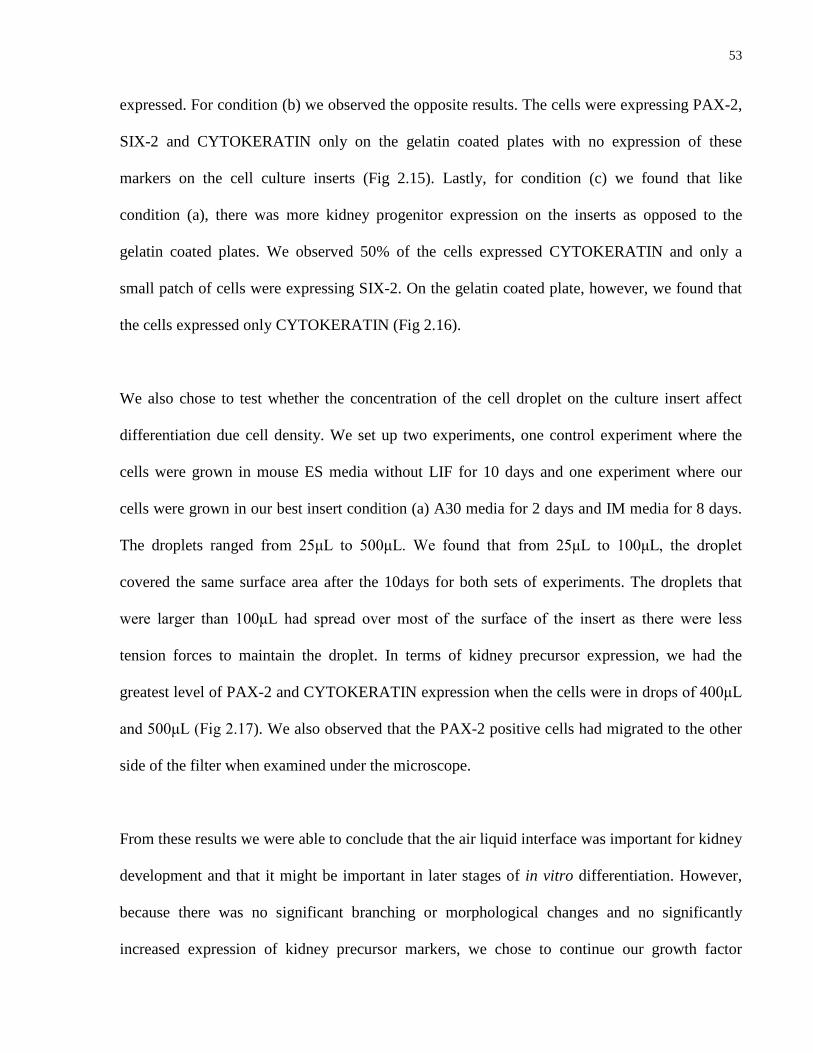
53
expressed. For condition (b) we observed the opposite results. The cells were expressing PAX-2,
SIX-2 and CYTOKERATIN only on the gelatin coated plates with no expression of these
markers on the cell culture inserts (Fig 2.15). Lastly, for condition (c) we found that like
condition (a), there was more kidney progenitor expression on the inserts as opposed to the
gelatin coated plates. We observed 50% of the cells expressed CYTOKERATIN and only a
small patch of cells were expressing SIX-2. On the gelatin coated plate, however, we found that
the cells expressed only CYTOKERATIN (Fig 2.16).
We also chose to test whether the concentration of the cell droplet on the culture insert affect
differentiation due cell density. We set up two experiments, one control experiment where the
cells were grown in mouse ES media without LIF for 10 days and one experiment where our
cells were grown in our best insert condition (a) A30 media for 2 days and IM media for 8 days.
The droplets ranged from 25μL to 500μL. We found that from 25μL to 100μL, the droplet
covered the same surface area after the 10days for both sets of experiments. The droplets that
were larger than 100μL had spread over most of the surface of the insert as there were less
tension forces to maintain the droplet. In terms of kidney precursor expression, we had the
greatest level of PAX-2 and CYTOKERATIN expression when the cells were in drops of 400μL
and 500μL (Fig 2.17). We also observed that the PAX-2 positive cells had migrated to the other
side of the filter when examined under the microscope.
From these results we were able to conclude that the air liquid interface was important for kidney
development and that it might be important in later stages of in vitro differentiation. However,
because there was no significant branching or morphological changes and no significantly
increased expression of kidney precursor markers, we chose to continue our growth factor

54
experiments on gelatin coated plates and chose to test the differentiation potential of additional
growth factors and molecules cited to be important during kidney development.
2.3.14 Varying Concentrations and Combinations of LIF, GDNF and
FGF2 in Mouse ES media to Induce Metanephric Mesenchyme
and Ureteric Bud Differentiation
The literature cited that LIF and FGF2 were important initiators of cell migration and
aggregation seen in metanephric mesenchyme induction and that GDNF (Glial cell line-derived
neurotrophic factor) was an in important molecule to induce branching (Pohl, Stuart et al. 2000;
Basson, Watson-Johnson et al. 2006; Michos 2009). We chose to use these growth factors in
different combinations using the literature as a guide for optimal concentrations (Stuart, Bush et
al. 2003). Cells were cultured in A30 medium for two days, IM medium for two days and then
mouse ES media supplemented with various combinations of growth factors for 3.5 days. From
our results we found that the conditions that resulted in some kidney precursor expression were
(a) 125ng/mL of GDNF and 1000 U/mL of LIF (b) 125ng/mL of FGF2 and (c) 125ng/mL of
GDNF, 1000 U/mL of LIF and 125ng/mL of FGF2. For condition (a) mouse ES media was
supplemented with 125ng/mL GDNF and 1000 U/mL of LIF for 3.5 days. We observed some
branching and organization (Fig 2.18), with CYTOKERATIN expression in 40% of cells. For
condition (b) mouse ES media was supplemented with 125ng/mL of FGF2 for 3.5 days. We
observed some expression of SIX-2 in an area where cells had grown in a monolayer and about
25% expression of CYTOKERATIN (Fig 2.19). Lastly, for condition (c) mouse ES media was
supplemented with 125ng/mL GDNF, 1000 U/mL of LIF and 125ng/mL of FGF2 for 3.5 days.

55
This condition produced SIX-2 in an area where cells were growing in a monolayer. We also
found that about 50% of our cells were expressing CYTOKERATIN (Fig 2.20).
From these experiments we learned that the high 20% serum concentration in mouse ES media
was causing the cells to grow quickly making the wells overly confluent without the presence of
monolayers. We also observed the importance of FGF2 as an inducer of SIX-2 (Fig 2.19 & Fig
2.20). We concluded that it was important for our successive experiments to use a media with
less serum and to continue designing the MM (metanephric mesenchyme) differentiation
medium supplemented with FGF2.
2.3.15 Inducing SIX-2 Expression with FGF2 in a Medium with Less
Serum
With FGF2 being important to the induction of SIX-2 and with mouse ES media no longer a
candidate base medium, we chose to test our A30 and IM base media (see Section 2.2.4 and 2.2.6
for details) supplemented with FGF2 at 100ng/mL to investigate the SIX-2 differentiation
potential of these two media. From this experiment, we observed that FGF2 in A30 base medium
resulted in high cell death with no induction of SIX-2 or CYTOKERATIN. The IM base
medium, however, allowed the cells to grow in monolayers, which is integral to differentiation
and we were able to achieve SIX-2 and CYTOKERATIN induction (Fig 2.21). Most
interestingly, we observed double staining of CYTOKERATIN and SIX-2 (Fig 2.21a). While
this has not yet been documented to occur in the embryo, because these cells are grown in culture
without any barriers between different cell types, it is possible that this particular population of
double stained cells is a phenomenon that only rarely occurs in the embryo (Stuart, Bush et al.
2003; Shah, Tee et al. 2009).

56
We also used PCR to confirm induction of metanephric mesenchyme and found that Eya-1, Wt-1
and Cited1 were being expressed by these cells (Fig 2.22). The Eya-1 gene is important
throughout kidney development and is expressed early in development once tissues have been
fated towards the kidney lineage, specifically metanephric mesenchyme (Dressler 2009). The
Wt-1 gene is also expressed throughout kidney development and controls a number of processes
including induction of metanephric mesenchyme and podocyte development (Reidy and
Rosenblum 2009). The gene Cited1 is important for metanephric mesenchyme growth and
development, it is expressed in metanephric mesenchymal tissues when they are invaded by the
ureteric bud (Boyle, Shioda et al. 2007). This particular marker is specific for metanephric
mesenchyme cells that localize after invasion of the ureteric bud and form cells of the cap
mesenchyme lineage which will later develop into the kidney capsule (Dressler 2009). The
expression of these genes confirms our findings that the ES culture in our differentiation media
are indeed fated towards the kidney lineage. Unexpectedly, these cells were also expressing Gsc,
a “pre-mesoderm” marker that is responsible for regulating the expression of the mesoderm
marker BRACHYURY (Thisse, Thisse et al. 1994; Nakaya, Murakami et al. 2008). However, as
a possible explanation of Gsc being expressed in the metanephric mesenchyme, it may play a
role in mesenchyme development and growth because it has been suggested that it is expressed
by tissues fated towards mesenchymal cell lineages (Gaunt, Blum et al. 1993). Furthermore, its
expression this far in development may not be inaccurate because Gsc has been shown to be
expressed in ear tissue up until day E14.5 (Zhu, Yamada et al. 1997). Further study on the
expression of this marker and its importance in kidney development will be examined.
In conclusion, we found that these experiments clearly demonstrated the importance of FGF2 as
a more potent inducer of metanephric mesenchyme and further reinforced the importance of

57
monolayers for efficient differentiation. Our lab will continue to develop and modify our media
and choose other biologically relevant growth factors to induce other kidney precursors.
However, with A30 and IM media firmly established, the next step was to test the potency of
these media on human ES cells.
2.3.16 Returning to our CA-1 cell line and Applying Established A30 and
IM media to Human ES Cells
Upon establishing the intermediate mesoderm induction protocol using mouse ES cells, we chose
to test the potential of these media on human ES cells. We cultured the CA-1 cell line under the
same temporal and media conditions that we determined for mouse ES cells and found that we
could achieve greater expression of BRACHYURY (Fig 2.23) with A30 differentiation medium
as compared with earlier experiments with FGF2 and BMP-4 (Fig 2.1). We were also able to
achieve PAX-2 induction using IM differentiation medium and observed expression of
CYTOKERATIN, a marker of early ureteric bud (Unbekandt and Davies 2010) (Fig 2.24). With
this in mind, we will investigate whether the amount of time the cells were in culture in
differentiation media would play a role in increasing PAX-2 and BRACHYURY efficiency in
future studies.
2.4 Discussion
One of the biggest hurdles of controlled differentiation of stem cells is mimicking the embryonic
environment in a dish. For the first stage of our differentiation protocol, we determined from the
literature what the most likely and relevant growth factors were involved in mesoderm
formation. We concluded that a combination of BMP-4, Activin A and FGF2 would be

58
worthwhile investigating and we designed our experiments to determine the optimal
concentration and combination of these growth factors to induce mesoderm and intermediate
mesoderm. We used a human ES cell line and found that after 4 days in a growth factor cocktail
of FGF2, BMP-4 and Activin, we achieved mesoderm induction, however at a very low yield.
We duplicated these conditions on a mouse ES cell line and did not achieve BRACHYURY
expression. We postulated that this was because applying a protocol designed for the more
complex human ES cells may not work on mouse ES cells. However, mouse ES cell work has
been cited to be easier to apply to higher organisms such as human ES cells. We therefore
modified our medium accordingly and designed it for mouse ES cells. We determined that our
A30 differentiation media with only Activin A at a concentration of 30ng/mL for 48 hours
resulted in the most efficient population of mesoderm for mouse ES cell lines. When we applied
these conditions to a human ES cell line and our preliminary results showed that there was
greater mesoderm induction at the 48 hour time point with the A30 differentiation media. This
suggests that designing protocols for less complex organisms can be applied to higher organisms.
Interestingly, these studies, and as literature has confirmed, showed that human ES cells in
culture have the same development time line to that of mouse ES cells (Van Hoof, D'Amour et
al. 2009). Therefore, our observations of BRACHYURY expression at the end of 2 days in both
the human ES cell line and mouse ES cell line are perhaps not surprising. We also tested the
mouse ES cells for other mesoderm markers including Gsc, a mesendoderm marker, and found
that these cells were expressing this gene. Interestingly, these cells were also expressing Wt-1,
which is normally expressed in intermediate mesoderm (Artinger, Blitz et al. 1997; Kreidberg
2010). These cells also expressed Eya-1, while this particular gene is important in early kidney
development, is not generally expressed in the mesoderm. These results suggest that there may
have been a population of cells that were further along in development while in culture. It also

59
suggests that these cells have the potential to be fated preferentially towards the kidney lineage
and argues that the differentiation medium is well suited for supporting mesoderm cells and their
kidney derivatives.
The next stage of kidney organogenesis is the development of intermediate mesoderm, which
proved to be the biggest challenge. We determined that the developing paraxial mesoderm
played a vital role in intermediate mesoderm induction through the secretion of RA and in
conjunction with other literature citing the additional importance of Activin A secretion, we
determined the most likely growth factor candidates for intermediate mesoderm induction
(Mauch, Yang et al. 2000; James and Schultheiss 2003; Cartry, Nichane et al. 2006). We applied
this information to our studies on mouse ES cell lines. After numerous medium modifications
and cell density changes, we observed the greatest and most consistent levels of intermediate
mesoderm differentiation in the BRY-GFP mouse cell line. Furthermore, in areas where the cells
were in monolayers, there was greater PAX-2 expression when compared with areas where the
cells were expressing OCT-4 and forming tight colonies. We explored the use of inhibitors and
whether it was possible to increase PAX-2 induction by reducing the expression of OCT-4 and
by extension increasing monolayer to allow the cells to differentiate. We concluded that our IM
differentiation medium yielded far more PAX-2 expression and we continued to modify our IM
differentiation medium and tested various levels of confluency. We determined that when cells
were at a density of 40,000 cell/mL in A30 differentiation medium and continued differentiation
in our modified IM differentiation medium, we were able to induce PAX-2 expression in all
surviving cells when cultured for 48 hours. We also tested these cells with RT-PCR to confirm
that they were indeed fated towards the kidney lineage and found that they were expressing Wt-1.
As previously stated, Wt-1 is an important marker throughout kidney development and is first

60
expressed in intermediate mesoderm cells (Dressler 2009). This confirms our immunostaining
suggesting that the cells are indeed intermediate mesoderm cells.
As with the A30 differentiation medium, we extrapolated the mouse ES cell studies to human ES
cells and found the cells were expressing PAX-2, however not as efficiently as with the mouse
ES cell line when cultured for two days in IM medium. We hope to continue to adapt our mouse
ES cell work to human ES cells and design a protocol to induce an efficient level of PAX-2
positive cells. We believe that the human ES cells may need to be cultured in IM medium longer
in order to increase efficiency PAX-2 expression.
The literature has suggested that once at the intermediate mesoderm stage, cells are “determined”
to continue to direct their own differentiation beyond intermediate mesoderm to produce its
derivatives if they are cultured in the conditions that support intermediate mesoderm cells (James
and Schultheiss 2003). We tested this theory by growing intermediate mesoderm cells on cell
culture inserts using our established protocol for intermediate mesoderm induction, followed by
3.5 days in mouse ES media without LIF to investigate whether the cells were “determined’
towards the kidney cell fate and whether they could direct their own differentiation without
additional growth factor supplements. We tested for expression of SIX-2, the marker for
metanephric mesenchyme and CYTOKERATIN, the marker for early ureteric bud. We observed
little to no expression of both these markers. We hypothesized that it is possible that the cells
required longer time in the media and additional growth factors to induce metanephric
mesenchyme and ureteric bud marker expression. We investigated the effects of growing the
cells for a total of 10 days on cell culture inserts and on a gelatin coated well in growth factor
supplemented conditions and found varying results. This could be because the cells are able to

61
differentially express various markers when grown on different substrates. Therefore it is
possible that at this stage of development, the air liquid interface may be important, however
further exploration of this is required. Specifically, testing the possibility of cell drop volume
with regards to the optimal concentration of cells that is required for the air liquid interface to be
effective is a possible area of further study. It is also likely that the cells required additional
growth factors that are relevant to this particular stage in kidney development in order to
differentiate to metanephric mesenchyme and ureteric bud.
From the literature, it had been suggested that there were a number of potential candidate
molecules that were important for the next stage in kidney development, namely FGF2, GDNF
and LIF, among others (Vainio and Muller 1997; Sariola and Sainio 1998; Dudley, Godin et al.
1999; Qiao, Sakurai et al. 1999; Burrow 2000; James and Schultheiss 2003). FGF2 and LIF are
important factors in the initiation of differentiation events after intermediate mesoderm.
Furthermore, GDNF is a ligand that is produced by the metanephric mesenchyme that binds to its
receptor Ret, located on the ureteric bud tips (Dressler 2006). Signaling by GDNF once bound to
Ret allows for branching to occur (Dressler 2002). Therefore, since both metanephric
mesenchyme and ureteric bud are derived from a population of intermediate mesoderm cells, we
chose to add these growth factors in varying combinations to determine which growth factors
would induce SIX-2 and CYTOKERATIN expression. We observed the greatest induction of
SIX-2 in conditions that were supplemented with FGF2, furthermore expression of SIX-2 was
localized to areas where cells were growing in monolayers. This argues that FGF2 is indeed an
important inducer of SIX-2 and these conditions may be supportive of metanephric mesenchyme
cells, however, due to the importance of monolayers coupled with these cells grown in a serum
rich medium, one of the first changes we made to offset increased confluency was to instead use

62
A30 base medium and IM base medium, both of which were low in serum. We added FGF2 to
investigate whether we could reduce the level of confluency by using a less supportive and
serum deficient medium. We found a few areas where the cells were growing in monolayers and
therefore had greater expression of SIX-2, and only the IM base medium appeared to support the
cells. We also tested these cells with RT-PCR and found that they were expressing Eya-1,
Cited1 and Wt-1, all of which are important for metanephric mesenchyme development. This
suggested these cells were indeed fated towards the kidney lineage. Cited1, for example, is a
known marker of cells of the cap mesenchyme (Dressler 2009). The observation that Cited1 does
not appear to be expressed by mesoderm and intermediate mesoderm differentiated cells
confirms that these cells are cap mesenchyme. Furthermore, the expression of Eya-1 and Wt-1,
both of which interact with other kidney genes including Pax-2 and Six-1 to regulate kidney
marker expression further verify our immunostaining data. However, we will continue our
studies on designing a new MM differentiation medium. From our preliminary data, we can
conclude that because of FGF2, the cells became confluent regardless of the lack of serum in the
IM base medium. An area of further investigation will be to explore the possibility of inhibiting
the self-renewal pathway of FGF2 while leaving the pathway involved in Six-2 induction
unaffected. We will also be determining whether we can establish a new base medium for
metanephric mesenchyme and ureteric bud induction.

63
2.5 Chapter 2 Figures
Figure 2.1 CA-1 human ES cells expressing BRACHYURY (20x). CA-1 cells were grown in
DMEM/F12, PenStrep and 0.5% serum replacement with additional FGF2 at a concentration of
500ng/mL and BMP-4 5ng/mL for 3 days. Only 5-10% of living cells were expressing
BRACHYURY.
Figure 2.1 CA-1 human ES cells expressing BRACHYURY (20x).
(A) CA-1 (B) CA-1 DAPI + BRACHYURY BRACHYURY

64
Figure 2.2 CA-1 human ES cells expressing BRACHYURY (20x). CA-1 cells were grown in
DMEM/F12, PenStrep and 0.5% serum replacement with Activin A at a concentration of
10ng/mL and BMP-4 1ng/mL for 3 days. Only 5% of living cells were expressing
BRACHYURY.
Figure 2.2 CA-1 human ES cells expressing BRACHYURY (20x).
(A) CA-1 (B) CA-1 DAPI + BRACHYURY BRACHYURY

65
Figure 2.3 CA-2 human ES cells double stained for BRACHYURY and HNF3β (20x). CA-2
cells were grown in DMEM/F12, PenStrep and 0.5% serum replacement with FGF2 at a
concentration of 20ng/mL for 3 days. Of the cells expressing HNF3β only 50% of cells double
stained for BRACHYURY and this shows that our medium is able to produce two separate
populations of cells and that the BRACHYURY single positive cells were fated towards
mesoderm.
Figure 2.3 CA-2 human ES cells double stained for BRACHYURY and HNF3β (20x).
(A) CA-1 (B) CA-1 DAPI BRACHYURY + HNF3β

66
Figure 2.4 CA-1 Human ES Cells stained for OCT-4 (a,b) in the absence of LY294002
and(c,d) in the presence of LY294002 (5x). Human CA-1 cells were grown in DMEM/F12,
PenStrep and 0.5% serum replacement with BMP-4 at a concentration of 25ng/mL for 24 hours
in the presence or absence of LY294002 at 10mM. While there was no BRACHYURY
expression, OCT-4 expression was reduced in the condition with LY294002 suggesting that the
OCT-4 gene is being suppressed as evidence by only DAPI staining.
Figure 2.4 CA-1 human ES cells stained for OCT-4 (a,b) in the absence of
LY294002 and(c,d) in the presence of LY294002 (5x).
(A) CA-1 –LY294002 (B) CA-1 –LY294002 DAPI + OCT-4 OCT-4
(C) CA-1 +LY294002 (D) CA-1 + LY294002 DAPI + OCT-4 OCT-4

67
Figure 2.5 B6 eGFP and BRY-GFP mouse ES cells stained positive for BRACHYURY (5x).
Mouse ES cells were grown on gelatin coated plates for 2 days Activin A 30ng/mL in
DMEM/F12, 0.5% serum replacement and PenStrep. Both cell lines produced 80% positive
mesoderm cells.
Figure 2.5 B6 EGFP and BRY-GFP mouse ES cells stained positive for BRACHYURY (5x).
(A) B6 eGFP BRACHYURY
(C) BRY-GFP BRACHYURY
(B) B6 eGFP DAPI
(D) BRY-GFP DAPI

68
Figure 2.6 B6 eGFP mouse ES cells Stained Positive for PAX-2 (b) (20x). B6 eGFP Mouse
ES cells were grown in Activin A 30ng/mL + RA 100nM in DMEM/F12, 0.5% serum
replacement and PenStrep. Results show that all living cells expressed the intermediate
mesoderm marker PAX-2 however there was also a high level of cell death.
Figure 2.6 B6 eGFP mouse ES cells stained positive for PAX-2 (b) (20x).
(A) B6 eGFP DAPI
(B) B6 eGFP PAX-2
(C) B6 eGFP GFP
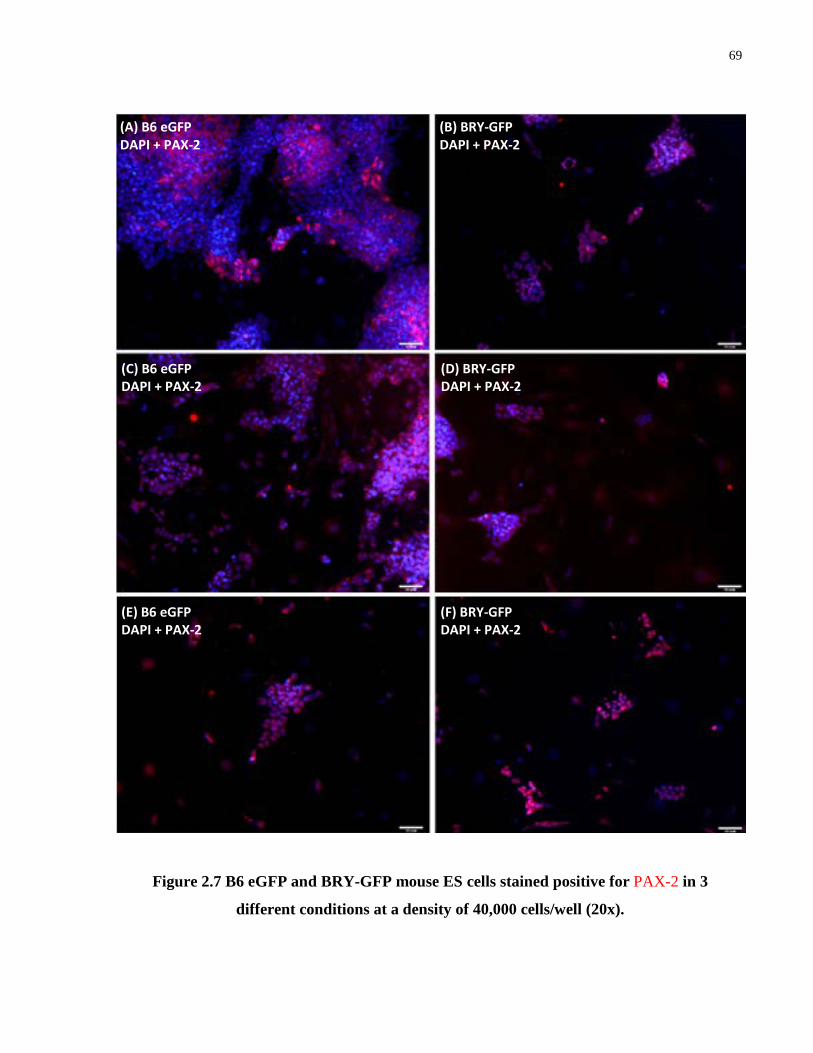
69
Figure 2.7 B6 eGFP and BRY-GFP mouse ES cells stained positive for PAX-2 in 3
different conditions at a density of 40,000 cells/well (20x).
(A) B6 eGFP DAPI + PAX-2
(C) B6 eGFP DAPI + PAX-2
(E) B6 eGFP DAPI + PAX-2
(B) BRY-GFP DAPI + PAX-2
(D) BRY-GFP DAPI + PAX-2
(F) BRY-GFP DAPI + PAX-2

70
Figure 2.7 B6 EGFP and BRY-GFP mouse ES cells stained positive for PAX-2 in 3 different
conditions at a density of 40,000 cells/well (20x). Both cell lines expressed PAX-2 when plated
at a density of 40 000 cells/well. Cells were grown in DMEM High Glucose, 10% FBS, ʟ-
glutamine and β-mercaptoethanol in (a,b) Activin A at 40ng/mL, RA 100nM (c,d) Activin A at
10ng/mL, RA at 100nM and BMP-7 at 50ng/mL (e,f) Activin A at 30ng/mL, RA 100nM. While
both cell lines were expressing PAX-2, observations showed that PAX-2 expression was in cell
monolayers.

71
Figure 2.8 BRY-GFP ES cells in new basal media with 100nM RA concentrations
yielding most PAX-2 induction.
(A) BRY-GFP DAPI + PAX-2
(C) BRY-GFP DAPI + PAX-2
(E) BRY-GFP DAPI + PAX-2
(G) BRY-GFP DAPI + PAX-2
(B) BRY-GFP PAX-2
(D) BRY-GFP PAX-2
(F) BRY-GFP PAX-2
(H) BRY-GFP PAX-2

72
Figure 2.8 BRY-GFP ES cells in new basal media with 100nM RA concentrations yielding
most PAX-2 induction. (a,b) BRY-GFP mouse ES cells grown with Activin A at 30ngmL and
RA at 10nM, (c,d) mouse ES cells Activin A 30ng/mL + RA 1000nM, (e,f) BRY-GFP mouse ES
cells Activin A 30ng/mL+ RA 100nM + BMP-7 50ng/mL, (g,h) BRY-GFP mouse ES cells
Activin A 30ng/mL+ RA 100nM. Condition (g) yielded most consistently efficient induction of
PAX-2 with RA at 100nM. Furthermore, optimal RA concentration was determined to be 100nM
for PAX-2 induction. Lastly, it is important for cells to be in monolayers in order for
differentiation to occur.

73
Figure 2.9 Diminished expression of OCT-4 in PAX-2 positive regions. While cells were
expressing PAX-2 and differentiating, areas where there were colonies, OCT-4 expression was
extremely high suggesting that these cells remained pluripotent and in their stem cell state. PAX-
2 expression is expressed in areas where the cells are in a monolayer, therefore because of
crosstalk between cells in tight colonies, it is possible that if cells were not grown in colonies,
they would differentiate more efficiently when exposed to exogenous growth factors that may
efficiently override any crosstalk between cells in monolayers.
Figure 2.9 Diminished expression of OCT-4 in PAX-2 positive regions.
(A) BRY-GFP DAPI + PAX-2
(B) BRY-GFP OCT-4 + PAX-2

74
Figure 2.10 Addition of inhibitors did not reduce OCT-4 expression or increase PAX-2.
High cell death occurred across all inhibitor conditions, however, it is possible that all
undifferentiated cells account for the high level of cell death. There was a similar population of
PAX-2 positive cells with no OCT-4 with and without inhibitors. Monolayer is important for
differentiation and PAX-2 induction. Inhibitors achieve the same level of PAX-2 induction as
without inhibitors with OCT-4 expression equally reduced with and without inhibitors in
differentiated areas
Figure 2.10 Addition of inhibitors did not reduce OCT-4 expression or increase PAX-2.
(A) LY294002 25μM DAPI + OCT-4 + PAX-2
(C) Stauprimide 250nM DAPI + OCT-4 + PAX-2
(B) LY294002 75μM DAPI + OCT-4 + PAX-2
(D) No inhibitors DAPI + OCT-4 + PAX-2

75
Figure 2.11 Cells expression PAX-2 at lower serum concentrations with addition of ROCK
inhibitor. With lowered serum in addition to ROCK inhibitor, the same level of PAX-2
induction was achieved with 4% serum as with 10% serum without addition of ROCK. It is
important to reduce serum as much as possible to decrease the effect of growth factors found in
serum that may affect cell differentiation therefore ROCK inhibition helps to promote cell
survival in low serum.
Figure 2.11 Cells expression PAX-2 at lower serum concentrations with addition of ROCK
inhibitor.
(A) 1% Serum + ROCK inhibitor DAPI + PAX-2
(C) 2% Serum + ROCK inhibitor DAPI + PAX-2
(E) 4% Serum + ROCK inhibitor DAPI + PAX-2
(B) 8% Serum + ROCK inhibitor DAPI + PAX-2
(D) 10% Serum + ROCK inhibitor DAPI + PAX-2
(F) 10% Serum - ROCK inhibitor DAPI + PAX-2

76
Figure 2.12 All living cells expressing PAX-2 at 4% serum concentrations with addition of
ROCK inhibitor in monolayers. With monolayers OCT-4 expression persists at very low levels
and 100% of cells expressed PAX-2 were observed when grown in IM induction media for 2
days. Confluency plays a vital role in determining how efficiently cells differentiate. OCT-4
expression can be reduced without inhibitors and we can achieve the better results with our
conditions and when cells are grown in monolayers
Figure 2.12 All living cells expressing PAX-2 at 4% serum concentrations with addition of
ROCK inhibitor in monolayers.
(A) BRY-GFP PAX-2
(B) BRY-GFP DAPI + PAX-2

77
Figure 2.13 When grown in mouse ES media for 3.5 days after intermediate mesoderm
induction, 20% of cells grown on gelatin coated plates were expressing CYTOKERATIN
with some organization occurring. When mouse ES cells were grown for two days in A30
differentiation medium, two days in IM differentiation medium and mouse ES medium without
LIF, CYTOKERATIN was expressed in 20% of cells, organization of cells was also observed
(a,c) however, cells did not express SIX-2. Cells likely require additional growth factors.
Figure 2.13 When grown in mouse ES media for 3.5 days after intermediate mesoderm
induction, 20% of cells grown on gelatin coated plates were expressing CYTOKERATIN with
some organization occurring.
(A) Gelatin DAPI
(B) Gelatin DAPI + CYTOKERATIN
(C) Gelatin DAPI - Structure

78
Figure 2.14 Mouse ES cells grown gelatin plate for 2 days A30 differentiation medium, 8
days IM differentiation medium expressed PAX-2, SIX-2 and CYTOKERATIN with
differential expression patterns on both substrates. Results showed better growth and
differentiation on inserts at the air-liquid interface with an efficiency of 20% CYTOKERATIN
expression on gelatin and filters, however, <5% PAX-2 and only SIX-2 expression on filters as
shown above. This suggests that the air liquid interface may provide better differentiation of
cells. Furthermore, additional growth factors appeared to be important to add to cells to control
differentiation. Lastly, longer time in media was important for differentiation towards kidney
precursors to occur.
Figure 2.14 Mouse ES cells grown gelatin plate for 2 days A30 differentiation medium, 8
days IM differentiation medium expressed PAX-2, SIX-2 and CYTOKERATIN with
differential expression patterns on both substrates.
(A) Gelatin (10x) DAPI + CYTOKERATIN
(C) Inserts (20x) DAPI + PAX-2
(B) Inserts (10x) DAPI + CYTOKERATIN
(D) Inserts (20x) DAPI + SIX-2

79
Figure 2.15 Mouse ES cells grown in IM differentiation medium for 10 days express SIX-2,
PAX-2 and CYTOKERATIN more efficiently on cell culture inserts. When cells were grown
in intermediate mesoderm differentiation medium for 10 days on filters and gelatin, cells grown
on gelatin expressed PAX-2, CYTOKERATIN and SIX-2. The cells grown on filters did not
express any of the above markers. There also appeared to be layering of the cells although there
was no apparent organization of cells. It is possible that the increased time in medium with
growth factors is important for differentiation, however cells were very confluent by the end of
the experiment and as previous data has shown, confluency affects efficiency of differentiation.
Figure 2.15 Mouse ES cells grown in IM differentiation medium for 10 days express SIX-2,
PAX-2 and CYTOKERATIN more efficiently on cell culture inserts.
(A) Gelatin DAPI + CYTOKERATIN
(B) Gelatin DAPI + PAX-2
(C) Gelatin DAPI + SIX-2

80
Figure 2.16 Mouse ES cells grown on gelatin plate for 2 days in A30 differentiation
medium, 2 days in IM differentiation medium and 6 days in mouse ES media without LIF
expressed more kidney markers on filters when compared with gelatin coated plates. Cells
appeared to express SIX-2 and CYTOKERATIN more efficiently at the air liquid interface under
this condition. It is possible that the air liquid interface is important for differentiation of certain
cell types. Furthermore, because SIX-2 is expressed, we can also infer that CYTOKERATIN
may likely be indicative of the presence of ureteric bud cells. CYTOKERATIN was expressed in
about 50% of cells on gelatin and filters, however, SIX-2 expression was found in that area as
shown above.
Figure 2.16 Mouse ES cells grown on gelatin plate for 2 days in A30 differentiation medium,
2 days in IM differentiation medium and 6 days in mouse ES media without LIF expressed
more kidney markers on filters when compared with gelatin coated plates.
(A) Gelatin (5x) DAPI + CYTOKERATIN
(B) Inserts (20x) DAPI + CYTOKERATIN
(C) Inserts (20x) DAPI + PAX-2

81
Figure 2.17 Mouse ES cells grown for 2 days in A30 differentiation medium, 8 days in IM
differentiation medium showed better growth and differentiation on inserts with cell
volumes at 400μL and 500μL. The results have shown that for cell volumes of 400μL and
larger, there is 10% expression of CYTOKERATIN on gelatin and filters and <5% PAX-2. This
indicates that that cells need to be at these two volumes in order to differentiate. This could be
because of the cell interactions that occur at this cell concentration which leads to more
communication between cells. These concentrations also allow the cells to grow in monolayers.
Figure 2.17 Mouse ES cells grown for 2 days in A30 differentiation medium, 8 days in IM
differentiation medium showed better growth and differentiation on inserts with cell
volumes at 400μL and 500μL.
(A) 300μL (20x) DAPI + CYTOKERATIN
(B) 400μL (20x) DAPI + CYTOKERATIN
(C) 400μL (20x) DAPI + PAX-2
(D) 500μL (20x) DAPI + CYTOKERATIN
(E) 500μL (20x) DAPI + PAX-2

82
Figure 2.18 Mouse ES cells grown on gelatin plate for 2 days in A30 differentiation
medium, 2 days in IM differentiation medium, 3.5 days in mouse ES media 125ng/mL
GDNF + LIF 1000U/mL express CYTOKERATIN and may also be forming organized
structures. GDNF is known to initiate branching of the ureteric bud, from the figure some
branching and organization can be observed. While there was no SIX-2 induction,
CYTOKERATIN was expressed in 40% of cells.
Figure 2.18 Mouse ES cells grown on gelatin plate for 2 days in A30 differentiation medium,
2 days in IM differentiation medium, 3.5 days in mouse ES media 125ng/mL GDNF + LIF
1000U/mL express CYTOKERATIN and may also be forming organized structures.
(A) BRY-GFP CYTOKERATIN
(B) BRY-GFP DAPI + CYTOKERATIN
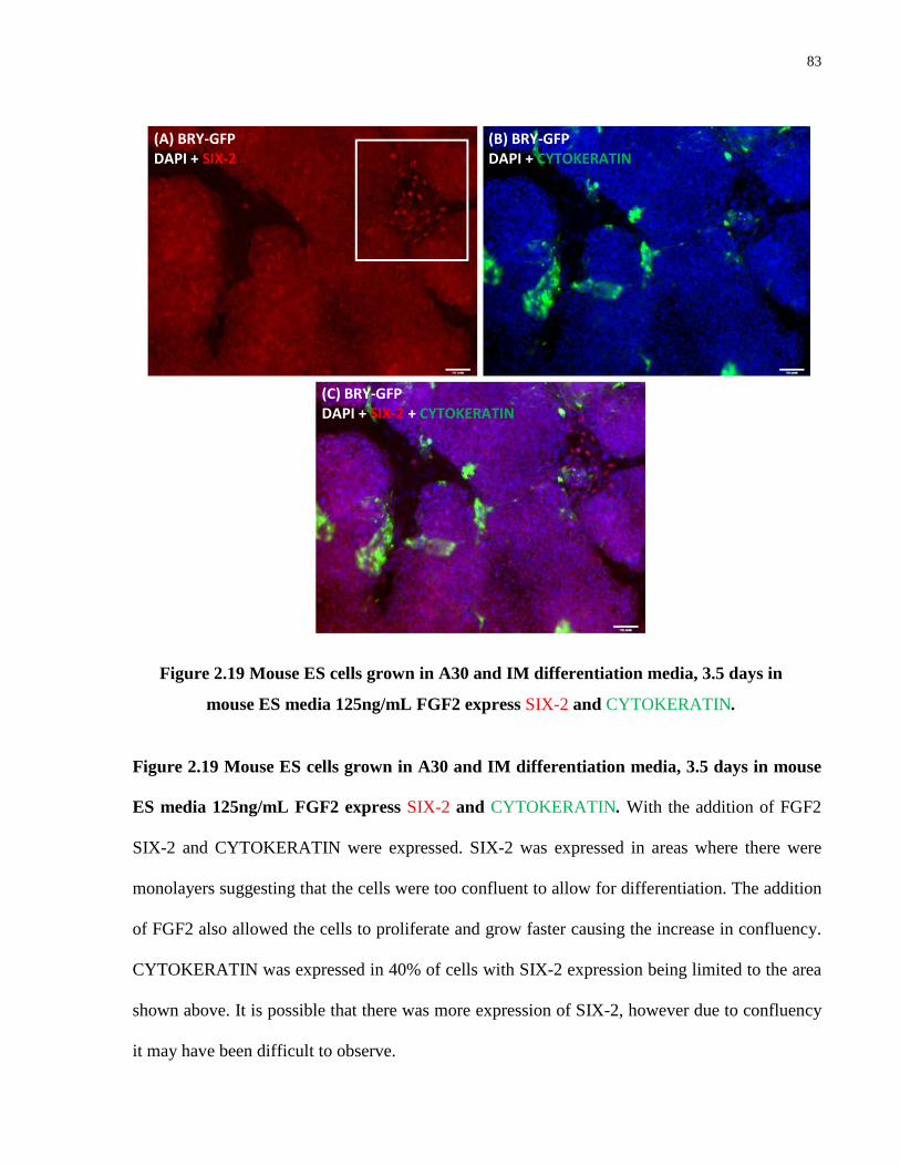
83
Figure 2.19 Mouse ES cells grown in A30 and IM differentiation media, 3.5 days in mouse
ES media 125ng/mL FGF2 express SIX-2 and CYTOKERATIN. With the addition of FGF2
SIX-2 and CYTOKERATIN were expressed. SIX-2 was expressed in areas where there were
monolayers suggesting that the cells were too confluent to allow for differentiation. The addition
of FGF2 also allowed the cells to proliferate and grow faster causing the increase in confluency.
CYTOKERATIN was expressed in 40% of cells with SIX-2 expression being limited to the area
shown above. It is possible that there was more expression of SIX-2, however due to confluency
it may have been difficult to observe.
Figure 2.19 Mouse ES cells grown in A30 and IM differentiation media, 3.5 days in
mouse ES media 125ng/mL FGF2 express SIX-2 and CYTOKERATIN.
(A) BRY-GFP DAPI + SIX-2
(B) BRY-GFP DAPI + CYTOKERATIN
(C) BRY-GFP DAPI + SIX-2 + CYTOKERATIN

84
Figure 2.20 Mouse ES cells grown in A30 and IM differentiation media, 3.5 days in mouse
ES media 125ng/mL FGF2 , GDNF, LIF 1000U/mL showed expression of SIX-2 and
CYTOKERATIN. With the addition of the three growth factors, SIX-2 expression was localized
to area growing in monolayers, however, it is possible that confluent areas could also be
expressing SIX-2 but due to cell density it is difficult to observe. Furthermore, there also appears
to be double staining of SIX-2 and CYTOKERATIN. CYTOKERATIN was expressed by 50%
of cells while SIX-2 positive cells are shown above. FGF2 is an important inducer of SIX-2 and
may provide a more robust positive population of SIX-2 in different base media with less serum
because of the overgrowth seen above.
Figure 2.20 Mouse ES cells grown in A30 and IM differentiation media, 3.5 days in mouse
ES media 125ng/mL FGF2 , GDNF, LIF 1000U/mL showed expression of SIX-2 and
CYTOKERATIN.
(A) BRY-GFP DAPI + SIX-2
(B) BRY-GFP DAPI + CYTOKERATIN
(C) BRY-GFP DAPI + SIX-2 + CYTOKERATIN

85
Figure 2.21 Mouse ES cells in A30 and IM differentiation media and 3.5 days in new media
with 100ng/mL FGF2 were expressing SIX-2 and CYTOKERATIN with some double
staining. FGF2 appears to be an important promoter of SIX-2 induction as <1% of cells were
expressing SIX-2. About 20% of cells were expressing CYTOKERATIN. However there was
also double staining of CYTOKERATIN with SIX-2 suggesting that this particular population of
cells may have been a “pre metanephric and ureteric bud” population. Furthermore cells were
expressing SIX-2 in areas where they were growing in monolayers.
Figure 2.21 Mouse ES cells grown in A30 and IM differentiation media and 3.5 days in
new media with 100ng/mL FGF2 were expressing SIX-2 and CYTOKERATIN with
some double staining.
(A) BRY-GFP CYTOKERATIN + SIX-2
(B) BRY-GFP DAPI + CYTOKERATIN
(C) BRY-GFP DAPI + SIX-2
86
Figure 2.22 Mesoderm (Meso), intermediate mesoderm (IM) and metanephric mesenchyme
(MM) differentiated cells express Gsc, Eya-1, Cited1 and Wt-1. ES cell derived mesoderm
cells expressed Gsc, Eya-1 and Wt-1 when grown in A30 differentiation medium and confirm
immunostaining of BRACHYURY. ES cell derived intermediate mesoderm cells expressed Wt-1
when grown in IM differentiation medium and confirmed PAX-2 immunostaining. ES cell
derived metanephric mesenchyme cells expressed Gsc, Eya-1, Cited1 and Wt-1 when grown in
preliminary MM differentiation medium and confirmed CYTOKERATIN and SIX-2
immunostaining.
Figure 2.22 Mesoderm (Meso), intermediate mesoderm (IM) and metanephric
mesenchyme (MM) differentiated cells express Gsc, Eya-1, Cited1 and Wt-1.

87
Figure 2.23 CA-1 human ES cells grown in A30 differentiation medium for two days
express PAX-2, BRACHYURY, β-CATENIN and CYTOKERATIN. When compared with
previous results, when grown in A30 differentiation medium ~30% CA-1 cells expressed
BRACHYURY, <2% PAX-2, ~40% β-CATENIN and CYTOKERATIN. The cells were grown
in monolayers and stained negative for OCT-4. Cells double stained for PAX-2 and
CYTOKERATIN (b).
Figure 2.23 CA-1 human ES cells grown in A30 differentiation medium for two days
express PAX-2, BRACHYURY, β-CATENIN and CYTOKERATIN.
(A) CA-1 DAPI + BRACHYURY
(B) CA-1 DAPI + PAX-2 + CYTOKERATIN
(C) CA-1 DAPI + β-CATENIN

88
Figure 2.24 CA-1 human ES cells grown in A30 media for two days and IM media for 2
days expressed PAX-2 and CYTOKERATIN. Established protocol produces similar results to
mouse ES cells although time point for human ES cells must be established. Cells were also
easily grown in monolayers and therefore differentiated efficiently. PAX-2 was expressed by
30% of cells, while CYTOKERATIN was expressed by 40% of cells with some resemblance of
tubular staining (b).
Figure 2.24 CA-1 human ES cells grown in A30 media for two days and IM media for
2 days expressed PAX-2 and CYTOKERATIN.
(A) CA-1 DAPI + PAX-2
(B) CA-1 DAPI + CYTOKERATIN

89
3 Chapter 3 Applying Differentiated and Undifferentiated ES Cells to Test for
Mechanical and Biological Roles Played by a Decellularized Extracellular Matrix
3.1 Introduction
The extracellular matrix (ECM) is a vital component of organ and tissue organization (Tsang,
Cheung et al. 2010). While its structure is conserved across species, its composition varies and is
dependent on the organ or tissue type, the cells that interact with and generate the ECM, and the
complexity of the organism itself (Har-el and Tanzer 1993). The ECM consists of two separate
regions: the basement membrane and the interstitial matrix (Bosman and Stamenkovic 2003).
While the general composition of these two separate structures consists of similar components,
the proteins that make up these domains differ in their variety (Bosman and Stamenkovic 2003).
As a whole, the ECM consists of a number of components (Tsang, Cheung et al. 2010). These
components are classed into two types of proteins: fibrous proteins and proteoglycans (Frantz,
Stewart et al. 2010). Fibrous proteins include collagen, elastin, fibronectin and laminin, the
former two are classified as structural proteins, while the latter two are classified as specialized
proteins (Frantz, Stewart et al. 2010). The most readily produced component of the ECM is
collagen (Laurila and Leivo 1993). Collagen provides tissues and organs with structural integrity
and can control development of cells and tissues (Frantz, Stewart et al. 2010). There are currently
20 different documented types of collagen proteins that differ based on the chains that make up
the triple helical protein structure of collagen (Bosman and Stamenkovic 2003). The basement
membrane is composed primarily of collagen type IV (Bosman and Stamenkovic 2003). Elastin
provides tissues and organs with elasticity and it generally interacts with glycoproteins that help
to maintain elastin fibers (Frantz, Stewart et al. 2010). Fibronectin is important for cell adhesion

90
interactions as well as arranging the ECM (Tayebjee, MacFadyen et al. 2003). Laminin acts as an
anchoring protein and plays a supportive role in relation to cell-ECM interactions, it also
interacts with other fibrous proteins such as collagen type IV and is an important component of
the basement membrane (Bosman and Stamenkovic 2003). There are currently 12 different
laminin proteins that have been identified and with their unique arrangement of α, β, γ chains,
they are able to participate in a number of cellular activities including cell attachment and
differentiation (Bosman and Stamenkovic 2003). Proteoglycans, are proteins that can be sub-
classed into (a) proteins that form a network with glycosaminoglycans to form a unique group of
proteins that are involved in a number of cellular functions including regulating proliferation of
fibroblasts and (b) a protein composed primarily of leucine residues that interact with
glycosaminoglycans in order to interact with the TGFβ family of growth factors (Bosman and
Stamenkovic 2003). Heparan sulphate is an example of an important proteoglycan component of
the matrix and are involved in a number of activities including differentiation and cell migration
(Bosman and Stamenkovic 2003). The most important aspect of the ECM, however, is its
dynamic nature.
It is this dynamic nature that make the ECM an important substrate in relation to helping to
differentiate developing organs and tissues (Furness 1996). This interaction between cells and
the matrix occur through receptor-ligand activities including integrins (Chen, Fitzgerald et al.
2007). Integrins are receptors that come in many forms based on their protein structures and
interact with many different ligands to control cellular activities including differentiation (Chen,
Fitzgerald et al. 2007). Drawing on this important aspect, regenerative medicine studies have
opted to use a decellularized ECM as a substrate on which to grow various types of cells
including stem cells. Recent research advances in regenerative medicine have also uncovered the

91
importance of co-culture systems to improve differentiation protocols when producing artificial
organs and tissues from stem cells. As such, the ECM has recently garnered attention in relation
to its role in directing differentiation during embryogenesis.
An important aspect of using extracellular matrices is technical in nature, where the removal of
native cells and reseeding with new cells can be a challenge. A number of decellularization
protocols have been developed to ensure complete removal of cellular components from the
tissue of choice while retaining the biomolecules that can support and direct differentiation of
cells (Crapo, Gilbert et al. 2011). For the kidney, literature has shown that because of the high
density of this tissue, a solution of 0.1% sodium dodecyl sulfate (SDS), an ionic detergent, is
currently the most effective method to remove cells from the tissue (Gilbert, Sellaro et al. 2006).
A number of protocols, including our lab’s methods, have been set up where kidneys are
cannulated through the artery in order to set up a constant flow rate of the 0.1% SDS solution via
a peristaltic pump to ensure complete removal of cellular debris and efficient decellularization.
Other protocols have opted to use thick kidney sections which are soaked in the same 0.1% SDS
solution. Our method to decellularize kidney sections involves the use of a peristaltic pump to
circulate the detergent and allow for efficient decellularization by establishing a constant flow
rate that removes cellular waste.
In this study we aim to test whether ES cell derived intermediate mesoderm cells as well as
undifferentiated stem cells can be supported on a decellularized kidney section to confirm the
mechanical nature of the ECM. We will also be testing for whether the ECM is able to direct
differentiation on the pretense that the decellularized scaffold will retain its biomolecules. We
will be using an adult decellularized scaffold as well as a day 8 postnatal decellularized kidney

92
scaffold to determine whether age-matched kidneys have comparable results in relation to
providing support and directing differentiation.
3.2 Materials and Methods
3.2.1 Reagents, Growth Factors, Media and Dilution Buffers
For details see Section 2.2.1
3.2.2 Mouse ES media
For details see Section 2.2.2
3.2.3 Growing Mouse ES cells
For details see Section 2.2.3
3.2.4 Passaging Mouse ES cells:
For details see Section 2.2.4
3.2.5 A30 Base Medium
For details see Section 2.2.5
3.2.6 A30 Differentiation Medium:
For details see Section 2.2.6

93
3.2.7 Intermediate Mesoderm Base Medium:
For details see Section 2.2.7
3.2.8 Intermediate Mesoderm Differentiation Medium:
For details see Section 2.2.8
3.2.9 Preliminary Metanephric Mesenchyme Differentiation Media:
For details see Section 2.2.12
3.2.10 Differentiation Protocol for Mouse ES cells:
For details see Section 2.2.13
3.2.11 Decellularizing Neonatal and Adult Kidneys
Kidneys from the pups of CD-1 time-mated pregnant mice were collected postnatally. Kidneys
were dissected. Adult kidneys were collected from perfused NOD-SCID gamma mice. Neonatal
and adult kidneys were embedded in 6% low melting agarose gel and were cooled at 4°C until
gel hardened. 1000μm to 2000μm transverse sections were cut using a Leica Vibratome at a
speed of 0.3mm/second. Sections were soaked in 0.1% SDS using peristaltic pump supplying
fresh SDS at a flow rate range of 0.4 – 0.8 rpm for 3 days. All decellularized kidneys were
soaked in 100x PenStrep for 1 hour. Kidneys were collected, transferred to a 50mL falcon tube in
1% PenStrep in PBS -/- and stored at 4°C until use (See Fig 3.1 for set up).

94
3.2.12 Recellularizing Adult and Neonatal Kidneys
Thick adult and neonatal kidney sections were soaked in 100x PenStrep for 15 minutes and
washed in PBS -/- 2x for 15 minutes. Sections were transferred to 6-well cell culture inserts with
3μm pore size. Insert membrane was punctured using a 32 ½ gauge needle.
Cells were collected from an 80% confluent 10 cm plate, detached from plate (see section 2.2.3)
and resuspended in 1500μL of mouse ES supplemented with LIF. Using a 32 ½ gauge needle,
100μL of ES cells were injected into decellularized matrices. Approximately 2mL of mouse ES
media without LIF or A30 differentiation media were added below the filter and 1mL of media
was added on the filter membrane. Recellularized kidney sections were grown for 2-14 days and
were incubated at 37°C, 5% CO2. Recellularized sections were fed every other day.
3.2.13 Processing Kidney Tissues:
At the conclusion of experiments, kidney sections were fixed with formalin for 2 hours and
placed in 70% ethanol overnight at 4°C. Dehydration protocol was conducted as follows: 80%
ethanol for 30 minutes, 95% ethanol for 45 minutes, 100% ethanol for 1 hour, 100% ethanol for
1 hour, toluene I for 1 hour, toluene II for 1 hour, wax I for 30 minutes, wax II for 45 minutes
and wax III for 1 hour. Thick sections were paraffin embedded. 5μm sections were cut using the
Leica Microtome and put onto ᴌ-poly-lysine coated slides. Slides were stored in slide chambers
until use.

95
3.2.14 Immunohistochemistry:
Paraffin embedded thick kidney sections were dewaxed using Leica Staining Machine. Slides
were washed once with PBS. Slides were autoclaved using the liquid cycle for antigen retrieval
in 0.1% TBE buffer. Slides were allowed to cool and washed with PBS1-/- two times. Blocking
buffer (10% goat serum, 0.1% triton in PBS -/-) was added in 100μL drop to each section and
was left in slide chamber for 1-3 hours. Slides were washed with PBS. Primary antibody against
mouse made in rabbit β-CATENIN (Santa Cruz sc7199), goat BRACHYURY N-19 (Santa Cruz
sc-17743), mouse CYTOKERATIN (Sigma p2871), rabbit and goat HNF3β respectively
(Millipore 07-633 and Santa Cruz SC9187), mouse Oct-3/4 (Santa Cruz sc-8628), rabbit PAX-2
(Covance PRB-276P), rabbit SIX-2 (ProteinTech, 11562-AP), goat SOX-17 (R&D Systems
AF1924), mouse Tubulin (Chemicon MAB1637), rabbit WT-1 (Abcam ab89901) were added to
sections at a dilution of 1/100 and diluted with antibody dilution buffer (1% goat serum, 0.1%
triton made in PBS -/-) and left overnight at 4°C. Slides were washed with PBS -/- and incubated
with secondary antibody for 1 hour in slide chamber. Secondary antibodies used were Alexa
Fluor 549 and Alexa Fluor 488 goat anti-mouse IgG, Alexa Fluor 594 and 488 goat anti-rabbit
IgG, Alexa Fluor 594 and 488 donkey anti-goat IgG. Secondary antibodies were diluted using
antibody dilution buffer at 1/500. Slides were placed in DAPI to detect nuclei and were cover
slipped using 10% Dabco made in 50% Glycerol in PBS -/-.
3.2.15 Immunocytochemistry:
For details see Section 2.2.14

96
3.2.16 Microscopy
Axioplan, Carl Zeiss AG microscope was used to examine slides. Images were captured using
QIClick Mono 12 Bit, QImaging camera. Leica DM IL microscope with the Hamamatsu ORCA-
03G camera was used to capture images of cells grown on plates.
3.3 Results
Our goal was to determine whether a decellularized kidney ECM was (a) mechanical in nature
and could therefore be treated as any other substrate or (b) possessed biological properties to
drive differentiation of mouse ES cells or ES cell derived intermediate mesoderm cells. One of
the most important aspects of this goal was first to overcome technical obstacles involved in
reseeding decellularized matrices. We also chose to grow the cells on thick acellular-kidney
sections to test multiple medium conditions.
Literature has shown that a number of organs including heart and lung can successfully be
decellularized and reseeded (Vunjak-Novakovic, Tandon et al. 2010; Nichols, Niles et al. 2012).
However, the kidney, being a much more dense and complex organ required more consideration.
The kidney contains many different cell types that arise from the mesoderm germ layer
(Papaioannou 2004). As such, determining what cell type to load on to a decellularized matrix
proved to be a challenge, therefore we opted to use our developed differentiation protocol to
drive differentiation to the intermediate mesoderm stage. This is the earliest kidney specific stage
that would give rise to virtually every cell of the kidney (Dressler 2009). We used these cells to
reseed our decellularized matrices. We therefore had to determine the most effective way to
detach the cells from the dish in which they were cultured in without disrupting the cells.

97
3.3.1 Dissociating Mesoderm and Intermediate Mesoderm Differentiated
BRY-GFP ES Cells
Based on literature regarding kidney development, logic dictated that the most relevant cell type
that would give rise to all potential kidney cells was the intermediate mesoderm. However, we
first had to determine whether ES cell derived mesoderm and intermediate mesoderm could be
dissociated and then reattach to a well so that we could evaluate the likelihood of the cells
reattaching to the acellular-kidney matrix.
Our first series of experiments involved dissociating the cells at the mesoderm stage. We grew
the ES cells on 10cm culture dishes to 90-100% confluency, trypsinized the cells with 0.25%
trypsin, collected and resuspended the cells in our A30 differentiation media on gelatin coated
plates for two days to differentiate them to mesoderm. We then trypsinized these cells using (a)
AccutaseTM (b) TrypLETM and (c) 0.25% trypsin in order of increasing harshness. AccutaseTM is
a detachment agent that is least harsh than and consists of collagenolytic and protelolytic
enzymes (Bajpai, Lesperance et al. 2008). We observed the cells under the microscope to ensure
they were detached but remained in colonies. We also used a cell scraper to ensure that we were
collecting all differentiated cells. Cells were then centrifuged at 1200rpm for 4 minutes. We
resupended the cell pellet in (a) A30 media to allow the cells to recover and (b) IM medium in
order to remain on our differentiation schedule. We replated these cells on a new gelatin coated
plate. After leaving the cells overnight to allow them to reattach to the well we observed that we
had very little to no reattachment for both sets of conditions.
Therefore, we then tested the ability of intermediate mesoderm differentiated cells to survive
dissociation and replating. We repeated the protocol as outlined above but dissociated the cells at

98
the end of the intermediate mesoderm induction stage. After two days in IM media we used (a)
AccutaseTM (b) TrypLETM and (c) 0.25% trypsin to detach the cells from the well. We
centrifuged the cells, resupended them in IM media and replated them on a new gelatin coated
plate. After leaving the cells overnight, we found that we had virtually no cell reattachment.
We also tried a number of experiments where we repeated the detachment procedure, but
resuspended our cells in our A30 media with 0.1% gelatin added to the media. We hoped that
this additional gelatin would better support the cells and allow them to reattach. However, we
found that the cells did not reattach to the well when left overnight. We repeated this experiment
at the end of our intermediate mesoderm stage and resuspended our cell pellet in IM media with
0.1% gelatin. We observed little or no reattachment of our differentiated cells to the well.
These results argue that the cells required close contact with each other once they had been
differentiated in order to survive. Furthermore, even with the presence of ROCK (Rho-associated
protein kinase) inhibitor in our media, we still had a very high level of cell death. Therefore, we
postulated that the cells were too early in their differentiation stage to be dissociated and
replated. However, we also hypothesized that the kidney matrix might be able to offset this high
level of cell death when the cells are differentiated because of the supportive environment of a
decellularized matrix. Therefore we decided to investigate whether growing the cells was
possible with decellularized adult and day 8 neonate kidney matrices.
3.3.2 Reseeding a Decellularized Adult and Day 8 Neonatal Kidney
Matrix with ES Derived Intermediate Mesoderm Cells
One of the most important aspects of using a decellularized organ and reseeding cells on it is to
determine whether cells and respective ECMs should be age-matched. For example, intermediate

99
mesoderm cells are derived prior to the establishment of the kidney anlage. Therefore it was
expected that when ES derived intermediate mesoderm cells are loaded onto an adult matrix,
they would not survive because a mature matrix would not necessarily be supportive.
Conversely, reseeding a decellularized juvenile kidney matrix that is undergoing development
would be more biologically relevant for providing cell support.
Using this knowledge and drawing from the literature, we cultured ES cells in our differentiation
media on a 10 cm plate and one 12-well well which we stained for PAX-2 to ensure the cells
were committed to the intermediate mesoderm lineage. We detached the intermediate mesoderm
cells using 0.25% trypsin. We chose trypsin over other detachment agents because our
dissociation studies showed the cells did not preferentially favor one agent over another. After
centrifugation, we resuspended the cell pellet in IM media and injected the cells into an adult
kidney thick section and a day 8 neonatal kidney section. We also replated the cells on an
uncoated well to observe whether the cells could reattach. After growing these cells for 10 days,
we found that these cells had not survived on the matrix. From these experiments we concluded
that intermediate mesoderm differentiated cells were not the ideal stage at which the cells should
be dissociated. However, literature has shown that cells isolated and dissociated at the
metanephric mesenchyme stage are in fact able to re-aggregate and survive once cultured
(Auerbach and Grobstein 1958). Therefore, the most ideal and earliest stage when cells can be
dissociated may be the metanephric mesenchyme stage. Nonetheless, we decided to continue our
reseeding experiments with undifferentiated mouse ES in conjunction with our differentiation
media to determine whether differentiation could be controlled on the ECM.

100
3.3.3 Differentiation of Mouse ES cells to Intermediate Mesoderm using
a Combination of Acellular Kidney and Specific Growth Factors
We hypothesized that a decellularized kidney matrix was only a mechanical structure that could
be treated like any other substrate. Therefore our early reseeding experiments involved injecting
undifferentiated mouse ES cells into neonatal and adult decellularized kidney sections. The ES
cells were grown on a 10cm culture dish to 80% confluency after which the cells were collected.
The cells were resuspended in 1mL of mouse ES media supplemented with LIF and ~200μL of
cells was injected into each kidney section. We placed these sections on cell culture inserts. We
chose this method because it allowed greater stability and ease during feedings, we also
punctured holes through the filter membrane for greater media exchange. We conducted a
staggered experiment to monitor cell growth at the end of each stage of differentiation, namely,
mesoderm, intermediate mesoderm and metanephric mesenchyme stage. For our first
experiment, we placed recellularized kidneys on culture inserts in the A30 differentiation
medium for two days. We processed kidney sections and observed little to no cell survival after
DAPI staining.
For the second experiment, we cultured kidney sections for 2 days in A30 differentiation media
and 2 days in IM differentiation media. After processing we stained the sections with DAPI and
found little to no cell survival. At the end of the third stage, once we had cultured our cells in
A30 for 2 days, IM for 2 days and our preliminary MM (metanephric mesenchyme media) for 4
days we processed the kidney sections. After DAPI staining, we observed the same results, that
being little to no cell survival. We chose to continue the experiment after having observed cell
death during the first two stages because we hoped to observe some cell recovery after the IM
stage because the IM medium is more supportive as observed in our differentiation experiments

101
on 12 well plates. While it is entirely possible that the injected cells may have leaked out of the
matrix, we also conducted control experiments. These controls were run parallel with the
aforementioned experiments. These recellularized kidney sections were grown in Mouse ES
supplemented with LIF. We observed positive DAPI staining and higher cell survival when
compared with our experiments when kidney sections were grown in differentiation media. In
addition, we also attempted to reseed a whole kidney with cells that had been resuspended in
A30 media and we obtained similar results, that being little to no cell survival. In fact, DAPI
staining showed the presence of debris (Fig 3.2). Interestingly (as detailed in Chapter 2) these
cells were capable of surviving and differentiating in A30, IM and MM media when grown on
plates. However, when cells are grown on an ECM in these same differentiation media, the cells
appear to die. This argues that there is a possibility the kidney matrix possesses biomolecules
that are bioreactive. This bioreactivity could cause the matrix to signal the ES cells to
differentiate, in contrast to the growth factors in the differentiation media causing the cells to
undergo apoptosis because of these mixed signals. Therefore the ECM may not allow
differentiation of cells when placed in media with additional growth factors. We further
investigated the bioreactive property of the ECM in experiments described below.
3.3.4 The Role of the Acellular Kidney Matrix in Directing Differentiation
Since there was a possibility that the kidney matrix retained its biomolecules, we tested this
theory. As previously described, we grew ES cells on a 10cm culture dish. We detached these
cells, resupended them in 1mL of mouse ES supplemented with LIF and injected 200μL of cells
in kidney sections. We placed these sections on filters and fed them with mouse ES media
without LIF. This is because Mouse ES media is supportive of a majority of cell types and does
not direct or control differentiation. We cultured these sections for 10 to 14 days after which the

102
sections were processed and stained for kidney precursor markers. Upon staining we observed
that the cells were expressing kidney precursor markers including WT-1, CYTOKERATIN, β-
CATENIN, PAX-2, as well as OCT-4 (Fig 3.3). Although the efficiency of these markers was
low (<10% of DAPI stained cells), we could conclude that these cells were being directed to
differentiate to early kidney cells. There was also OCT-4 expression however, it was also very
low (<5% of DAPI stained cells), and it did not appear to be positive in areas where cells were
expressing differentiation markers. To ensure that these cells were not spontaneously
differentiating, we also tested for endoderm markers including SOX-17 and HNF3β and
observed no expression of these markers. This argued that the cells were not spontaneously
differentiating on the matrix and differentiation was indeed being directed by the matrix.
In order to investigate whether the age of the matrix played a role in directing differentiation we
repeated the experiment but used a day 8 neonate matrix. The ES cells were grown for 10 days
and stained for the same markers mentioned above, including endoderm markers. We found that
the cells only expressed kidney markers CYTOKERATIN and β-CATENIN (Fig 3.4).
Interestingly none of the cells expressed OCT-4, arguing that the day 8 neonatal matrix may in
fact be more bioreactive and better at directing differentiation as evidenced by the elimination of
OCT-4 expression. The loss of OCT-4 expression is an important factor of differentiation
efficiency because, as mentioned in chapter 2, mouse ES cells continually express OCT-4
throughout differentiation protocols, making it difficult to achieve an efficient population of
differentiated cells (Wong and Rogers 2009). Despite this using inhibitors or other growth
factors and the fact that a day 8 neonatal matrix is capable of eliminating all OCT-4 expression
argues in favor of its bioreactivity. Further study is required in this area of research.

103
Furthermore, to ensure that the data confirming the bioreactive and directive nature of the kidney
matrix was indeed genuine, we chose to conduct a control experiment in parallel with the
reseeding experiments. For this experiment, we injected adult kidney sections with
undifferentiated mouse ES in mouse ES media supplemented with LIF. We put these sections on
filters and changed the medium to mouse ES media without LIF and cultured the cells for 4 days.
For the control experiment, we grew the mouse ES cells in mouse ES media without LIF on
gelatin coated 12 well plates for 4 days. We stained for kidney precursor markers including WT-
1, CYTOKERATIN, β-CATENIN, PAX-2, as well as OCT-4. We also stained for endoderm
markers: SOX-17 and HNF3β on both experiments. From our results, we found that there was no
expression of SOX-17 and HNF3β on the cells that were grown on the matrix, there was,
however, expression of PAX-2 as well as OCT-4. In contrast, on the control experiment, we
observed expression of SOX-17 and HNF3β and OCT-4 expression but no expression of any
kidney markers (Fig 3.5). This further confirmed our data illustrating the bioreactive nature of
the matrix as kidney markers were only detected on the matrix and no markers of foreign tissues
were detectable. In addition to these results, it was also important to determine whether the SDS
wash was affecting the differentiation efficiency of the matrix. Therefore, we investigated the
possibility of SDS being harmful to the matrix and its ability to efficiently direct differentiation.
3.3.5 Determining Whether a Triton Wash Following Decellularization
with SDS Improves Differentiation on Adult Kidney Sections
One of the most debated aspects in the literature regarding the use of decellularized matrices is
the possibility that the ionic detergent (SDS) used to lyse and remove cells from tissues could be
detrimental to the survival of cells reseeded on to the matrix post decellularization. There is also

104
other literature that tested the effects of SDS on cell survival and found there to be little to no
difference in cell survival when SDS was washed off with the anionic detergent, triton x-100
(Cebotari, Tudorache et al. 2010; Sullivan, Mirmalek-Sani et al. 2012). To test this, we
decellularized the adult kidney matrices with SDS for 3 days at a flow rate of 0.4 rpm. At the end
of the third day, we collected half of the kidney sections and washed them with 0.1% triton for 3
hours, PenStrep once for 15 minutes and put them in PBS stored at 4°C. The other half, we
washed with PenStrep and stored in PBS at 4°C. We loaded SDS and triton washed sections
following the same procedures as detailed in the earlier section. We stained the cells for kidney
precursor markers including WT-1, CYTOKERATIN, β-CATENIN, PAX-2, as well as OCT-4
(Fig 3.6). We observed no differential expression of these markers when we compared the two
decellularization procedures. We concluded that we would continue our experiments without the
triton wash because SDS did not adversely affect cell survival or differentiation.
3.3.6 Future Directions on Further ECM Studies
The data we have presented supports our theory that the matrix retains biomolecules that have
the ability to direct differentiation of undifferentiated ES cells. From this data, we can confirm
that the kidney matrix will not only provide structural support, but will also play an important
role as a bioreactive scaffold. Using this information, our lab will continue studying the ECM
and determine whether a better age-matched matrix will yield a higher efficiency of kidney
progenitor cells. Furthermore, in accordance with our earlier directed differentiation
experiments, we hope to differentiate mouse ES cells to the optimal stage in kidney development
when the cells can be dissociated and survive on the matrix. We feel that the optimal stage for
this may be the metanephric mesenchyme stage based on what literature has shown (Auerbach
and Grobstein 1958). Furthermore, experiments in our lab have confirmed that it is possible for

105
isolated human fetal kidney cells to undergo tubulogenesis. However, since these experiments
were conducted on filters, it is possible that once dissociated from a well and replated on a cell
culture insert at the air-liquid interface, dissociated cells may be able to better survive the process
of dissociation. Therefore, this may be another area to investigate with intermediate mesoderm
differentiated cells to determine if they require the air-liquid interface to survive after
dissociation. These theories will be further discussed in the succeeding section.
3.4 Discussion
One of the most difficult aspects of working with decellularized matrices is the optimization of
decellularization techniques specific to the organ of choice and an efficient method of loading
cells. Specifically for the kidney, because of its density and the nature of its function, it was
important to develop a protocol that was able to remove all the cellular components while
retaining the embedded growth factors, molecules and structural integrity of the ECM. Previous
experiments done in our lab on whole kidneys showed that 0.1% SDS was the most efficient
means by which an adult mouse kidney could be decellularized. However, our studies involved
testing multiple conditions, therefore, we opted to conduct our experiments on thick transverse
kidney sections with a similar set up as with our other decellularization experiments. We set up a
peristaltic pump so that there was constant removal of cellular debris for efficient
decellularization (Fig 3.1). Since SDS is an ionic detergent and triton is an anionic detergent, we
postulated that with the SDS completely removed from the matrix, the cells would survive and
differentiate. We tested this by adding a wash step with 0.1% triton after SDS treatment to
determine if there was an effect on the growth and differentiation of the cells. We found, as the
literature has also shown, that there was no significant difference (Cebotari, Tudorache et al.
2010; Sullivan, Mirmalek-Sani et al. 2012). Furthermore, with any means of decellularization,

106
there is some level of damage to the ECM and it is important to achieve a balance between these
two aspects. We concluded that we would continue our experiments without a triton wash step
because we observed no difference in the level of kidney precursor expression when we
compared SDS and triton washed kidney sections.
The first aspect of ECM studies was to determine if the ES cell derived intermediate mesoderm
cells could be dissociated, loaded on to an ECM and be supported or directed to differentiate by
the ECM. We found that at this stage in kidney development, dissociating and loading cells on to
the ECM or replating the cells on a well resulted in cell death. This may not be surprising
because at this early stage in kidney development, it is possible that the cells require interaction
in order to survive and differentiate. Furthermore, the literature has also shown that when
differentiated cells are enzymatically dissociated, they lose their ability to differentiate and can
undergo apoptosis (Gross, Muller et al. 1977). While there is very little data on the growth and
development of ES cell derived intermediate mesoderm, we concluded that this stage was not the
optimal stage at which cells should be dissociated. However, the literature has shown that when
metanephric mesenchyme and ureteric bud cells are isolated and are enzymatically dissociated,
these cells are able to reaggregate, differentiate and undergo branching (Auerbach and Grobstein
1958; Shah, Sakurai et al. 2010; Unbekandt and Davies 2010). Therefore, for future experiments,
we hope to improve our metanephric mesenchyme induction protocol and increase the efficiency
of SIX-2 and CYTOKERATIN expression in order to test whether these cells are able to be
dissociated and reseeded on to the matrix. This is an important step because these cells will be
kidney specific and may perhaps be better suited to differentiate in an environment that may
contain biomolecules that are specifically geared towards supporting kidney cells.

107
Our next approach was to determine whether the matrix was limited to providing just mechanical
support or whether it possessed some bioreactive properties, since it was important for the cells
to be differentiated to a specific developmental stage in order for the ECM to direct
differentiation. Furthermore, studies with the embryo have shown that when cells are
differentiated to intermediate mesoderm, they are essentially “determined” to continue to
differentiate towards derivatives of the intermediate mesoderm if in an environment that supports
these cells (James and Schultheiss 2003). We chose to test this theory by growing IM
differentiated ES cells on the ECM in supportive media with no exogenous growth factors. We
injected ES cells into decellularized adult kidney matrices and grew them in (a) A30
differentiation medium for two days (b) A30 medium for two days and IM medium for two days
(c) A30 medium for two days and IM medium for two days and mouse ES medium with without
LIF for three days. At the end of the first two days, we found that there was little to no cell
survival; but because the second medium was considered more supportive and allowed cells to
recover in vitro, we chose to continue the experiment. However, there was little to no cell
survival at the end of four days. From these results, we assumed that condition (c) would yield
similar results. We concluded that it was not possible to differentiate ES cells when co-cultured
with a decellularized matrix. It is also entirely possible that the notion of cells being
“determined” may not be as accurate as cited by the literature. Our lab’s in vitro studies have
also shown that when ES cell derived intermediate mesoderm cells are grown in mouse ES
media, they do not appear to direct their own differentiation and additional growth factor
supplementation is required in order to direct differentiation to metanephric mesenchyme. This
might be equally true for cells grown on the ECM in differentiation media. However, there could
also be cues in the embryonic environment that are required to “determine” intermediate
mesoderm cells and we may not have mimicked these exact conditions which is why our culture

108
media require additional growth factors. We also concluded that because our differentiation
media were not detrimental to cell survival in vitro, but was causing high cell death when
reseeded matrices were grown in these media, it argued that it was possible that the exogenous
growth factors as well as the bioreactive molecules possessed by the matrix could be negatively
interacting leading to high cell death in these experiments.
Therefore, our next approach was to determine whether the ECM did possess bioreactive
properties and what role they played in supporting or directing differentiation. Our initial
experiments followed the same procedure as we outlined above, where undifferentiated ES cells
were collected and injected into transverse decellularized adult kidney sections and grown in
mouse ES media without LIF. These recellularized sections were grown for 4 days, 10 days and
14 days to assess the growth and differentiation rates of the cells. At the end of 4 days, we found
that while we did not achieve complete recellularization, differentiated cells were expressing
PAX-2, while other cells were expressing OCT-4. It is possible that some of these OCT-4
positive cells were double positive for markers that we did not test. At the end of 10 days and 14
days, we found that there were more cells on the matrix; however this could be the result of
either cell proliferation or a technical issue. The biggest hurdle we currently face is reseeding an
efficient amount of cells to get as many cells on the matrix as possible; however, it is difficult to
control what happens to the cells once they are injected, it is possible that they could leak out
from the puncture created by the needle. Furthermore, the cells expressed a number of kidney
markers including PAX-2, CYTOKERATIN, β-CATENIN, as well as the pluripotency marker
OCT-4. For all three time points, we immunostained for other non-kidney markers including
HNF3β and SOX-17, all three time points stained negative for these two markers. In addition, in
order to confirm that our results were caused by the ECM directing differentiation as opposed to

109
being spontaneous, we conducted a control where we cultured undifferentiated cells for 4 days
on gelatin coated plates in mouse ES media. With cells only expressing non-kidney markers, we
concluded that the ECM was directing differentiation and expression of kidney markers was not
spontaneous. Other studies on the heart and lung have shown that a decellularized matrix is
capable of supporting differentiated cells, however, very few studies have been able to confirm
whether differentiation of stem cells on the matrix can be directed (Ott, Matthiesen et al. 2008).
Our results show that it is possible for the ECM to do so.
The most crucial aspect of this study was to determine whether it was important to grow cells on
an age-matched matrix. One particular study on age-matched matrices was able to show a
significant difference in the growth of isolated structures on juvenile matrices when compared
with adult matrices (Nakayama, Batchelder et al. 2011). Based on this theory, we chose to
conduct further ECM experiments on a day 8 neonatal mouse kidney. We sectioned and
decellularized the kidney as we had for the adult kidneys. We injected the mouse ES cells into
the section and grew them in mouse ES media for 10 days, which was our best differentiation
time point from adult kidney studies. While we did not observe any expression of PAX-2, we did
not observe OCT-4 expression. We also observed expression of β-CATENIN, suggesting that the
cells had differentiated to perhaps a more mature cell type. Furthermore, the lack of OCT-4
expression was something we did not observe at the 10 day time point in the adult studies,
suggesting that the day 8 matrix could possibly be more bioreactive than the adult matrix. It is
also possible that the ES cells differentiate better and survive better once reseeded on the day 8
matrix. We will, therefore, continue our studies on the ECM and investigate the effects of
growing undifferentiated cells on younger matrices, potentially a day 5 neonatal matrix, to
determine whether the younger matrix is more bioreactive and better directs differentiation.

110
3.5 Chapter 3 Figures
Figure 3.1 Transverse kidney section decellularization set up. This image depicts the general
set up we have used to decellularize kidney sections. Kidney sections are placed in a petri dish
and a constant flow rate allows for removal of cellular debris allowing for a more efficient
decellularization. Cells are injected into the tissue and are submerged in various media and tested
for the expression of kidney markers.
Figure 3.1 Transverse kidney section decellularization set up.

111
Fig 3.2 Mesoderm differentiated mouse ES cells grown in decellularized kidneys for 8 days
(20x) showed some DAPI staining. B6-EGFP cells seeded through ureter after resuspension in
A30 differentiation medium showed little to no survival.
Fig 3.2 Mesoderm differentiated mouse ES cells grown in decellularized kidneys for 8 days
(20x) showed some DAPI staining
(A) B6 eGFP DAPI
(B) B6 eGFP Adult ECM
(C) B6 eGFP DAPI + Adult ECM

112
Figure 3.3 Mouse ES cells on decellularized kidney sections for 10 to 14 days in mouse ES
media without LIF expressed β-CATENIN, WT-1 and CYTOKERATIN. Staining shows
successful reseeding of cells on to an adult kidney scaffold. Positive staining of kidney markers
listed above, negative staining for non-kidney markers (i.e. SOX-17 and HNF3β). The adult
matrix appears to direct differentiation, however, it is important to ensure that this is not
spontaneous. OCT-4 is expressed in some cells, however, it did not appear to double stain with
differentiated markers. The efficiency of differentiation is as shown above.
Figure 3.3 Mouse ES cells on decellularized kidney sections for 10 to 14 days in
mouse ES media without LIF expressed β-CATENIN, WT-1 and CYTOKERATIN.
(A) BRY-GFP – Adult ECM DAPI + β-CATENIN
(B) BRY-GFP – Adult ECM DAPI + WT-1
(C) BRY-GFP – Adult ECM DAPI + CYTOKERATIN
(D) BRY-GFP – Adult ECM DAPI + OCT-4

113
Figure 3.4 Mouse ES cells grown on decellularized day 8 neonatal kidney sections for 10
days in mouse ES media without LIF and stained positive for CYTOKERATIN and β-
CATENIN, however negative for OCT-4. We achieved successful reseeding, survival and
differentiation on day 8 neonatal kidneys. We also observed positive staining of kidney markers
CYTOKERATIN and β-CATENIN but negative staining for non-kidney markers, SOX-17 and
HNF3β. There also appeared to be higher cell death on day 8 scaffolds and no OCT-4
expression. Neonatal scaffold appears to be more bioreactive than the adult scaffold because it is
able to better direct differentiation of cells and eliminates OCT-4 positive cells.
Figure 3.4 Mouse ES cells grown on decellularized day 8 neonatal kidney sections
for 10 days in mouse ES media without LIF and stained positive for
CYTOKERATIN and β-CATENIN, however negative for OCT-4.
(A) BRY-GFP – Day 8 Neonatal ECM DAPI + CYTOKERATIN
(B) BRY-GFP – Day 8 Neonatal ECM DAPI + β-CATENIN

114
Figure 3.5 Mouse ES cells grown on decellularized kidney sections for 4 days in mouse ES
media without LIF express PAX-2 and OCT-4 but no SOX-17 or HNF3β. Differentiation of
ES cells on ECM may not be spontaneous as SOX-17 and HNF3β staining was not positive. The
matrix appears to be bioreactive and may direct differentiation. When compared to the control
where cells were grown on gelatin coated plates, there was no kidney marker expression,
however there was spontaneous expression of SOX-17 and HNF3β. Neonatal matrices may be
more beneficial for growing ES cells because the matrix may still be developing.
Figure 3.5 Mouse ES cells grown on decellularized kidney sections for 4 days in mouse
ES media without LIF express PAX-2 and OCT-4 but no SOX-17 or HNF3β.
(A) BRY-GFP – Adult ECM DAPI + OCT-4
(B) BRY-GFP – Adult ECM DAPI + PAX-2
(C) BRY-GFP [Gelatin Control] DAPI + SOX-17
(D) BRY-GFP – [Gelatin Control] DAPI + HNF3β

115
Figure 3.6 Mouse ES cells grown in decellularized triton x-100 washed kidney sections
grown on filters for 10 days in mouse ES media (10x) expressed OCT-4, PAX-2 and
CYTOKERATIN. Cell differentiation and expression of kidney markers on triton x-100 was
similar to that of marker expression of cells grown on SDS washed matrices (see Fig. 3.3, 3.4
and 3.5) suggesting that the SDS detergent is not harmful to the matrix or the cells.
Figure 3.6 Mouse ES cells grown in decellularized triton x-100 washed kidney sections
grown on filters for 10 days in mouse ES media (10x) expressed OCT-4, PAX-2 and
CYTOKERATIN.
(A) Triton Washed Adult ECM DAPI + OCT-4
(B) Triton Washed Adult ECM DAPI + PAX-2
(C) Triton Washed Adult ECM DAPI + CYTOKERATIN

116
4 Chapter 4 Conclusions, Summary and Future Directions
4.1 Summary and Conclusions
Since their discovery and isolation, stem cells were the answer to regenerative medicine’s
questions. Stem cells have the capability to give rise to every cell type in the body, controlling
their progression towards a particular lineage remains the biggest challenge.
Specifically deriving cells of the kidney lineage have recently garnered attention. We have
outlined a protocol for deriving an efficient population of intermediate mesoderm cells. This
population of cells is what gives rise to virtually the entire population of kidney cells. The other
subpopulations of mesoderm namely lateral plate mesoderm and paraxial mesoderm, give rise to
multiple organs. The intermediate mesoderm, however, is unique in that it generates the entire
urogenital system. Therefore, differentiating stem cells to this stage and producing an efficient
population of intermediate mesoderm cells was the most crucial step when designing a kidney
differentiation protocol. Beyond the most accurate concentration and combination of growth
factors, the most important lesson that we learned while developing this protocol was the
significance of achieving a monolayer of cells. As with other cell types, stem cells require
interaction with neighboring cells in order to participate in crosstalk. This crosstalk is important
in providing signals for various cellular activities including self-renewal and differentiation.
However, this crosstalk can also hinder controlled differentiation experiments because the
proteins being secreted due to this crosstalk can override any growth factors or molecules added
to differentiation media. On the other hand, when cells are grown at a low confluency, cell death
occurs because there is little communication between cells to maintain their growth. Therefore, it

117
is extremely important to determine the optimal level of confluency in order to achieve a balance
between cell health and differentiation. We found that this is most important at the earliest stage
of differentiation. If at the mesoderm differentiation step, the cells were too confluent, they
would not be well suited to continue to the next step as the plate would be overgrown at the end
of the intermediate mesoderm stage. This occurred a number of times during our experiments
and we concluded that if we collected cells that were 90% confluent and were then plated at a
density of <40,000 cells per well at the first stage, the cells would likely be able to grow at a rate
that would yield the most efficient population of intermediate mesoderm cells. Once we obtained
this population, we began preliminary experiments to determine whether the cells could direct
their own differentiation to produce metanephric mesenchyme and ureteric bud cells, which
occurs after the intermediate mesoderm stage. We determined that the cells require additional
growth factors and that the media would also have to be optimized for these two stages of kidney
development. Our most important discovery was that FGF2 appears to be very important to
induce SIX-2 expression, which is primarily a metaneprhic mesenchyme marker. We plan to
optimize the correct concentration of FGF2 and perhaps determine what other molecules and
growth factors will be important. For example, studies have shown the BMP-7 is a very
important growth molecule throughout kidney development and may be involved at this
particular stage of development (Dressler 2006; Reidy and Rosenblum 2009). One key
observation from the use of FGF2, however, was that the cells begin to proliferate uncontrollably
and increase confluency. As we have previously detailed, this is an extremely important aspect of
developing differentiation protocols and our experiments have shown that SIX-2 expression is
localized to areas where the cells are in a monolayer. Therefore, we will also investigate which
particular pathway FGF2 is activated to induce SIX-2 expression in order to determine whether
there is an alternative small molecule that may activate the same pathway without the added

118
proliferation effect of FGF2. If we cannot find an appropriate substitute, we will investigate the
use of inhibitors to inhibit the proliferation pathway of FGF2 so that we may use FGF2 in media
without having uncontrolled proliferation.
4.2 Bridging Differentiation and ECM Experiments
Differentiating the cells to intermediate mesoderm was extremely important for our extracellular
experiments. We hypothesized that this was the ideal stage at which we should reseed a
decellularized kidney in order to test for whether the matrix was solely mechanical in nature or
whether it possessed biomolecules and could therefore direct differentiation. Since the majority
of cells arise from intermediate mesoderm cells, we felt that if the matrix could direct
differentiation, this would be the population that would produce all cell types of the kidney.
However, while this particular stage was the most biologically relevant stage to carry our
extracellular matrix (ECM) experiments forward, we were unable to achieve survival of these
cells after reseeding. From this point, we chose to test the ability of the ECM to support
undifferentiated cells to confirm whether the ECM only provided mechanical support. Our
experiments on adult matrices showed that the decellularized adult matrix not only provided cells
with support, but also appeared to be directing differentiation through the expression of various
kidney markers including PAX-2, β-CATENIN and CYTOKERATIN. We confirmed that these
results were not spontaneous with control experiments where we tested for these and other
markers including those for endoderm, SOX-17 and HNF3β. From our control experiments, we
observed expression of SOX-17 and HNF3β as well as CYTOKERATIN and β-CATENIN ,
we did not, however, observe expression of PAX-2.

119
Furthermore, as Nakayama et al. showed, cells survive and proliferate differentially based on the
age of the scaffold on which they are grown (Nakayama, Batchelder et al. 2011). Therefore,
drawing from the data available on the growth and development of kidney basement membranes,
we then tested the possibility of an underdeveloped matrix providing greater support to cells and
thereby better directing differentiation. We used day 8 postnatal matrices and found that OCT-4
expression was non-existent; however, we did not observe the same diversity of kidney marker
expression as on the adult matrix. We observed the expression of CYTOKERATIN and β-
CATENIN.
From these experiments, we concluded a number of things. Firstly, we would have to
differentiate stem cells to metanephric mesenchyme in a dish where they would be likely to
survive the dissociation process so that we could seed these differentiated cells on to an ECM.
Secondly, we would also have to improve our loading methods in order to increase the cell
number on the matrix so we may be able to produce a more robust population of healthy cells to
better analyze kidney marker expression. Lastly, we would also have to test growth and
differentiation of cells on younger matrices to determine whether there is a significant difference
between differentiation efficiencies between the adult and juvenile matrix.
4.3 Using Alternative Substrates and Cell Lines
In addition, we have also been able to apply our differentiation protocol to human ES cells. We
have found that we are able to achieve mesoderm and intermediate mesoderm differentiation of
human ES cells, however, since the timeline of the developing kidney is different from that of a
mouse, our future experiments will involve optimizing for a time point that allows for the peak
expression of mesoderm and intermediate mesoderm specific to human ES cells.

120
Lastly, we have also conducted preliminary cell culture insert studies to determine whether it is
possible to culture and differentiate cells at the air liquid interface to allow cell migration and
cell interaction, as this becomes important in later stages of kidney development. Past
experiments done in our lab have shown that when human fetal kidney cells are grown at the air
liquid interface, they are able to migrate through the filter and they are also able to differentiate.
We will be conducting further experiments in this area to determine if there is an optimal cell
concentration volume that will allow cells to migrate, differentiate and undergo branching
morphogenesis. In addition, we would also like to investigate whether concentration of cells in a
droplet is important in relation to three dimensional organization. Past experiments done in our
lab have shown this to be important for 3D organization for islet differentiation protocols and
islet organization; we would like to determine if this is also true for kidney cell development and
organization in vitro.
Furthermore, we would also like to investigate whether cells can be dissociated at the
intermediate mesoderm stage and replated or grown on a matrix when they are collected from a
cell culture insert. Although we have not extensively compared differentiation of ES cells at the
air liquid interface with our experiments in wells, we feel that this is also a relatively new area of
study which may have significance in kidney development and directed differentiation.
Specifically because of the fact that cells migrate on filters, this may allow for greater interaction
and perhaps three-dimensional organization of cells.
4.4 What We Have Learned from the Literature
While much of the current literature has shown successful recellularization of decellularized
matrices, there are, however, a number of technical pitfalls that researchers have to avoid and

121
master. For example, loading cells on to a decellularized matrix, while seems straightforward, is
in actuality, the most difficult task in the entire process. As the matrix of a decellularized organ
is extremely fragile, tissues must be handled with care and loading a sample of cells on to a
matrix and allowing them to settle on a thin slice is difficult when there are disruptions that can
cause the cells to flow off the section rendering differentiation and proliferation experiments
impractical. Another aspect that requires further research is determining whether a mature or an
immature scaffold is most useful for growing ES cells. As Nakayama et al. showed, cell survival
and differentiation is contingent on the maturity of the scaffold on which they are grown
(Nakayama, Batchelder et al. 2011). Therefore, drawing from the data available on the growth
and development of kidney basement membranes, such an observation is understandable as stem
or fetal cells grown on an adult matrix may not differentiate as efficiently as they would on a
younger matrix which may possess bioreactive molecules that are more beneficial for cells early
in their development. Different stages of kidney development possess basement membranes of
varying compositions to support the growth and development of cells of those stages, as such,
while an adult kidney scaffold may only be able to provide mechanical support and minimal
biological support to cells, it is quite possible that acquiring a decellularized kidney matrix at
stage X of development coupled with differentiated cells at a similar stage of development or
cells at an earlier stage of development than stage X may in fact drive differentiation more
accurately and efficiently. In addition, it is also possible that because the kidney may not contain
a progenitor population as some other organs do, it is quite possible that an adult kidney scaffold
will only contain biomolecules that support the growth and differentiation of adult cells as
opposed to stem or partially differentiated cells.

122
Another important issue to address is the type of cell that should be loaded on to scaffolds as
well as the type of media they must be resuspended in. Hence, referring to the previous example
of maturity of the ECM, specific stages of development give rise to extracellular matrices that
possess the corresponding biomolecules and machinery to allow the cells to differentiate to the
next stage, therefore, perhaps growing cells on a neonatal scaffold will require cells that are
further along in development, perhaps at the S-shaped body stage. We have tested a very basic
and supportive media type; however, it is possible that a more defined medium may perhaps
better support cells whose differentiation is being controlled by the matrix.
Therefore, as an aside to decellularization protocols, it is also important to develop directed
differentiation protocols that give rise to ES cell derived cells that are readily available and
clinically applicable. We have successfully developed a protocol that has yielded an efficient
population of intermediate mesoderm cells; however, from our dissociation results it appears that
further differentiation is required.
As such, currently there are a number of studies involving directed differentiation of mouse and
human ES cells to early kidney precursors; however, an effective protocol for efficient and
directed intermediate mesoderm differentiation has yet to be established. For example, most
recently, a group published a detailed protocol specifying controlled differentiation of ES cells to
the intermediate mesoderm stage, after which they collected conditioned media from isolated
ureteric bud cells as well as mesenchyme cells, to further differentiate and commit these cells to
the renal lineage (Nishikawa, Yanagawa et al. 2012). Another group was also able to provide
evidence for the potential of ES cells to commit to the renal lineage through the use of
conditioned media (Ren, Zhang et al. 2010). In contrast, Mae et al., chose to employ the use of

123
inhibitors as opposed to relying solely on grow factors to prime and direct differentiation of
murine ES cells to the intermediate mesoderm stage (Mae, Shirasawa et al. 2010). However,
these inhibitors appear to be involved in pathways that allow for stem cell self-renewal (Cuenda,
Rouse et al. 1995; Takemoto, Mulloy et al. 1997; Carballada, Yasuo et al. 2001; Lucet, Fantino
et al. 2006; Evelyn, Wade et al. 2007) and therefore, may not be directing differentiation towards
a renal lineage, instead, these inhibitors may in fact be priming stem cells from self-renewing
and allowing growth factor additions to have a greater impact on differentiation efficiency. Most
recently, a Pax-2 reporter cell line was established that has been useful in tracking the induction
of intermediate mesoderm through the addition of various growth factors and conditions (Bruce,
Rea et al. 2007). The most unique aspect of this line, however, is the fact that the line is attached
to a green fluorescent protein that is only expressed when Pax-2 positive cells that will give rise
to kidney precursors are being induced (Bruce, Rea et al. 2007). This is extremely important to
regulate and define as Pax-2 is also a marker of mid and hind brain development (Pfeffer, Payer
et al. 2002). Therefore, it is a valuable tool to confirm our cells staining positive for Pax-2 is
truly a kidney precursor cell. Unfortunately, research in the directed differentiation of human ES
cells to renal lineages is not as extensive. Many other groups have not yet been able to determine
the most efficient combination of growth factors and/or inhibitors that will induce cells of stages
beyond intermediate mesoderm. Therefore this is an extremely relevant area that requires study
in order to drive differentiation beyond the intermediate mesoderm stage so that a bridge can be
established between ES cell studies in a dish and the ECM.

124
4.5 Can we Differentiate ES cells to Ureteric Bud and
Metanephric Mesenchyme?
During kidney development, the intermediate mesoderm stage gives rise to two distinct
populations of cells: early ureteric bud cells and metanephric mesenchyme cells. Both these cell
types must interact with each other in order for a functional kidney to develop. The ureteric bud
cells invaginate the metanephric mesenchyme cell clusters in order to induce mesenchymal to
epithelial transition to occur to give rise to virtually all the epithelial cells of the nephron, the
functional unit of the kidney. Signals from the metanephric mesenchyme induce ureteric bud
cells, through the interactions between GDNF (a growth factor produced by metanephric
mesenchyme) and its receptor Ret (located on ureteric bud cells), to undergo branching
morphogenesis and tubulogenesis to give rise to the duct system of the kidney. These two
important processes involve migration and free movement of cells so that they are able to
interact with each other. Therefore, one of the key aspects of our differentiation studies will be to
determine whether we need to optimize media to produce these two populations separately and
then mix them together for reseeding matrix experiments and further differentiation experiments
or whether it is possible to produce both populations in a single well. Furthermore, we will also
have to determine whether it is more efficient to use cell culture inserts so that cells have the
ability to interact with each other without being attached to a well. Studies in our lab on pancreas
differentiation have shown that organization of cells into three dimensional structures is
contingent on the concentration of cells when a droplet is placed on a culture insert. Our
preliminary studies have shown this to be true; however, further investigation on cell droplet
concentrations is required.

125
4.6 Can we Further Optimize Reseeding Experiments and Use
Younger Matrices?
One of the most crucial aspects of ECM experiments that we have observed in our lab is whether
a kidney matrix can be reseeded with cells. We have been able to successfully decellularize a
postnatal day 5 matrix and have used injection techniques to load the matrix. However, we need
to optimize current loading methods to increase the amount of cells that successfully reseed the
matrix. This will be important for future experiments as a greater number of cells will be
required to produce a partially functional recellularized organ. It is important to mention the
most clinically relevant candidate for therapeutic use is an adult kidney so that we may study the
co-culture effects of cells with therapeutic potential while grown on a decellularized scaffold.
Thus we will also need to investigate and develop clinically relevant methods in relation to
studying kidney development to use for regenerative therapies.
4.7 Final Remarks
The purpose of our study was to allow us to study the interaction between kidney cells at
different stages of development and the ECM from decellularized kidneys with the goal of
determining the role of extracellular proteins in kidney development. While this study has
answered many questions, it has also raised an equal number of questions that still need to be
answered. As such, since these studies are in an early stage of research and understanding, there
are many challenges that are yet to be overcome and understood. Using the matrix as a three
dimensional substrate to allow for better organization and interaction of cells has just recently
become coupled with stem cell research. There are still many aspects of kidney development and
ECM interactions that need to be understood before we can apply this research in a clinical

126
manner. Combining ECM co-culture with specific growth factors given exogenously throughout
culture should help us to understand kidney development and aid in the development of cell
based therapies for the treatment of kidney disease.

127
Bibliography
Allegrucci, C. and L. E. Young (2007). "Differences between human embryonic stem cell lines." Human reproduction update 13(2): 103-120.
Artinger, M., I. Blitz, K. Inoue, U. Tran and K. W. Cho (1997). "Interaction of goosecoid and brachyury in Xenopus mesoderm patterning." Mechanisms of development 65(1-2): 187-196.
Auerbach, R. and C. Grobstein (1958). "Inductive interaction of embryonic tissues after dissociation and reaggregation." Experimental cell research 15(2): 384-397.
Bajpai, R., J. Lesperance, M. Kim and A. V. Terskikh (2008). "Efficient propagation of single cells Accutase-dissociated human embryonic stem cells." Molecular reproduction and development 75(5): 818-827.
Basson, M. A., J. Watson-Johnson, R. Shakya, S. Akbulut, D. Hyink, F. D. Costantini, P. D. Wilson, I. J. Mason and J. D. Licht (2006). "Branching morphogenesis of the ureteric epithelium during kidney development is coordinated by the opposing functions of GDNF and Sprouty1." Developmental biology 299(2): 466-477.
Batchelder, C. A., C. C. Lee, D. G. Matsell, M. C. Yoder and A. F. Tarantal (2009). "Renal ontogeny in the rhesus monkey (Macaca mulatta) and directed differentiation of human embryonic stem cells towards kidney precursors." Differentiation; research in biological diversity 78(1): 45-56.
Bernardo, A. S., T. Faial, L. Gardner, K. K. Niakan, D. Ortmann, C. E. Senner, E. M. Callery, M. W. Trotter, M. Hemberger, J. C. Smith, L. Bardwell, A. Moffett and R. A. Pedersen (2011). "BRACHYURY and CDX2 mediate BMP-induced differentiation of human and mouse pluripotent stem cells into embryonic and extraembryonic lineages." Cell stem cell 9(2): 144-155.
Bikfalvi, A., S. Klein, G. Pintucci and D. B. Rifkin (1997). "Biological roles of fibroblast growth factor-2." Endocrine reviews 18(1): 26-45.
Blum, B., O. Bar-Nur, T. Golan-Lev and N. Benvenisty (2009). "The anti-apoptotic gene survivin contributes to teratoma formation by human embryonic stem cells." Nature biotechnology 27(3): 281-287.
Bollig, F., B. Perner, B. Besenbeck, S. Kothe, C. Ebert, S. Taudien and C. Englert (2009). "A highly conserved retinoic acid responsive element controls wt1a expression in the zebrafish pronephros." Development 136(17): 2883-2892.
Bosman, F. T. and I. Stamenkovic (2003). "Functional structure and composition of the extracellular matrix." The Journal of pathology 200(4): 423-428.
Bottcher, R. T. and C. Niehrs (2005). "Fibroblast growth factor signaling during early vertebrate development." Endocrine reviews 26(1): 63-77.

128
Bouchard, M., A. Souabni, M. Mandler, A. Neubuser and M. Busslinger (2002). "Nephric lineage specification by Pax2 and Pax8." Genes & development 16(22): 2958-2970.
Boyle, S., T. Shioda, A. O. Perantoni and M. de Caestecker (2007). "Cited1 and Cited2 are differentially expressed in the developing kidney but are not required for nephrogenesis." Developmental dynamics : an official publication of the American Association of Anatomists 236(8): 2321-2330.
Brown, B. N., C. A. Barnes, R. T. Kasick, R. Michel, T. W. Gilbert, D. Beer-Stolz, D. G. Castner, B. D. Ratner and S. F. Badylak (2010). "Surface characterization of extracellular matrix scaffolds." Biomaterials 31(3): 428-437.
Bruce, S. J., R. W. Rea, A. L. Steptoe, M. Busslinger, J. F. Bertram and A. C. Perkins (2007). "In vitro differentiation of murine embryonic stem cells toward a renal lineage." Differentiation; research in biological diversity 75(5): 337-349.
Brunskill, E. W., B. J. Aronow, K. Georgas, B. Rumballe, M. T. Valerius, J. Aronow, V. Kaimal, A. G. Jegga, J. Yu, S. Grimmond, A. P. McMahon, L. T. Patterson, M. H. Little and S. S. Potter (2008). "Atlas of gene expression in the developing kidney at microanatomic resolution." Developmental cell 15(5): 781-791.
Burrow, C. R. (2000). "Regulatory molecules in kidney development." Pediatric nephrology 14(3): 240-253.
Carballada, R., H. Yasuo and P. Lemaire (2001). "Phosphatidylinositol-3 kinase acts in parallel to the ERK MAP kinase in the FGF pathway during Xenopus mesoderm induction." Development 128(1): 35-44.
Carlson, B., M (2009). Human Embryology and Develomental Biology. Philadelphia, Mosby Elsevier.
Carlson, B. M. (2009). Human Embryology and Developmental Biology. Philadelphia, Elsevier.
Cartry, J., M. Nichane, V. Ribes, A. Colas, J. F. Riou, T. Pieler, P. Dolle, E. J. Bellefroid and M. Umbhauer (2006). "Retinoic acid signalling is required for specification of pronephric cell fate." Developmental biology 299(1): 35-51.
Cebotari, S., I. Tudorache, T. Jaekel, A. Hilfiker, S. Dorfman, W. Ternes, A. Haverich and A. Lichtenberg (2010). "Detergent decellularization of heart valves for tissue engineering: toxicological effects of residual detergents on human endothelial cells." Artificial organs 34(3): 206-210.
Chen, D., M. Zhao, S. E. Harris and Z. Mi (2004). "Signal transduction and biological functions of bone morphogenetic proteins." Frontiers in bioscience : a journal and virtual library 9: 349-358.
Chen, S. S., W. Fitzgerald, J. Zimmerberg, H. K. Kleinman and L. Margolis (2007). "Cell-cell and cell-extracellular matrix interactions regulate embryonic stem cell differentiation." Stem cells 25(3): 553-561.

129
Chen, Y. G., Z. Li and X. F. Wang (2012). "Where PI3K/Akt meets Smads: the crosstalk determines human embryonic stem cell fate." Cell stem cell 10(3): 231-232.
Cheung, C., A. S. Bernardo, M. W. Trotter, R. A. Pedersen and S. Sinha (2012). "Generation of human vascular smooth muscle subtypes provides insight into embryological origin-dependent disease susceptibility." Nature biotechnology 30(2): 165-173.
Chung, A. E. and M. E. Durkin (1990). "Entactin: structure and function." American journal of respiratory cell and molecular biology 3(4): 275-282.
Crapo, P. M., T. W. Gilbert and S. F. Badylak (2011). "An overview of tissue and whole organ decellularization processes." Biomaterials 32(12): 3233-3243.
Cuenda, A., J. Rouse, Y. N. Doza, R. Meier, P. Cohen, T. F. Gallagher, P. R. Young and J. C. Lee (1995). "SB 203580 is a specific inhibitor of a MAP kinase homologue which is stimulated by cellular stresses and interleukin-1." FEBS letters 364(2): 229-233.
Dahl, E., H. Koseki and R. Balling (1997). "Pax genes and organogenesis." BioEssays : news and reviews in molecular, cellular and developmental biology 19(9): 755-765.
Dosch, R., V. Gawantka, H. Delius, C. Blumenstock and C. Niehrs (1997). "Bmp-4 acts as a morphogen in dorsoventral mesoderm patterning in Xenopus." Development 124(12): 2325-2334.
Dressler, G. (2002). "Tubulogenesis in the developing mammalian kidney." Trends in cell biology 12(8): 390-395.
Dressler, G. R. (2006). "The cellular basis of kidney development." Annual review of cell and developmental biology 22: 509-529.
Dressler, G. R. (2009). "Advances in early kidney specification, development and patterning." Development 136(23): 3863-3874.
Dudley, A. T., R. E. Godin and E. J. Robertson (1999). "Interaction between FGF and BMP signaling pathways regulates development of metanephric mesenchyme." Genes & development 13(12): 1601-1613.
Evelyn, C. R., S. M. Wade, Q. Wang, M. Wu, J. A. Iniguez-Lluhi, S. D. Merajver and R. R. Neubig (2007). "CCG-1423: a small-molecule inhibitor of RhoA transcriptional signaling." Molecular cancer therapeutics 6(8): 2249-2260.
Fletcher, R. B. and R. M. Harland (2008). "The role of FGF signaling in the establishment and maintenance of mesodermal gene expression in Xenopus." Developmental dynamics : an official publication of the American Association of Anatomists 237(5): 1243-1254.
Fougerousse, F., P. Bullen, M. Herasse, S. Lindsay, I. Richard, D. Wilson, L. Suel, M. Durand, S. Robson, M. Abitbol, J. S. Beckmann and T. Strachan (2000). "Human-mouse differences in the embryonic expression patterns of developmental control genes and disease genes." Human molecular genetics 9(2): 165-173.

130
Frantz, C., K. M. Stewart and V. M. Weaver (2010). "The extracellular matrix at a glance." Journal of cell science 123(Pt 24): 4195-4200.
Furness, P. N. (1996). "Extracellular matrix and the kidney." Journal of clinical pathology 49(5): 355-359.
Gadue, P., T. L. Huber, M. C. Nostro, S. Kattman and G. M. Keller (2005). "Germ layer induction from embryonic stem cells." Experimental hematology 33(9): 955-964.
Gaunt, S. J., M. Blum and E. M. De Robertis (1993). "Expression of the mouse goosecoid gene during mid-embryogenesis may mark mesenchymal cell lineages in the developing head, limbs and body wall." Development 117(2): 769-778.
Gauthaman, K., C. Y. Fong, A. Subramanian, A. Biswas and A. Bongso (2010). "ROCK inhibitor Y-27632 increases thaw-survival rates and preserves stemness and differentiation potential of human Wharton's jelly stem cells after cryopreservation." Stem cell reviews 6(4): 665-676.
Gilbert, S. (2000). Developmental Biology. Sunderland, Sinauer Associates.
Gilbert, T. W., T. L. Sellaro and S. F. Badylak (2006). "Decellularization of tissues and organs." Biomaterials 27(19): 3675-3683.
Gross, W. O., C. A. Muller and E. H. Schlotmann (1977). "Loss of differentiation features in trypsin separated heart muscle cells." Anatomy and embryology 151(3): 341-350.
Gstraunthaler, G. (2003). "Alternatives to the use of fetal bovine serum: serum-free cell culture." ALTEX 20(4): 275-281.
Gunsilius, E., G. Gastl and A. L. Petzer (2001). "Hematopoietic stem cells." Biomedicine & pharmacotherapy = Biomedecine & pharmacotherapie 55(4): 186-194.
Hacker, U., K. Nybakken and N. Perrimon (2005). "Heparan sulphate proteoglycans: the sweet side of development." Nature reviews. Molecular cell biology 6(7): 530-541.
Har-el, R. and M. L. Tanzer (1993). "Extracellular matrix. 3: Evolution of the extracellular matrix in invertebrates." FASEB journal : official publication of the Federation of American Societies for Experimental Biology 7(12): 1115-1123.
Harland, R. (1988). "Growth factors and mesoderm induction." Trends in genetics : TIG 4(3): 62-63.
Heintz, B., G. Stocker, C. Mrowka, U. Rentz, H. Melzer, E. Stickeler, H. G. Sieberth, H. Greiling and H. D. Haubeck (1995). "Decreased glomerular basement membrane heparan sulfate proteoglycan in essential hypertension." Hypertension 25(3): 399-407.
Higuchi, C. and H. Yoshikawa (2004). "[Bone morphogenetic proteins]." Nihon rinsho. Japanese journal of clinical medicine 62 Suppl 2: 52-56.

131
Hong, Y. K., J. W. Shin and M. Detmar (2004). "Development of the lymphatic vascular system: a mystery unravels." Developmental dynamics : an official publication of the American Association of Anatomists 231(3): 462-473.
Hynes, R. O. (2002). "Integrins: bidirectional, allosteric signaling machines." Cell 110(6): 673-687.
James, R. G. and T. M. Schultheiss (2003). "Patterning of the avian intermediate mesoderm by lateral plate and axial tissues." Developmental biology 253(1): 109-124.
Jones, R. L., T. J. Kaitu'u-Lino, G. Nie, L. G. Sanchez-Partida, J. K. Findlay and L. A. Salamonsen (2006). "Complex expression patterns support potential roles for maternally derived activins in the establishment of pregnancy in mouse." Reproduction 132(5): 799-810.
Kam, R. K., Y. Deng, Y. Chen and H. Zhao (2012). "Retinoic acid synthesis and functions in early embryonic development." Cell & bioscience 2(1): 11.
Kessler, D. S. and D. A. Melton (1994). "Vertebrate embryonic induction: mesodermal and neural patterning." Science 266(5185): 596-604.
Kitagawa, M. and T. Era (2010). "Differentiation of mesodermal cells from pluripotent stem cells." International journal of hematology 91(3): 373-383.
Kreidberg, J. A. (2010). "WT1 and kidney progenitor cells." Organogenesis 6(2): 61-70.
Kreidberg, J. A., M. J. Donovan, S. L. Goldstein, H. Rennke, K. Shepherd, R. C. Jones and R. Jaenisch (1996). "Alpha 3 beta 1 integrin has a crucial role in kidney and lung organogenesis." Development 122(11): 3537-3547.
Kurosawa, H. (2007). "Methods for inducing embryoid body formation: in vitro differentiation system of embryonic stem cells." Journal of bioscience and bioengineering 103(5): 389-398.
Laurila, P. and I. Leivo (1993). "Basement membrane and interstitial matrix components form separate matrices in heterokaryons of PYS-2 cells and fibroblasts." Journal of cell science 104 ( Pt 1): 59-68.
Lawson, K. A., N. R. Dunn, B. A. Roelen, L. M. Zeinstra, A. M. Davis, C. V. Wright, J. P. Korving and B. L. Hogan (1999). "Bmp4 is required for the generation of primordial germ cells in the mouse embryo." Genes & development 13(4): 424-436.
Leivo, I. (1983). "Structure and composition of early basement membranes: studies with early embryos and teratocarcinoma cells." Medical biology 61(1): 1-30.
Leivo, I. and J. Wartiovaara (1989). "Basement membrane matrices in mouse embryogenesis, teratocarcinoma differentiation and in neuromuscular maturation." The International journal of developmental biology 33(1): 81-89.

132
Linton, J. M., G. R. Martin and L. F. Reichardt (2007). "The ECM protein nephronectin promotes kidney development via integrin alpha8beta1-mediated stimulation of Gdnf expression." Development 134(13): 2501-2509.
Loh, Y. H., Q. Wu, J. L. Chew, V. B. Vega, W. Zhang, X. Chen, G. Bourque, J. George, B. Leong, J. Liu, K. Y. Wong, K. W. Sung, C. W. Lee, X. D. Zhao, K. P. Chiu, L. Lipovich, V. A. Kuznetsov, P. Robson, L. W. Stanton, C. L. Wei, Y. Ruan, B. Lim and H. H. Ng (2006). "The Oct4 and Nanog transcription network regulates pluripotency in mouse embryonic stem cells." Nature genetics 38(4): 431-440.
Lucet, I. S., E. Fantino, M. Styles, R. Bamert, O. Patel, S. E. Broughton, M. Walter, C. J. Burns, H. Treutlein, A. F. Wilks and J. Rossjohn (2006). "The structural basis of Janus kinase 2 inhibition by a potent and specific pan-Janus kinase inhibitor." Blood 107(1): 176-183.
Mae, S., S. Shirasawa, S. Yoshie, F. Sato, Y. Kanoh, H. Ichikawa, T. Yokoyama, F. Yue, D. Tomotsune and K. Sasaki (2010). "Combination of small molecules enhances differentiation of mouse embryonic stem cells into intermediate mesoderm through BMP7-positive cells." Biochemical and biophysical research communications 393(4): 877-882.
Martin, G. R. and R. Timpl (1987). "Laminin and other basement membrane components." Annual review of cell biology 3: 57-85.
Mauch, T. J., G. Yang, M. Wright, D. Smith and G. C. Schoenwolf (2000). "Signals from trunk paraxial mesoderm induce pronephros formation in chick intermediate mesoderm." Developmental biology 220(1): 62-75.
McDowell, N. and J. B. Gurdon (1999). "Activin as a morphogen in Xenopus mesoderm induction." Seminars in cell & developmental biology 10(3): 311-317.
Michos, O. (2009). "Kidney development: from ureteric bud formation to branching morphogenesis." Current opinion in genetics & development 19(5): 484-490.
Mitalipov, S. and D. Wolf (2009). "Totipotency, pluripotency and nuclear reprogramming." Advances in biochemical engineering/biotechnology 114: 185-199.
Mitalipova, M., J. Calhoun, S. Shin, D. Wininger, T. Schulz, S. Noggle, A. Venable, I. Lyons, A. Robins and S. Stice (2003). "Human embryonic stem cell lines derived from discarded embryos." Stem cells 21(5): 521-526.
Mizoguchi, T., T. Izawa, A. Kuroiwa and Y. Kikuchi (2006). "Fgf signaling negatively regulates Nodal-dependent endoderm induction in zebrafish." Developmental biology 300(2): 612-622.
Muller, U. and A. W. Brandli (1999). "Cell adhesion molecules and extracellular-matrix constituents in kidney development and disease." Journal of cell science 112 ( Pt 22): 3855-3867.

133
Nakaya, K., M. Murakami and M. Funaba (2008). "Regulatory expression of Brachyury and Goosecoid in P19 embryonal carcinoma cells." Journal of cellular biochemistry 105(3): 801-813.
Nakayama, K. H., C. A. Batchelder, C. I. Lee and A. F. Tarantal (2011). "Renal tissue engineering with decellularized rhesus monkey kidneys: age-related differences." Tissue engineering. Part A 17(23-24): 2891-2901.
Niakan, K. K., H. Ji, R. Maehr, S. A. Vokes, K. T. Rodolfa, R. I. Sherwood, M. Yamaki, J. T. Dimos, A. E. Chen, D. A. Melton, A. P. McMahon and K. Eggan (2010). "Sox17 promotes differentiation in mouse embryonic stem cells by directly regulating extraembryonic gene expression and indirectly antagonizing self-renewal." Genes & development 24(3): 312-326.
Nichols, J. E., J. A. Niles and J. Cortiella (2012). "Production and utilization of acellular lung scaffolds in tissue engineering." Journal of cellular biochemistry 113(7): 2185-2192.
Nishikawa, M., N. Yanagawa, N. Kojima, S. Yuri, P. V. Hauser and O. D. Jo (2012). "Stepwise renal lineage differentiation of mouse embryonic stem cells tracing in vivo development." Biochemical and biophysical research communications 417(2): 897-902.
Onodera, T., T. Sakai, J. C. Hsu, K. Matsumoto, J. A. Chiorini and K. M. Yamada (2010). "Btbd7 regulates epithelial cell dynamics and branching morphogenesis." Science 329(5991): 562-565.
Ornitz, D. M. (2000). "FGFs, heparan sulfate and FGFRs: complex interactions essential for development." BioEssays : news and reviews in molecular, cellular and developmental biology 22(2): 108-112.
Ott, H. C., T. S. Matthiesen, S. K. Goh, L. D. Black, S. M. Kren, T. I. Netoff and D. A. Taylor (2008). "Perfusion-decellularized matrix: using nature's platform to engineer a bioartificial heart." Nature medicine 14(2): 213-221.
Papaioannou, A. (2004). Early Embryonic Mesoderm Development. Handbook of Stem Cells L. R, Elsevier Academic Press 1760. 1.
Papaioannou, A. (2004). Early Embryonic Mesoderm Development Elsevier Academic Press
Pauklin, S., R. A. Pedersen and L. Vallier (2011). "Mouse pluripotent stem cells at a glance." Journal of cell science 124(Pt 22): 3727-3732.
Pfeffer, P. L., B. Payer, G. Reim, M. P. di Magliano and M. Busslinger (2002). "The activation and maintenance of Pax2 expression at the mid-hindbrain boundary is controlled by separate enhancers." Development 129(2): 307-318.
Plisov, S. Y., K. Yoshino, L. F. Dove, K. G. Higinbotham, J. S. Rubin and A. O. Perantoni (2001). "TGF beta 2, LIF and FGF2 cooperate to induce nephrogenesis." Development 128(7): 1045-1057.

134
Pohl, M., R. O. Stuart, H. Sakurai and S. K. Nigam (2000). "Branching morphogenesis during kidney development." Annual review of physiology 62: 595-620.
Qiao, J., H. Sakurai and S. K. Nigam (1999). "Branching morphogenesis independent of mesenchymal-epithelial contact in the developing kidney." Proceedings of the National Academy of Sciences of the United States of America 96(13): 7330-7335.
Reidy, K. J. and N. D. Rosenblum (2009). "Cell and molecular biology of kidney development." Seminars in nephrology 29(4): 321-337.
Ren, X., J. Zhang, X. Gong, X. Niu, X. Zhang and P. Chen (2010). "Differentiation of murine embryonic stem cells toward renal lineages by conditioned medium from ureteric bud cells in vitro." Acta biochimica et biophysica Sinica 42(7): 464-471.
Riese, J., R. Zeller and R. Dono (1995). "Nucleo-cytoplasmic translocation and secretion of fibroblast growth factor-2 during avian gastrulation." Mechanisms of development 49(1-2): 13-22.
Ross, E. A., M. J. Williams, T. Hamazaki, N. Terada, W. L. Clapp, C. Adin, G. W. Ellison, M. Jorgensen and C. D. Batich (2009). "Embryonic stem cells proliferate and differentiate when seeded into kidney scaffolds." Journal of the American Society of Nephrology : JASN 20(11): 2338-2347.
Sariola, H. and K. Sainio (1998). "Cell lineages in the embryonic kidney: their inductive interactions and signalling molecules." Biochemistry and cell biology = Biochimie et biologie cellulaire 76(6): 1009-1016.
Shah, M. M., H. Sakurai, D. E. Sweeney, T. F. Gallegos, K. T. Bush, J. D. Esko and S. K. Nigam (2010). "Hs2st mediated kidney mesenchyme induction regulates early ureteric bud branching." Developmental biology 339(2): 354-365.
Shah, M. M., J. B. Tee, T. Meyer, C. Meyer-Schwesinger, Y. Choi, D. E. Sweeney, T. F. Gallegos, K. Johkura, E. Rosines, V. Kouznetsova, D. W. Rose, K. T. Bush, H. Sakurai and S. K. Nigam (2009). "The instructive role of metanephric mesenchyme in ureteric bud patterning, sculpting, and maturation and its potential ability to buffer ureteric bud branching defects." American journal of physiology. Renal physiology 297(5): F1330-1341.
Shiraki, N., Y. Higuchi, S. Harada, K. Umeda, T. Isagawa, H. Aburatani, K. Kume and S. Kume (2009). "Differentiation and characterization of embryonic stem cells into three germ layers." Biochemical and biophysical research communications 381(4): 694-699.
Smith, J. C. (1989). "Mesoderm induction and mesoderm-inducing factors in early amphibian development." Development 105(4): 665-677.
Sokol, S. Y. (2011). "Maintaining embryonic stem cell pluripotency with Wnt signaling." Development 138(20): 4341-4350.

135
Song, B., A. M. Smink, C. V. Jones, J. M. Callaghan, S. D. Firth, C. A. Bernard, A. L. Laslett, P. G. Kerr and S. D. Ricardo (2012). "The directed differentiation of human iPS cells into kidney podocytes." PloS one 7(9): e46453.
Song, H., Y. Wang and P. F. Goetinck (1996). "Fibroblast growth factor 2 can replace ectodermal signaling for feather development." Proceedings of the National Academy of Sciences of the United States of America 93(19): 10246-10249.
Song, J. J. and H. C. Ott (2011). "Organ engineering based on decellularized matrix scaffolds." Trends in molecular medicine 17(8): 424-432.
Stojkovic, M., M. Lako, T. Strachan and A. Murdoch (2004). "Derivation, growth and applications of human embryonic stem cells." Reproduction 128(3): 259-267.
Stuart, R. O., K. T. Bush and S. K. Nigam (2003). "Changes in gene expression patterns in the ureteric bud and metanephric mesenchyme in models of kidney development." Kidney international 64(6): 1997-2008.
Sullivan, D. C., S. H. Mirmalek-Sani, D. B. Deegan, P. M. Baptista, T. Aboushwareb, A. Atala and J. J. Yoo (2012). "Decellularization methods of porcine kidneys for whole organ engineering using a high-throughput system." Biomaterials 33(31): 7756-7764.
Taipale, J. and J. Keski-Oja (1997). "Growth factors in the extracellular matrix." FASEB journal : official publication of the Federation of American Societies for Experimental Biology 11(1): 51-59.
Tajbakhsh, S. (2009). "Stem cell: What's in a name." Nature Reports. Stem Cells.
Takemoto, S., J. C. Mulloy, A. Cereseto, T. S. Migone, B. K. Patel, M. Matsuoka, K. Yamaguchi, K. Takatsuki, S. Kamihira, J. D. White, W. J. Leonard, T. Waldmann and G. Franchini (1997). "Proliferation of adult T cell leukemia/lymphoma cells is associated with the constitutive activation of JAK/STAT proteins." Proceedings of the National Academy of Sciences of the United States of America 94(25): 13897-13902.
Tanzer, M. L. (2006). "Current concepts of extracellular matrix." Journal of orthopaedic science : official journal of the Japanese Orthopaedic Association 11(3): 326-331.
Tayebjee, M. H., R. J. MacFadyen and G. Y. Lip (2003). "Extracellular matrix biology: a new frontier in linking the pathology and therapy of hypertension?" Journal of hypertension 21(12): 2211-2218.
Teo, A. K., Y. Ali, K. Y. Wong, H. Chipperfield, A. Sadasivam, Y. Poobalan, E. K. Tan, S. T. Wang, S. Abraham, N. Tsuneyoshi, L. W. Stanton and N. R. Dunn (2012). "Activin and BMP4 synergistically promote formation of definitive endoderm in human embryonic stem cells." Stem cells 30(4): 631-642.
Thisse, C., B. Thisse, M. E. Halpern and J. H. Postlethwait (1994). "Goosecoid expression in neurectoderm and mesendoderm is disrupted in zebrafish cyclops gastrulas." Developmental biology 164(2): 420-429.

136
Torres, M., E. Gomez-Pardo, G. R. Dressler and P. Gruss (1995). "Pax-2 controls multiple steps of urogenital development." Development 121(12): 4057-4065.
Tremml, G., M. Singer and R. Malavarca (2008). "Culture of mouse embryonic stem cells." Current protocols in stem cell biology Chapter 1: Unit 1C 4.
Tripathi, B. J., R. C. Tripathi, A. M. Livingston and N. S. Borisuth (1991). "The role of growth factors in the embryogenesis and differentiation of the eye." The American journal of anatomy 192(4): 442-471.
Tsang, K. Y., M. C. Cheung, D. Chan and K. S. Cheah (2010). "The developmental roles of the extracellular matrix: beyond structure to regulation." Cell and tissue research 339(1): 93-110.
Unbekandt, M. and J. A. Davies (2010). "Dissociation of embryonic kidneys followed by reaggregation allows the formation of renal tissues." Kidney international 77(5): 407-416.
Vainio, S. and U. Muller (1997). "Inductive tissue interactions, cell signaling, and the control of kidney organogenesis." Cell 90(6): 975-978.
Van Hoof, D., K. A. D'Amour and M. S. German (2009). "Derivation of insulin-producing cells from human embryonic stem cells." Stem cell research 3(2-3): 73-87.
Vasilyev, A., Y. Liu, S. Mudumana, S. Mangos, P. Y. Lam, A. Majumdar, J. Zhao, K. L. Poon, I. Kondrychyn, V. Korzh and I. A. Drummond (2009). "Collective cell migration drives morphogenesis of the kidney nephron." PLoS biology 7(1): e9.
von der Mark, K., H. von der Mark and S. Goodman (1992). "Cellular responses to extracellular matrix." Kidney international 41(3): 632-640.
Vunjak-Novakovic, G., N. Tandon, A. Godier, R. Maidhof, A. Marsano, T. P. Martens and M. Radisic (2010). "Challenges in cardiac tissue engineering." Tissue engineering. Part B, Reviews 16(2): 169-187.
Wong, C. J. and I. Rogers (2009). "Cell therapy for Alport syndrome." Journal of the American Society of Nephrology : JASN 20(11): 2279-2281.
Xia, Y. and A. L. Schneyer (2009). "The biology of activin: recent advances in structure, regulation and function." The Journal of endocrinology 202(1): 1-12.
Yoshioka, K., M. Takata, T. Taniguchi, H. Yamanaka and K. Sekikawa (1998). "Differential expression of activin subunits, activin receptors and follistatin genes in bovine oocytes and preimplantation embryos." Reproduction, fertility, and development 10(3): 293-298.
Zhang, P., J. Li, Z. Tan, C. Wang, T. Liu, L. Chen, J. Yong, W. Jiang, X. Sun, L. Du, M. Ding and H. Deng (2008). "Short-term BMP-4 treatment initiates mesoderm induction in human embryonic stem cells." Blood 111(4): 1933-1941.
137
Zhang, X., G. Mernaugh, D. H. Yang, L. Gewin, M. B. Srichai, R. C. Harris, J. M. Iturregui, R. D. Nelson, D. E. Kohan, D. Abrahamson, R. Fassler, P. Yurchenco, A. Pozzi and R. Zent (2009). "beta1 integrin is necessary for ureteric bud branching morphogenesis and maintenance of collecting duct structural integrity." Development 136(19): 3357-3366.
Zhu, C. C., G. Yamada and M. Blum (1997). "Correlation between loss of middle ear bones and altered goosecoid gene expression in the branchial region following retinoic acid treatment of mouse embryos in vivo." Biochemical and biophysical research communications 235(3): 748-753.
Zhu, S., H. Wurdak, J. Wang, C. A. Lyssiotis, E. C. Peters, C. Y. Cho, X. Wu and P. G. Schultz (2009). "A small molecule primes embryonic stem cells for differentiation." Cell stem cell 4(5): 416-426.
Zorn, A. M. and J. M. Wells (2009). "Vertebrate endoderm development and organ formation." Annual review of cell and developmental biology 25: 221-251.