controlled deposition of nanoparticle clusters by electrohydrodynamic atomization
TRANSCRIPT

Controlled deposition of nanoparticle clusters by electrohydrodynamic atomization
This article has been downloaded from IOPscience. Please scroll down to see the full text article.
2004 Nanotechnology 15 1519
(http://iopscience.iop.org/0957-4484/15/11/025)
Download details:
IP Address: 128.148.252.35
The article was downloaded on 03/05/2013 at 08:02
Please note that terms and conditions apply.
View the table of contents for this issue, or go to the journal homepage for more
Home Search Collections Journals About Contact us My IOPscience

INSTITUTE OF PHYSICS PUBLISHING NANOTECHNOLOGY
Nanotechnology 15 (2004) 1519–1523 PII: S0957-4484(04)84121-7
Controlled deposition of nanoparticleclusters by electrohydrodynamicatomizationS N Jayasinghe, M J Edirisinghe1 and D Z Wang
Department of Materials, Queen Mary, University of London, Mile End Road,London E1 4NS, UK
E-mail: [email protected]
Received 2 August 2004, in final form 20 August 2004Published 10 September 2004Online at stacks.iop.org/Nano/15/1519doi:10.1088/0957-4484/15/11/025
AbstractA suspension containing 20 nm silica particles in ethylene glycol wassubjected to electrohydrodynamic atomization (EHDA) in the stable cone-jetmode using a ring-shaped ground electrode. The droplets produced weresized by laser diffraction and were in the range 0.5–20 µm. Immediatelyafter deposition, droplet relics were analysed by optical microscopy andwere found to be in the size range 1–80 µm. Subsequently, using a pointedrod-electrode (rather than a ring), and by increasing the intensity of theelectric field and by reducing the flow rate of suspension subjected toEHDA, relics of ∼50 µm in size were deposited using a patterning device.In both of the above instances, the relics contained two distinct zones, anouter ring of ethylene glycol and a much smaller dense inner region of silicananoparticles. These results show that, by using EHDA, a novel controlleddeposition method of nanosuspensions has been developed.
(Some figures in this article are in colour only in the electronic version)
1. Introduction
Electrohydrodynamic atomization (EHDA), which is alsoknown as electrospraying, has increasingly attracted theattention of materials scientists in recent years because ofits capability of handling the processing of concentratedsuspensions containing advanced materials.
EHDA is a novel and cost effective processing technologyin which a suspension is released at a controlled flow ratethrough a needle exposed to an electric field and the resulting jetdisintegrates into fine droplets [1]. The flow of the suspensionbeneath the needle takes many shapes or forms known as modesof atomization [2]. Microdripping, spindle, cone-jet and multi-jet are some of many modes, which occur as a result of theproperties of the suspension (electrical conductivity, viscosity,relative permittivity, surface tension and density), the electricfield (applied voltage) and the flow rate [2, 3]. The cone-jet mode is where a cone forms at the exit of the needle and
1 Author to whom any correspondence should be addressed.
a jet evolves from the cone apex and later propagates intoa spray [4]. This mode is most desirable, as it is knownto produce a near mono-dispersion of droplets and depositeddroplets (relics) [4, 5].
EHDA-based materials processing currently competeswith more developed, jet-based technology, namely ink-jetprinting (IJP). Materials scientists are constantly searching fornovel methods of processing suspensions containing a highparticulate volume from which the quest is to generate thefinest possible droplets/relics for precision deposition. EHDAhas two significant advantages over IJP. First, EHDA is capableof processing suspensions containing a higher volume fractionof powder [6, 7]. Second, using needles almost an order ofmagnitude higher in diameter compared with those used inIJP, it is capable of producing finer relics (approximately anorder of magnitude less than those generated by IJP [8]), fromconcentrated suspensions with less risk of blockage.
Recently, electrostatic atomization printing (EAP) ofadvanced materials was innovated for forming predetermined2D architectures from suspensions containing micrometre size
0957-4484/04/111519+05$30.00 © 2004 IOP Publishing Ltd Printed in the UK 1519

S N Jayasinghe et al
particles [7], and subsequently this technique was developedfurther into a multiple needle configuration to simultaneouslyform several 2D architectures [9]. Sprays produced byEHDA have also been used for processing open-cell ceramicfoam [10], coatings [11], thin films of advanced ceramics [12]and biological polymers [13], bio-ceramics [14] for tissueengineering and for electrospinning of uniaxially alignednanofibre arrays [15].
Generating and depositing fine regular-size relics fromnanosuspensions in a systematic array is of utmost importanceif EAP is to supersede IJP technology. The realization ofsuch relics could extend considerably the solid freeformingtechnologies for the preparation of fine structures fromnanoparticles contained in concentrated suspensions. In thispaper, we explore the capability of such a process.
2. Experimental details
2.1. Suspension
The nanoparticle suspension was prepared and supplied byNyacol Nano Technologies, Inc., USA. It contained 30 wt%of SiO2 (grade 7631-86-9) with a particle size of 20 nm,suspended in 70 wt% of ethylene glycol (molecular weight62). This is equivalent to 17 vol% of SiO2, and the viscosity,electrical conductivity (K ), relative permittivity (β), surfacetension and density of the suspension are 93 mPa s, 13 µS m−1,33, 51 mN m−1 and 1304 kg m−3 respectively. Theseproperties were measured according to methods describedpreviously [16]. The suspension is very stable and nosedimentation occurs on standing indefinitely.
2.2. Electrohydrodynamic atomization and printing
The equipment used for EHDA (figure 1) consists of a stainlesssteel needle, having an inner and outer diameter of ∼200and ∼500 µm respectively, which was held in epoxy resin.A ring-shaped ground electrode was held 10 mm below theneedle exit. In later experiments, the ring ground electrodewas replaced by a pointed rod-electrode in order to carry outelectrostatic atomization printing (EAP). Full details of thedevice and method used for EAP are given in [7]. Briefly,it consists of a X–Y stepper motor driven two-axis system.The X and Y tables are mounted directly on one anotherkeeping the two-axis profile very low, allowing a theoreticalresolution of 2.5 µm. A datum and an end of travel limitsensor are fitted on each of the tables to trigger the controllerwhen a respective carriage reaches a limit. The two-axissystem is controlled using a programmable motion-controller,which communicates directly with a PC. A Perspex table ismounted firmly on the two-axis system. The top of the tableaccommodates a frame for holding firmly up to an A4 sheetof substrate. Using Motion Planner software, geometry can becreated using X and Y coordinates and downloaded to the two-axis controller, allowing the two-axis system to follow the pathof the coordinates given, to enable printing. When the two-axissystem was fully set-up, the needle and pointed rod-electrodewere fitted firmly in place and kept in line with each other. Aquartz glass substrate (1 mm thick) was placed ∼1 mm belowthe needle exit, and throughout printing the rod-electrode waskept at a distance of∼1 mm below the substrate. A high voltage
Portable PC
Power Supply
Syringe Pump
High-speed camera with microscope lens Fibre optic
light source
Silicone Tubing
Needle held in resin
Ring shaped ground electrode
Portable PC
Transmitting laser beam/optics
Receiving laser optics Laser beam having diameter 2.2mm
Sympatec Helos system
Figure 1. EHDA equipment assembly (ring ground electrode)incorporating the Sympatec laser diffraction system.
power supply, which was capable of applying up to 30 kV, wasused to apply the electric field between the electrodes. The inletof the needle was connected to a syringe pump using a siliconerubber tube, which allowed flow rates to the needle to be variedbetween 10−12 and 10−8 m3 s−1. A computer-controlled high-speed camera was used with a portable computer to observethe suspension undergoing EHDA.
2.3. Droplets and relics
The droplet size distributions resulting from EHDA of thesuspension using a ring-like ground electrode was measuredusing a helium laser optical spectrometer (Model BF, SympatecLtd, System-Partikel-Technik, Bury, UK), which recorded andsubsequently plotted the data collected. The laser system wasincorporated in the EHDA equipment by placing it so that the2.2 mm diameter laser beam was 40 mm below the ring asillustrated in figure 1. Further details of the system used havebeen published elsewhere [16].
The data acquisition rate of the spectrometer was set at2000 Hz, allowing the dynamics of spray production to betaken into account. Measurements were synchronized withthe appearance of the droplets in the measurement zone by
1520
Controlled deposition of nanoparticle clusters by electrohydrodynamic atomization
Cone
Jet Jet break-up, propagation of a 3-D spray
500µm
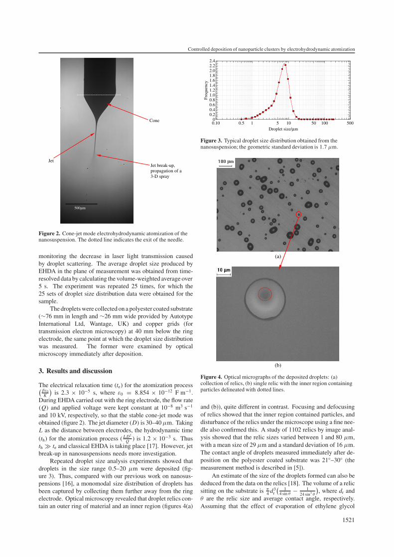
Figure 2. Cone-jet mode electrohydrodynamic atomization of thenanosuspension. The dotted line indicates the exit of the needle.
monitoring the decrease in laser light transmission causedby droplet scattering. The average droplet size produced byEHDA in the plane of measurement was obtained from time-resolved data by calculating the volume-weighted average over5 s. The experiment was repeated 25 times, for which the25 sets of droplet size distribution data were obtained for thesample.
The droplets were collected on a polyester coated substrate(∼76 mm in length and ∼26 mm wide provided by AutotypeInternational Ltd, Wantage, UK) and copper grids (fortransmission electron microscopy) at 40 mm below the ringelectrode, the same point at which the droplet size distributionwas measured. The former were examined by opticalmicroscopy immediately after deposition.
3. Results and discussion
The electrical relaxation time (te) for the atomization process(βε0
K
)is 2.3 × 10⁻⁵ s, where ε0 = 8.854 × 10⁻¹² F m−1.
During EHDA carried out with the ring electrode, the flow rate(Q) and applied voltage were kept constant at 10−8 m3 s−1
and 10 kV, respectively, so that the stable cone-jet mode wasobtained (figure 2). The jet diameter (D) is 30–40 µm. TakingL as the distance between electrodes, the hydrodynamic time(th) for the atomization process ( L D2
Q ) is 1.2 × 10⁻³ s. Thusth � te and classical EHDA is taking place [17]. However, jetbreak-up in nanosuspensions needs more investigation.
Repeated droplet size analysis experiments showed thatdroplets in the size range 0.5–20 µm were deposited (fig-ure 3). Thus, compared with our previous work on nanosus-pensions [16], a monomodal size distribution of droplets hasbeen captured by collecting them further away from the ringelectrode. Optical microscopy revealed that droplet relics con-tain an outer ring of material and an inner region (figures 4(a)
0.10 0.5 1 5 10 50 100 500Droplet size/µm
Fre
quen
cy
00.20.40.60.81.01.21.41.61.82.02.22.4
Figure 3. Typical droplet size distribution obtained from thenanosuspension; the geometric standard deviation is 1.7 µm.
(a)
(b)
Figure 4. Optical micrographs of the deposited droplets: (a)collection of relics, (b) single relic with the inner region containingparticles delineated with dotted lines.
and (b)), quite different in contrast. Focusing and defocusingof relics showed that the inner region contained particles, anddisturbance of the relics under the microscope using a fine nee-dle also confirmed this. A study of 1102 relics by image anal-ysis showed that the relic sizes varied between 1 and 80 µm,with a mean size of 29 µm and a standard deviation of 16 µm.The contact angle of droplets measured immediately after de-position on the polyester coated substrate was 21◦–30◦ (themeasurement method is described in [5]).
An estimate of the size of the droplets formed can also bededuced from the data on the relics [18]. The volume of a relicsitting on the substrate is π
4 d3r
(1
4 sin θ− 1
24 sin3 θ
), where dr and
θ are the relic size and average contact angle, respectively.Assuming that the effect of evaporation of ethylene glycol
1521

S N Jayasinghe et al
(a)
(b)
(c)
Figure 5. (a) and (b) show examples of transmission electron micrographs of the inner regions of the relics and (c) is a typical energydispersive x-ray map of the relics in (a) and (b).
1522
Controlled deposition of nanoparticle clusters by electrohydrodynamic atomization
PEG Silica
Figure 6. Relics generated by printing the nanosuspension. Thevoltage applied to the needle was 11.2 kV and the flow rate of thenanosuspension exiting the needle was 3.2 × 10⁻¹¹ m3 s−1. PEGabbreviates polyethylene glycol.
during atomization is negligible, the corresponding dropletdiameter (DD) is given by DD ≈ 0.91dr
(1
4 sin θ− 1
24 sin3 θ
)1/3.
DD values predicted in this way correlate well with the dropletsizes measured in figure 3.
Further investigations of the relics deposited on coppergrids by transmission electron microscopy confirmed that theinner dense regions contained assembled SiO2 nanoparticles(figures 5(a)–(c); in figure 5(b) the thinnest section is resolvedfurther). The size of the images in the transmission electronmicrographs ranged from 200 nm in figure 5(a) to 15 µm infigure 5(b), correlating well with the size of inner regionsseen in the optical micrographs. Energy dispersive x-rayanalysis (figure 5(c)) confirmed that, indeed, the assembledparticles were SiO2. Brinker et al [19–21] have prepared relicsof the nanoparticles using micro-pen lithography [19, 20],ink-jet printing [19, 20], dip-coating [19, 20] and aerosol-assisted methods [21]. However, the structures produced weremuch coarser [19, 20] and tailored to create meso-porousstructures [21]. In contrast, the structures presented in thispaper represent a much finer scale.
Experiments carried out with the pointed rod-electrodeenabled the controlled deposition of the finer dropletsgenerated under a higher electric field of ∼4 kV mm−1 anda lower flow rate of ∼10−11 m3 s−1, as shown in figure 6.The use of a pointed electrode allows the droplets generatedby EHDA to converge rather than form a plume like in thecase of the ring electrode. These relics also show clearlythe inner core of silica nanoparticles and the outer layerof ethylene glycol, as indicated in figure 4, confirming ourdeductions made above in the experiments carried out with thering electrode. The ethylene glycol can be easily removed bythermolysis. Hence, we have shown that controlled depositionof nanoparticle clusters <50 µm in size according to apredetermined architecture can be realized in this way.
4. Conclusions
The use of electrohydrodynamic atomization to producefine droplets of a nanosuspension has been demonstrated.
On deposition, the nanoparticles separate from the organicmedium used to suspend them, and form clusters. By usinga printing device, which incorporates electrohydrodynamicatomization, we have been able to deposit the droplets inregular arrays, generating nanoparticle relics <50 µm in size.
Acknowledgments
Dr Zophia Luklinska and Mr Mick Willis of the ElectronMicroscopy Unit at the Materials Department at QMUL arethanked for their generous help with the transmission electronmicroscopy. The authors take this opportunity to thank MrAndy Smith of Sympatec, Dr David Catone and Mr RogerCook of Nyacol Nano Technologies, Inc. for collaboratingwith Queen Mary in this work. We acknowledge gratefullyfunding provided by The Royal Society and The Engineeringand Physical Sciences Research Council of the UK forelectrohydrodynamic atomization research at Queen Mary; inparticular, for the two-year research fellowship position forSNJ supported by the Platform Grant (GR/S52636) awardedby the latter.
References
[1] Taylor G I 1964 Proc. R. Soc. 280 383[2] Cloupeau M and Prunet-Foch B 1999 J. Electrost. 25 165[3] Jaworek A and Krupa A 1999 J. Aerosol Sci. 30 873[4] Hartman R P A, Borra J-P, Brunner D J,
Marijnissen J C M and Scarlett B 1999 J. Electrost. 47 143[5] Jayasinghe S N and Edirisinghe M J 2002 J. Aerosol Sci. 33
1379[6] Teng W D, Huneiti Z A, Machowski W, Evans J R G,
Edirisinghe M J and Balachandran W 1997 J. Mater. Sci.Lett. 16 1017
[7] Jayasinghe S N, Edirisinghe M J and de Wilde T 2003 Mater.Res. Innovat. 7 62
[8] Zhao X, Evans J R G, Edirisinghe M J and Song J H 2002J. Mater. Sci. 37 1987
[9] Jayasinghe S N and Edirisinghe M J 2002 Mater. Res. Innovat.6 92
[10] Jayasinghe S N and Edirisinghe M J 2002 J. Porous Mater. 9265
[11] Grigoriev D A, Edirisinghe M J, Bao X, Evans J R G andLuklinska Z B 2001 Phil. Mag. Lett. 81 285
[12] Balachandran W, Miao P and Xiao P 2001 J. Electrost. 50 249[13] Jayasinghe S N and Edirisinghe M J 2003 J. Mater. Sci. Lett.
22 1443[14] Huang J, Best S M, Bonfield W, Brooks R A, Rushton N,
Jayasinghe S N and Edirisinghe M J 2004 Mater. Sci.:Mater. Med. 15 441
[15] Li D, Wang Y and Xia Y 2003 Nanoletters 3 1167[16] Jayasinghe S N and Edirisinghe M J 2004 J. Appl. Ceram.
Tech. 1 140[17] Ganan-Calvo A M, Davila J and Barrero A 1997 J. Aerosol
Sci. 28 249[18] Jayasinghe S N, Edirisinghe M J and Kippax P G 2004 Appl.
Phys. A 78 343[19] Fan H, Lu Y, Stump A, Reed S T, Baer T, Schunk R,
Perez-Luna V, Lopez G P and Brinker C J 2000 Nature 40556
[20] Fan H, Reed S T, Baer T, Schunk R, Lopez G P andBrinker C J 2001 Microporous Mesoporous Mater. 44/45625
[21] Lu Y, Fan H, Stump A, Ward T L, Rieker T and Brinker C J1999 Nature 398 223
1523