controlled delivery of ropinirole hydrochloride … delivery of ropinirole hydrochloride through...
TRANSCRIPT
2013
http://informahealthcare.com/drtISSN: 1061-186X (print), 1029-2330 (electronic)
J Drug Target, Early Online: 1–13! 2013 Informa UK Ltd. DOI: 10.3109/1061186X.2012.757768
ORIGINAL ARTICLE
Controlled delivery of ropinirole hydrochloride through skin usingmodulated iontophoresis and microneedles
Neha D. Singh and Ajay K. Banga
College of Pharmacy and Health Sciences, Mercer University, Atlanta, GA, USA
Abstract
The objective of this study was to investigate the effect of modulated current application usingiontophoresis- and microneedle-mediated delivery on transdermal permeation of ropinirolehydrochloride. AdminPatch� microneedles and microchannels formed by them were charac-terized by scanning electron microscopy, dye staining and confocal microscopy. In vitropermeation studies were carried out using Franz diffusion cells, and skin extraction was used toquantify drug in underlying skin. Effect of microneedle pore density and ions in donorformulation was studied. Active enhancement techniques, continuous iontophoresis(74.13� 2.20mg/cm2) and microneedles (66.97� 10.39 mg/cm2), significantly increased thepermeation of drug with respect to passive delivery (8.25� 2.41 mg/cm2). Modulatediontophoresis could control the amount of drug delivered at a given time point with thehighest flux being 5.12� 1.70 mg/cm2/h (5–7 h) and 5.99� 0.81 mg/cm2/h (20–22 h).Combination of modulated iontophoresis and microneedles (46.50� 6.46mg/cm2) showedsignificantly higher delivery of ropinirole hydrochloride compared to modulated iontophoresisalone (84.91� 9.21mg/cm2). Modulated iontophoresis can help in maintaining precise controlover ropinirole hydrochloride delivery for dose titration in Parkinson’s disease therapy anddeliver therapeutic amounts over a suitable patch area and time.
Keywords
Dose titration, iontophoresis, modulateddelivery, Parkinson’s disease, ropinirole,skin microporation, transdermal
History
Received 17 August 2012Revised 2 December 2012Accepted 9 December 2012Published online 11 January 2013
Introduction
Parkinson’s Disease Foundation (PDF) estimates that around
7–10 million of world population is affected by Parkinson’s
disease [1]. Diagnosis of the disease pathophysiology reveals
degeneration of dopamine cells mainly in the substantia nigra
region of the brain due to genetic or environmental factors and
leads to progressive worsening of the motor functions [2,3].
Parkinson’s disease is marked by symptoms such as tremor,
bradykinesia, rigidity and postural instability which disrupt the
daily life of a patient [4,5]. Levodopa is the first line of therapy
in Parkinson’s disease and considered a ‘‘gold standard’’ in the
treatment regimen. However, as this disease worsens with time,
long-term therapy with levodopa gives rise to side effects like
dyskinesia and fluctuation in motor responses [6].
Ropinirole is a D2 receptor agonist and acts by dopaminer-
gic stimulation of the central and peripheral receptors to offer
symptomatic relief in Parkinson’s disease. Ropinirole hydro-
chloride is available as an oral tablet, both immediate and
extended release, prescribed to alleviate the limitations of
levodopa therapy [7]. The dose range administered to the
patients increases from 0.25 up to 25 mg per day as the disease
progresses thus emphasizing one of the most integral aspects of
Parkinson’s disease management, i.e. dose titration [8,9]. Inter-
patient variability due to differences in age, sex and tolerance
as well as intra-patient variability resulting from constant
neurodegeneration makes customized dosing regimens the
most feasible approach [10]. Since patients are prescribed
multiple oral medications it makes compliance an important
concern and stresses the need for a less frequent/prolonged
administration [11].
Transdermal route of delivery bypasses first pass metabo-
lism (bioavailability of ropinirole is 50%) and can achieve
stable plasma levels when applied once daily. Rotigotine,
another dopamine agonist, which is formulated as a patch for
passive delivery offers the advantage of once daily use and
prolonged effect [12,13]; also a patch can be withdrawn at the
occurrence of side effects like nausea, dizziness, hallucinations
and orthostatic hypotension associated with peripheral receptor
stimulation of ropinirole hydrochloride [14,15]. However, like
oral administration a patch formulation limits the use to
specified doses and does not allow the physician to make
adjustments to the dose tailored to the needs of a patient’s
condition. Ropinirole, which is also a small molecular weight
dopamine agonist, will face a similar challenge when admin-
istered by transdermal route. Ropinirole base is unstable;
hence, formulation of a passive transdermal system is difficult
[16]. A topical formulation or patch of specific strength will
limit the use to a narrow dose range and not according toAddress for correspondence: Ajay K. Banga, College of Pharmacy andHealth Sciences, Mercer University, Atlanta, GA 30341, USA. Tel: 678-547-6243. Fax: 678-547-6423. E-mail: [email protected]
disease progression in a patient. Transdermal administration of
ropinirole can be compared to extended release tablets as in
both the cases dose flexibility is reduced. The lag time
associated with passive delivery will be higher which leads to
delay in alleviating symptoms. Active transdermal techniques,
on the other hand, can help overcoming these limitations.
Iontophoresis is an active enhancement technique where
low current intensities are applied to the skin for topical or
transdermal delivery of charged or neutral molecules. It works
on the principle of electrorepulsion, i.e. like repels like and
electroosmosis where neutral molecules are transported from
anode to cathode along with the bulk solvent flow [17–19]. A
programmed iontophoretic system can allow precise and
controlled delivery of therapeutic agent. Transdermal delivery
of dopamine agonists like ropinirole hydrochloride, apomor-
phine, rotigotine and 5-OH-DPAT using iontophoresis has been
studied due the advantages offered by this technique [20–25].
Therapeutic levels of drug are achieved when current is
applied; however, the level decreases in the post-iontophoretic
phase. However, modulation of the current applied can help in
delivering the required dose and thereafter in maintaining it
based on the disease progression of the patient.
Microneedle-mediated transdermal delivery is a minimally
invasive technique used for hydrophilic and large molecular
weight compounds [26]. Different methods and fabrication
techniques have been described in literature for fabrication of
microneedles made from materials like stainless steel, glass,
polyethylene glycol, carboxymethyl cellulose and maltose to
name a few, as the type used depends on the application [27].
Length and density of pores are important parameters which
determine the efficiency of microchannel formation as well as
the safety concerns. Microchannels remain open up to duration
of 72 h under occluded conditions in vivo and this can be
helpful for delivering drug for prolonged duration at the
microporated site [28]. Combination of iontophoresis and
microneedles has been investigated and encouraging
results have been reported for large molecular weight com-
pounds [29,30].
In this study the enhancement of transdermal permeation
of ropinirole hydrochloride across porcine ear skin model
using iontophoresis and microneedles was investigated.
Continuous iontophoresis (4 h) and its combination with
microneedles were also studied. However, the main objective
was to see if a predictable dose could be delivered if the
duration of iontophoresis was modulated, i.e. 4 h were split
into 2 h application time at two intervals. Modulated
iontophoresis was thus applied and the control over delivery
of drug delivered at a given time point was assessed. We
also studied the effect of combination of modulated
iontophoresis and microneedle on the delivery profile of
the drug at later time points. Although other studies in
literature have used iontophoresis for delivery of antiparkin-
son’s agents, this is the first study which uses modulated
iontophoresis and its combination with microneedles to
achieve the desired delivery at a given time point in
accordance with the dose titration requirement. Effect of
microneedle pore density and presence of ions in solution on
delivery of ropinirole hydrochloride delivery were also
studied. Skin extraction method was used to quantify drug in
underlying skin.
Materials and methods
Materials
Ropinirole hydrochloride was obtained from Hangzhou
Uniwise International Co. Ltd, Hangzhou, China.
AdminPatch� array 600-mm microneedles (1-cm2 patch
area) were purchased from AdminMed. Sodium chloride,
sodium phosphate monobasic, sodium phosphate dibasic and
ammonium acetate buffer were obtained from Fisher
Scientific (Pittsburg, PA). Acetonitrile and centrifuge tubes
for skin extraction studies were obtained from MedSupply
Partners (Atlanta, GA). Silver wire and silver chloride used for
preparing iontophoresis electrodes were obtained from Sigma
Aldrich (St. Louis, MO). 3 M Transpore� tape for tape
stripping was purchased from 3 M (St. Paul, MN). Calcein
(Fluoresoft�, 0.35%) used for imaging micropores was
obtained from Holles Laboratories, Inc. (Cohasset, MA).
Methylene blue used as the hydrophilic dye for characterizing
micropores was obtained from Eastman Kodak Co.
(Rochester, NY). Fluospheres� (0.2 mm) for confocal imaging
studies was purchased from Invitrogen� (Carlsbad, CA). All
the solutions used in this study and HPLC solvents for analyses
were prepared using deionized water. Porcine ears were
obtained from slaughter house.
Methods
Skin preparation
Porcine ear skin was freshly excised, cleaned thoroughly
stored at �80 �C until further use. On the day of experiment,
skin was thawed to room temperature, thoroughly cleaned
with deionized water to remove subcutaneous fat and hair was
carefully cut using scissors. Skin pieces were cut into required
sizes and mounted onto vertical Franz diffusion cells.
Microneedle insertion
A custom-made syringe applicator was used for microneedle
insertion into the skin to help in uniform insertion of 1-cm2
AdminPatch�. The microneedle patch was stuck onto the
circular end of the plunger of a 5-ml syringe using a double
sided tape. The barrel provided an easy grip on the entire
assembly so that uniform force could be applied from all sides
while inserting the microneedles in the skin. Skin was stretched
and microneedles was inserted for 1 min prior to a study.
Characterization of microneedle dimensions by scanning
electron microscopy
Dimensions of AdminPatch� microneedles were confirmed
using a Hitachi S-3700N variable pressure scanning electron
microscope (Hitachi High-Technologies, Maidenhead
Berkshire, UK). Microneedle array was placed in an emission-
field scanning electron microscopy (SEM) with an accelerated
voltage of 15 kV. Images were taken from various distances and
angle to measure the microneedle height and tip sharpness.
Characterization of microchannels created by
AdminPatch� microneedles
Microporated skin was treated with hydrophilic dyes meth-
ylene blue and calcein to visualize pores created by
2 N. D. Singh & A. K. Banga J Drug Target, Early Online: 1–13
AdminPatch� microneedles. Microneedles were inserted into
the skin pieces using a custom-made applicator. Methylene
blue solution (1% w/v solution of water) and fluorescent dye
calcein were placed onto the microporated skin for duration of
1 min. Methylene blue dye was removed from the skin using
kimwipes and alcohol swabs. Proscope HR video microscope
(Bodelin Technologies, Lake Oswego, OR) was used to
capture images of the stained micropores. For calcein dye, the
skin was cleaned with kimwipes to remove excess dye and
images were immediately taken using a fluorescent camera
(Nikon camera integrated with a macrolens and 525-nm long-
pass filter). The images from calcein dye studies were further
analyzed by Fluoropore software which measures fluorescent
intensity around each pore and calculates a value called as
pore permeability index (PPI).
Confocal microscopy was performed to measure the depth
of microchannels formed by AdminPatch� microneedles.
Porcine ear skin was microporated followed by application of
FluoSpheres� for 40 s. Excess solution was cleared by
kimwipes, the skin piece was mounted onto a slide and
observed under Zeiss confocal microscope. Images were
obtained using X–Z sectioning at excitation/emission wave-
length of 495/515 nm, respectively.
Preparation of electrodes
Silver wire was manually coiled into circles and used as
anode. Silver chloride coated onto a thin silver wire served as
the cathode. To briefly describe this procedure, silver chloride
was melted by heating in a crucible. Silver wire was coiled at
one end and dipped into the solution to ensure sufficient and
uniform coating over the surface.
Permeation studies
In vitro studies (n� 3) were carried out on porcine ear skin
using vertical Franz diffusion cells. The diffusion cells were
surrounded by a water jacket and temperature of the water
bath was maintained at 37� 1 �C. The receptor compartment
was washed prior to each experiment and filled with a 5-ml
phosphate buffered saline (50 mM) which was used as the
receptor buffer. Skin samples for passive and iontophoresis
treatment were mounted onto the Franz diffusion cells
(effective surface area of 0.64 cm2) such that the dermal
side faced receptor compartment and donor was placed on the
epidermal side. For microneedle and combination studies skin
samples were microporated according to the procedure
mentioned and then mounted onto the Franz cells in a
manner similar to passive and iontophoresis. Donor cells were
placed on the skin and the entire assembly was maintained
together using clamps. Donor formulation (500 ml) was then
placed on the skin surface. Donor formulation consisted of
1 mg/ml solution of ropinirole hydrochloride in phosphate
buffer and 75 mM sodium chloride. Anodal iontophoresis was
used for studying iontophoretic delivery of ropinirole hydro-
chloride across skin. Silver wire coil (anode) was immersed in
the donor compartment and it was ensured that the coil did
not touch skin surface and sides of donor compartment. Silver
chloride wire (cathode) was placed in the receptor chamber
arm. Current supply (Keithley Instruments, Cleveland, OH)
was maintained at 0.2 mA/cm2 for a total duration of 4 h;
however, the time points of current application were varied for
continuous versus modulation experiments. For combination
studies, microporated skin was first mounted onto Franz
diffusion cell; donor was placed on the skin surface followed
by application of anodal iontophoresis. Passive permeation
study was performed on untreated skin as a control for
enhancement using active techniques iontophoresis and
microneedles. Samples were withdrawn from receptor com-
partment (500ml) at predetermined time points and equal
volume of fresh receptor buffer was added immediately.
Samples were analyzed using a validated HPLC assay.
Continuous versus modulated iontophoresis
Iontophoresis was applied for a total duration of 4 h; however,
the pattern of application was changed. Continuous iontopho-
resis was applied at a stretch from 0 to 4 h. Modulated
iontophoresis was applied in intervals, from 0 to 2 h followed
by 3 h of passive diffusion and then again from 5 to 7 h.
Samples were taken up to 24 h and passive diffusion of
ropinirole hydrochloride through the skin post-iontophoresis
was also studied. In another experiment modulated iontopho-
resis was applied from 0 to 2 h and 20 to 22 h and sampling
was carried until 32 h.
Effect of microneedle pore density
Skin was microporated with microneedles one time, two times
and three times over the same area (0.64 cm2) to study the
effect of increasing pore density on delivery of ropinirole
hydrochloride, when using microneedles alone. The effect of
increasing pore density was also studied for combination
approach where skin was microporated once and twice
followed by application of iontophoresis over the same area.
Effect of ions in solution
Donor formulations consisting of phosphate buffer (50 mM)
and distilled water were compared for studying the effect of
ions on iontophoresis-mediated delivery of ropinirole hydro-
chloride. Sodium chloride (75 mM) was added to both donors.
pH of the donors was noted using a MI-410 combination pH
electrode (Microelectrodes, Inc., Bedford, NH).
Skin extraction
Drug present in the skin was quantified using a skin extraction
assay. Following permeation study, the donor formulation was
pipetted from the skin surface. The mounted skin was cleaned
using Q-tips and receptor solution three times to ensure
removal of excess donor formulation. The skin piece was then
dabbed with kimwipes to dry the remaining moisture. The
permeation area was then tape stripped with 3 M Transpore
tape one time to clean any excess formulation resulting in
overestimation of ropinirole hydrochloride. Skin pieces were
then minced using a pair of scissors into centrifuge tubes.
Extraction solvent (3 ml of distilled water) was added to each
centrifuge tube and the samples were sonicated using Fisher
Scientific mechanical ultrasonic cleaner FS110 for 20 min.
Overnight extraction was carried out by placing the samples
on a roller shaker (New Brunswick Scientific Co. Inc.,
Edison, NJ) at a speed of 200 rpm. Samples were again
DOI: 10.3109/1061186X.2012.757768 Controlled delivery of ropinirole hydrochloride 3
sonicated for 20 min the next day followed by centrifugation
(Sorvall RT6000 refrigerated centrifuge) at 4000 rpm for
10 min to separate the supernatant extract from skin pieces.
The extract was filtered, diluted as required and analyzed
using HPLC method.
Recovery studies
Extraction efficiency was determined in vitro in porcine ear
skin using recovery study. Ropinirole hydrochloride standard
solutions (200, 500 and 1000 mg/ml) were made and 50 ml of
each solution was injected into porcine ear skin (n¼ 3).
Injected samples were incubated for 4 h and the skin
extraction procedure mentioned in detail above was followed.
Amount of drug extracted was quantified using a standard
curve and the extraction recovery was found to be
78.45� 4.56%. This recovery value was used when quanti-
fying drug for skin samples from in vitro permeation
experiments (total amount of drug calculated from high-
performance liquid chromatography (HPLC) quantification
was divided by 0.7845 to accurately predict drug in skin).
Quantitative analysis
Ropinirole hydrochloride was quantified using HPLC by
using modified assay from literature. Analysis was performed
on Perkin Elmer System (Waltham, MA) with a UV detector
operating at 245 nm. The column used was Phenomenex
Gemini-NX 5 m C18 110 A; 250� 4.6 mm. Mobile phase
consisted of ammonium acetate buffer, 50 mM pH 7.8:
acetonitrile (50:50%, v/v). Isocratic elution was performed
at flow rate of 0.6 ml/min after injecting 10 ml of sample. The
total run time was 7 min and the retention time of ropinirole
hydrochloride was around 5 min. Standards were prepared in
the range of 0.1–100mg and the assay was sensitive to
detection within this range.
Statistical analysis
Statistical significance was determined using analysis of
variance (ANOVA) and Student’s t-test. Tukey’s honestly
significant difference post hoc test was performed for specific
comparison.
Results
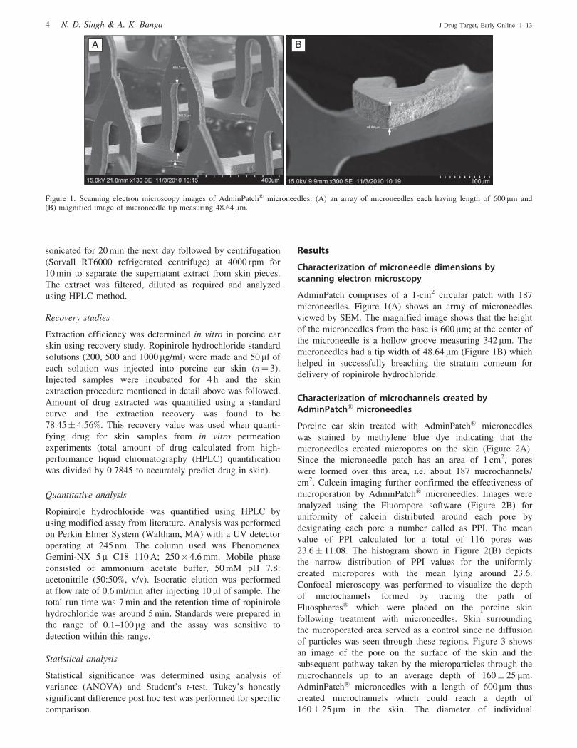
Characterization of microneedle dimensions byscanning electron microscopy
AdminPatch comprises of a 1-cm2 circular patch with 187
microneedles. Figure 1(A) shows an array of microneedles
viewed by SEM. The magnified image shows that the height
of the microneedles from the base is 600 mm; at the center of
the microneedle is a hollow groove measuring 342 mm. The
microneedles had a tip width of 48.64 mm (Figure 1B) which
helped in successfully breaching the stratum corneum for
delivery of ropinirole hydrochloride.
Characterization of microchannels created byAdminPatch� microneedles
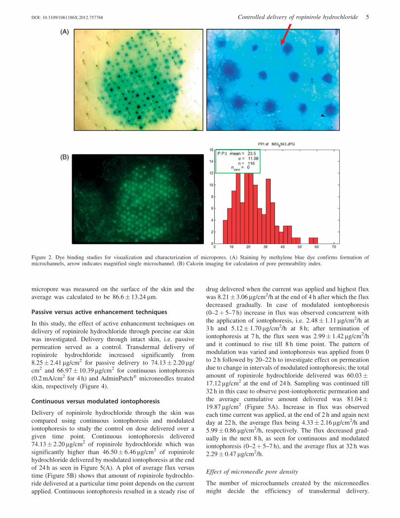
Porcine ear skin treated with AdminPatch� microneedles
was stained by methylene blue dye indicating that the
microneedles created micropores on the skin (Figure 2A).
Since the microneedle patch has an area of 1 cm2, pores
were formed over this area, i.e. about 187 microchannels/
cm2. Calcein imaging further confirmed the effectiveness of
microporation by AdminPatch� microneedles. Images were
analyzed using the Fluoropore software (Figure 2B) for
uniformity of calcein distributed around each pore by
designating each pore a number called as PPI. The mean
value of PPI calculated for a total of 116 pores was
23.6� 11.08. The histogram shown in Figure 2(B) depicts
the narrow distribution of PPI values for the uniformly
created micropores with the mean lying around 23.6.
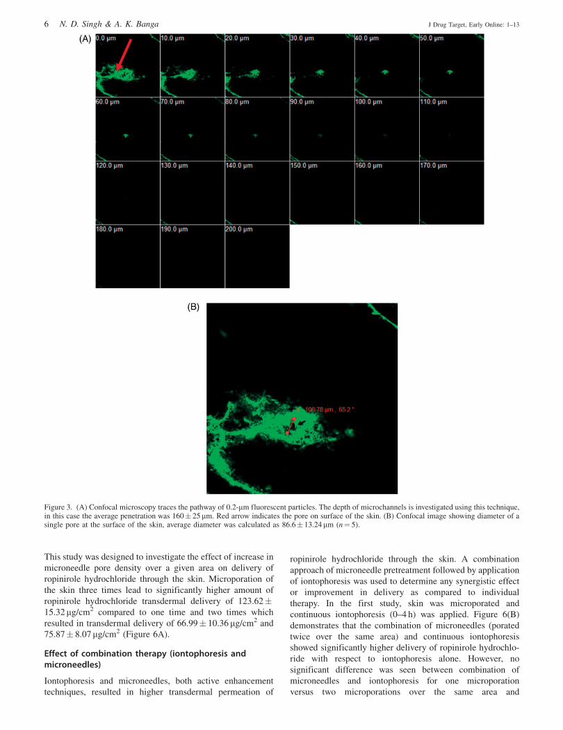
Confocal microscopy was performed to visualize the depth
of microchannels formed by tracing the path of
Fluospheres� which were placed on the porcine skin
following treatment with microneedles. Skin surrounding
the microporated area served as a control since no diffusion
of particles was seen through these regions. Figure 3 shows
an image of the pore on the surface of the skin and the
subsequent pathway taken by the microparticles through the
microchannels up to an average depth of 160� 25 mm.
AdminPatch� microneedles with a length of 600mm thus
created microchannels which could reach a depth of
160� 25 mm in the skin. The diameter of individual
A B
Figure 1. Scanning electron microscopy images of AdminPatch� microneedles: (A) an array of microneedles each having length of 600 mm and(B) magnified image of microneedle tip measuring 48.64 mm.
4 N. D. Singh & A. K. Banga J Drug Target, Early Online: 1–13
micropore was measured on the surface of the skin and the
average was calculated to be 86.6� 13.24 mm.
Passive versus active enhancement techniques
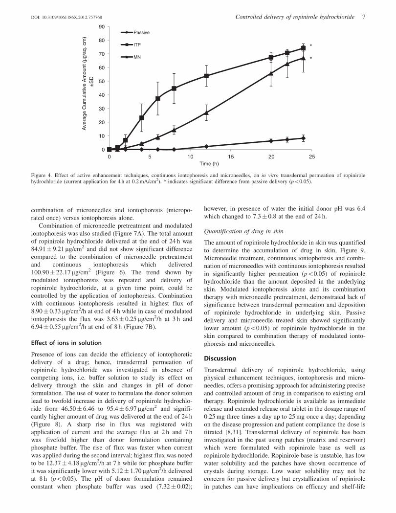
In this study, the effect of active enhancement techniques on
delivery of ropinirole hydrochloride through porcine ear skin
was investigated. Delivery through intact skin, i.e. passive
permeation served as a control. Transdermal delivery of
ropinirole hydrochloride increased significantly from
8.25� 2.41 mg/cm2 for passive delivery to 74.13� 2.20 mg/
cm2 and 66.97� 10.39mg/cm2 for continuous iontophoresis
(0.2 mA/cm2 for 4 h) and AdminPatch� microneedles treated
skin, respectively (Figure 4).
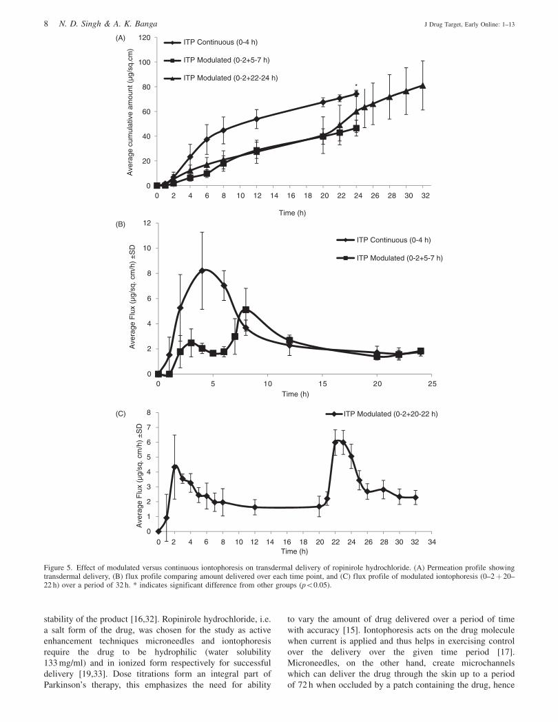
Continuous versus modulated iontophoresis
Delivery of ropinirole hydrochloride through the skin was
compared using continuous iontophoresis and modulated
iontophoresis to study the control on dose delivered over a
given time point. Continuous iontophoresis delivered
74.13� 2.20 mg/cm2 of ropinirole hydrochloride which was
significantly higher than 46.50� 6.46 mg/cm2 of ropinirole
hydrochloride delivered by modulated iontophoresis at the end
of 24 h as seen in Figure 5(A). A plot of average flux versus
time (Figure 5B) shows that amount of ropinirole hydrochlo-
ride delivered at a particular time point depends on the current
applied. Continuous iontophoresis resulted in a steady rise of
drug delivered when the current was applied and highest flux
was 8.21� 3.06 mg/cm2/h at the end of 4 h after which the flux
decreased gradually. In case of modulated iontophoresis
(0–2þ 5–7 h) increase in flux was observed concurrent with
the application of iontophoresis, i.e. 2.48� 1.11 mg/cm2/h at
3 h and 5.12� 1.70 mg/cm2/h at 8 h; after termination of
iontophoresis at 7 h, the flux seen was 2.99� 1.42 mg/cm2/h
and it continued to rise till 8 h time point. The pattern of
modulation was varied and iontophoresis was applied from 0
to 2 h followed by 20–22 h to investigate effect on permeation
due to change in intervals of modulated iontophoresis; the total
amount of ropinirole hydrochloride delivered was 60.03�17.12 mg/cm2 at the end of 24 h. Sampling was continued till
32 h in this case to observe post-iontophoretic permeation and
the average cumulative amount delivered was 81.04�19.87 mg/cm2 (Figure 5A). Increase in flux was observed
each time current was applied, at the end of 2 h and again next
day at 22 h, the average flux being 4.33� 2.16mg/cm2/h and
5.99� 0.86 mg/cm2/h, respectively. The flux decreased grad-
ually in the next 8 h, as seen for continuous and modulated
iontophoresis (0–2þ 5–7 h), and the average flux at 32 h was
2.29� 0.47 mg/cm2/h.
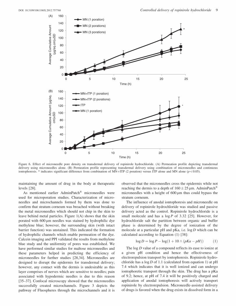
Effect of microneedle pore density
The number of microchannels created by the microneedles
might decide the efficiency of transdermal delivery.
Figure 2. Dye binding studies for visualization and characterization of micropores. (A) Staining by methylene blue dye confirms formation ofmicrochannels, arrow indicates magnified single microchannel. (B) Calcein imaging for calculation of pore permeability index.
DOI: 10.3109/1061186X.2012.757768 Controlled delivery of ropinirole hydrochloride 5
This study was designed to investigate the effect of increase in
microneedle pore density over a given area on delivery of
ropinirole hydrochloride through the skin. Microporation of
the skin three times lead to significantly higher amount of
ropinirole hydrochloride transdermal delivery of 123.62�15.32mg/cm2 compared to one time and two times which
resulted in transdermal delivery of 66.99� 10.36 mg/cm2 and
75.87� 8.07 mg/cm2 (Figure 6A).
Effect of combination therapy (iontophoresis andmicroneedles)
Iontophoresis and microneedles, both active enhancement
techniques, resulted in higher transdermal permeation of
ropinirole hydrochloride through the skin. A combination
approach of microneedle pretreatment followed by application
of iontophoresis was used to determine any synergistic effect
or improvement in delivery as compared to individual
therapy. In the first study, skin was microporated and
continuous iontophoresis (0–4 h) was applied. Figure 6(B)
demonstrates that the combination of microneedles (porated
twice over the same area) and continuous iontophoresis
showed significantly higher delivery of ropinirole hydrochlo-
ride with respect to iontophoresis alone. However, no
significant difference was seen between combination of
microneedles and iontophoresis for one microporation
versus two microporations over the same area and
Figure 3. (A) Confocal microscopy traces the pathway of 0.2-mm fluorescent particles. The depth of microchannels is investigated using this technique,in this case the average penetration was 160� 25 mm. Red arrow indicates the pore on surface of the skin. (B) Confocal image showing diameter of asingle pore at the surface of the skin, average diameter was calculated as 86.6� 13.24 mm (n¼ 5).
6 N. D. Singh & A. K. Banga J Drug Target, Early Online: 1–13
combination of microneedles and iontophoresis (micropo-
rated once) versus iontophoresis alone.
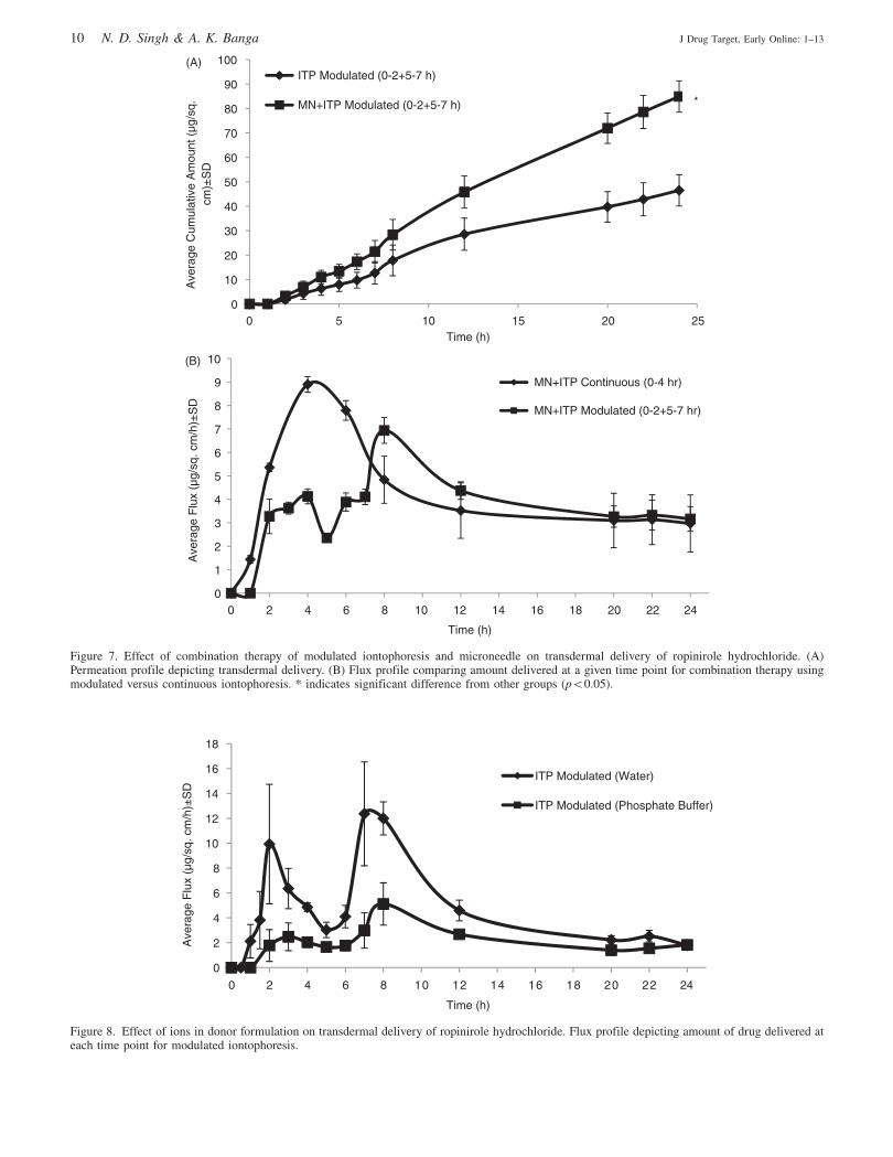
Combination of microneedle pretreatment and modulated
iontophoresis was also studied (Figure 7A). The total amount
of ropinirole hydrochloride delivered at the end of 24 h was
84.91� 9.21 mg/cm2 and did not show significant difference
compared to the combination of microneedle pretreatment
and continuous iontophoresis which delivered
100.90� 22.17mg/cm2 (Figure 6). The trend shown by
modulated iontophoresis was repeated and delivery of
ropinirole hydrochloride, at a given time point, could be
controlled by the application of iontophoresis. Combination
with continuous iontophoresis resulted in highest flux of
8.90� 0.33 mg/cm2/h at end of 4 h while in case of modulated
iontophoresis the flux was 3.63� 0.25 mg/cm2/h at 3 h and
6.94� 0.55 mg/cm2/h at end of 8 h (Figure 7B).
Effect of ions in solution
Presence of ions can decide the efficiency of iontophoretic
delivery of a drug; hence, transdermal permeation of
ropinirole hydrochloride was investigated in absence of
competing ions, i.e. buffer solution to study its effect on
delivery through the skin and changes in pH of donor
formulation. The use of water to formulate the donor solution
lead to twofold increase in delivery of ropinirole hydrochlo-
ride from 46.50� 6.46 to 95.4� 6.97 mg/cm2 and signifi-
cantly higher amount of drug was delivered at the end of 24 h
(Figure 8). A sharp rise in flux was registered with
application of current and the average flux at 2 h and 7 h
was fivefold higher than donor formulation containing
phosphate buffer. The rise of flux was faster when current
was applied during the second interval; highest flux was noted
to be 12.37� 4.18 mg/cm2/h at 7 h while for phosphate buffer
it was significantly lower with 5.12� 1.70 mg/cm2/h delivered
at 8 h (p50.05). The pH of donor formulation remained
constant when phosphate buffer was used (7.32� 0.02);
however, in presence of water the initial donor pH was 6.4
which changed to 7.3� 0.8 at the end of 24 h.
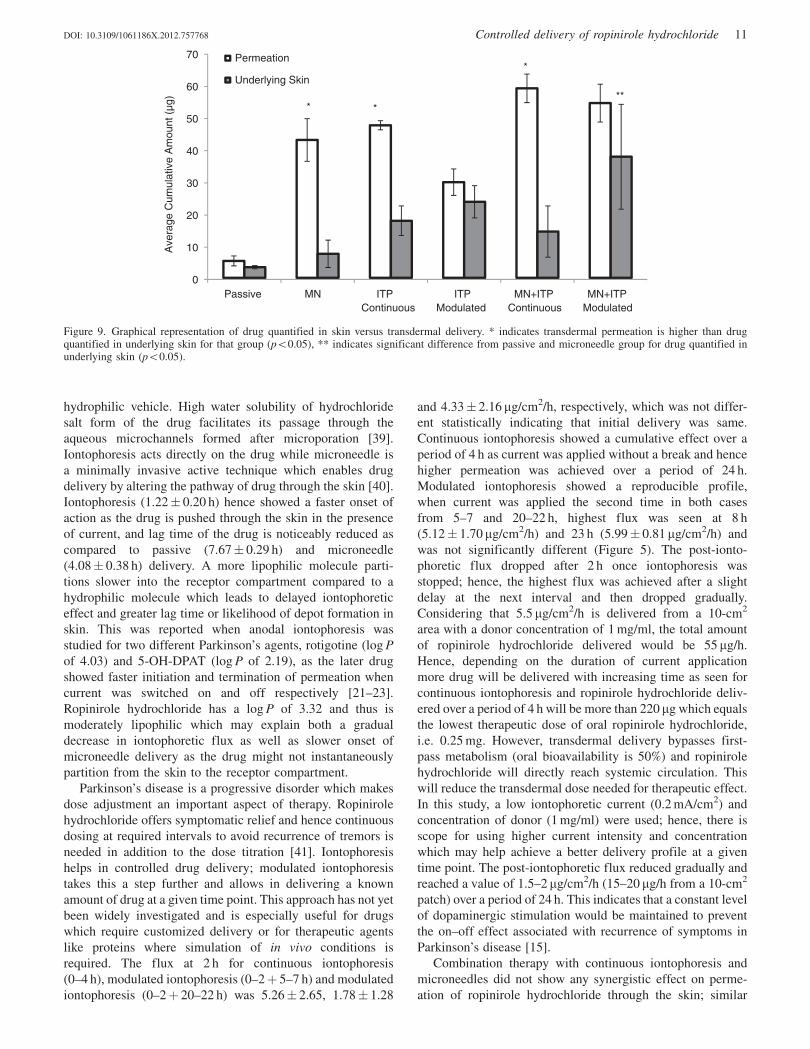
Quantification of drug in skin
The amount of ropinirole hydrochloride in skin was quantified
to determine the accumulation of drug in skin, Figure 9.
Microneedle treatment, continuous iontophoresis and combi-
nation of microneedles with continuous iontophoresis resulted
in significantly higher permeation (p50.05) of ropinirole
hydrochloride than the amount deposited in the underlying
skin. Modulated iontophoresis alone and its combination
therapy with microneedle pretreatment, demonstrated lack of
significance between transdermal permeation and deposition
of ropinirole hydrochloride in underlying skin. Passive
delivery and microneedle treated skin showed significantly
lower amount (p50.05) of ropinirole hydrochloride in the
skin compared to combination therapy of modulated ionto-
phoresis and microneedles.
Discussion
Transdermal delivery of ropinirole hydrochloride, using
physical enhancement techniques, iontophoresis and micro-
needles, offers a promising approach for administering precise
and controlled amount of drug in comparison to existing oral
therapy. Ropinirole hydrochloride is available as immediate
release and extended release oral tablet in the dosage range of
0.25 mg three times a day up to 25 mg once a day; depending
on the disease progression and patient compliance the dose is
titrated [8,31]. Transdermal delivery of ropinirole has been
investigated in the past using patches (matrix and reservoir)
which were formulated with ropinirole base as well as
ropinirole hydrochloride. Ropinirole base is unstable, has low
water solubility and the patches have shown occurrence of
crystals during storage. Low water solubility may not be
concern for passive delivery but crystallization of ropinirole
in patches can have implications on efficacy and shelf-life
0
10
20
30
40
50
60
70
80
90
0 5 10 15 20 25
Ave
rage
Cum
ulat
ive
Am
ount
(µg
/sq.
cm
) ±
SD
Time (h)
Passive
ITP
MN
*
*
Figure 4. Effect of active enhancement techniques, continuous iontophoresis and microneedles, on in vitro transdermal permeation of ropinirolehydrochloride (current application for 4 h at 0.2 mA/cm2). * indicates significant difference from passive delivery (p50.05).
DOI: 10.3109/1061186X.2012.757768 Controlled delivery of ropinirole hydrochloride 7
stability of the product [16,32]. Ropinirole hydrochloride, i.e.
a salt form of the drug, was chosen for the study as active
enhancement techniques microneedles and iontophoresis
require the drug to be hydrophilic (water solubility
133 mg/ml) and in ionized form respectively for successful
delivery [19,33]. Dose titrations form an integral part of
Parkinson’s therapy, this emphasizes the need for ability
to vary the amount of drug delivered over a period of time
with accuracy [15]. Iontophoresis acts on the drug molecule
when current is applied and thus helps in exercising control
over the delivery over the given time period [17].
Microneedles, on the other hand, create microchannels
which can deliver the drug through the skin up to a period
of 72 h when occluded by a patch containing the drug, hence
0
20
40
60
80
100
120(A)
(B)
(C)
0 2 4 6 8 10 12 14 16 18 20 22 24 26 28 30 32
Ave
rage
cum
ulat
ive
amou
nt (
µg/s
q.cm
)
Time (h)
ITP Continuous (0-4 h)
ITP Modulated (0-2+5-7 h)
ITP Modulated (0-2+22-24 h)
0
2
4
6
8
10
12
0 5 10 15 20 25
Ave
rage
Flu
x (µ
g/sq
. cm
/h)
±S
D
Time (h)
ITP Continuous (0-4 h)
ITP Modulated (0-2+5-7 h)
*
0
1
2
3
4
5
6
7
8
0 2 4 6 8 10 12 14 16 18 20 22 24 26 28 30 32 34
Ave
rage
Flu
x (µ
g/sq
. cm
/h)
±S
D
Time (h)
ITP Modulated (0-2+20-22 h)
Figure 5. Effect of modulated versus continuous iontophoresis on transdermal delivery of ropinirole hydrochloride. (A) Permeation profile showingtransdermal delivery, (B) flux profile comparing amount delivered over each time point, and (C) flux profile of modulated iontophoresis (0–2þ 20–22 h) over a period of 32 h. * indicates significant difference from other groups (p50.05).
8 N. D. Singh & A. K. Banga J Drug Target, Early Online: 1–13
maintaining the amount of drug in the body at therapeutic
levels [28].
As mentioned earlier AdminPatch� microneedles were
used for microporation studies. Characterization of micro-
needles and microchannels formed by them was done to
confirm that stratum corneum was breached without breaking
the metal microneedles which should not chip in the skin to
leave behind metal particles. Figure 1(A) shows that the skin
porated with 600 mm needles was stained by hydrophilic dye
methylene blue; however, the surrounding skin (with intact
barrier function) was unstained. This indicated the formation
of hydrophilic channels which enable permeation of the dye.
Calcein imaging and PPI validated the results from methylene
blue study and the uniformity of pores was established. We
have performed similar studies for maltose microneedles and
these parameters helped in predicting the efficiency of
microneedles for further studies [28,34]. Microneedles are
designed to disrupt the epidermis for transdermal delivery;
however, any contact with the dermis is undesirable as this
layer comprises of nerves which are sensitive to needles; pain
associated with hypodermic needles is due to this reason
[35–37]. Confocal microscopy showed that the microneedles
successfully created microchannels. Figure 3 depicts the
pathway of Fluospheres through the microchannels and it is
observed that the microneedles cross the epidermis while not
reaching the dermis to a depth of 160� 25 mm. AdminPatch�
microneedles with a height of 600 mm thus could bypass the
stratum corneum.
The influence of anodal iontophoresis and microneedle on
delivery of ropinirole hydrochloride was studied and passive
delivery acted as the control. Ropinirole hydrochloride is a
small molecule and has a log P of 3.32 [25]. However, for
hydrochloride salt the partition between organic and buffer
phase is determined by the degree of ionization of the
molecule at a particular pH and pKa, i.e. log D which can be
calculated according to Equation (1) [38]:
log D ¼ log P� log 1þ 10 ^ pKa� pHð Þð Þ ð1Þ
The log D value of a compound reflects its ease to ionize at
a given pH condition and hence the effectiveness of
electrorepulsion transport by iontophoresis. Ropinirole hydro-
chloride has a log D of 1.1 (calculated from equation 1) at pH
7.4 which indicates that it is well ionized and can undergo
iontophoretic transport through the skin. The drug has a pKa
of 9.2; hence, at pH of 7.4 it will be positively charged and
application of anodal iontophoresis will actively transport
ropinirole by electrorepulsion. Microneedle-assisted delivery
of drugs is favored when the drug exists in dissolved form in a
0
20
40
60
80
100
120
140
160(A)
(B)
0 5 10 15 20 25
Ave
rage
Cum
ulat
ive
Am
ount
(µ
g/sq
.cm
)±S
D
Time (h)
MN (1 poration)
MN (2 porations)
MN (3 porations)
0
20
40
60
80
100
120
140
160
0 5 10 15 20 25
Ave
rage
Cum
ulat
ive
Am
ount
(µg
/sq.
cm
)±S
D
Time (h)
MN+ITP (1 poration)
MN+ITP (2 porations)
ITP
MN (1 poration)
*
*
Figure 6. Effect of microneedle pore density on transdermal delivery of ropinirole hydrochloride. (A) Permeation profile depicting transdermaldelivery using microneedles alone. (B) Permeation profile representing transdermal delivery using combination of microneedles and continuousiontophoresis. * indicates significant difference from combination of MNþITP (2 poration) versus ITP alone and MN alone (p50.05).
DOI: 10.3109/1061186X.2012.757768 Controlled delivery of ropinirole hydrochloride 9
0
10
20
30
40
50
60
70
80
90
100(A)
(B)
0 5 10 15 20 25
Ave
rage
Cum
ulat
ive
Am
ount
(µg
/sq.
cm
)±S
D
Time (h)
ITP Modulated (0-2+5-7 h)
MN+ITP Modulated (0-2+5-7 h)
0
1
2
3
4
5
6
7
8
9
10
0 2 4 6 8 10 12 14 16 18 20 22 24
Ave
rage
Flu
x (µ
g/sq
. cm
/h)±
SD
Time (h)
MN+ITP Continuous (0-4 hr)
MN+ITP Modulated (0-2+5-7 hr)
*
Figure 7. Effect of combination therapy of modulated iontophoresis and microneedle on transdermal delivery of ropinirole hydrochloride. (A)Permeation profile depicting transdermal delivery. (B) Flux profile comparing amount delivered at a given time point for combination therapy usingmodulated versus continuous iontophoresis. * indicates significant difference from other groups (p50.05).
0
2
4
6
8
10
12
14
16
18
0 2 4 6 8 10 12 14 16 18 20 22 24
Ave
rage
Flu
x (µ
g/sq
. cm
/h)±
SD
Time (h)
ITP Modulated (Water)
ITP Modulated (Phosphate Buffer)
Figure 8. Effect of ions in donor formulation on transdermal delivery of ropinirole hydrochloride. Flux profile depicting amount of drug delivered ateach time point for modulated iontophoresis.
10 N. D. Singh & A. K. Banga J Drug Target, Early Online: 1–13
hydrophilic vehicle. High water solubility of hydrochloride
salt form of the drug facilitates its passage through the
aqueous microchannels formed after microporation [39].
Iontophoresis acts directly on the drug while microneedle is
a minimally invasive active technique which enables drug
delivery by altering the pathway of drug through the skin [40].
Iontophoresis (1.22� 0.20 h) hence showed a faster onset of
action as the drug is pushed through the skin in the presence
of current, and lag time of the drug is noticeably reduced as
compared to passive (7.67� 0.29 h) and microneedle
(4.08� 0.38 h) delivery. A more lipophilic molecule parti-
tions slower into the receptor compartment compared to a
hydrophilic molecule which leads to delayed iontophoretic
effect and greater lag time or likelihood of depot formation in
skin. This was reported when anodal iontophoresis was
studied for two different Parkinson’s agents, rotigotine (log P
of 4.03) and 5-OH-DPAT (log P of 2.19), as the later drug
showed faster initiation and termination of permeation when
current was switched on and off respectively [21–23].
Ropinirole hydrochloride has a log P of 3.32 and thus is
moderately lipophilic which may explain both a gradual
decrease in iontophoretic flux as well as slower onset of
microneedle delivery as the drug might not instantaneously
partition from the skin to the receptor compartment.
Parkinson’s disease is a progressive disorder which makes
dose adjustment an important aspect of therapy. Ropinirole
hydrochloride offers symptomatic relief and hence continuous
dosing at required intervals to avoid recurrence of tremors is
needed in addition to the dose titration [41]. Iontophoresis
helps in controlled drug delivery; modulated iontophoresis
takes this a step further and allows in delivering a known
amount of drug at a given time point. This approach has not yet
been widely investigated and is especially useful for drugs
which require customized delivery or for therapeutic agents
like proteins where simulation of in vivo conditions is
required. The flux at 2 h for continuous iontophoresis
(0–4 h), modulated iontophoresis (0–2þ 5–7 h) and modulated
iontophoresis (0–2þ 20–22 h) was 5.26� 2.65, 1.78� 1.28
and 4.33� 2.16 mg/cm2/h, respectively, which was not differ-
ent statistically indicating that initial delivery was same.
Continuous iontophoresis showed a cumulative effect over a
period of 4 h as current was applied without a break and hence
higher permeation was achieved over a period of 24 h.
Modulated iontophoresis showed a reproducible profile,
when current was applied the second time in both cases
from 5–7 and 20–22 h, highest flux was seen at 8 h
(5.12� 1.70mg/cm2/h) and 23 h (5.99� 0.81 mg/cm2/h) and
was not significantly different (Figure 5). The post-ionto-
phoretic flux dropped after 2 h once iontophoresis was
stopped; hence, the highest flux was achieved after a slight
delay at the next interval and then dropped gradually.
Considering that 5.5 mg/cm2/h is delivered from a 10-cm2
area with a donor concentration of 1 mg/ml, the total amount
of ropinirole hydrochloride delivered would be 55 mg/h.
Hence, depending on the duration of current application
more drug will be delivered with increasing time as seen for
continuous iontophoresis and ropinirole hydrochloride deliv-
ered over a period of 4 h will be more than 220 mg which equals
the lowest therapeutic dose of oral ropinirole hydrochloride,
i.e. 0.25 mg. However, transdermal delivery bypasses first-
pass metabolism (oral bioavailability is 50%) and ropinirole
hydrochloride will directly reach systemic circulation. This
will reduce the transdermal dose needed for therapeutic effect.
In this study, a low iontophoretic current (0.2 mA/cm2) and
concentration of donor (1 mg/ml) were used; hence, there is
scope for using higher current intensity and concentration
which may help achieve a better delivery profile at a given
time point. The post-iontophoretic flux reduced gradually and
reached a value of 1.5–2 mg/cm2/h (15–20 mg/h from a 10-cm2
patch) over a period of 24 h. This indicates that a constant level
of dopaminergic stimulation would be maintained to prevent
the on–off effect associated with recurrence of symptoms in
Parkinson’s disease [15].
Combination therapy with continuous iontophoresis and
microneedles did not show any synergistic effect on perme-
ation of ropinirole hydrochloride through the skin; similar
0
10
20
30
40
50
60
70
Passive MN ITP Continuous
ITP Modulated
MN+ITP Continuous
MN+ITP Modulated
Ave
rage
Cum
ulat
ive
Am
ount
(µg
)
Permeation
Underlying Skin
*
*
***
Figure 9. Graphical representation of drug quantified in skin versus transdermal delivery. * indicates transdermal permeation is higher than drugquantified in underlying skin for that group (p50.05), ** indicates significant difference from passive and microneedle group for drug quantified inunderlying skin (p50.05).
DOI: 10.3109/1061186X.2012.757768 Controlled delivery of ropinirole hydrochloride 11
trend has been reported in the literature for other small
molecular weight compounds including deuterium oxide,
methylene blue, theophylline and fluorescein sodium [29,42].
On the other hand, large molecular weight drugs/therapeutic
agents show increased delivery as compared to individual
treatment [30]. Small molecular weight drugs can be easily
transported by iontophoresis through shunt pathways in the
skin; however, this does not apply to high molecular weight
compounds. Micron-sized pores formed by microneedles can
deliver molecules as large in size as a monoclonal antibody,
vaccines, proteins and peptides. For small molecular weight
compound, additional pathways created by combination
therapy may not significantly drive higher amounts of drug
through them as compared to individual active techniques
[35]. The reason for this can be an efficient transport of drug
through existing pathways when using iontophoresis and
microneedles alone so that further enhancement of delivery
cannot be seen at same concentration and microneedle pore
density. However, exceptions to this have been reported [43]
and further work is needed to establish mechanism behind the
same. Higher flux was seen with iontophoresis at initial time
points which was concurrent with the application of current,
whereas microneedles achieved greater flux at later time
points after a gradual increase in delivery at start of the
experiment. Combination therapy on the other hand showed
increase in flux when iontophoresis was applied and also
helped in maintaining higher flux at later time points as
compared to iontophoresis alone. Modulation of iontophoresis
during combination therapy, however, resulted in significantly
higher permeation of ropinirole hydrochloride with respect to
modulated iontophoresis (p50.05). The presence of micro-
channels may be a reason for better delivery by modulated
combination therapy. When using modulated iontophoresis
alone, the delivery only depends on application of current and
flux remains low in the absence of iontophoresis whereas for
modulated combination therapy drug continues permeating
through the microchannels in the absence of current, main-
taining the flux until it rises at next iontophoretic interval.
This can avoid fluctuations in steady state levels of ropinirole
hydrochloride associated with appearance of side-effects
during ‘‘off’’ period of therapy [15].
The permeation of ropinirole hydrochloride was signifi-
cantly increased when the skin was porated thrice over the
same area compared to once and twice. This can be attributed
to the increase in number of microchannels formed over the
exposed permeation area. However, no significant difference
was seen between one time and two time poration. It has been
reported before that the amount of drug delivered increases
with the number of pores formed over an area and our results
confirm this [35,44]; however, the increase may not be linear
and depends on the total number of pores formed over that
area as seen in the case of one poration versus two porations.
Combination of iontophoresis and skin microporated twice
over the same area lead to significantly higher permeation of
ropinirole hydrochloride compared to combination of ionto-
phoresis and skin microporated once which confirms that
microneedle pore density does affect permeation of ropinirole
hydrochloride.
The efficiency of iontophoretic transport depends on the
transport number of the charged drug species being delivered
under the electrode [45]. In presence of small molecular
weight charged competing ions the transport number of drug
decreases thus affecting the flux [46].
J ¼ I � t � F � z, ð2Þ
where J equals flux, I is the current applied, F is Faraday’s
constant and z is the valency of drug.
Our finding confirms these results as the use of water in
presence of 75 mM sodium chloride leads to twofold higher
delivery. The rise of flux was faster and when current was
applied during the second interval; highest flux was noted to
be 12.37� 4.18 mg/cm2/h at 7 h while for phosphate buffer it
was significantly lower with 5.12� 1.70 mg/cm2/h delivered
at 8 h (p50.05). The pH of donor formulation without buffer
showed a drift, even though it was in acceptable range for this
experiment it emphasizes the need for finding the right
balance between eliminating the competition from other ions
(buffer) and maintaining the stability of formulation.
Quantification of ropinirole hydrochloride in the skin
established that stratum corneum was a barrier to the delivery
of this drug since negligible amounts were detected in the
skin after passive delivery. The application of microneedles
and continuous iontophoresis (both alone and in combination)
efficiently delivered ropinirole hydrochloride into the receptor
compartment; however, modulated iontophoresis showed a
trend of depositing equal amount of drug in the skin [47,48].
The reason behind this can be duration and continuity of
iontophoretic application. In case of continuous iontophore-
sis, 4 h is sufficient for drug to partition from skin into the
receptor under the influence of current; on the other hand,
modulated iontophoresis is applied only for 2 h at both
intervals (0–2þ 5–7 h). Hence the drug might be still
partitioning into the receptor when the current is stopped
hindering any further delivery. Ropinirole hydrochloride has a
log P of 3.32, as mentioned earlier and this may further affect
effective transport from skin to the receptor in the absence of
current.
Conclusion
Iontophoresis has been used for delivering dopamine agonists
but application of modulated iontophoresis to suit dose
titration schedules in Parkinson’s therapy has been used for
the first time. Modulated iontophoresis showed a predictable
flux profile at two different intervals and amount of drug
delivered at a time point could be controlled. Combination
therapy of modulated iontophoresis with microneedles lead to
increased delivery of ropinirole hydrochloride; higher flux
was maintained even at later time points which is desirable for
preventing recurrence of symptoms associated with
Parkinson’s disease. Optimization of microneedle density
and ions present in solution further helped in improving
transdermal delivery of ropinirole hydrochloride. Therapeutic
amounts of ropinirole hydrochloride can be delivered with
precision by applying the right current density over a
stipulated time and patch area to allow customized treatment
of Parkinson’s disease.
Declaration of interest
The authors report no declarations of interest.
12 N. D. Singh & A. K. Banga J Drug Target, Early Online: 1–13
References
1. Parkinson’s Disease Foundation I. Statistics on Parkinson’s. 2012ed. 2012.
2. Schapira AH. Neurobiology and treatment of Parkinson’s disease.Trends Pharmacol Sci 2009;30:41–7.
3. Warner TT, Schapira AH. Genetic and environmental factors in thecause of Parkinson’s disease. Ann Neurol 2003;53:S16–23; discus-sion S23–5.
4. Samii A, Nutt JG, Ransom BR. Parkinson’s disease. Lancet2004;363:1783–93.
5. Mamikonyan E, Siderowf AD, Duda JE, et al. Long-term follow-upof impulse control disorders in Parkinson’s disease. Mov Disord2008;23:75–80.
6. Obeso JA, Olanow CW, Nutt JG. Levodopa motor complications inParkinson’s disease. Trends Neurosci 2000;23:S2–7.
7. Im JH, Ha JH, Cho IS. Lee MC. 2003. Ropinirole as an adjunct tolevodopa in the treatment of Parkinson’s disease: a 16-weekbromocriptine controlled study. J Neurol 2003;250:90–6.
8. Adler CH, Sethi KD, Hauser RA, et al. Ropinirole for the treatmentof early Parkinson’s disease. The Ropinirole Study Group.Neurology 1997;49:393–9.
9. Rascol O, Brooks DJ, Brunt ER, et al. Ropinirole in the treatment ofearly Parkinson’s disease: a 6-month interim report of a 5-yearlevodopa-controlled study. 056 Study Group. Mov Disord1998;13:39–45.
10. Levy G. Predicting effective drug concentrations for individualpatients. Determinants of pharmacodynamic variability. ClinPharmacokinet 1998;34:323–33.
11. Nashatizadeh MM, Lyons KE, Pahwa R. A review of ropiniroleprolonged release in Parkinson’s disease. Clin Interv Aging2009;4:179–86.
12. Kim HJ, Jeon BS, Lee WY, et al. Overnight switch from ropiniroleto transdermal rotigotine patch in patients with Parkinson disease.BMC Neurol 2011;11:100.
13. Sanford M, Scott LJ. Rotigotine transdermal patch: a review of itsuse in the treatment of Parkinson’s disease. CNS Drugs2011;25:699–719.
14. Schrag AE, Brooks DJ, Brunt E, et al. The safety of ropinirole, aselective nonergoline dopamine agonist, in patients withParkinson’s disease. Clin Neuropharmacol 1998;21:169–75.
15. Pahwa R, Stacy MA, Factor SA, et al. Ropinirole 24-hourprolonged release: randomized, controlled study in advancedParkinson disease. Neurology 2007;68:1108–15.
16. Uchida N, Takagi Y, Takada Y. Adhesive patch-containingpackage bag and method for storing adhesive patch. EuropeanPatent Application 2012;13/265:418.
17. Banga AK. Electrically assisted transdermal and topical drugdelivery. Bristol, PA: Taylor and Francis; 1998.
18. Pikal MJ. The role of electroosmotic flow in transdermaliontophoresis. Adv Drug Deliv Rev 2001;46:281–305.
19. Kalia YN, Naik A, Garrison J, Guy RH. Iontophoretic drugdelivery. Adv Drug Deliv Rev 2004;56:619–58.
20. Vemulapalli V, Yang Y, Siddoju S, et al. In vitro and in vivoiontophoretic transdermal delivery of an anti-parkinsonian agent.Int J Pharm 2011;420:20–5.
21. Nugroho AK, Li G, Grossklaus A, et al. Transdermal iontophore-sis of rotigotine: influence of concentration, temperature andcurrent density in human skin in vitro. J Control Release2004;96:159–67.
22. Nugroho AK, Li GL, Danhof M., et al. Transdermal iontophoresisof rotigotine across human stratum corneum in vitro: influence ofpH and NaCl concentration. Pharm Res 2004;21:844–50.
23. Nugroho AK, Li L, Dijkstra D, et al. Transdermal iontophoresis ofthe dopamine agonist 5-OH-DPAT in human skin in vitro. J ControlRelease 2005;103:393–403.
24. Van Der Geest R, Danhof M, Bodde HE. Iontophoretic delivery ofapomorphine. I: in vitro optimization and validation. Pharm Res1997;14:1798–803.
25. Luzardo-Alvarez A, Delgado-Charro MB, Blanco-Mendez J.Iontophoretic delivery of ropinirole hydrochloride: effect of currentdensity and vehicle formulation. Pharm Res 2001;18:1714–20.
26. Prausnitz MR. Microneedles for transdermal drug delivery. AdvDrug Deliv Rev 2004;56:581–7.
27. Singh N, Kalluri H, Herwadkar A, et al. Transcending the skinbarrier to deliver peptides and proteins using active technologies.Crit Rev Ther Drug Carrier Syst 2012;29:265–98.
28. Kalluri H. Banga AK. Formation and closure of microchannels inskin following microporation. Pharm Res 2011;28:82–94.
29. Wu XM, Todo H, Sugibayashi K. Enhancement of skin permeationof high molecular compounds by a combination of microneedlepretreatment and iontophoresis. J Control Release 2007;118:189–95.
30. Katikaneni S, Badkar A, Nema S, Banga AK. Molecular chargemediated transport of a 13 kD protein across microporated skin. IntJ Pharm 2009;378:93–100.
31. Matheson AJ, Spencer CM. Ropinirole: a review of its use in themanagement of Parkinson’s disease. Drugs 2000;60:115–37.
32. Beier C, Wilhelm M. Transdermal therapeutic system (reservoir-tts)for using pramipexole and ropinirole. 2004. 10/485043.
33. Banga AK. Microporation applications for enhancing drug delivery.Expert Opin Drug Deliv 2009;6:343–54.
34. Kolli CS, Banga AK. Characterization of solid maltose microneedlesand their use for transdermal delivery. Pharm Res 2008;25:104–13.
35. Li G, Badkar A, Nema S, et al. In vitro transdermal delivery oftherapeutic antibodies using maltose microneedles. Int J Pharm2009;368:109–15.
36. Badran MM, Kuntsche J, Fahr A. Skin penetration enhancement bya microneedle device (Dermaroller) in vitro: dependency on needlesize and applied formulation. Eur J Pharm Sci 2009;36:511–23.
37. Kaushik S, Hord AH, Denson DD, et al. Lack of pain associatedwith microfabricated microneedles. Anesth Analg 2001;92:502–4.
38. Kah M. Brown CD. LogD: lipophilicity for ionisable compounds.Chemosphere 2008;72:1401–8.
39. Banks SL, Pinninti RR, Gill HS, et al. Flux across [corrected]microneedle-treated skin is increased by increasing charge ofnaltrexone and naltrexol in vitro. Pharm Res 2008;25:1677–85.
40. Banga AK. Transdermal and intradermal delivery of therapeuticagents: Application of physical technologies. London: Taylor andFrancis; 2011.
41. Steiger M. Constant dopaminergic stimulation by transdermaldelivery of dopaminergic drugs: a new treatment paradigm inParkinson’s disease. Eur J Neurol 2008;15:6–15.
42. Garland MJ, Caffarel-Salvador E, Migalska K. Dissolving poly-meric microneedle arrays for electrically assisted transdermal drugdelivery. J Control Release 2012;159:52–9.
43. Vemulapalli V, Yang Y, Friden PM, Banga AK. Synergistic effectof iontophoresis and soluble microneedles for transdermal deliveryof methotrexate. J Pharm Pharmacol 2008;60:27–33.
44. Badkar AV, Smith AM, Eppstein JA, Banga AK. Transdermaldelivery of interferon alpha-2B using microporation and iontopho-resis in hairless rats. Pharm Res 2007;24:1389–95.
45. Mudry B, Guy RH, Delgado-Charro MB. Transport numbers intransdermal iontophoresis. Biophys J 2006;90:2822–30.
46. Phipps JB, Gyory JR. Transdermal ion migration. Adv Drug DelivRev 1992;9:137–76.
47. Fatouros DG, Groenink HW, De Graaff AM. Visualization studiesof human skin in vitro/in vivo under the influence of an electricalfield. Eur J Pharm Sci 2006;29:160–70.
48. Calatayud-Pascual MA. Balaguer-Fernandez C, Serna-Jimenez CE,Effect of iontophoresis on in vitro transdermal absorption ofalmotriptan. Int J Pharm 2011;416:189–94.
DOI: 10.3109/1061186X.2012.757768 Controlled delivery of ropinirole hydrochloride 13