control of the extension-flexion cycle of human knees ... · control of the extension-flexion cycle...
TRANSCRIPT

Control of the Extension-Flexion Cycle of HumanKnees During Bicycle Riding by a Synergy of Solitary
Muscular Excitations and Contractions
Zoran Gojkovic∗ and Tijana Ivancevic†
Abstract
A Hill-type model is proposed for the extension-flexion cycle of human knees dur-ing bicycle riding. The extension-flexion cycle is controlled by a synergy of muscularexcitations and contractions of the knee musculature. Muscular action potentialsare modeled by Sine-Gordon kinks, while titin-influenced actomyosin contractionsare modeled by Korteweg-de Vries solitons. As an application, the total knee arthro-plasty is discussed.
Keywords: Knee extension-flexion cycle, Hill-type model, muscular excitationsand contractions, Sine-Gordon kinks, Korteweg-de Vries solitons
∗Clinical Centre Vojvodina, Serbia; e-mail: [email protected]†Tesla Science Evolution Institute, Adelaide, Australia; e-mail: ti-
1
arX
iv:1
606.
0096
5v1
[q-
bio.
TO
] 3
Jun
201
6
Contents
1 Introduction 2
2 Neuromuscular Background 32.1 Sherrington’s Reciprocal Neuromuscular Excitation and Inhibition . . . . . 32.2 Sliding Filaments of Muscular Contraction: Actin, Myosin and Titin . . . . 4
3 Kink-Soliton Model for Excitations and Contractions of the Knee Ex-tensors and Flexors 63.1 Isolated Sine-Gordon Excitations/Inhibitions of the Knee Actuators . . . . 63.2 Isolated Korteweg-de Vries Contractions/Relaxations of the Knee Actuators 83.3 Kink-Soliton: Excitation-Contraction Coupling of the Knee Actuators . . . 9
4 Hill-Type Synergetic Control of the Knee Extension-Flexion Cycle 94.1 Three-Component Joint Model Driven by the Kink-Soliton Excitation-
Contraction . . . . . . . . . . . . . . . . . . . . . . . . . . . . . . . . . . . 94.2 Qualitative Model Validation: Force-Velocity Relation . . . . . . . . . . . . 11
5 Application to the Implantation of the Knee Joint Endoprosthesis 12
6 Conclusion 12
1 Introduction
Human knee joint has a complex anatomical structure (including three articulations: tibio-femoral, tibio-fibular and patelo-femoral) and a highly nonlinear biomechanical function(see Figure 3.20 in [1] and the related references, as well as [2]). Its primary dynamicalfunction is a single dominant degree-of-freedom (DOF): flexion/extension in the sagittalplane, actuated by the two of the largest muscular groups in the human body: m. quadri-ceps femoris performing knee extension and m. hamstrings performing knee flexion.1
Modelling and simulation studies of the knee dynamics have usually been based onmulti-body models and/or finite element method (see [4, 5] and the references therein).In the present paper we extend this approach by including soliton models of excitationand contraction of the extensor-quadriceps and flexor-hamstrings. We understand that,
1In reality, human knee is much more flexible than the humanoid robot knee: it also allows a restrictedlatero-medial rotation in the semi-flexed position (which allows voluntary steering in alpine skiing), aswell as three micro-translations in the tibio-femoral, tibio-fibular and patelo-femoral articulations, whichare the primary cause of the knee injuries - when they become macro-translations [3].
2
apart from muscular actuators and internal knee-joint structure, many other factors arealso important for a thorough knee biomechanics, including fascia, hormonal, neural andother factors, but for the sake of simplicity we are omitting them at this stage and planthem for the larger future research.
The exercise of riding a stationary bicycle ergometer is a natural movement performedunder safe and controlled conditions, well-suited for rehabilitation in patients after theknee-replacement surgery. Although in reality this cyclic movement includes more DOFsthan only two flexions and extensions in the knees (both hips and ankles are also involved,together with some support from the arms and upper body), for the purpose of this paperwe focus on the two angular movements in the knees only. Still, all the neurophysiological,spatiotemporal dynamical and biomechanical concepts and models presented in this paperapply to the musculature of the hips and ankles as well.
2 Neuromuscular Background
2.1 Sherrington’s Reciprocal Neuromuscular Excitation and In-hibition
Classical Sherrington’s2 theory of reciprocal neuromuscular excitation and inhibition canbe applied to the bicycle riding movement as follows. Let us assume that we start thebicycle by pressing-down the pedal with the left leg. This voluntary action is performed byexcitation and subsequent contraction of the knee-extensor muscles (quadriceps femorisgroup) of the left leg. This voluntary “command intent” is immediately followed by thefollowing three reflex actions, one performed by the same (left) leg and the other twoperformed by the other (right) leg. On the leading left leg, we can observe the inhibitionand subsequent relaxation of the knee-flexor muscles (hamstrings group). At the sametime, the right leg is automatically pulling-up the pedal, by exciting-and-contracting itsknee-flexors and simultaneously inhibiting-and-relaxing its knee-extensors.
This is the general way how the Central Nervous System (mainly Cerebellum) coor-dinates any kind of energy-efficient cyclic motion in both humans and legged animals,like walking, running, flying, swimming, etc. If we want to artificially enhance the pa-tient’s performance in the bicycle riding (and in the process increase the speed of thepatient’s recovery) we can include the electro-muscular stimulation (EMS) of the quadri-ceps and hamstrings groups in both legs, by carefully designing the EMS-control patternin such a way to naturally blend into Sherrington’s reciprocal neuromuscular excitationand inhibition.
On the nonlinear dynamics side of this problem, we recognize the need for modellingtwo pairs of actions performed by the knee musculature on both legs: excitation/inhibitionand contraction/relaxation. All these spatiotemporal actions can be modeled as appro-priate solitons, which are functions of time and muscular shortening/stretching. In this
2Nobel Laureate Sir Charles S. Sherrington was the father of the integral neurophysiology (beyondthe simple reflex arc), first published in his seminal work [6].
3
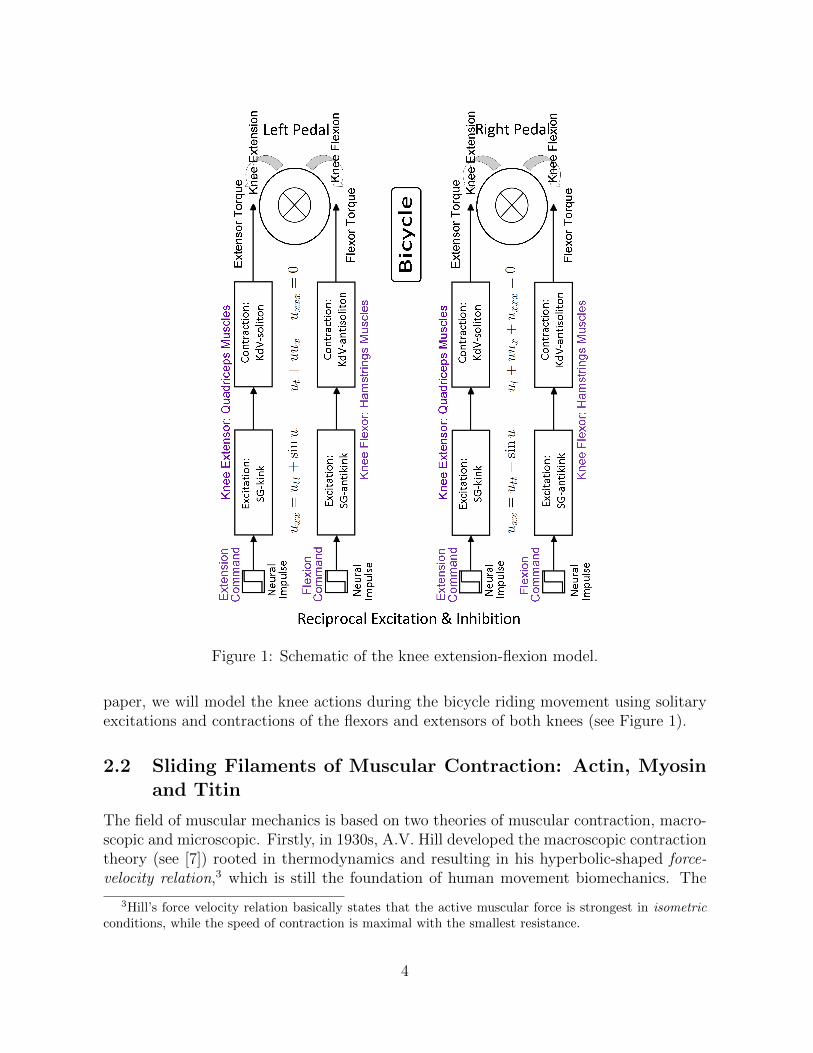
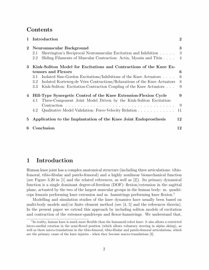
Figure 1: Schematic of the knee extension-flexion model.
paper, we will model the knee actions during the bicycle riding movement using solitaryexcitations and contractions of the flexors and extensors of both knees (see Figure 1).
2.2 Sliding Filaments of Muscular Contraction: Actin, Myosinand Titin
The field of muscular mechanics is based on two theories of muscular contraction, macro-scopic and microscopic. Firstly, in 1930s, A.V. Hill developed the macroscopic contractiontheory (see [7]) rooted in thermodynamics and resulting in his hyperbolic-shaped force-velocity relation,3 which is still the foundation of human movement biomechanics. The
3Hill’s force velocity relation basically states that the active muscular force is strongest in isometricconditions, while the speed of contraction is maximal with the smallest resistance.
4
microscopic contraction theory is called the sliding filament theory, developed in 1954jointly by two Huxleys [8, 9], who proposed that the muscular contraction force is gen-erated by the relative motion/interaction between two kinds of protein filaments, thickmyosin filaments and thin actin filaments, biochemically fueled by the ATP-release, withinthe basic muscular micro-units called sarcomeres (bounded by the Z-discs; for a recentreview, see, e.g. [10]). This microscopic contraction theory is usually captured in the(skewed) bell-shaped force-length relation of the concentric muscular contraction.
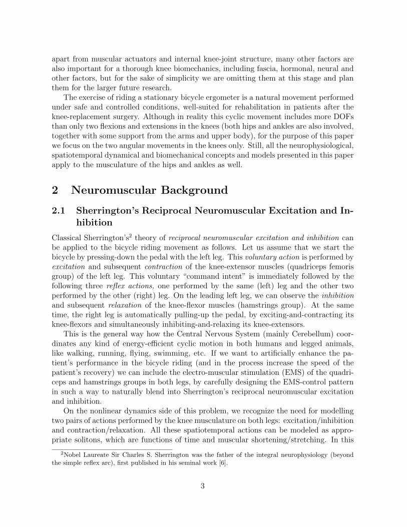
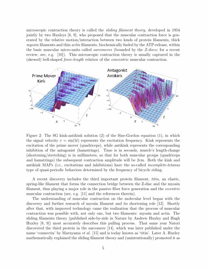
Figure 2: The SG kink-antikink solution (2) of the Sine-Gordon equation (1), in whichthe signal velocity v = sin(5t) represents the excitation frequency. Kink represents theexcitation of the prime mover (quadriceps), while antikink represents the correspondinginhibition of the antagonist (hamstrings). Time is in seconds, muscle’s length-change(shortening/stretching) is in millimeters, so that for both muscular groups (quadricepsand hamstrings) the subsequent contraction amplitude will be 2cm. Both the kink andantikink MAPs (i.e., excitations and inhibitions) have the so-called incomplete-tetanustype of quasi-periodic behaviors determined by the frequency of bicycle riding.
A recent discovery includes the third important protein filament, titin, an elastic,spring-like filament that forms the connection bridge between the Z-disc and the myosinfilament, thus playing a major role in the passive fiber force generation and the eccentricmuscular contraction (see, e.g. [11] and the references therein).
The understanding of muscular contraction on the molecular level began with thediscovery and further research of myosin filament and its shortening role [12]. Shortlyafter that, with improved technology came the realization that the process of muscularcontraction was possible with, not only one, but two filaments: myosin and actin. Thesliding filaments theory (published side-by-side in Nature by Andrew Huxley and HughHuxley [8, 9]) most accurately describes this pulling process. That same year Natoridiscovered the third protein in the sarcomere [14], which was later published under thename ‘connectin’ by Maruyama et al. [15] and is today known as ‘titin’. Later A. Huxleymathematically explained the sliding filament theory and (unintentionally) promoted it as
5
the ‘cross-bridged theory’ [13] (he had an active correspondence with the Science Editorwho encouraged him to publish his mathematical contribution as the molecular theory ofmuscular contraction) [24].
Parallel to the story of the three proteins involved in muscular contraction, there arethree types of contraction. The concentric contraction perfectly fitted in the cross bridgetheory and gave the microscopic foundation to classical Hill’s concentric force-velocityrelationship [7]. The isometric contraction was in similar way explained by Gordon (A.Huxley’s co-worker) [16] in 1966. The eccentric contraction remained inappropriately ex-plained for a long time, until its important role in muscular contraction was realized; itwas clearly noticeable through the force-length relationship curve and its connection withthe sarcomere, myosin and actin [24]. A prolific work by Herzog put the light into myste-rious explanation of eccentric contraction [17]-[25] and the role of titin, through active andpassive stretching of the sarcomere, and its bond with calcium during passive stretching.The protein titin attracted the attention of scientific community, which resulted in thestudies about its mechanical properties [26], its ‘complex structure-dependant elasticity’[27]. Titin’s palindromic assembly was formulated (titin-richest structures in the bodyare sarcomeres of striated and cardiac muscles [28]; titin is the largest protein moleculein the body, with the role in control of structure and extensibility of sarcomeres and thesource of actin stiffness [29]).
3 Kink-Soliton Model for Excitations and Contrac-
tions of the Knee Extensors and Flexors
In this section, we will first present a kink excitation/inhibition model for the antago-nistic pair of knee muscles, then we will present the simplest possible soliton contrac-tion/relaxation model for the same pair of knee muscles, and finally we will couple theminto a working kink-soliton model of excitation-contraction, to be subsequently used tomodel bicycle riding movement. For general overview of solitons and kinks, see [40]. Thecoupled excitation-contraction model will be used to generate the normalized force-timerelation for various muscular length changes. In the next section, this force-time relationwill be used to generate the resulting torque in the knee joint, that is, to control theextension-flexion cycle in the human knee.
3.1 Isolated Sine-Gordon Excitations/Inhibitions of the KneeActuators
To start with the solitary modeling of muscular excitations and contractions, recall thata thermal wave alternative to the classical, Nobel-awarded, Hodgkin-Huxley electricalneural-conduction theory [31, 32] was proposed a decade ago by [33, 34], which wasrecently generalized in [35, 36], where various forms of the neural action potential havebeen represented by a variety of solitons, kinks and breathers of the (1+1) Sine-Gordon
6

Figure 3: The soliton-antisoliton solution (4) of the KdV Eq. (3) with τ = 0.1s with thegenerated force amplitude normalized to 1. Again, time is in seconds, muscle’s length-change is in millimeters, giving the total contraction amplitude of 2cm for both muscles.Soliton represents the normalized contraction of the prime mover (quadriceps), while anti-soliton represents the corresponding normalized relaxation of the antagonist (hamstrings).
(SG) equation:uxx = utt + sinu, (1)
where u = u(x, t) is the real spatiotemporal function (a scalar field) and indices denotepartial derivatives (e.g., uxx ≡ ∂2u/∂x2).
Using the fact that muscles are excitable tissues similar to nerves (only about 100times slower), we can apply Eq. (1) to model various forms of muscular action potential(MAP), or muscular excitation, by interpreting the driving term sinu as either inducedinternally by neural stimuli, or externally by electro-muscular stimulation (see [1] and thereferences therein).
To describe the excitation/inhibition action of a pair of mutually antagonistic kneemuscles (i.e., quadriceps femoris and hamstrings groups) as the actuators in the kneejoint, a particularly useful solution of Eq. (1) is the following SG kink-antikink pair (seeFigure 2):
u(x, t) = 4 tan−1[± exp
(x− tv√1− v2
)], (2)
where the positive sign corresponds to the prime mover’s kink and negative sign to theantagonists’s antikink, and v represents the excitation/inhibition signal velocity that isa function of the neural (or, electro-muscular) stimulation frequency. For example, if weuse the SG-kink MAP to describe the simultaneous excitation of the left quadriceps andthe right hamstrings, then the SG-antikink MAP corresponds to the inhibition of the lefthamstrings and the right quadriceps.
7

Figure 4: The kink-soliton model (5) for the excitation-contraction coupling (left) withthe corresponding inhibition/relaxation coupling (right) of the knee extensors (quadriceps)and flexors (hamstrings), representing the normalized muscular force-length-time relation.Again, time is in seconds, muscle’s length-change is in millimeters, giving the contractionamplitude of 2cm for both muscles.
3.2 Isolated Korteweg-de Vries Contractions/Relaxations of theKnee Actuators
Next, we come to the isolated of muscular contractions/inhibitions. To model the titin-influenced actomyosin contractions, we recall that solitary models of muscular contrac-tions were pioneered by Davydov in [37] and subsequently elaborated in [38]. These mod-els, representing oscillations of peptide group Amid I inside spiral structures of myosinfilaments, can be represented by the solutions of the Korteweg-de Vries (KdV) equation:
ut + uux + uxxx = 0. (3)
Out of a variety of known sech, tanh and cnoidal solutions of the KdV Eq. (3), to modelthe contraction/inhibition action of the knee extensor-flexor pair (with the normalizedamplitude), we start with the simplest solution (see [40]) in the form of the followingsoliton-antisoliton sech-pair:
u(x, t) = ± sech(x− t+ τ). (4)
where the positive sign corresponds to the prime mover’s contraction soliton and negativesign to the antagonist’s inhibition antisoliton, and τ represents the time-delay betweenthe previous excitation and the current contraction (see Figure 3).
In particular, the positive sech soliton u(x, t) = sech(x− t+ τ) can be interpreted as anormalized 3D model for the muscular force-length relation (see Figures 6 and 7 in [11]).
8
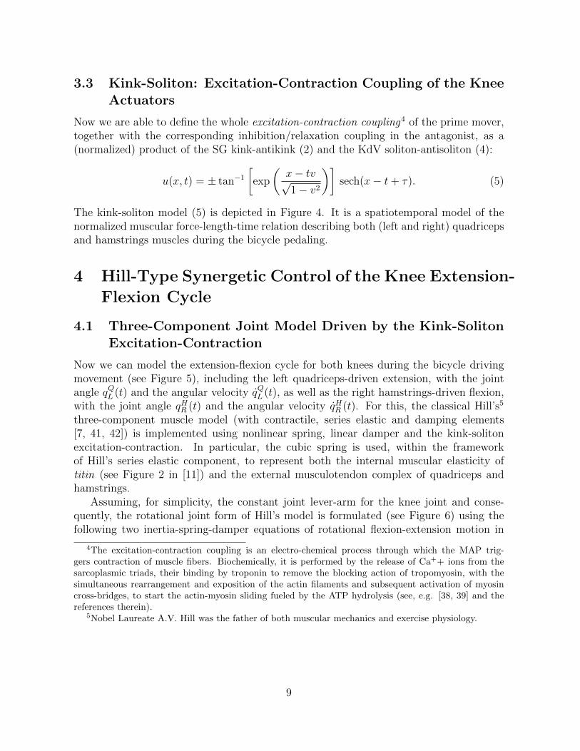
3.3 Kink-Soliton: Excitation-Contraction Coupling of the KneeActuators
Now we are able to define the whole excitation-contraction coupling4 of the prime mover,together with the corresponding inhibition/relaxation coupling in the antagonist, as a(normalized) product of the SG kink-antikink (2) and the KdV soliton-antisoliton (4):
u(x, t) = ± tan−1[exp
(x− tv√1− v2
)]sech(x− t+ τ). (5)
The kink-soliton model (5) is depicted in Figure 4. It is a spatiotemporal model of thenormalized muscular force-length-time relation describing both (left and right) quadricepsand hamstrings muscles during the bicycle pedaling.
4 Hill-Type Synergetic Control of the Knee Extension-
Flexion Cycle
4.1 Three-Component Joint Model Driven by the Kink-SolitonExcitation-Contraction
Now we can model the extension-flexion cycle for both knees during the bicycle drivingmovement (see Figure 5), including the left quadriceps-driven extension, with the jointangle qQL (t) and the angular velocity q̇QL (t), as well as the right hamstrings-driven flexion,with the joint angle qHR (t) and the angular velocity q̇HR (t). For this, the classical Hill’s5
three-component muscle model (with contractile, series elastic and damping elements[7, 41, 42]) is implemented using nonlinear spring, linear damper and the kink-solitonexcitation-contraction. In particular, the cubic spring is used, within the frameworkof Hill’s series elastic component, to represent both the internal muscular elasticity oftitin (see Figure 2 in [11]) and the external musculotendon complex of quadriceps andhamstrings.
Assuming, for simplicity, the constant joint lever-arm for the knee joint and conse-quently, the rotational joint form of Hill’s model is formulated (see Figure 6) using thefollowing two inertia-spring-damper equations of rotational flexion-extension motion in
4The excitation-contraction coupling is an electro-chemical process through which the MAP trig-gers contraction of muscle fibers. Biochemically, it is performed by the release of Ca++ ions from thesarcoplasmic triads, their binding by troponin to remove the blocking action of tropomyosin, with thesimultaneous rearrangement and exposition of the actin filaments and subsequent activation of myosincross-bridges, to start the actin-myosin sliding fueled by the ATP hydrolysis (see, e.g. [38, 39] and thereferences therein).
5Nobel Laureate A.V. Hill was the father of both muscular mechanics and exercise physiology.
9

Figure 5: A synergy of two angular Hill-type muscular models driving the bicycle.
the knee joint:
Left knee extension : J q̈QL (t) + a q̇QL (t) + b qQL (t)3 = c T (t, x), (for x=1, ..., 8; τ=0.1) ,
Right knee flexion : J q̈HR (t) + a q̇HR (t) + b qHR (t)3 =c
2T (t− τ, x), (6)
driven by the torque : T (t, x) = tan−1
[exp
(x−tv√1−v2
)]sech(x− t+ τ), (7)
with normalized values of all included muscular parameters (lower leg’s moment of inertiaJ , Hill’s angular spring a, damper b and torque amplitude c): J=a=b=c=1.6
6Hamstrings is naturally significantly weaker than quadriceps (which is an anti-gravitational muscle).This difference/disbalance is increased in athletes because of the squats-with-weights exercises for thequadriceps. This situation causes frequent hamstrings injuries among sprinters (and football/soccerplayers), partly because under fatigue hamstrings relaxation is slower than quadriceps contraction andpartly because of the shoes with pins that put even more stress on the hamstrings.
10
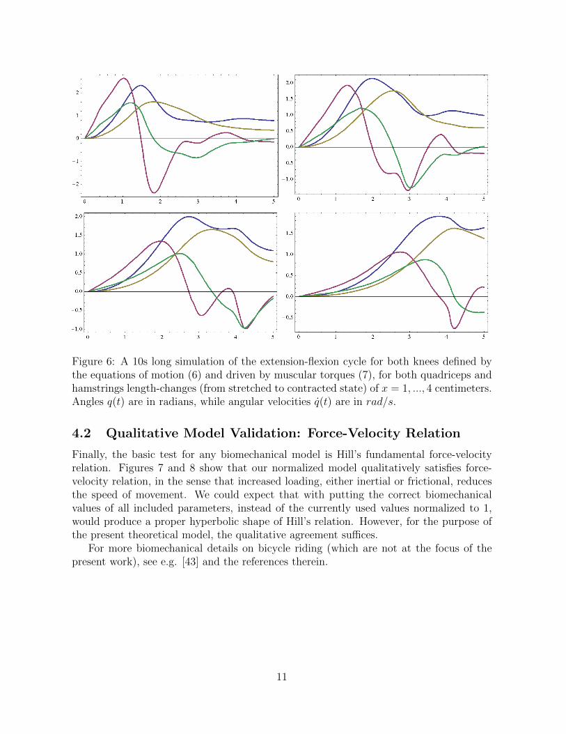
Figure 6: A 10s long simulation of the extension-flexion cycle for both knees defined bythe equations of motion (6) and driven by muscular torques (7), for both quadriceps andhamstrings length-changes (from stretched to contracted state) of x = 1, ..., 4 centimeters.Angles q(t) are in radians, while angular velocities q̇(t) are in rad/s.
4.2 Qualitative Model Validation: Force-Velocity Relation
Finally, the basic test for any biomechanical model is Hill’s fundamental force-velocityrelation. Figures 7 and 8 show that our normalized model qualitatively satisfies force-velocity relation, in the sense that increased loading, either inertial or frictional, reducesthe speed of movement. We could expect that with putting the correct biomechanicalvalues of all included parameters, instead of the currently used values normalized to 1,would produce a proper hyperbolic shape of Hill’s relation. However, for the purpose ofthe present theoretical model, the qualitative agreement suffices.
For more biomechanical details on bicycle riding (which are not at the focus of thepresent work), see e.g. [43] and the references therein.
11
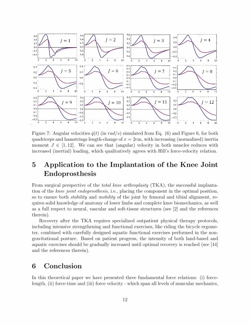
Figure 7: Angular velocities q̇(t) (in rad/s) simulated from Eq. (6) and Figure 6, for bothquadriceps and hamstrings length-change of x = 2cm, with increasing (normalized) inertiamoment J ∈ [1, 12]. We can see that (angular) velocity in both muscles reduces withincreased (inertial) loading, which qualitatively agrees with Hill’s force-velocity relation.
5 Application to the Implantation of the Knee Joint
Endoprosthesis
From surgical perspective of the total knee arthroplasty (TKA), the successful implanta-tion of the knee joint endoprosthesis, i.e., placing the component in the optimal position,so to ensure both stability and mobility of the joint by femoral and tibial alignment, re-quires solid knowledge of anatomy of lower limbs and complete knee biomechanics, as wellas a full respect to neural, vascular and soft-tissue structures (see [2] and the referencestherein).
Recovery after the TKA requires specialized outpatient physical therapy protocols,including intensive strengthening and functional exercises, like riding the bicycle ergome-ter, combined with carefully designed aquatic functional exercises performed in the non-gravitational posture. Based on patient progress, the intensity of both land-based andaquatic exercises should be gradually increased until optimal recovery is reached (see [44]and the references therein).
6 Conclusion
In this theoretical paper we have presented three fundamental force relations: (i) force-length, (ii) force-time and (iii) force velocity - which span all levels of muscular mechanics,
12
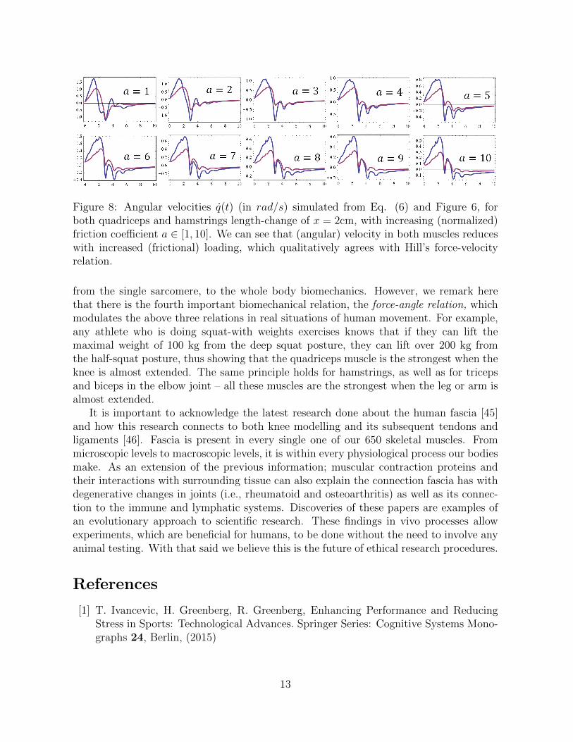
Figure 8: Angular velocities q̇(t) (in rad/s) simulated from Eq. (6) and Figure 6, forboth quadriceps and hamstrings length-change of x = 2cm, with increasing (normalized)friction coefficient a ∈ [1, 10]. We can see that (angular) velocity in both muscles reduceswith increased (frictional) loading, which qualitatively agrees with Hill’s force-velocityrelation.
from the single sarcomere, to the whole body biomechanics. However, we remark herethat there is the fourth important biomechanical relation, the force-angle relation, whichmodulates the above three relations in real situations of human movement. For example,any athlete who is doing squat-with weights exercises knows that if they can lift themaximal weight of 100 kg from the deep squat posture, they can lift over 200 kg fromthe half-squat posture, thus showing that the quadriceps muscle is the strongest when theknee is almost extended. The same principle holds for hamstrings, as well as for tricepsand biceps in the elbow joint – all these muscles are the strongest when the leg or arm isalmost extended.
It is important to acknowledge the latest research done about the human fascia [45]and how this research connects to both knee modelling and its subsequent tendons andligaments [46]. Fascia is present in every single one of our 650 skeletal muscles. Frommicroscopic levels to macroscopic levels, it is within every physiological process our bodiesmake. As an extension of the previous information; muscular contraction proteins andtheir interactions with surrounding tissue can also explain the connection fascia has withdegenerative changes in joints (i.e., rheumatoid and osteoarthritis) as well as its connec-tion to the immune and lymphatic systems. Discoveries of these papers are examples ofan evolutionary approach to scientific research. These findings in vivo processes allowexperiments, which are beneficial for humans, to be done without the need to involve anyanimal testing. With that said we believe this is the future of ethical research procedures.
References
[1] T. Ivancevic, H. Greenberg, R. Greenberg, Enhancing Performance and ReducingStress in Sports: Technological Advances. Springer Series: Cognitive Systems Mono-graphs 24, Berlin, (2015)
13
[2] Z. Gojkovic, Implantation of the knee joint endoprosthesis, Med. Survay, Novi Sad,LXVIII (11-12), 367-369, (2015)
[3] V. Ivancevic, New mechanics of generic musculo-skeletal injury, Biophys. Rev. Let.4(3), 273-287, (2009)
[4] M. Machado, P. Flores, J.C.P. Claro, J. Ambrø’sio, M. Silva, A. Completo, H.M.Lankarani, Development of a planar multibody model of the human knee joint, Non-linear Dyn. 60, 459-478, (2010)
[5] D. Weed, L.G. Maqueda, M.A. Brown, B.A. Hussein, A.A. Shabana, A new nonlinearmultibody/finite element formulation for knee joint ligaments. Nonlinear Dyn. 60,357-367, (2010)
[6] C.S. Sherrington, The Integrative Action of the Nervous System. Oxford Uni. Press,(1906)
[7] A.V. Hill, The heat of shortening and the dynamic constants of muscle. Proc. R. Soc.B76, 136-195, (1938)
[8] A.F. Huxley, R. Niedergerke, Structural changes in muscle during contraction: In-terference microscopy of living muscle fibres. Nature 173, 97117973 (1954)
[9] H.E. Huxley, J. Hanson, Changes in the cross-striations of muscle during contractionand stretch and their structural interpretation, Nature 173, 97317976 (1954)
[10] J.L. Krans, The Sliding Filament Theory of Muscle Contraction, Nature Educ. 3(9),66, (2010)
[11] K.M. Wisdom, S.L. Delp, E. Kuhl, Use it or lose it: multiscale skeletal muscle adap-tation to mechanical stimuli, Biomech. Mod. Mechanobiol, 14(2), 195-215, (2015)
[12] H.E. Huxley, Electron microscope studies of the organization of the filaments instriated muscle. Biochim. Biophys. Acta 12, 387-394, (1953)
[13] H.E. Huxley, The mechanism of muscular contraction. Science 164, 1356-1366, (1969)
[14] R. Natori, Skinned Fibres of Skeletal Muscle and the Mechanism of Muscle Contrac-tion – A Chronological Account of the Author’s Investigations into Muscle Physiology,Jikeikai Med. J. 54(1), (1954)
[15] K. Maruyama, S. Matsubara, R. Natori, Y. Nonomura, S. Kimura, Connectin, anelastic protein of muscle. Characterization and Function, J. Biochem. 82(2), 317-37,(1977)
[16] A.M. Gordon, A.F. Huxley, F.J. Julian, The variation in isometric tension withsarcomere length in vertebrate muscle fibres, J. Physiol. 184, 170-192, (1966)
14
[17] W. Herzog, Mechanisms of enhanced force production in lengthening (eccentric) mus-cle contractions. J. Appl. Physiol. 116, 1407-1417, (1985)
[18] W. Herzog, M. Duvall, T.R. Leonard, Molecular mechanisms of muscle force regula-tion: a role for titin? Exerc. Sport Sci. Rev. 40, 50-57, (2012)
[19] W. Herzog, E.J. Lee, D.E. Rassier, Residual force enhancement in skeletal muscle,J. Physiol. 574, 635-642, (2006)
[20] W. Herzog, T. Leonard, V. Joumaa, M. Duvall, A. Panchangam, The three filamentmodel of skeletal muscle stability and force production. Mol. Cell. Biomech. 9, 175-191, (2012)
[21] W. Herzog, T.R. Leonard, Force enhancement following stretching of skeletal muscle:a new mechanism. J. Exp. Biol. 205, 1275-1283, (2002)
[22] W. Herzog, T.R. Leonard, Response to the (Morgan and Proske) Letter to the Editorby Walter Herzog and Tim Leonard (on behalf of the authors). J. Physiol 578(2),617-620, (2006)
[23] W. Herzog, T.R. Leonard, Residual force enhancement: the neglected property ofstriated muscle contraction. J. Physiol. 591(8), 2221, (2013)
[24] W. Herzog, K. Powers, K. Johnston, M. Duvall, A new paradigm for muscle contrac-tion, Frontiers in Physiol. 6, 174, (2015)
[25] R. Fortuna, et al., Residual force enhancement following shortening is speed-dependent. Sci. Rep. 6, 21513, (2016)
[26] P. Williams, et al., Hidden complexity in the mechanical properties of titin Nature422, 446-440, (2003)
[27] H. Li, et al., Reverse engineering of the giant muscle protein titin, Nature 418,998-1002, (2002)
[28] P. Zou, Palindromic assembly of the giant muscle protein titin in the sarcomericZ-disk, Nature 439, 229-233, (2006)
[29] L. Tskhovrebova, J.Trinick, Titin: properties and family relationships, Nature Rev.Mol. Cell Biol. 4, 679-689, (2003)
[30] M. Mireille, et al., Actin-binding proteins sensitively mediate F-actin bundle stiffness,Nature Materials 5, 748-753, (2006)
[31] A.L. Hodgkin, A.F. Huxley, A quantitative description of membrane current andapplication to conduction and excitation in nerve, J. Physiol. (London) 117, 500-544, (1952)
15

[32] A.L. Hodgkin, The Conduction of the Nervous Impulse. Liverpool: Liverpool Univ.Press, (1964)
[33] T. Heimburg, A.D. Jackson, On soliton propagation in biomembranes and nerves,Proc. Natl. Acad. Sci. US, 102(28), pp. 9790-9795, 2005.
[34] T. Heimburg, A.D. Jackson, On the action potential as a propagating density pulseand the role of anesthetics, Biophys. Rev. Let., 2, 57-78, (2007)
[35] V. Ivancevic, T. Ivancevic, Sine-Gordon Solitons, Kinks and Breathers as PhysicalModels of Nonlinear Excitations in Living Cellular Structures, J. Geo. Sym. Phys.31, 1-56, (2013)
[36] V. Ivancevic, D. Reid, Controlled Complexity in Pulse Conduction: Traveling Soli-tons from Neural to Optical Fibers, Math. in Engineering, Sci. and Aerospace (MESAjournal), 5(1), 17-32, (2014)
[37] A.S. Davydov, Solitons in Molecular Systems. (2nd ed), Kluwer, Dordrecht, (1991)
[38] V. Ivancevic, T. Ivancevic, Human-Like Biomechanics. Springer, Dordrecht, (2005)
[39] V. Ivancevic, T. Ivancevic, Natural Biodynamics. World Scientific, Singapore, (2006)
[40] M.J. Ablowitz, P.A. Clarkson, Solitons, Nonlinear Evolution Equations and InverseScattering. Cambridge University Press, (1991)
[41] A.V. Hill, The dynamic constants of human muscle. Proc. R. Soc. B128, 263-274,(1940)
[42] A.V. Hill, The series elastic component of muscles. Proc. R. Soc B137, 273-280,(1950)
[43] J.W. Rankin, R.R. Neptune, A theoretical analysis of an optimal chainring shapeto maximize crank power during isokinetic pedaling, J. Biomech. 41, 1494171502,(2008)
[44] F. Pozzi, L. Snyder-Mackler, J. Zeni, Physical exercise after knee arthroplasty: asystematic review of controlled trials, Eur. J. Phys. Rehabil. Med. 49(6), 877-92,(2013)
[45] J.C. Guimberteau, Armtrong, C., Architecture of Human Living Fascia, HandspringPublishing, UK, (2015)
[46] N. Hagemeister, N. Duval, L. Yahia, W. Krudwig, U. Witzel, J.A. de Guise,Computer-based method for the 3-D kinematic analysis of posterior cruciate liga-ment nad posterior-lateral corner lesions, The Knee, 9, 301–308, (2002)
16